vwhpv d qg 5hioh[lqj, qwhuidfhv - unimi.ithomes.di.unimi.it/pizzi/paperi/annmicrotubuli.pdf ·...
TRANSCRIPT

Franco OrsucciUniversity College London, UK & Institute for Complexity Studies, Italy
Nicoletta SalaUniversity of Lugano, Switzerland
Complexity Science, Living Systems, and Reflexing Interfaces:New Models and Perspectives
Complexity science, living systems, and reflexing interfaces : new models and perspectives / Franco Orsucci and Nicoletta Sala, editors. p. cm. Includes bibliographical references and index. ISBN 978-1-4666-2077-3 (hardcover) -- ISBN 978-1-4666-2078-0 (ebook) -- ISBN 978-1-4666-2079-7 (print & perpetual access) 1. Complexity (Philosophy) 2. Biocomplexity. I. Orsucci, Franco. II. Sala, Nicoletta. Q175.32.C65C654 2013 577--dc23 2012015948
British Cataloguing in Publication DataA Cataloguing in Publication record for this book is available from the British Library.
All work contributed to this book is new, previously-unpublished material. The views expressed in this book are those of the authors, but not necessarily of the publisher.
Managing Director: Lindsay JohnstonBook Production Manager: Jennifer RomanchakPublishing Systems Analyst: Adrienne FreelandManaging Editor: Joel GamonDevelopment Editor: Myla MerkelAssistant Acquisitions Editor: Kayla WolfeTypesetter: Lisandro GonzalezCover Design: Nick Newcomer
Published in the United States of America by Information Science Reference (an imprint of IGI Global)701 E. Chocolate AvenueHershey PA 17033Tel: 717-533-8845Fax: 717-533-8661 E-mail: [email protected] site: http://www.igi-global.com
Copyright © 2013 by IGI Global. All rights reserved. No part of this publication may be reproduced, stored or distributed in any form or by any means, electronic or mechanical, including photocopying, without written permission from the publisher.Product or company names used in this set are for identification purposes only. Inclusion of the names of the products or companies does not indicate a claim of ownership by IGI Global of the trademark or registered trademark.
Library of Congress Cataloging-in-Publication Data
78
Copyright © 2013, IGI Global. Copying or distributing in print or electronic forms without written permission of IGI Global is prohibited.
Chapter 5
R. PizziUniversità degli Studi di Milano, Italy
S. FiorentiniUniversità degli Studi di Milano, Italy
G. StriniUniversità degli Studi di Milano, Italy
M. PregnolatoUniversità degli Studi di Pavia, Italy
Exploring Structural and Dynamical Properties
Microtubules by Means of Artificial Neural Networks
ABSTRACT
Microtubules (MTs) are cylindrical polymers of the tubulin dimer, are constituents of all eukaryotic cells cytoskeleton and are involved in key cellular functions and are claimed to be involved as sub-cellular information or quantum information communication systems. The authors evaluated some biophysical properties of MTs by means of specific physical measures of resonance and birefringence in presence of electromagnetic field, on the assumption that when tubulin and MTs show different biophysical behav-iours, this should be due to their special structural properties. Actually, MTs are the closest biological equivalent to the well-known carbon nanotubes (CNTs), whose interesting biophysical and quantum properties are due to their peculiar microscopic structure. The experimental results highlighted a physical behaviour of MTs in comparison with tubulin. The dynamic simulation of MT and tubulin subjected to electromagnetic field was performed via MD tools. Their level of self-organization was evaluated using artificial neural networks, which resulted to be an effective method to gather the dynamical behaviour of cellular and non-cellular structures and to compare their physical properties.
DOI: 10.4018/978-1-4666-2077-3.ch005
79
Exploring Structural and Dynamical Properties Microtubules by Means of Artificial Neural Networks
INTRODUCTION
Background
Microtubules
Microtubules (MTs) are key constituents of all eukaryotic cells cytoskeleton. They are involved in the regulation of essential cellular functions such as the transport of materials within the cell, the movement of cytoplasmic organelles or vesicles and the cell division (Hyams & Lloyd, 1994).
MTs are stiff cytoskeletal filaments character-ized by a tubelike structure. The building block of a MT is a 110-kDa heterodimeric protein said tubulin, that is the association product of two different subunits, designated α and β tubulin (Postingl, Krauhs, Little, & Kempf 1981, Krauhs, Little, Kempf, Hofer-Warbinek, Ade, & Postingl 1981) and encoded by separate genes. The word tubulin always refers to the αβ heterodimer, that is usually considered as one unit, although the as-sociation is only due to non-covalent interactions. Each monomer of α and β tubulin is a compact ellipsoid of approximate dimensions 46 x 40 x 65 A° (width, height, and depth, respectively); while dimensions of α β-heterodimer are 46 x 80 x 65 A°. Both α- and β- tubulin is composed of approximately 450 amino acids.
Recently important information about tubulin conformational changes during the MTs po-lymerization have been obtained through X-ray crystallography (Ravelli, Gigant, Curmi, Jourdain, Lachkar, Sobel, & Knossow 2004).
The general structure of MTs has been es-tablished experimentally (Amos & Amos1991, Chrétien & Wade 1991). MTs have been consid-ered as helical polymers and they are built by the self-association of the αβ-heterodimer through a process of polymerization and depolymerization.
This dynamic nature makes MTs sensitive to several pharmacological agents, i.e. some classes of anticancer agents that are able to destroy or stabilize their structure.
The polymerization occurs in a two-dimen-sional process that involves two types of contacts between tubulin subunits. The first process involve head-to-tail binding of heterodimers and it results in polar protofilaments that run along the length of the MT. The second process involve lateral interactions between parallel protofilaments and it completes the MT wall to form a hollow tube (Nogales, Whittaker, Milligan & Downing 1999). The longitudinal contacts along protofilaments appear to be much stronger than those between adjacent protofilaments (Mandelkow, Mandelkow, & Milligan 1991).
All protofilaments in a MT have the same orientation.
Assembly mechanism of α- and β- tubulin gives rise in vitro to a variety of cylindrical structures that differ by their protofilament and monomer helix-start numbers (Binder & Rosenbaum, 1978, Burton & Himes 1978, Chrétien, Metoz, Verde, Karsenti & Wade 1992, Linck & Langevin 1981, Pierson, Burton & Himes 1978, Chrétien 2000). In contrast, most MTs assembled in vivo seem to be composed of 13 protofilaments, although many exceptions have been noted in different species and cell types.
The lengths of MTs vary but commonly reach 5-10 μm dimensions; and their diameter depends on the protofilament number. For example in the case of 13 protofilaments the tube has an outer diameter of 23 nm and an inner diameter of roughly 15 nm.
Microtubules Quantum Theories
In the last decade many theories and papers have been published concerning the biophysical properties of MTs including the hypothesis of MTs implication in coherent quantum states in the brain evolving in some form of energy and information transfer.
The most discussed theory on quantum effects involving MTs has been proposed by Hameroff
80
Exploring Structural and Dynamical Properties Microtubules by Means of Artificial Neural Networks
and Penrose that published the OrchOR Model in 1996 (Hameroff & Penrose, 1996,Hameroff & Penrose, 1996).
They supposed that quantum-superposed states develop in tubulins, remain coherent and recruit more superposed tubulins until a mass-time-energy threshold, related to quantum gravity, is reached (up to 500 msec). This model has been discussed and refined for more than 10 years, mainly focus-ing attention to the decoherence criterion after the Tegmark critical paper of 2000 (Tegmark, 2000a, Tegmark, 2000b) and proposing several methods of shielding MTs against the environment of the brain (Woolf & Hameroff, 2001, Hagan, Hamer-off & Tuszynski, 2002, Hameroff, 2007a). In the Hameroff model MTs perform a kind of quantum computation through the tubulins working like cellular automata. The MTs interior works as an electromagnetic wave guide, filled with water in an organized collective states, transmitting information through the brain (Hameroff, 2007b).
In the same years Nanopoulos et al adopted the string theory to develop a so called QED-Cavity model predicting dissipationless energy transfer along MTs as well as quantum teleportation of states at near room temperature (Nanopoulos & Mavromatos, 1996).
Mavromatos 2000, Mavromatos, Mershin & Nanopoulos, 2002). The Tuszynski approach is based on the biophysical aspects of MTs. Tubulins have electric dipole momeCNTs due to asymmet-ric charges distribution and MTs can be modeled as a lattice of orientated dipoles that can be in random phase, ferroelectric (parallel-aligned) and an intermediate weakly ferroelectric phase like a spin-glass phase (Tuszynski, Hameroff, Satarić, Trpišová & Nip, 1995, Trpišová, Sept & Satarić,1997, Tuszynski, Brown & Hawrylak, 1998). The model has been sustained by Faber, Portugal & Rosa (2006), who considered a MT as a classical subneuronal information processor.
In 1994 Jibu Hagan, Hameroff, Pribram and Yasue (1994) suggested that the Fröhlich dynamics of ordered water molecules and the quantizated
electromagnetic field confined inside the hollow MTs core can give rise to the collective quantum optical modes responsible for the phenomenon of superradiance by which any incoherent molecular electromagnetic energy can be transformed in a coherent photon inside the MTs. These photons propagate along the internal hollow core as if the optical medium were transparent and this quantum theoretical phenomenon is called “self-induced transparency”.
A decade before, applying quantum field theory (QFT), Del Giudice, Doglia, Milani, Vitiello (1982, 1983) reported that electromagnetic energy penetrating into cytoplasm would self-focus inside filaments whose diameter depend on symmetry breaking (Bose condensation) of ordered water dipoles. The diameter calculated was exactly the inner diameter of MTs (15 nm).
In any case, all phenomena occurring within the brain, both at macroscopic or microscopic level, can be related to some form of phase transition and a number of authors (Pessa, 2007, Alfinito, Viglione, & Vitiello, 2001) pointed out the in-consistence of a quantum mechanical framework based only on traditional computational schemata. It is to be recalled, in this regard, that these sche-mata have been introduced to deal with particles, atoms, or molecules, and are unsuitable when ap-plied to biological phenomena. In particular Pessa suggested that adopting a wider framework of QFT and, in particular, the dissipative version of it, relying on the doubling mechanism, we could achieve a generalization of QFT able to account for change phenomena in the biological world (Vitiello, 1995, Pessa & Vitiello, 2004, Freeman & Vitiello, 2006).
Carbon Nanotubes and Microtubules
The electronics industry is going to evolve from the technology based on silicon towards innova-tive materials with new physical properties. These new materials include the carbon nanotubes which
81
Exploring Structural and Dynamical Properties Microtubules by Means of Artificial Neural Networks
currently represent one of the most promising al-ternatives to overcome the current limits of silicon.
Currently, with a large commitment of aca-demic and industrial scientists, the research is developing nanoscale materials with extremely advanced and useful properties, as they can act both as semiconductors and as superconductors. Thanks to the structure of these materials, their properties are not restricted to classical physics, but presents a wide range of quantum mechanical effects. These may lead to an even more efficient tool for information transfer.
In particular, carbon nanotubes (CNTs) display a wide range of physical effects among them electronic properties are particularly attractive Quantum transport properties of CNTs has been re-viewed by Roche, Akkermans, Chauvet, Hekking, Issi, Martel, Montambaux, & Poncharal (2006) both from a theoretical and experimental view. Recently has been described the low-temperature spin relaxation time measurement in a fully tun-able CNT double quantum dots. This is an inter-esting study for new microwave-based quantum information processing experiments with CNTs (Sapmaz, Meyer, Beliczynski, Jarillo-Herrero & Knowenhoven, 2006).
According to Pampaloni & Florin (2008) CNTs are the closest equivalent to MTs among the known nanomaterials. Although their elastic moduli are different, MTs and CNTs have similar mechanical behaviours. They are both exceptionally resilient and form large boundless with improved stiffness.
Nanobiotechnology can move towards a next generation of materials with a wide range of functional properties. As suggest by Michette, Mavromatos, Powell, Holwill, and Pfauntsch (2004), MTs associated with carbon chemistry will allow to build complex macromolecular assemblies for sharing the exciting electronic properties of semi- and super-conductors
Purpose of this Work
Taking into account the connection between struc-tural and physical properties in CNTs and their structural similarity to MTs, our basic assumption in this research was that when tubulin and MTs show different biophysical behaviours, this should be due to the special structural properties of MTs.
In the study of the physical properties of MTs compared with those of CNTs, it is desired to search and analyze a possible reaction to microwaves, observing any ability of MTs to absorb or emit like antennas. The MTs, as well as CNTs, may behave as oscillators, this could make them superreactive receivers able to amplify the signals.
In order to validate this hypothesis we carried out a set of in vitro experiments on MTs and tu-bulin, then we explored the possible meaning of the obtained findings by simulating and compar-ing the dynamic evolution of MTs, tubulin and CNTs. To this purpose two different procedures based on Artificial Neural Networks models were developed and applied. This paper describes and discussed both procedures and the achieved results.
METHODS
Resonance and Birefringence Experiments
Antennas are devices capable to transform an electromagnetic field into an electrical signal, or to radiate, in the form of electromagnetic field, the electrical signal they are fed by. When powered by an electrical signal to their ends, antennas absorb energy and return it in the surrounding space as electromagnetic waves (transmitting antenna), or absorb energy from an electromagnetic wave and generate a voltage to their ends (receiving antenna). On theoretical bases any conductive object acts as an antenna, regardless of the elec-
82
Exploring Structural and Dynamical Properties Microtubules by Means of Artificial Neural Networks
tromagnetic wave frequency they are hit or the signal that is fed by. In particular, any tubular conductor cable, resonating mechanically, acts as a cavity antenna.
The magnitude of the effect becomes sig-nificant when the frequency corresponds to the resonance frequency and in this case the output voltage can be used for receiving and transmitting radio waves.
Resonance is a physical condition that occurs when a damped oscillating system is subjected to a periodic solicitation with a frequency equal to the system oscillation. A resonance phenomenon causes a significant increase in the extent of the oscillations that corresponds to a remarkable ac-cumulation of energy within the oscillator.
The other physical property taken into account in this research is birefringence. Birefringence is an optical property of materials that arises from the interaction of light with oriented molecular andstructural components (Huang & Knighton, 2005). Birefringence is the decomposition of a beam of light into two rays that occurs when the light crosses specific anisotropic media depending on the polarization of the light. The interaction between light and magnetic field in a medium results in the rotation of the plane of polarization proportional to the intensity of the magnetic field component in the direction of the beam of light (Faraday effect).
Recent observations and experiments on CNTs have led to the development of an array of CNTs able to act as antennas (Wang, Kempa, Kimball, Carlson, Benham, Li, Kempa, Rybczynski, Her-czynski & Ren, 2004). These, instead to transmit and receive radio waves (measured in meters), due to their scale capture wavelengths at the nanoscale (measured in nanometers).
Our group carried out an experiment (Pizzi, Strini, Fiorentini, Pappalardo & Pregnolato, submitted) intended to verify the existence of mechanical resonance in MTs, in analogy with the CNTs, at the frequency that amplifies the wave.
During the experiment we identified a differ-ence in the peak amplitude of the solution with MTs at a frequency of 1510 MHz, whereas the solution with tubulin and the control solution did not show any reaction. The lack of response in tubulin and control can be considered a hint that the peculiar structure of microtubules could be the cause of the observed signal.
Considering the nanoscopic size of MTs, the resonance analysis would be more effective if carried out on much higher frequencies (up to 100 GHz), with suitable instrumentation. But the presence of a small but sharp resonance effect at a low frequency could be the hint of a much evident effect at higher frequencies.
To assess the physical behaviour of MTs under the effect of electric and magnetic fields we per-formed in vitro birefringence experiments (Pizzi, Strini, Fiorentini, Pappalardo & Pregnolato, sub-mitted) on different samples of MTs and tubulin.
By means of a polarized light and a suitable detection apparatus, it is possible to observe the associated birefringence and, therefore, the index of orientation of MTs subjected either to transverse electric fields and to transverse and longitudinal magnetic fields (Oldenbourg, Salmon & Tran, 1998).
MTs and tubulin were put in stabilizing buffer solution, and we measured the polarization under controlled conditions in order to determine dif-ferent effects in the interaction of almost static electromagnetic fields. For our comparative ex-periments the variation of the refraction index is important because it is a function of the wavelength of the electromagnetic radiation and the nature of the crossed material.
Behavioural differences observed between samples of tubulin and MTs, would lead us to understand weather the cavity structure in the MT reacts in a peculiar way in response to specific stimuli or not.
Actually, the analysis of the results of birefrin-gence experiment highlights that the MTs react to electromagnetic fields in a different way than
83
Exploring Structural and Dynamical Properties Microtubules by Means of Artificial Neural Networks
tubulin. In particular, electric field and longitudinal magnetic field show opposite effects in the two types of proteins. Anyway in spite of the effect under electric field is the same as with no field, an unexpected and interesting effect is shown in the case of longitudinal magnetic field.
The achieved results, supported by statistical significance, suggest that the tubular structure of MTs might be responsible for the different behaviour in respect to free tubulins.
Dynamical Simulation
In order to constitute a significant progress in the comprehension of the hypothesized peculiar properties of MTs, the experimental findings must match the estimations of a consistent model.
To this purpose, we performed a dynamic simulation of the molecular structures of tubulin and MTs subjected to different levels of electro-magnetic field and in the absence of field, com-pared with the similar behavior in terms of carbon nanotubes (CNTs) and buckyballs (BBs), globular nanostructured elements (Kroto,Heath, O’Brien, Curl & Smalley,1985). whose relationship with CNTs can be compared to the relationship between tubulin and MTs.
We adopted the simulation environment As-calaph (Ascalaph,website): due to the possibility to perform simulations for wide molecular structures with a large number of parameterizations. The simulations were carried out as follows:
• 1st simulation: zero electric field, A = 0• 2nd simulation: A = 2 V/cm, F = 90 Hz• 3rd simulation: A = 90 V/cm, F = 90 Hz
The structures were immersed in water at 298.15 °K. The simulation duration was 7000 ps. We adopted the AMBER (Assisted Model Build-ing and Energie Refinement) default force field.
The tertiary structure of tubulin was ob-tained from Protein Data Bank (Protein Data Bank,website), MTs from the website of the
NANO-D) research group at INRIA Grenoble-Rhone-Alpes (NANO-D,website), BBs and CNTs were directly obtained from Ascalaph.
After the end of simulation and a suitable dy-namical optimization, the graphical visualization of the structures appears as in Figure 1.
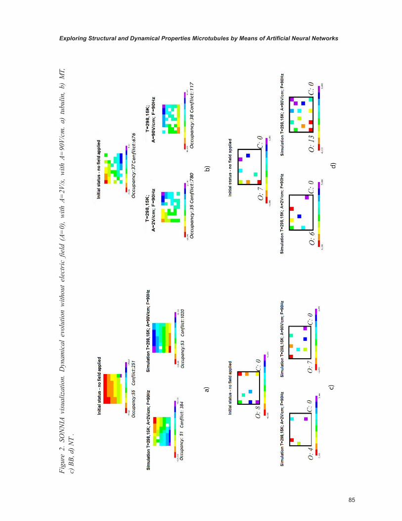
Artificial Neural Network Processing
In order to evaluate the results of the dynamical simulations, we conceived a novel methods based on Artificial Neural Networks (ANNs). ANNs are intrinsically non-linear models able to classify complex patterns. In particular, the self-organizing networks as the Kohonen’s Self Organizing Map (SOM) is well-known as a natural non-linear clas-sifiers (Kohonen,1990, Ritter, & Schulten 1988).
We submitted the structural data obtained by the MD evolution to two different SOM-based models, then compared their results.
The first adopted model was SONNIA. SON-NIA is a powerful Artificial Neural Networks environment, very useful in the field of drug dis-covery and protein prediction (SONNIA, website). It allows to classify a series of data sets, providing both supervised and unsupervised learning.
The output maps are represented by a set of colored boxes, one for each output neuron. The boxes configuration highlightes two interesting parameters:
1. Occupancy, i.e. the number of patterns that have been mapped onto the same neuron, indicating similarities in the input domain.
2. Conflicts or conflict neurons, i.e. neurons that refer to inputs belonging to different classes.
3. In general, there are always at least a few conflicts such as with any other modeling technique there are false positives or false negatives.

84
Exploring Structural and Dynamical Properties Microtubules by Means of Artificial Neural Networks
For our case study we chose a Kohonen rect-angular network structure with 9x6 neurons and a random initialization.
Besides, we used another self-organizing artificial network developed by our group, the ITSOM (Inductive Tracing Self-Organizing Map), to discriminate the dynamical behaviour of the structures under investigation, on the basis of the chaotic attractors determinated by the sequences of its winning neurons (Pizzi, Inama, Durin & Pedrinazzi, 2007).
In fact an analysis on the SOM has shown that such a sequence, provided to keep the learn-ing rates steady (instead of gradually decreasing them), constitutes chaotic attractors that repeat
“nearly” exactly in time with the epochs suc-ceeding, and that, once codified by the network, univocally characterize the input element that has determined them.
An attractor can be defined as a generalization of the steady state point, and represents the trajec-tory in a portion of state space where a dynamical system is attracted to (Ruelle,1981).
We tried to highlight the presence of dynami-cal attractors in the described structures using MATLAB and its SIMULINK module for the dynamical systems simulation.
In the following (Figure2, Figure 3) we show a comparison of the two different visualizations (SONNIA and ITSOM).
Figure 1. 3D visualization of the structures after dynamical simulation under A= 90 V/cm electric field: a) tubulin, b) MT, c) BB, d) NT
85
Exploring Structural and Dynamical Properties Microtubules by Means of Artificial Neural Networks
Figu
re 2
. SO
NN
IA v
isua
lizat
ion.
Dyn
amic
al e
volu
tion
with
out
elec
tric
fie
ld (
A=0)
, w
ith A
=2V
/s,
with
A=
90V/
cm.
a) t
ubul
in,
b) M
T,
c) B
B, d
) NT
.
86
Exploring Structural and Dynamical Properties Microtubules by Means of Artificial Neural Networks
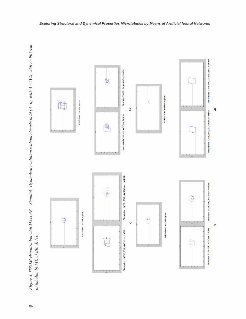
Figu
re 3
. ITS
OM
vis
ualiz
atio
n w
ith M
ATLA
B –
Sim
ulin
k. D
ynam
ical
evo
lutio
n w
ithou
t ele
ctri
c fie
ld (A
=0)
, with
A =
2V/s
, with
A=
90V/
cm.
a) tu
bulin
, b) M
T, c
) BB,
d) N
T.
87
Exploring Structural and Dynamical Properties Microtubules by Means of Artificial Neural Networks
RESULTS
In the conditions of zero field the tubulin shows a high occupancy value, and a rather consistent number of conflicts.
The stabilization of the neural network is achieved with the greatest difficulty with respect to all other examined structures, to highlight a lack of native dynamic organization in relationship to the other structures.
By applying a weak electric field the tubulin tends to restrict its configuration space, while maintaining similar rates of occupancy and conflict with respect to the absence of field.
With a 90 V/cm field the configuration space and the occupancy don’t change, but the number of conflicts is increased.
In absence of field the MT shows a much more restricted occupancy than tubulin, especially considering that its dimensions are much greater. The configuration space is well confined. With weak electric field the situation does not change, the MT appears spatially and structurally stable.
With a stronger electric field the occupancy does not change, while decreasing the number of conflicts.
The low occupancy values and the absence of conflicts in all configurations of BB and NT is due to the low number of their components if compared to the size of the network and to their extremely regular structure.
By applying a weak electric field to the BB, occupancy tends to decline, the BB tends to stabi-lize in a range of values. The spatial configuration tends to shrink. But as the electric field grows, the occupancy tends to return to the same levels as in the absence of field.
Although the NT structure is bigger than that of a BB, the occupancy is low and similar to the BB one, symbolizing the strong stability of the structure.
With weak electric field the situation does not change, even though there is a spatial displace-ment of the structure.
With higher field the structure tends to go back to the positions obtained without field, although in a more distributed way, as occupancy tends to be more distributed.
CONCLUSION AND FUTURE TRENDS
This research presents a novel use of Artificial Neural Networks in the evaluation of the dynami-cal organization of MTs versus tubulin and CNTs. After performing a MD simulation, we compared it with the evolution of two different models of self-organized neural networks.
The results obtained by SONNIA reflect the same behaviour observed during the Ascalaph dynamical simulation. In fact during the dynamical simulations we observed that both BBs and CNTs move with a dynamic axial motion, which becomes a real pulse in the presence of electric field.
The NT, which in the dynamic evolution at zero field tends to move off its initial position, with the influence of the electric field tends to return to the starting position and to stabilize.
The behavior of the neural network reflects this trend, which shows the extreme regularity of these nanostructures and an interesting already known behavior of NT in the presence of electric field.
The tubulin, despite its symmetric structure, seems to have different internal forces that tend to resist a dynamic stabilization. However, in the presence of electric field, although it tends to squash, it does not show any particular reaction.
The dynamic simulation confirms the lack of specific characterization. The neural simulation shows final graphs that clearly indicate a MTs dynamic organization much stronger than the tubulin one, that is not altered by the presence of electric field even in its spatial configuration.
However, the significant reduction of conflicts indicates a dramatic increase in the spatial orga-nization. On the other hand, the graphs obtained by the ITSOM network confirm the SONNIA
88
Exploring Structural and Dynamical Properties Microtubules by Means of Artificial Neural Networks
neural network analysis and the dynamic simula-tions. The attractors generated by BB both in the absence of field and with low electric field are extremely cyclical and regular, even though with higher field it tends to present a regular compact-ness, and to broaden its values, as described by the SONNIA output.
The attractor regularity is clearly present even in the CNTs, and the electric field tends to increase both spatial range and regularity. The tubulin, which is initially well-structured (although with a much more complex pattern of NT and BB), maintains a structured shape even in presence of electric field, although with an increase of disor-der. The MTs, however, despite their structural complexity, show a strong dynamic stability, which the electric field, after an initial transient, improves significantly. The field increase further stabilizes the structural dynamics and the spatial configuration of MTs.
It is worth noting that all three methods con-verge in emphasizing the dynamic stability of these four structures, but show that only CNTs and MTs exhibit a significant behavior in pres-ence of electric field, in the direction of a stronger structural and spatial organization.
These results confirm those already obtained in the cited previous experiments on real samples of tubulin and MTs in conditions of resonance and birefringence.
For this reason, the research on these interest-ing structures will continue with further studies.
However, the use of simulation methods can help to motivate at a microscopic level the ex-perimental evidences and justify the agreement with theoretical assumptions.
These positive results encourage us to continue also our experimental research. In particular we will carry out in the future a replication of the already performed tests on MTs and tubulins interacting with different ligands.
The experimental results will be coupled with the MD simulation of the protein folding binding different ligands, to study the emerging
conformational differences. These studies would support hypotheses on the origin of the different biophysical behaviour in relationship with con-formational changes.
ACKNOWLEDGMENT
PRIN 2009 project on tubulins is acknowledged. We thank D. Rossetti, M. Eng., and Dr. S. Man-ziana, Università degli Studi di Milano, for their valuable work.
REFERENCES
Alfinito, E., Viglione, R., & Vitiello, G. (2001). The decoherence criterion. Retrieved from http://arxiv.org/PS_cache/quant-ph/pdf/0007/0007020v2.pdf
Amos, L. A., & Amos, W. B. (1991). Molecules of the cytoskeleton. London, UK: MacMillan Press.
Amos, L. A., & Schlieper, D. (2005). Microtubules and MAPs. Advances in Chemistry, 71, 257.
Ascalaph. (n.d.). Retrieved from http://www.agilemolecule.com/Products.html
Binder, L. I., & Rosenbaum, J. L. (1978). The in vitro assembly of flagellar outer doublet tubulin. The Journal of Cell Biology, 79, 500–515.
Burton, P. R., & Himes, R. H. (1978). Electron microscope studies of pH effects on assembly of tubulin free of associated proteins. The Journal of Cell Biology, 77(1), 120–133.
Chrétien, D. (2000). Microtubules switch occa-sionally into unfavorable configuration during elongation. Journal of Molecular Biology, 298, 663–676.
89
Exploring Structural and Dynamical Properties Microtubules by Means of Artificial Neural Networks
Chrétien, D., Metoz, F., Verde, F., Karsenti, E., & Wade, R. H. (1992). Lattice defects in mi-crotubules: Protofilament numbers vary within individual microtubules. The Journal of Cell Biology, 117(5), 1031–1040.
Chrétien, D., & Wade, R. H. (1991). New data on the microtubule surface lattice. Biology of the Cell, 71(1-2), 161–174.
Del Giudice, E., Doglia, M., & Milani, M. (1982). Self-focusing of Fröhlich waves and cytoskeleton dynamics. Physical Review Letters, 90A, 104–106.
Del Giudice, E., Doglia, S., Milani, M., & Vitiello, G. (1983). Spontaneous symmetry breakdown and boson condensation in biology. Physical Review Letters, 95A, 508–510.
Faber, J., Portugal, R., & Rosa, L. P. (2006). In-formation processing in brain microtubules. Bio Systems, 83(1), 1–9.
Freeman, W. J., & Vitiello, G. (2006). Nonlinear brain dynamics as macroscopic manifestation of underlying many-body field dynamics. Physics of Life Reviews, 3(2), 93–118.
Fukushige, T., Siddiqui, Z. K., Chou, M., Culotti, J. G., Gogonea, C. B., Siddiqui, S. S., & Hamelin, M. (1999). MEC-12, an α-tubulin required for touch sensitivity in C. elegans. Journal of Cell Science, 112, 395–403.
Hagan, S., Hameroff, S. R., & Tuszynski, J. A. (2002). Quantum computation in brain micro-tubules: Decoherence and biological feasibility. Physical Review E: Statistical, Nonlinear, and Soft Matter Physics, 65, 061901–061911.
Hameroff, S. R. (2007a). The brain is both neu-rocomputer and quantum computer. Cognitive Science, 31, 1035–1045.
Hameroff, S. R. (2007b). Orchestrated reduction of quantum coherence in brain microtubules. NeuroQuantology, 5(1), 1–8.
Hameroff, S. R., & Penrose, R. (1996a). Or-chestrated reduction of quantum coherence in brain microtubules: A model for consciousness. Mathematics and Computers in Simulation, 40, 453–480.
Hameroff, S. R., & Penrose, R. (1996b). Conscious events as orchestrated space-time selection. Jour-nal of Consciousness Studies, 3, 36–53.
Huang, X.-R., & Knighton, R. W. (2005). Microtu-bules contribute to the birefringence of the retinal nerve fiber layer. Investigative Ophthalmology & Visual Science, 46(12), 4588–4593.
Hyams, J. S., & Lloyd, C. W. (Eds.). (1994). Mi-crotubules. New York, NY: Wiley-Liss.
Jibu, M., Hagan, S., Hameroff, S. R., Pribram, K. H., & Yasue, K. (1994). Quantum optical coher-ence in cytoskeletal microtubules: Implications for brain function. Bio Systems, 32, 195–209.
Kohonen, T. (1990). The self-organizing map. Proceedings of the IEEE, 78, 1464–1480.
Krauhs, E., Little, M., Kempf, T., Hofer-Warbinek, R., Ade, W., & Postingl, H. (1981). Complete amino acid sequence of β-tubulin from porcine brain. Proceedings of the National Academy of Sciences of the United States of America, 78, 4156–4160.
Kroto, H. W., Heath, J. R., O’Brien, S. C., Curl, R. F., & Smalley, R. E. (1985). C60: Buckminster-fullerene. Nature, 318, 162–163.
Linck, R. W., & Langevin, G. L. (1981). Reas-sembly of flagellar B (αβ) tubulin into singlet microtubules: Consequences for cytoplasmic microtubule structure and assembly. The Journal of Cell Biology, 89, 323–337.
Lowe, J., & Amos, L. A. (1998). Crystal structure of the bacterial cell-division protein FtsZ. Nature, 391, 203–206.
90
Exploring Structural and Dynamical Properties Microtubules by Means of Artificial Neural Networks
Lowe, J., Li, H., Downing, K. H., & Nogales, E. (1998). Refined structure of αβ-Tubulin at 3.5 A° resolution. Journal of Molecular Biology, 313, 1045–1057.
Mandelkow, E. M., Mandelkow, E., & Milligan, R. A. (1991). Microtubules dynamics and mi-crotubules caps: A time-resolved cryoelectron microscopy study. The Journal of Cell Biology, 114, 977–991.
Mavromatos, N. (2000). Cell microtubules as cavities: Quantum coherence and energy trans-fer? Retrieved from http://arxiv.org/pdf/quant-ph/0009089
Mavromatos, N., Mershin, A., & Nanopoulos, D. V. (2002). QED-cavity model of microtubules implies dissipationless energy transfer and bio-logical quantum teleportation. Retrieved from http://arxiv.org/pdf/quant-ph/0204021
Michette, A. G., Mavromatos, N., Powell, K., Holwill, M., & Pfauntsch, S. J. (2004). Nano-tubes and microtubules as quantum information carriers. Proceedings of the Society for Photo-Instrumentation Engineers, 522, 5581.
NANO-D. (n.d.). Retrieved from http://nano-d.inrialpes.fr/
Nanopoulos, D. V. (1995).Theory of brain func-tion, quantum mechanics and superstrings. Re-trieved from http://arxiv.org/abs/hep-ph/9505374
Nanopoulos, D. V., & Mavromatos, N. (1996). A non-critical string (Liouville) approach to brain microtubules: State vector reduction, memory coding and capacity. Retrieved from http://arxiv.org/abs/quant-ph/9512021
Nogales, E. (1998). Structure of the αβ-tubulin dimer by electron crystallography. Letters to Nature, 391, 192–203.
Nogales, E., Whittaker, M., Milligan, R. A., & Downing, K. H. (1999). High-resolution model of the microtubule. Cell, 96, 79–88.
Oldenbourg, R., Salmon, E. D., & Tran, P. T. (1998). Birefringence of single and bundled mi-crotubules. Biophysical Journal, 74, 645–654.
Pampaloni, F., & Florin, E. L. (2008). Microtu-bule architecture: inspiration for novel carbon nanotube-based biomimetic materials. Trends in Biotechnology, 26(6), 302–310.
Pessa, E. (2007). Phase transition in biological matter. In Licata, I., & Sakaji, A. (Eds.), Physics of emergence and organization (pp. 165–228). Singapore: World Scientific.
Pessa, E., & Vitiello, G. (2004). Quantum noise induced entanglement and chaos in the dissipative quantum model of brain. International Journal of Modern Physics B, 18(6), 841–858.
Pierson, G. B., Burton, P. R., & Himes, R. H. (1978). Alterations in number of protofilameCNTs in microtubules assembled in vitro. The Journal of Cell Biology, 76, 223–228.
Pizzi, R., Inama, G., Durin, O., & Pedrinazzi, C. (2007). Non.invasive assessment of risk for severe tachyarrhythmias by means of non-linear analysis techniques. Chaos and Complexity Letters, 3(3).
Pizzi, R., Strini, G., Fiorentini, S., Pappalardo, V., & Pregnolato, M. (in press). Evidences of new biophysical properties of microtubules. [in press]. NanoBiotechnology.
Postingl, H., Krauhs, E., Little, M., & Kempf, T. (1981). Complete amino acid sequence of α-tubulin from porcine brain. Proceedings of the National Academy of Sciences of the United States of America, 78, 2757–2761.
Protein Data Bank. (n.d.). Retrieved from http://www.rcsb.org/pdb/home/home.do
Ravelli, R., Gigant, B., Curmi, P. A., Jourdain, I., Lachkar, S., Sobel, A., & Knossow, M. (2004). Insight into tubulin regulation from a complex with colchicine and a stathmin-like domain. Letters to Nature, 428, 198–202.
91
Exploring Structural and Dynamical Properties Microtubules by Means of Artificial Neural Networks
Ritter, H., & Schulten, H. (1988). Convergence properties of Kohonen’s topology conserving maps: Fluctuations, stability, and dimension selec-tion. Biological Cybernetics, 60, 59–71.
Roche, S., Akkermans, E., Chauvet, O., Hekking, F., Issi, J.-P., & Martel, R. … Poncharal, P. (2006). Transport properties. understanding carbon nanotubes. In A. Loiseau, P. Launois, P. Petit, S. Roche, & J.-P. Salvetat (Eds.), Lecture Notes in Computer Science, 677, 335–437.
Ruelle, D. (1981). Small random perturbations of dynamical systems and the definition of attrac-tors. Communications in Mathematical Physics, 82, 137–151.
Sapmaz, S., Meyer, C., Beliczynski, P., Jarillo-Herrero, P., & Knowenhoven, L. P. (2006). Excited state spectroscopy in carbon nanotube double quantum dots. Nano Letters, 6(7), 1350–1355.
Savage, C., Hamelin, M., Culotti, J. G., Coulson, A., Albertson, D. G., & Chalfie, M. (1989). MEC-7 is a β-tubulin gene required for the production of 15-protofilament microtubules in Caenorhabditis elegans. Genes & Development, 3, 870–881.
SONNIA. (n.d.). Retrieved from http://www.molecular-networks.com/
Tegmark, M. (2000a). The importance of quantum decoherence in brain processes. Physical Review E: Statistical Physics, Plasmas, Fluids, and Re-lated Interdisciplinary Topics, 61(4), 4194–4206.
Tegmark, M. (2000b). Why the brain is probably not a quantum computer. Information Science, 128(3-4), 155–179.
Tuszynski, J., Hameroff, S. R., Satarić, M. V., Trpišová, B., & Nip, M. L. A. (1995). Ferroelectric behavior in microtubule dipole lattices: Implica-tions for information processing, signaling and assembly/disassembly. Journal of Theoretical Biology, 174, 371–380.
Tuszynski, J. A., Brown, J. A., Crawford, E., & Carpenter, E. J. (2005). Molecular dynamics simulations of tubulin structure and calculations of electrostatic properties of microtubules. Mathe-matical and Computer Modelling, 41, 1055–1070.
Tuszynski, J. A., Brown, J. A., & Hawrylak, P. (1998). Dielectric polarization, electrical con-duction, information processing and quantum computation in microtubules: Are they plausible? Philosophical Transactions of the The Royal So-ciety of London A, 356(1743), 1897–926.
Tuszynski, J. A., Trpišová, B., Sept, D., & Satarić, M. V. (1997). The enigma of microtubules and their self-organization behavior in the cytoskeleton. Bio Systems, 42, 153–175.
Vitiello, G. (1995). Dissipation and memory ca-pacity in the quantum brain model. International Journal of Modern Physics B, 9(8), 973–989.
Wang, Y., Kempa, K., Kimball, B., Carlson, J. B., Benham, G., & Li, W. Z. (2004). Receiving and transmitting light-like radio waves: Antenna effect in arrays of aligned carbon nanotubes. Applied Physics Letters, 85(13), 2607–2609.
Woolf, N. J., & Hameroff, S. R. (2001). A quan-tum approach to visual consciousness. Trends in Cognitive Sciences, 5(11), 472–478.