visual field organization and retinal resolution in the beluga whale delphinapterus leucas (pallas)
TRANSCRIPT

0012-4966/01/1112- $25.00 © 2001 MAIK “Nauka /Interperiodica”0555
Doklady Biological Sciences, Vol. 381, 2001, pp. 555–558. Translated from Doklady Akademii Nauk, Vol. 381, No. 5, 2001, pp. 705–708.Original Russian Text Copyright © 2001 by Mass.
Comparative studies on the organization of visualfields in various mammals make it possible to under-stand specific characteristics of their orientation andbehavior. The organization of visual fields directlyrelates to the retinal topography and is usuallydescribed in terms of the size, shape, and location of theareas with the highest resolving power. The location ofsuch areas is correlated with the position of eyes,behavioral traits, and habitat properties [3].
We previously studied some cetaceans and foundthat they can serve as an unusual example of the visualfield organization and of the roles that specific areas ofthe visual fields play in orientation. Retinal topographymapping showed that, unlike the retina of terrestrialmammals, which contains only one area of high gan-glion cell density (known as the fovea, or area centra-lis), the retina of whales, whether toothed (Odontoceti)or baleen (Mysticeti), contains two such areas, one inthe temporal part and the other in the nasal part [1]. Pre-sumably, they are differently involved in the aerial andunderwater vision. In the underwater vision, both areasare used, whereas the aerial vision relies mostly on oneof the two. The use of both areas ensures a wide field ofmonocular vision under water, which may compensatefor the limited range of head movements typical ofmost dolphins and whales.
A question arises as to whether the presence of twoareas of high ganglion cell density in the cetacean ret-ina is a systematic trait or it is due to other factors. Inthis context, an interesting object of study is the belugawhale
Delphinapterus leucas
(Pallas), which belongsto the family Monodontidae and inhabits Arctic oceansand seas. Compared with dolphins or mysticetes, thebeluga whale is less limited in the range of head move-ments. In addition, this species is characterized by a lat-eroventral (rather than ventral, as in other cetaceans)position the eyes [9].
The results reported in this study were obtained byretinal topography mapping, which is useful in thecases when behavioral techniques for assessing theresolving power of animals are difficult or impossibleto implement (e.g., when large rare species are con-cerned). For mapping ganglion cells in the retina, theyare selectively stained and examined by light micros-copy. This method allows a researcher to determine theganglion cell distribution over the entire surface of theretina and to assess the cell density in the high-densityareas variously located in the retina. Knowing the gan-glion cell counts in the high-density areas makes it pos-sible to estimate the retinal resolution. This estimate, inturn, allows the visual acuity to be calculated. The goalof this study was to determine the visual acuity of thebeluga whale
Delphinapterus leucas
using retinalwhole mounts.
Observation of beluga whales in captivity leads tothe conclusion that they have good visual capabilities[5, 9]. However, their retinal resolution was estimatedin only one study conducted on a single retinal wholemount [7]. Therefore, those data need to be expandedand confirmed.
The material for investigation was isolated fromfour animals (body length, 225–420 cm) caught in theOkhotsk Sea. A license for their capture was obtainedfrom the Glavrybvod.
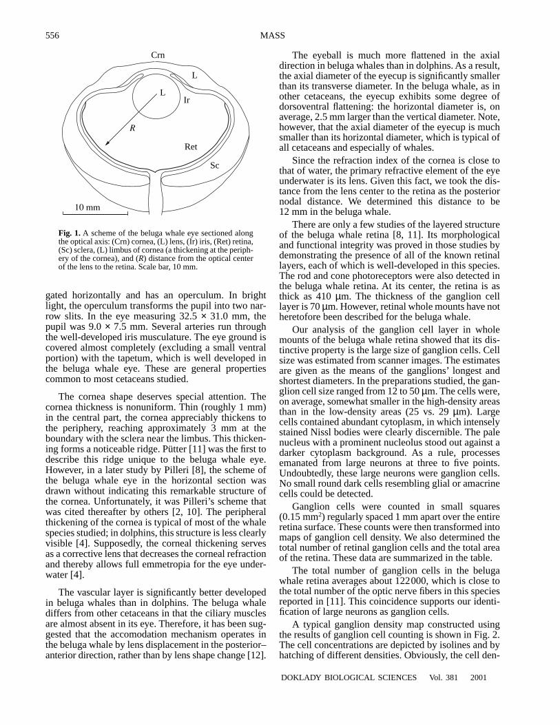
One eye was used to study its macromorphologicalcharacteristics and to determine the main geometricoptical parameters, including the posterior nodal dis-tance, which enters the formula for calculating the ret-inal resolution. The measurements were taken fromscanner images of the sagittal section of the eye fixed in10% formalin and frozen. The eye macromorphologyin the beluga whale inferred from these measurementsis shown schematically in Fig. 1.
Many features of the beluga whale eye are commonto most of the marine cetacean species studied; how-ever, there are also some distinctions.
As can be seen from Fig. 1, the sclera is thick in thebeluga whale. The anterior chamber is shallow. Thelens is round, with a slight degree of flattening in theanterior–posterior direction. Its frontal and axial diam-eters are 8 and 7 mm, respectively. The pupil is elon-
GENERAL BIOLOGY
Visual Field Organization and Retinal Resolution in the Beluga Whale
Delphinapterus leucas
(Pallas)
A. M. Mass
Presented by Academician D.S. Pavlov August 28, 2001
Received September 4, 2001
Severtsov Institute of Ecology and Evolution, Russian Academy of Sciences, Leninskii pr. 33, Moscow, 117071 Russia
556
DOKLADY BIOLOGICAL SCIENCES
Vol. 381
2001
MASS
gated horizontally and has an operculum. In brightlight, the operculum transforms the pupil into two nar-row slits. In the eye measuring 32.5
×
31.0 mm, thepupil was 9.0
×
7.5 mm. Several arteries run throughthe well-developed iris musculature. The eye ground iscovered almost completely (excluding a small ventralportion) with the tapetum, which is well developed inthe beluga whale eye. These are general propertiescommon to most cetaceans studied.
The cornea shape deserves special attention. Thecornea thickness is nonuniform. Thin (roughly 1 mm)in the central part, the cornea appreciably thickens tothe periphery, reaching approximately 3 mm at theboundary with the sclera near the limbus. This thicken-ing forms a noticeable ridge. Pütter [11] was the first todescribe this ridge unique to the beluga whale eye.However, in a later study by Pilleri [8], the scheme ofthe beluga whale eye in the horizontal section wasdrawn without indicating this remarkable structure ofthe cornea. Unfortunately, it was Pilleri’s scheme thatwas cited thereafter by others [2, 10]. The peripheralthickening of the cornea is typical of most of the whalespecies studied; in dolphins, this structure is less clearlyvisible [4]. Supposedly, the corneal thickening servesas a corrective lens that decreases the corneal refractionand thereby allows full emmetropia for the eye under-water [4].
The vascular layer is significantly better developedin beluga whales than in dolphins. The beluga whalediffers from other cetaceans in that the ciliary musclesare almost absent in its eye. Therefore, it has been sug-gested that the accomodation mechanism operates inthe beluga whale by lens displacement in the posterior–anterior direction, rather than by lens shape change [12].
The eyeball is much more flattened in the axialdirection in beluga whales than in dolphins. As a result,the axial diameter of the eyecup is significantly smallerthan its transverse diameter. In the beluga whale, as inother cetaceans, the eyecup exhibits some degree ofdorsoventral flattening: the horizontal diameter is, onaverage, 2.5 mm larger than the vertical diameter. Note,however, that the axial diameter of the eyecup is muchsmaller than its horizontal diameter, which is typical ofall cetaceans and especially of whales.
Since the refraction index of the cornea is close tothat of water, the primary refractive element of the eyeunderwater is its lens. Given this fact, we took the dis-tance from the lens center to the retina as the posteriornodal distance. We determined this distance to be12 mm in the beluga whale.
There are only a few studies of the layered structureof the beluga whale retina [8, 11]. Its morphologicaland functional integrity was proved in those studies bydemonstrating the presence of all of the known retinallayers, each of which is well-developed in this species.The rod and cone photoreceptors were also detected inthe beluga whale retina. At its center, the retina is asthick as 410
µ
m. The thickness of the ganglion celllayer is 70
µ
m. However, retinal whole mounts have notheretofore been described for the beluga whale.
Our analysis of the ganglion cell layer in wholemounts of the beluga whale retina showed that its dis-tinctive property is the large size of ganglion cells. Cellsize was estimated from scanner images. The estimatesare given as the means of the ganglions’ longest andshortest diameters. In the preparations studied, the gan-glion cell size ranged from 12 to 50
µ
m. The cells were,on average, somewhat smaller in the high-density areasthan in the low-density areas (25 vs. 29
µ
m). Largecells contained abundant cytoplasm, in which intenselystained Nissl bodies were clearly discernible. The palenucleus with a prominent nucleolus stood out against adarker cytoplasm background. As a rule, processesemanated from large neurons at three to five points.Undoubtedly, these large neurons were ganglion cells.No small round dark cells resembling glial or amacrinecells could be detected.
Ganglion cells were counted in small squares(0.15 mm
2
) regularly spaced 1 mm apart over the entireretina surface. These counts were then transformed intomaps of ganglion cell density. We also determined thetotal number of retinal ganglion cells and the total areaof the retina. These data are summarized in the table.
The total number of ganglion cells in the belugawhale retina averages about 122000, which is close tothe total number of the optic nerve fibers in this speciesreported in [11]. This coincidence supports our identi-fication of large neurons as ganglion cells.
A typical ganglion density map constructed usingthe results of ganglion cell counting is shown in Fig. 2.The cell concentrations are depicted by isolines and byhatching of different densities. Obviously, the cell den-
L
Crn
L
Sc
Ret
R
10 mm
Ir
Fig. 1.
A scheme of the beluga whale eye sectioned alongthe optical axis: (Crn) cornea, (L) lens, (Ir) iris, (Ret) retina,(Sc) sclera, (L) limbus of cornea (a thickening at the periph-ery of the cornea), and (
R
) distance from the optical centerof the lens to the retina. Scale bar, 10 mm.

DOKLADY BIOLOGICAL SCIENCES
Vol. 381
2001
VISUAL FIELD ORGANIZATION AND RETINAL RESOLUTION 557
sity in the ganglion cell layer is relatively low in thebeluga whale; notwithstanding, sparse and dense areascan be distinguished, among which two high-densityareas are the most conspicuous. One of them is in thenasal segment of the retina, close to its horizontal diam-eter; and the other is in the temporal retina. Both areasappear as vertically elongated spots. In the retinalwhole mount shown, the peak cell densities observed inthe temporal and nasal segments were 479 and352 cells/mm
2
, respectively. As can be seen from thetable, the cell density averaged over all whole mountsstudied was higher in the temporal segment than in thenasal one (400 vs. about 300 cells/mm
2
). Being thehighest at the center of each spot, the cell abundancerapidly declined to its periphery and remained lowthroughout the rest of the retina. The ventral segment ofthe retina was especially sparse.
The location of the densely hatched zones (high-density areas) draws particular attention. They are far(12 to 16 mm) apart from the center of the retina andrelatively close to its periphery. In all of the mountsstudied, the high-density areas were similar in size,shape, and location. Knowing the ganglion cell densityin the high-density areas, we can assess the angular dis-tance between ganglion cells from the following for-mula:
a
= 180°/ ,
where
d
is the cell density (cells/mm
2
) and
r
is the pos-terior nodal distance (mm). Another commonly usedestimate of the retinal resolution (in cycles per degree)reads
f
= 1/2
a.
For
d
= 400 cells/mm
2
in the temporal high-densityarea,
d
= 300 cells/mm
2
in the nasal area, and
r
=12 mm, we obtain
a
= 14.4
′
(
f
= 2.2 cycles per degree)and
16.9
′
(
f
= 1.8 cycles per degree) for the temporaland nasal high-density areas, respectively. These esti-mates of the retinal resolution of the beluga whale eyehold for the underwater vision, when the primaryrefractive structure of the eye is its lens and, hence, thenodal point is the lens center. In the air, refraction shiftsthe nodal point to the surface of the cornea. Its positioncannot be determined without knowing the corneal cur-vature, which has not been measured accurately. How-
πr d
ever, given the water-to-air refraction index ratio of1.33, we can assume, as a first approximation, that theretinal image size underwater is 1.33 times that in air. Ifso, we have in the air
a
= 19.2
′
(
f
= 1.6 cycles perdegree) for the temporal high-density area and
a
= 22.5
′
(
f
= 1.3 cycles per degree) for the nasal high-densityarea.
Earlier, the same method was used to assess thevisual acuity in
Tursiops truncatus, Delphinus delphis,Pseudorca crassidens
(Delphinidae),
Phocoena phoc-oena, Phocoenoides dalli
(Phocoenidae), and in somemysticetes (
Eschrichtius gibbosus
and
Balaenopteraacutorostrata
[1, 7]. In those studies, their visual acuity
Table
Whole mount number
Temporal retina Nasal retina Total number
of cellscells per mm
2
resolution,arc minutes
resolution, cycles/degree cells per mm
2
resolution,arc minutes
resolution, cycles/degree
1 315 15.2 2.1 225 18.0 1.7 124600
2 479 12.6 2.4 352 15.3 2.0 138800
3 414 15.4 2.0 320 17.4 1.6 103700
Mean 403 14.4 2.2 299 16.9 1.8 122367
D
NT
V
10 mm
0 75 150 225 300 375 450
Fig. 2.
A ganglion density map of a whole mount of the bel-uga whale retina. Cell density (cells/mm
2
) corresponds tothe hatching density. The hatching density scale is given atthe bottom. The dorsal, ventral, nasal, and temporal poles ofthe retina are indicated by letters
D, V, N
, and
T,
respectively.Scale bar, 10 mm.

558
DOKLADY BIOLOGICAL SCIENCES
Vol. 381
2001
MASS
was estimated at
7–11
′
. Our data suggest that the bel-uga whale has a slightly poorer visual acuity. Our esti-mate does not coincide with that reported in [7] (
11.8
′
).Note, however, that the previously reported estimatewas obtained for a single retinal preparation.
Thus, two areas of high ganglion cell density wererevealed in the beluga whale retina. Its topography issimilar to that described in many other cetaceans [1].These data provide further evidence for our suggestionput forward previously that the presence of two high-density areas is typical of the retinal topography in allthe cetaceans studied, whatever their family. The onlyexclusion is the Amazonian dolphin, whose retina con-tains only one high-density area mapped to its ventralsegment [6].
The retinal topography described may relate to thepupil shape. The pupil is elongated horizontally and hasan operculum dividing it into two slits in bright light.The operculum covers the pupil and does not let brightlight through in the axial direction; therefore, the lightis projected to the two high-density areas in the tempo-ral and nasal segments of the retina. In these two areas,the resolving power is higher than in any other area ofthe retina.
Thus, retinal topography mapping provided anopportunity of revealing the distinctive features of thevisual field organization and of assessing the visualacuity in the beluga whale
D. leucas
, a poorly knowncetacean species.
ACKNOWLEDGMENTS
This work was supported by the Russian Foundationfor Basic Research (project no. 01-04-48071).
REFERENCES
1. Mass, A.M.,
Sensor. Sist.
, 1997, vol. 11, pp. 256–291.2. Yablokov, A.V., Bel’kovich, V.M., and Borisov, V.I.,
Kityi del’finy
(Whales and Dolphins), Moscow: Nauka,1972.
3. Hughes, A.,
Handbook of Sensory Physiology: TheVisual System in Vertebrates
, Berlin: Springer, 1977,vol.
7/5, pp. 613–756.4. Krüger, R.H.H. and Kirschfeld, K.,
Aquat. Mammals,
1994, vol. 20, pp. 99–107.5. Marino, L. and Stowe, C.M.,
Aquat. Mammals
, 1997,vol. 23, no. 2, pp. 101–104.
6. Mass, A.M. and Supin, A.Y.,
Aquat. Mammals
, 1989,vol. 15, pp. 49–56.
7. Murayama, T. and Somiya, H.,
Fisheries Sci.
, 1998,vol.
64, no. 1, pp. 27–30.8. Pilleri, G.,
Hvalradets Skr.
, 1964, vol. 47, pp. 1–16.9. Pilleri, G.,
Investig. Cetacea
, 1982, vol. 13, pp. 167–176.10. Von Neuhaus, W.,
Z. Saugetierkunde
, 1986, vol. 51,no.
5, pp. 266–273.11. Pütter, A.,
Zool. Jahrb. Abt. Anat. Ontog. Thiere
, 1903,vol. 17, pp. 99–402.
12. West, J.A., Sivak, J.G., Murphy, C.J., and Kovacs, K.M.,
Can. J. Zool.
, 1991, vol. 69, pp. 2594–2607.