visian icl (implantable collamer lens) for nearsightedness · page 3 of 29 1.0 introduction the...
TRANSCRIPT
Visian ICL™
(Implantable Collamer Lens)
For Nearsightedness
Facts You Need To Know About STAAR Surgical’s Visian ICL
SURGERY
PATIENT INFORMATION BOOKLET
For Nearsightedness (Myopia) between –3 to –20 Diopters
with 2.5 Diopters or less of Astigmatism
Please read this entire booklet. Discuss its contents with your eye doctor so that you
have all of your questions answered to your satisfaction. Ask any questions you may
have before you agree to this surgery.
Distributed by: Manufactured by:
STAAR Surgical Company STAAR Surgical, AG
1911 Walker Avenue Hauptstrasse 104
Monrovia, CA 91016 USA CH-2560 Nidau, Switzerland
Tel: (800) 352-7842 Tel: + (41) 32 332 8888
FAX: (800) 952-4923 FAX: + (41) 32 332 8899
Copyright 2006 by STAAR Surgical Company
This booklet may be reproduced only by a treating physician, for use with patients considering
Visian ICL Surgery. All other rights are reserved.

Page 1 of 29
This page left blank intentionally.
Page 2 of 29
TABLE OF CONTENTS
1.0 Introduction ................................................................................................................... 3
2.0 How Does VISIAN ICL Correct NEARSIGHTEDNESS? ......................................... 3
3.0 What Are the Benefits of THE VISIAN ICL for NEARSIGHTEDNESS? ................. 6
4.0 What Are the Risks of THE VISIAN ICL for NEARSIGHTEDNESS? ...................... 9
5.0 ALTERNATIVE TREATMENTS ............................................................................. 10
6.0 Contraindications ........................................................................................................ 11
7.0 Warnings ..................................................................................................................... 11
8.0 Precautions .................................................................................................................. 12
9.0 Are You a Good Candidate for VISIAN ICL for NEARSIGHTEDNESS Surgery? . 13
10.0 What Should You Expect During VISIAN ICL Surgery? ........................................ 14
11.0 Questions To Ask Your Doctor ................................................................................ 17
12.0 Self-Test .................................................................................................................... 18
13.0 Summary of Important Information .......................................................................... 19
14.0 GLOSSARY ............................................................................................................. 20
15.0 Patient Assistance Information ................................................................................. 28
Page 3 of 29
1.0 INTRODUCTION
The purpose of this booklet is to provide you with information on the Visian ICL
surgery for nearsightedness (myopia). Please read this entire booklet carefully. See
the “Glossary” (Section 14) for an explanation of words shown in italics. Discuss
your questions with a doctor trained in Visian ICL surgery. You need to understand
the benefits and risks of this surgery before making a decision to have this procedure.
You may have nearsightedness if you have trouble seeing objects clearly when they
are far away. Nearsightedness, which is also called myopia, is a type of condition
that causes blurred vision. Glasses, contact lenses or eye surgery can correct
nearsightedness and help you see distant objects more clearly.
Your eyeglass prescription is the usual way to tell how nearsighted you are. Your
doctor will use your eyeglass prescription with a thorough eye examination to
determine if you are a candidate for Visian ICL surgery. Discuss with your doctor
whether you are a good candidate for Visian ICL surgery.
Visian ICL surgery is permanent as long as the Visian ICL stays in your eye. The
Visian ICL can be removed at a future date. However, the residual effect of the
Visian ICL on your eye after it is removed is not known. If your physician removes
the Visian ICL, you will lose the benefit of your nearsightedness correction.
2.0 HOW DOES VISIAN ICL CORRECT NEARSIGHTEDNESS?
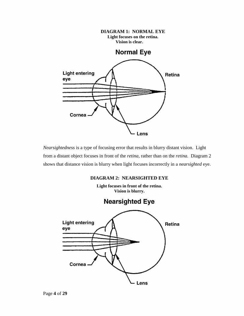
You see objects because your eye focuses light into images. Your eye works like a
camera. The camera lens focuses light to form clear images onto film. Both the
cornea and lens in the eye focus light onto the back surface of the eye, called the
retina. Diagram 1 shows that distant vision is clear when light focuses correctly.
Page 4 of 29
DIAGRAM 1: NORMAL EYE Light focuses on the retina.
Vision is clear.
Nearsightedness is a type of focusing error that results in blurry distant vision. Light
from a distant object focuses in front of the retina, rather than on the retina. Diagram 2
shows that distance vision is blurry when light focuses incorrectly in a nearsighted eye.
DIAGRAM 2: NEARSIGHTED EYE
Light focuses in front of the retina.
Vision is blurry.
Page 5 of 29
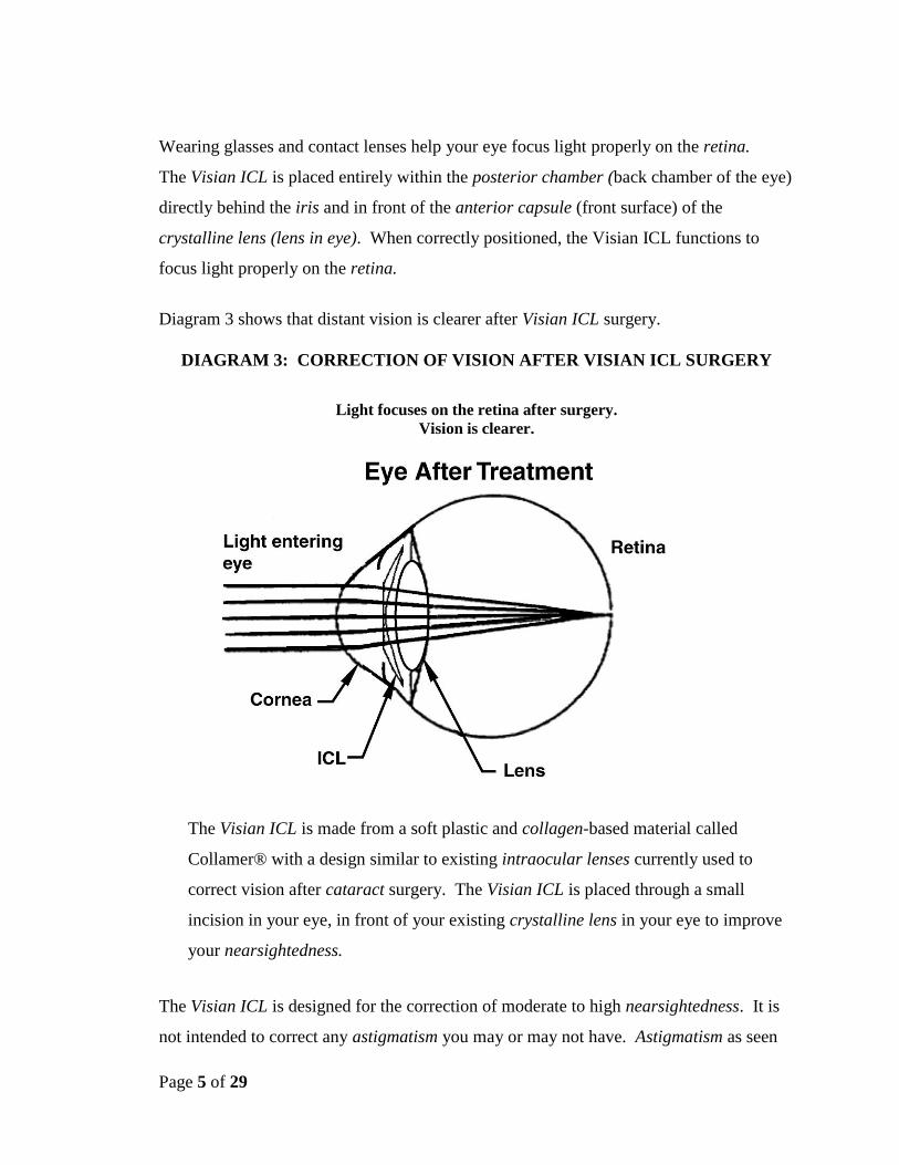
Wearing glasses and contact lenses help your eye focus light properly on the retina.
The Visian ICL is placed entirely within the posterior chamber (back chamber of the eye)
directly behind the iris and in front of the anterior capsule (front surface) of the
crystalline lens (lens in eye). When correctly positioned, the Visian ICL functions to
focus light properly on the retina.
Diagram 3 shows that distant vision is clearer after Visian ICL surgery.
DIAGRAM 3: CORRECTION OF VISION AFTER VISIAN ICL SURGERY
Light focuses on the retina after surgery.
Vision is clearer.
The Visian ICL is made from a soft plastic and collagen-based material called
Collamer® with a design similar to existing intraocular lenses currently used to
correct vision after cataract surgery. The Visian ICL is placed through a small
incision in your eye, in front of your existing crystalline lens in your eye to improve
your nearsightedness.
The Visian ICL is designed for the correction of moderate to high nearsightedness. It is
not intended to correct any astigmatism you may or may not have. Astigmatism as seen
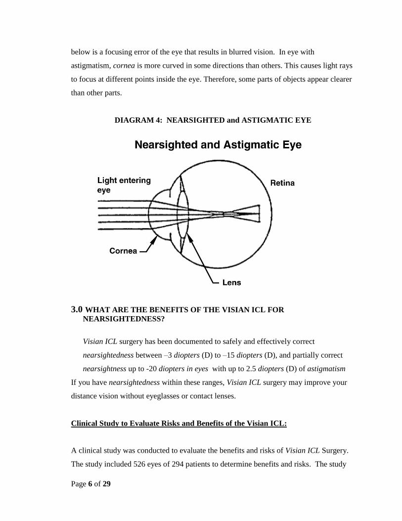
Page 6 of 29
below is a focusing error of the eye that results in blurred vision. In eye with
astigmatism, cornea is more curved in some directions than others. This causes light rays
to focus at different points inside the eye. Therefore, some parts of objects appear clearer
than other parts.
DIAGRAM 4: NEARSIGHTED and ASTIGMATIC EYE
3.0 WHAT ARE THE BENEFITS OF THE VISIAN ICL FOR
NEARSIGHTEDNESS?
Visian ICL surgery has been documented to safely and effectively correct
nearsightedness between –3 diopters (D) to –15 diopters (D), and partially correct
nearsightness up to -20 diopters in eyes with up to 2.5 diopters (D) of astigmatism
If you have nearsightedness within these ranges, Visian ICL surgery may improve your
distance vision without eyeglasses or contact lenses.
Clinical Study to Evaluate Risks and Benefits of the Visian ICL:
A clinical study was conducted to evaluate the benefits and risks of Visian ICL Surgery.
The study included 526 eyes of 294 patients to determine benefits and risks. The study
Page 7 of 29
results are discussed below and in “What are the Risks of the Visian ICL for
Nearsightedness?” (Section 4).
Description of Study Patient Group:
Most patients were Caucasian. Patients in the study ranged in age from 21 to 45
years of age and over half of the patients were female.
Nearsightedness before surgery ranged between –3.00D and –20.00D. The
average was –10.06D.
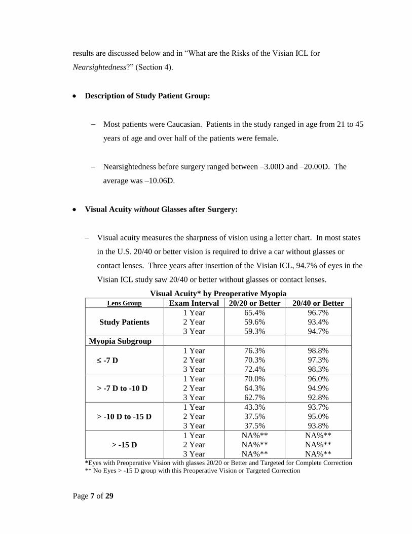
Visual Acuity without Glasses after Surgery:
Visual acuity measures the sharpness of vision using a letter chart. In most states
in the U.S. 20/40 or better vision is required to drive a car without glasses or
contact lenses. Three years after insertion of the Visian ICL, 94.7% of eyes in the
Visian ICL study saw 20/40 or better without glasses or contact lenses.
Visual Acuity* by Preoperative Myopia
Lens Group Exam Interval 20/20 or Better 20/40 or Better
Study Patients
1 Year
2 Year
3 Year
65.4%
59.6%
59.3%
96.7%
93.4%
94.7%
Myopia Subgroup
-7 D
1 Year
2 Year
3 Year
76.3%
70.3%
72.4%
98.8%
97.3%
98.3%
> -7 D to -10 D
1 Year
2 Year
3 Year
70.0%
64.3%
62.7%
96.0%
94.9%
92.8%
> -10 D to -15 D
1 Year
2 Year
3 Year
43.3%
37.5%
37.5%
93.7%
95.0%
93.8%
> -15 D
1 Year
2 Year
3 Year
NA%**
NA%**
NA%**
NA%**
NA%**
NA%** *Eyes with Preoperative Vision with glasses 20/20 or Better and Targeted for Complete Correction
** No Eyes > -15 D group with this Preoperative Vision or Targeted Correction
Page 8 of 29
In the clinical study of the Visian ICL, vision without glasses improved for all
eyes except in those eyes with the most extreme amount of nearsightedness that
the strongest Visian ICL could not completely correct and 1 case that developed a
retinal detachment where the uncorrected vision remained unchanged. Some
people still needed glasses or contact lenses after surgery to view distant objects .
Patient Satisfaction after Visian ICL Surgery:
Patients were asked to report their satisfaction with the Visian ICL procedure. Three years
after Visian ICL surgery, 92.1% of patients were very/extremely satisfied and 7.3% were
moderately/fairly satisfied with their vision. Only 0.6% of cases were unsatisfied.
Quality of Vision after Visian ICL Surgery:
Quality of vision reported by patients as very good/excellent improved from 55% before
the Visian ICL to 77% 3 years after the Visian ICL procedure. Patients reporting
poor/very poor vision dropped in half at 3 years (5.8%) compared to before the Visian
ICL (11.6%).
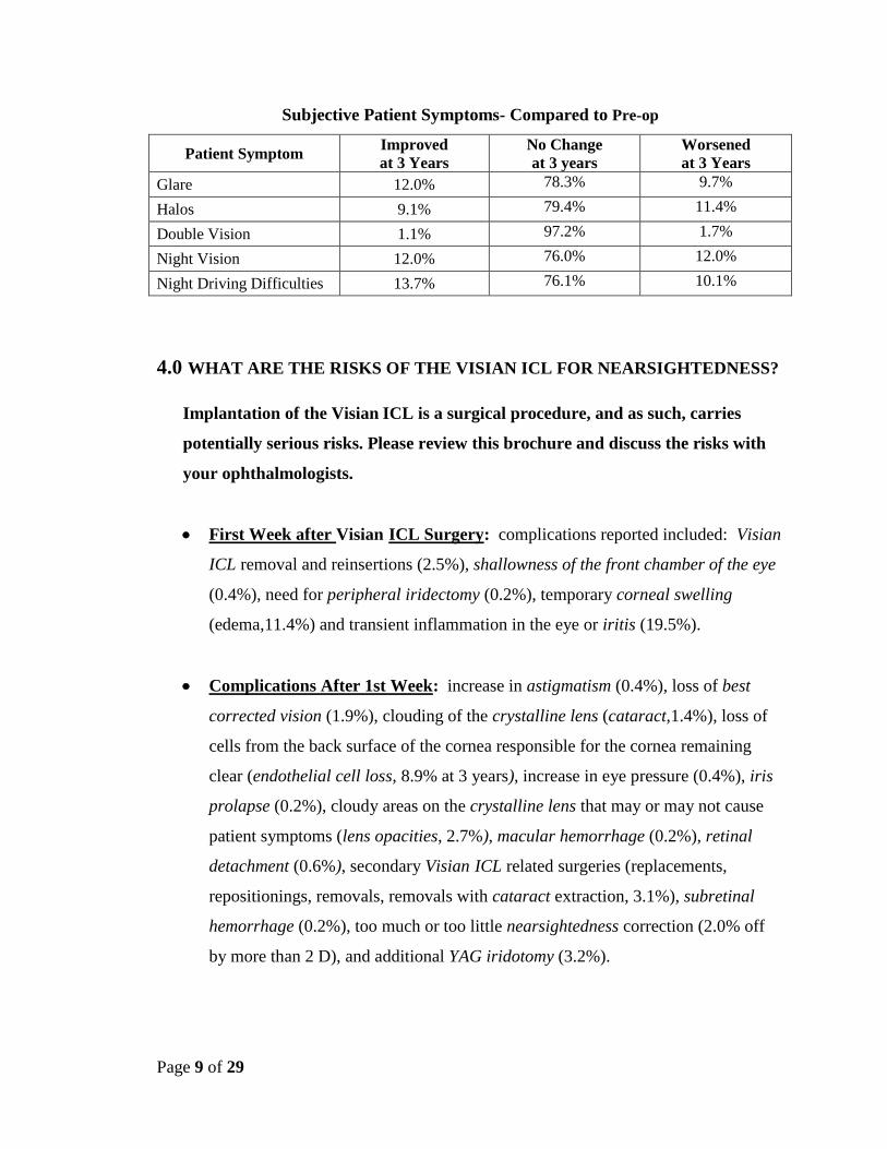
Patients were asked on a questionnaire to report on the following symptoms before and 3
years after the Visian ICL procedure. More patients rated the following symptoms absent
or mild at 3 years compared to before the Visian ICL: glare, night vision difficulties and
night driving difficulties. Halos and double vision percentages were similar before the
Visian ICL and at 3 years.
The higher the level of nearsightedness before the Visian ICL procedure, the more
frequent and more severe these symptoms were reported both before and after the Visian
ICL procedure.
Page 9 of 29
Subjective Patient Symptoms- Compared to Pre-op
Patient Symptom Improved
at 3 Years
No Change
at 3 years
Worsened
at 3 Years
Glare 12.0% 78.3% 9.7%
Halos 9.1% 79.4% 11.4%
Double Vision 1.1% 97.2% 1.7%
Night Vision 12.0% 76.0% 12.0%
Night Driving Difficulties 13.7% 76.1% 10.1%
4.0 WHAT ARE THE RISKS OF THE VISIAN ICL FOR NEARSIGHTEDNESS?
Implantation of the Visian ICL is a surgical procedure, and as such, carries
potentially serious risks. Please review this brochure and discuss the risks with
your ophthalmologists.
First Week after Visian ICL Surgery: complications reported included: Visian
ICL removal and reinsertions (2.5%), shallowness of the front chamber of the eye
(0.4%), need for peripheral iridectomy (0.2%), temporary corneal swelling
(edema,11.4%) and transient inflammation in the eye or iritis (19.5%).
Complications After 1st Week: increase in astigmatism (0.4%), loss of best
corrected vision (1.9%), clouding of the crystalline lens (cataract,1.4%), loss of
cells from the back surface of the cornea responsible for the cornea remaining
clear (endothelial cell loss, 8.9% at 3 years), increase in eye pressure (0.4%), iris
prolapse (0.2%), cloudy areas on the crystalline lens that may or may not cause
patient symptoms (lens opacities, 2.7%), macular hemorrhage (0.2%), retinal
detachment (0.6%), secondary Visian ICL related surgeries (replacements,
repositionings, removals, removals with cataract extraction, 3.1%), subretinal
hemorrhage (0.2%), too much or too little nearsightedness correction (2.0% off
by more than 2 D), and additional YAG iridotomy (3.2%).
Page 10 of 29
Only 2 eyes (0.5%) lost > 2 lines of best corrected vision (with glasses) compared
to 6.5% of eyes that gained > 2 lines of visual acuity with glasses. Of note, only
6.5% of eyes lost 1 line of best corrected visual acuity while 38.0% of eyes gained
1 line.
Potential Complications are not limited to those reported during the clinical study.
The following represent potential complications/adverse events reported in
conjunction with refractive surgery in general: conjunctival irritation, acute
corneal swelling, persistent corneal swelling, endophthalmitis (total eye
infection), significant glare and/or halos around lights, hyphema (blood in the
eye), hypopyon (pus in the eye), eye infection, Visian ICL dislocation, macular
edema, non-reactive pupil, pupillary block glaucoma, severe inflammation of the
eye, iritis, uveitis, vitreous loss and corneal transplant.
Overall, the higher the amount of nearsightedness before the Visian ICL, the higher
the incidence of complications/risks after Visian ICL surgery.
5.0 ALTERNATIVE TREATMENTS
The types of eye surgeries that are available to correct nearsightedness are Radial
Keratotomy (RK), Photorefractive Keratectomy (PRK), Laser Assisted in situ
Keratomileusis (LASIK) and Phakic Intraocular Lens surgery. The Implantable
Collamer Lens is a type of phakic intraocular lens. These surgeries may not meet
the vision requirements for some careers, such as military service.
Eye surgeries can be categorized by those that change the shape of the front
surface of your cornea, which is the clear layer at the front of your eye (including
RK, PRK and LASIK) and intraocular lens surgery that involves the insertion of a
lens within the eye.
Page 11 of 29
RK uses a scalpel to make fine cuts in the cornea.
PRK and LASIK use a laser to reshape the cornea. For LASIK, an instrument
called a microkeratome first cuts a thin flap of tissue from the front of your
cornea. This corneal flap is folded back and the laser removes tissue under the
flap to change the shape of the front surface of your eye (cornea). Then the flap
is put back in place for the eye to heal.
6.0 CONTRAINDICATIONS
You should NOT have Visian ICL surgery if you:
Have a narrow anterior chamber angle as determined by a special
examination by your eye doctor, or if your doctor determines that the shape of
your eye is not adequate to fit the Visian ICL (anterior chamber depth less
than 3 mm)
Are pregnant or nursing
Do not meet the minimum endothelial cell density for your age at the time of
implantation as determined by your eye doctor
7.0 WARNINGS
Two iridotomies (holes in the extreme outer edge of the colored portion of the
eye) must be performed 90º apart using a yttrium aluminum garnet (YAG) laser at
between 2 to 3 weeks before implantation of the Visian ICL.
The long-term effects on the corneal endothelium have not been established. You
should be aware of potential risk of corneal edema (swelling), possibly requiring
Page 12 of 29
corneal transplantation. Periodic checks of your endothelium are recommended
to monitor the long-term health of the cornea
The long-term rate of cataract formation (decrease clarity of your natural
crystalline lens) secondary to implantation / removal and/or replacement of the
Visian ICL are unknown.
The potential of the lens to alter the pressure in your eye and the long-term risks
of glaucoma, peripheral anterior synechiae and pigment dispersion are unknown.
8.0 PRECAUTIONS
1. Patients with higher amounts of nearsightedness had worse results with lower
effectiveness and higher risk of complications.
2. The effect of pupil size on visual symptoms is not known.
3. The relationship between the Visian ICL and future lens opacities and retinal
detachment is undetermined.
4. There currently is a lack of long-term data to assess cataract formation and
cataract progression following removal and/or replacement of the lens.
5. The effectiveness of ultraviolet absorbing lenses in reducing the incidence of
retinal disorders has not been established.
6. The safety and effectiveness of the Visian ICL for the correction of moderate to
high nearsightedness has NOT been established in patients:
with unstable or worsening nearsightedness
with history or clinical signs of iritis/uveitis
with diabetic retinopathy
Page 13 of 29
with glaucoma
with history of previous eye surgery
with serious (life-threatening) non-ophthalmic disease
with progressive sight-threatening disease other than nearsightedness
with a diagnosis of ocular hypertension(high eye pressure)
with insulin-dependent diabetes
with pseudoexfoliation
with pigment dispersion
with greater than -20 D of nearsightedness; greater than 2.5 D of astigmatism
9.0 ARE YOU A GOOD CANDIDATE FOR VISIAN ICL FOR
NEARSIGHTEDNESS SURGERY?
If you are considering Visian ICL surgery for nearsightedness you must:
be between the ages of 21 and 45
have between –3D and –20D of nearsightedness and no more than 2.5D of
astigmatism
understand that the Visian ICL is indicated for the correction of nearsightedness
between –3D and ≤ -15D and the reduction of nearsightedness between > -15D
and –20D
have an anterior chamber depth of 3.0 millimeters or greater
have a minimally acceptable endothelium cell density which will be determined
by your physician
have a refraction that has been stable for at least 1 year
Page 14 of 29
understand the risks and benefits of Visian ICL for nearsightedness surgery
compared to other available treatments for nearsightedness
be able to lie flat on your back
have no known allergies to any of the medications that your physician may
discuss will be used before, during and after your surgery
not be pregnant or nursing
understand that prior to implantation of the Visian ICL you will need to undergo
YAG iridotomy 2 to 3 weeks before Visian ICL surgery
be willing to sign an Informed Consent Form provided by your doctor.
10.0 WHAT SHOULD YOU EXPECT DURING VISIAN ICL SURGERY?
Before the Surgery
Before surgery, your doctor needs to determine your complete medical and eye history
and check the health of both your eyes. This exam will determine if your eyes are
healthy and if you are a good candidate for Visian ICL surgery. This examination will
include a measurement of the inner layer of your cornea (endothelium).
Tell your doctor if you take any medications, have any eye conditions, have undergone
previous eye surgery, have any medical conditions or have any allergies. Ask your
doctor if you should eat or drink right before the surgery. You should also arrange for
transportation since you must not drive immediately after surgery. Your doctor will
let you know when your vision is good enough to drive again.
Two to Three Weeks before Surgery
Page 15 of 29
Two to three weeks before your Visian ICL surgery, your eye doctor will schedule to
perform YAG laser iridotomy to prepare your eye for implantation of the Visian ICL.
This is necessary to make sure that the fluid flows properly from the back chamber to the
front chamber of the eye to prevent a buildup of pressure within the eye after Visian ICL
surgery. The doctor will usually apply numbing drops to the eye and make tiny openings
in the colored portion of the eye with a laser beam. Usually this doesn’t affect your
ability to drive home after this procedure but check with your eye doctor.
After the iridotomy procedure, your eye doctor will prescribe eye drops for you to use. It
is important that you follow-up all medication instructions. Your physician will instruct
you to discontinue the use of these medications before the day of surgery.
The Day of Surgery
The day of surgery, your eye doctor will place eye drops in your eye to dilate (enlarge)
the pupil in your eye.
Once your pupil is fully dilated, your eye doctor will put numbing eye drops in your eye
and/or use an injection of numbing medication and ask you to lie on your back on the
treatment table/chair in the treatment room. Your eye doctor may discuss alternative
anesthetic/sedation options with you before surgery.
A small incision is made into your cornea and the Visian ICL is inserted and positioned
in its proper position in the eye as illustrated in Diagram 3 at the beginning of this
booklet. The entire procedure will usually take approximately 20 to 30 minutes or less.
After the surgery is complete, your doctor will place some eye drops/ointment in your
eye. For your eye protection and comfort, your doctor may apply a patch or shield over
your eye. The procedure is painless because of the numbing medication. It is important
that you do not drive yourself home and make arrangements before the day of
surgery for transportation home.
Page 16 of 29
The First Days after Surgery
Your physician will need to see you the day after surgery for a check up which will
include monitoring the pressure in your eye.
You may be sensitive to light and have a feeling that something is in your eye.
Sunglasses may make you more comfortable. Also, your eye may hurt. Your doctor can
prescribe pain medication to make you more comfortable during the first few days after
the surgery. If you experience severe pain in the eye, please contact your doctor
immediately. You will need to use antibiotics and anti-inflammatory eye medications
(eye drops/ointments) in the first week.
IMPORTANT: Use the eye medications as directed by your eye doctor. (Your results
may depend upon your following your doctor’s instructions).
DO NOT rub your eyes especially for the first 3 to 5 days. If you notice any sudden
decrease in your vision, you should contact your doctor immediately.
Long Term Care: In a small number of cases Visian ICL replacement and/or removal
may become necessary. If your physician removes the Visian ICL, you will lose the
benefit of your nearsightedness correction. After Visian ICL surgery it is important that
you follow your physician’s recommendations for eye care and follow-up visits.
Page 17 of 29
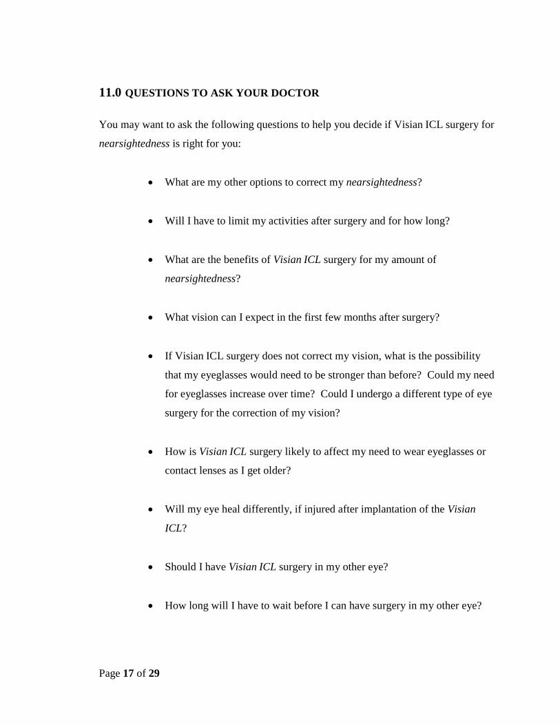
11.0 QUESTIONS TO ASK YOUR DOCTOR
You may want to ask the following questions to help you decide if Visian ICL surgery for
nearsightedness is right for you:
What are my other options to correct my nearsightedness?
Will I have to limit my activities after surgery and for how long?
What are the benefits of Visian ICL surgery for my amount of
nearsightedness?
What vision can I expect in the first few months after surgery?
If Visian ICL surgery does not correct my vision, what is the possibility
that my eyeglasses would need to be stronger than before? Could my need
for eyeglasses increase over time? Could I undergo a different type of eye
surgery for the correction of my vision?
How is Visian ICL surgery likely to affect my need to wear eyeglasses or
contact lenses as I get older?
Will my eye heal differently, if injured after implantation of the Visian
ICL?
Should I have Visian ICL surgery in my other eye?
How long will I have to wait before I can have surgery in my other eye?
Page 18 of 29
What vision problems might I experience if I have an Visian ICL only in 1
eye?
Discuss the cost of surgery and follow-up care needs with your doctor. Most
health insurance policies do not cover eye surgery for the correction of
nearsightedness.
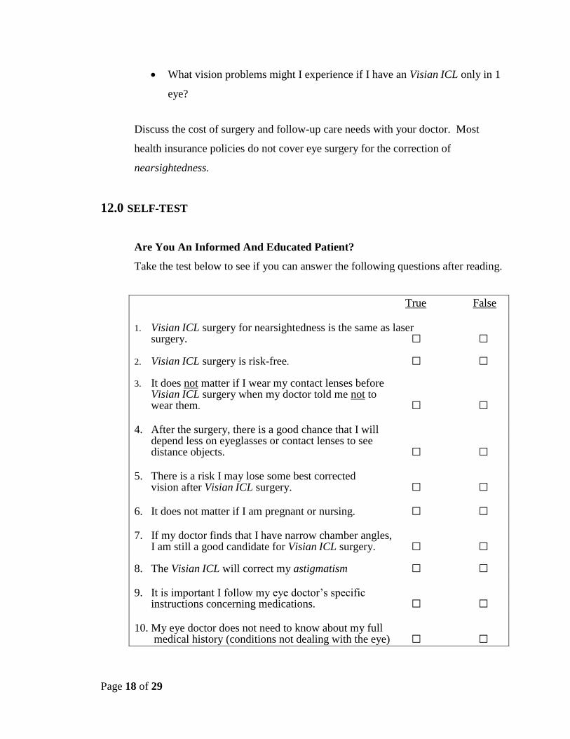
12.0 SELF-TEST
Are You An Informed And Educated Patient?
Take the test below to see if you can answer the following questions after reading.
True False
1. Visian ICL surgery for nearsightedness is the same as laser surgery. □ □ 2. Visian ICL surgery is risk-free. □ □
3. It does not matter if I wear my contact lenses before Visian ICL surgery when my doctor told me not to wear them. □ □ 4. After the surgery, there is a good chance that I will
depend less on eyeglasses or contact lenses to see distance objects. □ □
5. There is a risk I may lose some best corrected
vision after Visian ICL surgery. □ □ 6. It does not matter if I am pregnant or nursing. □ □ 7. If my doctor finds that I have narrow chamber angles, I am still a good candidate for Visian ICL surgery. □ □
8. The Visian ICL will correct my astigmatism □ □ 9. It is important I follow my eye doctor’s specific instructions concerning medications. □ □ 10. My eye doctor does not need to know about my full
medical history (conditions not dealing with the eye) □ □
Page 19 of 29
You can find the answers to Self-Test at the end of Section 13.
13.0 SUMMARY OF IMPORTANT INFORMATION
Visian ICL Surgery provides a permanent correction of your nearsightedness as
long as the Visian ICL remains in the eye. The Visian ICL may be removed. If
your physician removes the Visian ICL, you will lose the benefit of your
nearsightedness correction.
Visian ICL surgery does not eliminate the need for reading glasses, even if you
have never worn them before.
Your vision must be stable before Visian ICL surgery. You must provide written
evidence that your nearsightedness has changed no more than 0.50 D each year
for at least 1 year.
Pregnant and nursing women should wait until they are not pregnant and not
nursing to have the Visian ICL surgery.
Visian ICL surgery has some risks. Please read and understand this entire booklet
before you agree to the surgery. The sections on Benefits and Risks are especially
important to read carefully.
Some other options to correct nearsightedness include glasses, contact lenses, RK,
PRK and LASIK.
Before considering Visian ICL surgery you should:
a. have a complete eye examination.
Page 20 of 29
b. talk with at least one eye care professional about Visian ICL surgery,
especially the potential benefits, risks, and complications. You should
discuss the time needed for healing after surgery.
Certain eye diseases, eye conditions, previous eye surgery, systemic medical
conditions may have an impact on the results after Visian ICL surgery. It is
important that you provide your eye doctor with your complete medical history so
your eye doctor may determine if you are a good candidate for the Visian ICL for
correction of nearsightedness.
The Visian ICL is intended to improve your vision. However, because you are a
nearsighted patient you should consult with your eye doctor on a regular basis
(i.e., once a year) to verify the overall health of your eye.
Answers to Self-Test Questions:
1. F 6. F
2. F 7. F
3. F 8. F
4. T 9. T
5. T 10. F
14.0 GLOSSARY
This section summarizes important terms used in this information booklet or that your
eye doctor may discuss with you. Please discuss any related questions with your doctor.
Acute: Of sudden, rapid onset, usually with notable symptoms.
Acute Corneal Decompensation: A sudden opacification (clouding) of the usually clear
front surface of the eye (cornea).
Anterior Capsule: The front surface of the crystalline lens. It is a thin layer of skin
totally surrounding the lens much like the skin of a grape.
Page 21 of 29
Anterior Chamber: Front chamber of the eye; anterior chamber depth is the thickness
of the chamber
Antibiotic Medication: A drug used to treat or prevent infection. Your doctor may
prescribe this medication after Visian ICL surgery.
Anti-inflammatory Medication: A drug that reduces inflammation or the body’s
reaction to injury or disease. Any eye surgery can cause inflammation. Your doctor may
prescribe the medication after Visian ICL surgery.
Astigmatic Keratotomy: A type of eye surgery that changes the shape of the front
surface of the eye by making a special pattern of cuts in the cornea to correct
astigmatism.
Astigmatism: A focusing error that results in blurred distant and/or near vision. The
cornea is more curved in some directions than others, and causes light rays to focus at
different points inside the eye. Parts of objects appear clearer than other parts.
Best Corrected Vision: Best vision when wearing eyeglasses.
Cataract: Opacity, or clouding, of the crystalline lens inside the eye that can blur vision.
Collagen: A gel-like supporting substance found in the cornea, skin and other
connective tissue of the body.
Collamer: Hydroxyethyl methacrylate (HEMA)/porcine-collagen based polymer
material (STAAR proprietary product).
Page 22 of 29
Implantable Collamer Lens (ICL): A collagen based contact lens which is implanted
permanently in the rear chamber of the eye immediately in front of the crystalline lens in
order to correct nearsightedness. The Visian ICL can be removed.
Conjunctival Irritation: A reddening of the observable, white portion of the eyeball
and inner eyelid.
Contraindications: Any special conditions that result in the treatment not being
recommended.
Contrast Sensitivity: A measure of the ability of the eye to detect small lightness
differences between objects and the background in daylight and in dim light. For
example, black lines on a gray background are easier to see than gray lines on a gray
background. Objects in daylight are also easier to see than in dim light. Contrast
sensitivity is a way to determine how well patients can see in poor contrast conditions
such as very dim light, rain, snow and fog.
Cornea: The clear front layer of the eye. Surgery such as PRK, LASIK and RK
reshapes the front surface of the cornea to improve distant vision.
Corneal Swelling: Abnormal fluid build-up/swelling in the cornea. The condition is
usually temporary after surgery with no significant effect on vision. Persistent corneal
swelling may cause a loss of vision.
Corneal Flap: A thin slice of tissue on the surface of the cornea made with a
microkeratome at the beginning of a LASIK procedure. This flap is folded back before
the laser shapes the inner layer of the cornea.
Corneal Transplant: Removal and replacement of cornea
Page 23 of 29
Crystalline Lens: A structure inside the eye that helps to focus light onto the back
surface (retina) of the eye.
Diabetic Retinopathy: Damage to the back surface of the eye responsible for sensing
light due to diabetes.
Diopter: A unit of focusing power, used to describe the amount of nearsightedness and
astigmatism of an eye. Abbreviated as “D”.
Double Vision: Seeing multiple images of the object being looked at.
Endophthalmitis: Severe infection or inflammation of the entire eyeball.
Endothelium: Inner layer of the cornea.
Endothelial Cell Loss: Loss of cells in the endothelium. Endothelial cells are essential
in keeping the cornea clear and are essential for maintenance of good vision.
Excimer Laser: A type of laser used in LASIK and PRK to remove tissue from the
cornea.
Glare: A harsh or uncomfortable bright light. Glare symptoms are usually caused by a
distortion of light that would otherwise be tolerable without the distortion.
Glaucoma: An eye disease usually associated with high eye pressure. Glaucoma
damages the optic nerve of the eye and usually causes a progressive loss of vision.
Halos: Circular flares or rings of light that may appear around a headlight or other
lighted object. This symptom may occur after surgery.
Hyphema: Blood in the anterior chamber of the eye.
Page 24 of 29
Hypopyon: Pus in the front chamber of the eye.
ICL: Implantable Collamer Lens (see Device Description Section of this Booklet)
Inflammation of the Eye: The eye’s response to injury, infection or irritation which can
cause redness of the eye, pain, blurred vision and/or light sensitivity.
Intraocular Lenses: A lens that is placed in the eye after extraction of a cataract and
removal of the crystalline lens.
Intraocular Pressure (IOP): Pressure measurement monitored in your eye.
Iris: Colored part of the eye.
Iris Prolapse: A movement of the colored portion of the eye through a surgical wound
to a position outside the eye.
Iritis/Uveitis: Inflammation in the anterior chamber or other portion of the eye.
Laser Assisted In-Situ Keratomileusis (LASIK): A type of eye surgery that uses a
microkeratome and a laser to improve vision. The microkeratome creates a thin, hinged
flap of tissue on the cornea which is then folded back. The laser shapes the tissue under
the flap and the flap is put back on the eye so the tissue heals.
Lens: Natural crystalline lens in the eye which helps focus light properly into the back of
the eye.
Lens Opacities: A cloudiness of the crystalline lens.
Page 25 of 29
Lens Dislocation: A movement of the lens to an improper position.
Macular Edema: Swelling in the area responsible for fine (reading) vision on the back
surface of the eye (retina).
Macular Hemorrhage: Bleeding in the area responsible for fine (reading) vision on the
back surface of the eye (retina).
Manifest Refraction Spherical Equivalent (MRSE): A routine examination of the eye
to evaluate the amount of refractive error (i.e., amount of nearsightedness; amount of
astigmatism). Information used to prepare eyeglass/contact lens prescriptions.
Microkeratome: A surgical instrument used in LASIK to cut a thin flap of tissue from
the front surface of the eye (cornea) before the laser treatment is applied.
Myopia: A focusing error that results in blurrier vision at distance than near. Myopia is
also called nearsightedness.
Narrow Anterior Chamber Angle: A decrease in the size of the front chamber of the
eye which could block the flow of fluid from inside to outside of the eye resulting in a
raised eye pressure (glaucoma).
Nearsighted/Nearsightedness: A focusing error that results in blurrier vision at distance
than near. Nearsightedness is also called myopia.
Non-reactive Pupil: A condition where the colored portion of the eye does not get
larger or smaller when light is shined in the eye or removed.
Ocular Hypertension: Increased eye pressure.
Page 26 of 29
Peripheral Iridectomy: A small hole placed at the outer edge of the colored portion of
the eye.
Persistent: Lasts for a period of time during the study follow-up usually at least until the
end of the study.
Phakic Intraocular Lens: Placement of a man-made lens in a patient who still has their
natural crystalline lens.
Photorefractive Keratectomy (PRK): A type of eye surgery that uses an excimer laser
to reshape the front surface of the eye to improve vision. After the epithelium (outermost
layer) of the cornea is first scraped away, the laser removes tissue from the exposed
surface. After the surgery, the epithelium grows back.
Peripheral Anterior Synechiae: Scar tissue at the outer edges of the front chamber of
the eye.
Pigment Dispersion: An abnormal release of pigment particles from cells in the eye that
could get trapped in the fluid filtering mechanism (trabecular meshwork) possibly
causing an increase in pressure in the eye (glaucoma).
Posterior Chamber: Back or rear chamber of the eye.
Pseudoexfoliation: A condition where flakes of material can come off the surface of the
crystalline lens and block the drainage of fluid from the inside to the outside of the eye.
Pupil: The black part of the eye; fluctuates in size allowing varying degrees of light into
the eye.
Page 27 of 29
Pupillary Block Glaucoma: The inability of fluid to flow from the back chamber of the
eye to the front chamber frequently blocking drainage of fluid out of the eye and raising
the pressure in the eye (glaucoma).
Radial Keratotomy (RK): A type of eye surgery that changes the shape of the front
surface of the eye by making a special pattern of cuts in the cornea to correct
nearsightedness.
Retina: The layer of nerve tissue at the back of the eye that captures images, similar to
film in a camera, and sends information about these images to the brain. Light must be
focused correctly on the retina to form clear images.
Retinal Detachment: Separation of the retina from the rest of the back surface of the
eyeball.
Shallow Anterior Chamber: A flattening of the front chamber of the eye.
Subretinal Hemorrhage: Bleeding under the retina (see retina above).
Uveitis: Pathologic condition. Inflammation of any of the structures of the uvea: iris,
ciliary body, or choroid.
Visian ICL: Implantable Collamer Lens for the correction of nearsightedness (see
Device Description Section of this Booklet).
Visual Acuity: A measure of the sharpness of vision using a letter chart. Best Corrected
Visual Acuity (best vision with eyeglasses). Uncorrected Visual Acuity (best vision
without eyeglasses or contact lenses).
Vitreous Loss: The loss of a clear gel like material from the farthest back chamber of
the eye during a surgical procedure.
Page 28 of 29
YAG Laser: (Yttrium Aluminum Garnet) Laser Iridotomy: Production of a small
hole in the colored portion of the eye using a laser beam.
15.0 PATIENT ASSISTANCE INFORMATION
To be completed by you or your Primary Eye Care Professional as a reference.
Primary Eye Care Professional
Name:
Address:
Phone:
Visian ICL Doctor
Name:
Address:
Phone:
Treatment Location
Name:
Address:
Phone:
Page 29 of 29
Visian ICL Manufacturer:
STAAR Surgical, AG
Hauptstrasse 104
CH-2560 Nidau, Switzerland
Tel: + (41) 32 332 8888
FAX: + (41) 32 332 8899
U.S. Distributor:
STAAR Surgical Company
1911 Walker Avenue
Monrovia, CA 91016 USA
Tel: (800) 352-7842
FAX: (800) 952-4923
10-0001-41 Rev C
10000141 / C