vanadium determination in rat tissues and biological reference materials by neutron activation...
TRANSCRIPT

Journal o f Radioanalytical and Nuclear Chemistry, Articles, Vol. 141, No. 1 (1990)49-59
VANADIUM DETERMINATION IN RAT TISSUES AND BIOLOGICAL REFERENCE MATERIALS BY NEUTRON
ACTIVATION ANALYSIS +
J. KUI~ERA*, M. SIMKOVA,* J. LENER**, A. MRAVCOVA**, L. KINOVA***, I. PENEV***
*Nuclear Research Institute, 25068 l~e[ near Prague (Czechoslovakia) **Institute o f Hygiene and Epidemiology, ~robdrova 48, 10042 Prague (Czechoslovakia)
***Institute o f Nuclear Research and Nuclear Energy, bld. Lenin 72, 1184 Sofia (Bulgaria)
(Received November 14, 1989)
Vanadium was determined in adrenal gland, brain, ileum, kidney, liver, lung, muscle, myocard, skin, spleen, gonads, thyroid, and tibia of rats fed with normal diet and exposed to high vanadium doses in drinking water. Both radiochemical neutron activation analysis (RNAA) and instrumental neutron activation analysis (INAA) were employed. The RNAA procedure consisted in dry ashing samples prior to irradiation and vanadium separation from the irradiated samples by extraction with N-benzoyl-N-phenylhydroxylamine (BPHA) in toluene from 5 mol- 1-1 HCI. Vanadium accumulation a~ a function of a type of the tissue, exposure time, sex of rats, and administration of V(IV) or V(V) was studied. For quality as- surance purposes, the biological (standard) reference materials NBS SRM 1571 Orchard Leaves, NBS SRM 1577a Bovine Liver, IAEA H-4 Animal muscle, and Bowen's Kale were analyzed.
Introduction
In connect ion with the large quantifies o f vanadium released to the atmosphere by
burning o f fossil fuels, ~ increased exposure of humans to vanadium may occur. There-
fore, the assessment o f possible effects of vanadium on human health should be
considered because the element has been suspected of a possible role in air pol lut ion
disease? Until now, uncertainty persists about the toxic effects induced by chronic
industrial exposure. 3 - s The major symptoms of vanadium toxici ty in man are primari ly
in the respiratory tract, s
On the other hand, vanadium has been recognized as an essential trace element for
animals with roles in growth,~, 7 reproductive performance, bone and feather development,
induced resistance to dental caries 8 and as a possible essential trace element for many ,~ o
+Presented at the 2nd Balkan Conference on Activation Analysis and Nuclear-Related Analytical Techniques, Bled, 4 -6 October, 1989.
Elsevier Sequoia S. A., Lausanne Akaddmiai Kiad6, Budapest

J. KU~ERA et al.: VANADIUM DETERMINATION IN RAT TISSUES
Demonstrated biological effects of vanadium are also of inhibitory nature such as
depression of enzymatic activity of kinases, alkaline phosphatases, cholinesterase, sulfydril activity, cholesterol and cystin synthesis due to inhibition of the coenzyme A synthesis. 8 - 13 In experiments with lung, liver and kidney tissues of rats, it has
been found that, intracellularly, vanadium exhibits the highest affinity for the nuclear fraction, followed by the cytosol and mitochondrial fractions, s ,12,14 On the molecular
level, the interrelationship between vanadium and iron metabolism has been revealed to be due to vanadium association with nonheme, nonenzymatic iron-containing proteins such'as plasma transferrin, liver and spleen ferritin and milk lacto ferrin, s ,12 -1 s
Although research on the role of vanadium in organisms has increased consider- ably during the last few years, s ,9 -16 further studies on the element distribution and retention by tissues and organs are still needed. 8,16 Therefore, vanadium contents in 12-13 tissues of rats fed with normal diet or exposed to increased vanadium levels from drinking water was studied in this work. For quality assurance purposes, (standard) reference materials NBS SRM 1571 Orchard Leaves, NBS SRM 1577a Bovine Liver, Bowen's Kale, and IAEA H-4 Animal Muscle were analyzed.
Choice of NAA methods
Neutron activation analysis Provides a very low detection limit for vanadium due to the favourable nuclear characteristics of s 2 V formation by the 51V(n, ~,)52 V
reaction: Oo = 4.8 barns, 19 = 0.998, T = 3.76 min, E~ = 1434.4 keV. However, interferences from induced matrix activities in biological materials (24Na ' 3SCl ' 42 K, s ~Mn, etc.) and the short half-fife of S2V hamper vanadium determination by instru-
mental neutron activation analysis (INAA). For vanadium determination at the ppb level, radiochemical neutron activation analysis (RNAA) or a good preseparation of the element is required.
Preseparation techniques theoretically offer a lower detection limit because pro- cessing time after irradiation can be very short, but contamination and blank problems are unavoidable and thus the most important advantage of NAA can be lost. Reported blank values 17-21 vary in the range of 10 - s - 1 0 -9 g mainly as a function of the
quantity of nitric acid used as a critical reagent. 1 s Vanadium determination at the sub-ppb level is thus prevented.
In complete post-irradiation procedures, the detection limit depends on the dura- tion of sample decomposition and vanadium separation. While very fast, selective, and virtually quantitative vanadium separation by solvent extraction is available that can be accomplished within 1 minute, 9 1 o ,17 - 19,21,22 the most time-consuming
operation is the total decomposition of biological samples. This takes at least 4 to 5 minutes and the risk of incomplete decomposition or carrier equlibration has been
50

J. KU(~ERA et al.: VANADIUM DETERMINATION IN RAT TISSUES
reported? l Very recently, however, a totally post.irradiation RNAA method was
further developed and successfully applied to vanadium determination at the ultra- trace level in the 2nd Generation Human Serum Reference Material, to human serum of normal subjects, and also to other biological reference materials. 33
To facilitate the radiochemical processing, dry ashing samples prior to irradiation appears to be an attractive solution provided that no vanadium losses or contaminations
occur. However, at the ultratrace level, chlorine, which is not all removed on dry ashing, can interfere unless the procedure is designed to expel it from the dissolved ash. Evidence on vanadium losses during dry ashing is somewhat contradictory in the older literature)7,~ ~ ,23 Recently, proofs were given that dry ashing does not cause any significant vanadium losses from 48V metabolized or spiked animal tissues or botanical reference materials at temperatures ranging from 400 to 700 ~ 9 ~x 0,s 1,22
However, ashing at a temperature higher than 450 ~ has not been recommended as
serious etching of Spectrosil quartz vials occurred. 9,1~ Therefore, pre-ashing samples at 450 ~ and radiochemical vanadium separation
with N-benzoyl-N-phenylhydroxylamine (N-BPHA) according to BYRNE and KOSTA 22 has been chosen in this work for the determination of very low vanadium levels in bio- logical materials, whereas INAA has been employed for higher vanadium levels.
Experimental
Animals and their treatment
Rats of the Wistar strain 21 days old weighing 70-90 g at the beginning of ex- periments were used. Nonexposed male rats were fed ad libitum commercial food
pellets and water with a vanadium content of 414+28 ng �9 g-1 dry weight, i.e. 383+26 ng �9 g- 1 fresh weight (mean +- SD, n = 3) and < 2/ag �9 1-1, respectively as
determined by INAA. For the study of vanadium distribution in rat tissues as a func- tion of the exposure time, male rats were exposed to V2Os in drinking water with a vanadium content of 50 mg �9 1-1. After one and two months exposure, the vanadium
dose amounted to 224 mg and 415 mg per kg body weight, respectively. For the study of vanadium distribution as a function of the element valency and sex of the rats, the animals were exposed to V(tV) and V(V) compounds in drinking water for 50 days. The vanadium(V) dose supplied in the form of VsOs amounted to 231 mg �9 kg -1 and 317 mg �9 kg -~ for males and females, respectively, while the vanadium(IV) dose was 224 mg �9 kg -1 and 275 mg �9 kg -1 for males and females,
respectively, in the case of exposure to VOSO4. After the end of the experiments, the rats were sacrificed by decapitation, the tissues were removed and freeze-dried in polyethylene (PE) vials.
51

J. KU~ERA et al.: VANADIUM DETERMINATION IN RAT TISSUES
Sample and standard preparation
Freeze-dried rat tissues were used for INAA, while for RNAA the freeze<lried samples were dry ashed prior to irradiation.
Dry ashing of the 150-400 mg samples was carried out by heating in a special micro-oven in quartz vials precleaned by boiling in high purity HNO3 diluted 1:1
with deionized water and rinsing 3 times in deionized water. The oven was made of a resistance heated ceramic crucible insulated with porous aluminium oxide Alporit (Carborundum, Ben~tky n. J., Czechoslovakia) and asbestos. The temperature was first
kept at 150 ~ for 3 hours and then raised to 450 ~ at which it was maintained for 24 hours. The ratios of ash weight/dry weight were constant -+3% relative for particular tissues and in agreement with those given by IYENGAR, KOLLMER, BOWEN 24 for human tissues, indicating reproducible and efficient dry ashing.
Reference materials of weight 200-250 mg were analyzed in the "as received" state and their dry weight was determined on separate aliquots.
Freeze-dried samples, aliquots of dry ashed samples or reference materials were transferred to PE irradiation capsules which were cleaned prior to use by leaching in high purity HNO3 1:1 for 6 hours and washing with deionized water.
Vanadium standards for INAA and RNAA were prepared by pipetting 100 and 10/A aliquots, respectively, of a vanadium solution containing 0.01 mg �9 m1-1 obtained by dissolution of reagent grade ammonium vanadate (Lachema, Brno, Czechoslovakia) in deionized water. For INAA, the solution was pipetted into PE irradiation capsules and dried with the aid of an infrared lamp, whereas for RNAA the vanadium solution was pipetted into snap-cap polypropylene irradiation vials.
I~ad~on
For RNAA and a part of the INAA analyses, irradiations were carried out in the core of the WWR-S nuclear reactor of the NRI, l~e~., in which the thermal neutron flux density amounted to 5 �9 1013 n �9 era -2 �9 s -1 . A part of INAA work was
carried out by irradiation in a IRT-2000 reactor of the INRNE, Sofia, with a thermal neutron flux density 5 - 1012 n �9 cm -2 �9 s -~ . In both cases, the samples and
standards were irradiated separately with neutron fluence monitors (10 gg of gold and
a few mg of copper in l~e~ and Sofia, respectively) for 1 -2 minutes with the aid of pneumatic tube facilities.
INAA
At the NRI, l~e~, the irradiated samples and standards were counted after 2 minutes decay time for 7 minutes at a distance of 2 - 4 cm from the cap of a HPGe detector
52
J. KU~ERA et al. : VANADIUM DETERMINATION IN RAT TISSUES
(Ortec, relative efficiency 11%, resolution FWHM 1.7 keV for the 1332.5 keV photopeak of 60 Co.) The detector was coupled to a Nuclear Data gamma-spectroscopy system suited for high count rates, which has been described elsewhere.: s
At the INRNE, Sofia, counting was carried out after 1 minute decay time for 5 minutes at a distance of 0.5 cm from the cap of a Ge(Li) detector (NRI, l~e~, relative efficiency 8%, resolution FWHM 2.3 keV for 1332.5 keV photons of 6~ The v-ray spectra were collected with a Canberra 30 multichannel analyzer.
RNAA
The irradiated ash was dissolved within 1 -2 minutes in 10 ml of hot concentrated HC1 containing 50/.tg of vanadium carrier and about 20 kBq of 48V carrier-free
radiotracer. The solution obtained was diluted with water to obtain 5 mol �9 1-1 HC1, 1 ml of a KMnO4 solution (0.02 mol �9 1-1 ) was added to ensure that vanadium was in the pentavalent state. Vanadium was extracted with 10 ml of a 0.1% solution of N-BPHA in toluene for 1 minute. From the organic phase, a 9 ml aliquot was withdrawn into a syringe and transferred to a PE measurement vials and counted after 5 - 7 minute decay time for 7 minute with the NRI, l~e~ gamma-spectrometric system. The liquid vanadium standards were processed in the same way as the samples. Dry ashing losses and the yield of the vanadium radiochemical separation were studied on 48V spiked samples.
The 48V carrier-free radiotracer (T = 16 d, main E. r = 983.4 keV)was prepared by proton bombardment of a titanium foil in a cyclotron. The main nuclear reactions induced by proton bombardment of a natural titanium target were summarized by SABBIONI et al. 26 Separation from the titanium target and scandium radioisotopes (see Reference 26) was carried out as follows. The irradiated titanium foil was dissolved by heating with 2 ml of concentrated hydrofluoric acid with addition of a few drops of 30% H2 O2 and evaporated to dryness. This was repeated until the grey residue became white (usually 3 times). The white residue was dissolved in 2 ml of HF 2 mol �9 1-1 and ion-exchange chromatography using the strongly basic anion exchange
resin OSTION ADX8 (Spolek pro chemickou a hutni vyrobu, (.Jsti n. Labem, Czecho- slovakia), with similar properties to Dowex-1, was employed to separate vanadium. The activity of 48V was eluted with 10 ml of HF 0.5 mol �9 1-1 (separation yield 85%) while titanium and scandium remained bound to the resin.
53
J. KU~ERA et al.: VANADIUM DETERMINATION IN RAT TISSUES
Results and discussion
Dry ashing losses and the chemical yield of the vanadium separation were studied in preliminary experiments. The ashing of the 48V spiked samples revealed no detect- able losses of 4SV activity. The yield of vanadium separation by extraction with
N-BPHA from 5 mol �9 1-~ HC1 was found to be well reproducible and virtually quantitative (98+1%) so that there was no need to monitor it during each determina- tion. This was done occasionally only. The separation is also highly selective and the organic phase contained only minor contamination from 24Na and 38CI. A major
part of traces of these radionuclides can be removed by further washing the organic phase with 5 mol �9 1-1 HCI, which takes only about 30 seconds.
Blank values from irradiation and Counting the samples in the same PE capsules, or dry ashing the samples in vials from natural quartz, were also studied. The vanadium blank value for the cleaned PE capsules (mean +- SD) yielded 023_+0.15 ng�9 g-~ as determined from 4 replicates. This is in agreement with previous findings by KUCERA and SOUKAL 27 for PE Bralen and it can be neglected owing to the high vanadium contents being determined in this way. In the case of the quartz vials, it was worth- while to check whether contamination of the samples during ashing could occur. Since
no biological material was available with a known vanadium content in the'sub-ppb
range (recently, a value of 0.67+-0 05 n g � 9 g-i dry weight in the 2nd Generation Human Serum Reference Material became available), s3 it was decided to examine a
simulated "maximum" blank value by leaching the irradiated quartz vials with hot 12 mol �9 1-1 HC1 and processing the solution obtained as for the samples. In this way, a "maximum" blank value not exceeding 0.1 ng of vanadium was found. Obviously, the real blank value due to dry ashing the samples can be expected to be lower and it could also be neglected.
The accuracy and precision of the RNAA and INAA methods used were tested by analyses of biological (standard) reference materials. There were no biological reference materials available 2 a ,29 with a certified vanadium content in the range of
a few ng �9 g - 1 The most consistent values at this level were obtained for IAEA H-4 Animal Muscle that are listed in Table 1. Therefore, this reference material was chosen for verification of the RNAA procedure at very low vanadium levels. There is a better choice of reference material with vanadium levels that can be determined by INAA such as Bowen's Kale, NBS SRM-1577 Orchard Leaves, NBS SMR-1577a Bovine liver, 2a ,29 which were also analyzed in this work by the INAA and/or RNAA
methods. The results of the analyses of the above mentioned reference materials are given in Table 1 along with certified, information or literature values. A very good agreement was found between our INAA and RNAA results for Bowen's Kale, NBS SRM-1571, and NBS SRM-1577a (INAA only) and the respective reference values.
54

J. KU~ERA et al.: VANADIUM DETERMINATION IN RAT TISSUES
Table 1 Results of the vanadium determination in biological (standard) reference materials
(~tg �9 g-~ dry weight)*
Reference This work Reference Refer- Method
material x -+ s (N)** value ence
IAEA H-4 Animal Muscle
(ng�9 g- ~ ) RNAA 3.5 • 0.5 (7)
NBS SRM 1577a INAA 0.093 -+ 0.005 (3) Bovine liver
RNAA 0.384 • 0.019 (5) Bowen's Kale INAh~ 0.374 • 0.026 (7)
NBS SRM 1571 INAA 0.538 • 0.034 (3) Orchard Leaves
3 30
2.76 • 0.21 9
2.78 • 0.21 33
2.95 • 0.I0 34
3.09 • 0.64 35
3.2 • 0.2 36
3.0 + 0.2 37
0.099 + 0.008 31
0.386 + 0.058 28
0.500 + 0.110 32
*Unless otherwise stated. **Number of replicates.
An acceptable agreement was also found between our RNAA results and the IAEA
information and l i terature values for the IAEA H-4 Animal Muscle. This indicates
that no substantial vanadium contaminat ion occurred on dry ashing. Thus, accuracy
and sufficient precision o f the RNAA and I N A A methods used in this work was proved.
Results of the vanadium determination in 12 tissues of rats fed normal diet for
70 days are summarized in Table 2. The tissues obtained from 5 animals were analyzed;
however, in the case of small organs such as ileum and thyroid, the tissues from 2
animals were combined (1 organ left separately) and all 5 adrenal glands were combined
to prepare a sample for analysis. On the other hand, larger samples of kidney, muscle,
skin, and tibia were available. For liver, the organ was divided into two parts and
analyzed separately so that 10 samples were obtained. The tissues analyzed in this
work are ordered in Table 2 according to increasing vanadium content found and can
be divided into several groups. A vanadium level below the determination limit based
on a 3 o criterion was found for the adrenal gland, and very similar levels were deter-
mined for the pairs thyroid-muscle and liver-testis. Then, there is a group of organs
with similar vanadium contents consisting of spleen, ileum, skin, myokard, and kidney.
The highest vanadium levels were found in brain and tibia. In Table 2, for comparison
the values found by EDEL et al 16 are also given in tissues of rats of various age fed
55

J. KU~ERA et al.: VANADIUM DETERMINATION'IN RAT TISSUES
Table 2 Vanadium content in the tissues of rats fed normal diet (ng�9 g-~ dry weight)
This work Age variation according to Reference 16"*
Tissue 21 days 49 days 77 da~)s 115 days
~ • N* ~ • ~ • ~ • ~ •
Adrenal gland <0.1 1 . . . .
Thyroid 4.1 • 0.9 3 . . . . Muscle 4.6 • 1.6 9 - 21.2 • 11.0 24.1 • 15.4 26.0 • 15.0 Testis 7.3 • 0.9 5 26.0 • 7.5 25.5 • 16.0 29.5 • 20.5 20.5 • 2.5 Liver 7.6 • 1.7 10 58.3 • 15.8 17.3 • 5.8 10.8 • 8.3 11.5 • 5.0 Spleen 11.8 • 1.4 5 - 99.1 -+ 74.8 58.1 • 31.1 41.0 • 33.8 Ileum 12.4 • 2.9 3 . . . . Skin 13.0 • 3.6 9 . . . . Myocard 13.5 • 1 . 5 5 108.8 • 74.4 - 32.6 • 18.7 24.9 • 11.0 Kidneys 13.6 • 1.6 7 146.5 • 88.5 58.4 • 36.7 13.7 • .1.8 7.1 • 4.0 Brain 16.9 • 2.8 5 93.5 • 45.8 68.3 • 27.1 37.4 • 0.9 37.8 • 13.1 Tibia 23.3 • 2.6 8 . . . . Femur . . . . 26.0 • 4.8 Lung - 126.4 • 74.6 137.3 • 52.7 36.8 • 2.7 33.2 • 13.2 Blood - - 3.5 • 1.5 4.5.• 2.0 4 .0• 2.5 Fat*** - - 3.5 • 2.3 4.3 • 1.2 3.7 • 1.2
*Number oi ussues analyzed. **Recalculated from wet to dry weight using the factors given for human tissues in Reference 24.
***Wet weight.
w i th c h o w food and w a t e r w i t h v a n a d i u m c o n t e n t s o f 78 ng �9 g-1 a n d 2.8 ng �9 m 1 - 1 ,
respec t ive ly , The "rats we ighed a b o u t 50 g at the beg inn ing o f the e x p e r i m e n t . These
values suggest t h a t the " n o r m a l " levels o f v a n a d i u m m u s t a lways be def ined in con-
n e c t i o n w i t h age, especial ly for t issues such as k idney , liver, spleen, m y o c a r d , bra in ,
and l ung in w h i c h a s ignif icant decrease o f v a n a d i u m c o n c e n t r a t i o n occurs w i t h in-
creasing age. However , t he re is a d i f fe ren t p a t t e r n in the to ta l a m o u n t o f v a n a d i u m
in ra t organs. 16 Our values for k idney a n d b o n e agree well w i th those d e t e r m i n e d by
E D E L et al.16 in rats o f similar age. In general , howeve r , ou r values are s o m e w h a t
lower , a l t h o u g h the rats were fed wi th food h a v i n g a h i g h e r v a n a d i u m c o n t e n t . Thus,
the d i f ferences observed can be exp l a ined as due t o t h e h igher weight of our rats and
the i r organs b o t h a t the beg inn ing a n d at the e n d o f the e x p e r i m e n t .
The v a n a d i u m c o n t e n t s in 1 2 - 1 3 tissues o f ma le ra ts exposed to V 2 0 s in drink-
ing wa te r for 1 m o n t h a n d 2 m o n t h s are given in Table 3. In this tab le , the t issues
are o rde r ed acco rd ing to increas ing v a n a d i u m c o n t e n t s a f te r t he 2 - m o n t h exposure .
The v a n a d i u m levels cons iderab ly inc reased in all t issues c o m p a r e d t o those in rats
56
J. KU~ERA et al.: VANADIUM DETERMINATION IN RAT TISSUES
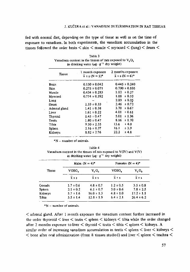
fed w i t h n o r m a l diet , d e p e n d i n g on the type o f t issue as well as on the t ime o f
exposure to v a n a d i u m . In b o t h e x p e r i m e n t s , t he v a n a d i u m a c c u m u l a t i o n in t he
t issues fo l lowed the order b ra in < skin < muscle < m y o c a r d < ( lung) < i l eum <
Table 3 Vanadium content in the tissues of rats exposed to V20 s
in drinking water ~ g - g-t dry weight)
Tissue 1 month exposure 2 months exposure • s (N = 5 ) * x • s (N = 6 ) *
Brain 0.150 • 0.042 0.445 • 0.240 Skin 0.275 • 0.071 0.700 • 0.105 Muscle 0.434 • 0.283 1.12 • 0.27
Myocard 0.774 • 0.392 1.88 • 0.33 Lung - 2.03 • 0.3~ Ileum 1133 • 0.33 2.46 • 0.72 Adrenal gland 1.41 • 0.38 3.70 • 0.67 Liver 1.61 • 0.22 4.01 • 0.61 Thyroid 1.45 • 0.47 5.01 • 1.36 Testis 1.80 • 0.47 8.16 • 1.70 Tibia 9.50 • 2.35 13.6 • 4.0 Spleen 2.16 • 0.37 16.1 • 3.9 Kidneys 8.82 • 2.76 22.2 • 4.8
*N - number of animals.
Table 4 Vanadium content in the tissues of rats exposed to V(IV) and V(V)
in drinking water ~ g �9 g-i dry weight)
Males (N = 4)* Females (N = 4)*
Tissue VOSO 4 V~ Os VOSO4 V20s
x • ~ • ~ • ~ •
Gonads 1.7 • 0.6 4.8 + 0.7 1.2 + 0.5 3.5 + 0.8 Spleen 2.1 • 0.2 6.1 • 0.7 3.0 • 0.6 7.8 + 2.5 Kidneys 5.7 • 1.6 16.0 • 1.3 4.8 • 1.0 17.2 • 1.1 Tibia 5.5 • 1.4 12.8 • 1.9 6.4 • 2.1 26.4 • 6.2
*N - number of animals.
< adrena l gland. Af t e r 1 m o n t h exposure the v a n a d i u m c o n t e n t f u r t he r increased in
the order t h y o r o i d < liver < test is < spleen < k idneys < t ib ia while the order c h a n g e d
af te r 2 m o n t h s exposu re to l iver < t h y r o i d < test is < t ib ia < spleen < k idneys . A
s imilar o rde r o f increas ing v a n a d i u m a c c u m u l a t i o n in tes t is < sp leen < liver < k idneys <
< bone af te r oral a d m i n i s t r a t i o n ( f rom 8 t issues s t ud i ed ) a n d liver < sp leen < t r a chea <
57
J. KU~ERA et aL: VANADIUM DETERMINATION IN RAT TISSUES
< kidneys < bone < lung after intratracheal instil lation (from 12 tissues studied) o f
both V(IV) and V(V) has recently been found by EDEL and SABBIONI. s
Vanadium accumulation in selected tissues of male and female rats as a function
of exposure to V2Os and VOSO4 in drinking water was also studied in this work.
In each group tissues from 4 animals were analyzed and the results are summarized
in Table 4. No sex dependence was observed for vanadium retention in the tissues
studied, except for tibia after exposure to V2Os. However, 2 - 4 times higher vanadium
contents were found in the tissues o f both male and female rats after exposure to
V 2 0 s compared to VOS04 although the total vanadium doses per kg body weight
were very similar. This finding is at variance with conclusions given in Ref. 5
on insignificant differences in the "retention of vanadium in rats after ingestion or
intratracheal instillation of V(IV) and V(V) in the form of vanadyl dichloride and
pentavanadate ions, respectively. The authors s concluded that the metabolic pathways
of tetravalent and pentavalent vanadium are independent of the route of vanadium
exposure and presumed the reduction of vanadate in vivo within 24 hours when 40 ng
and 100 ng of vanadium per rat as 4 S v ( v ) were intratracheally instilled and orally
administered, respectively. The results obtained in this work, however, would seem
to suggest that further s tudy is needed to elucidate accumulation of vanadium in rat
tissues after exposure to V(IV) and V(V) compounds with special regard to total
vanadium dose and exposure t ime.
Special thanks are due to Dr. E. SABBIONI and Dr. A. R. BYRNE for their helpful comments and providing the latest data related to this work.
References
1. E. SABBIONI, L. GOETZ, G. BIGNOLI, Sci. Total Environ., 40 (1984) 141. 2. D. H. K. LEE, Metallic Contaminants and Human Health, Academic Press, New York, 1972.
p. 153. 3. T. G. FAULKNER HUDSON, Vanadium - Toxicological and Biological Significance, Elsevier,
Amsterdam, 1964. 4. E. BROWNING, The Toxicity of Industrial Metals, Butterworths, London, 1969. 5. J. EDEL, E. SABBIONI, J. Trace Elem. Electrolytes Health Dis., 2 (1988) 23. 6. K. SCHWARZ, D. B. MILNE, Science, 174 (1971) 426. 7. L. L. HOPKINS, Jr., H. E. MOHR, in: Newer Trace Elements in Nutrition, W. MERTZ, W. E.
CORNATZER (Eds), Marcel Dekker, New York, 1971, p. 185. 8. V. BENCKO, M. CIKRT, J. LENER, Toxick~ kovy v pracovnim a ~ivotnim prostredi ~lov~ka,
Avicenum, Praha, 1984. 9. R. CORNELIS, L. MEES, J. HOSTE, J. RYCKENBUSH, J. VERSIECK, J. BARBIER, Proc.
Conf. Nuclear Activation Techniques in the Life Sciences, Vienna 1978, IAEA Vienna, 1979.
58

J. KU~ERA et al.: VANADIUM DETERMINATION IN RAT TISSUES
10. R. CORNELIS, J. VERSIECK, L. MEES, J. HOSTE, F, BARBIER, J. Radioanal. Chem., 55 (1980) 35.
11. A. R. BYRNE, L. KOSTA, Sci. Total Environ., 10 (1978) 17. 12. E. MARAFANTE, E. SABBIONI, Int. Conf. Management and Control of Heavy Metals in the
Environ., September 1979, London. 13. E. SABBIONI, E. MARAFANTE, J. Toxicol. Environ. Health, 8 (1981) 419. 14. E. SABBIONI, J. RADE, Toxicol. Letters, 5 (1980) 381. 15. E. SABBIONI, J. RADE, F. BERTOLERO, J. Inorg. Biochem., 12 (1980) 307. 16. J. EDEL, R. PIETRA, E. SABBIONI, E. MARAFANTE, A. SPRINGER, L. UBERTALLI, Che-
mosphere, 13 (1984) 87. 17. M. LEVSTEK, L. KOSTA, M. DERMELJ, A. R. BYRNE, Proc. Conf. Nuclear Activation Tech-
niques in the Life Sciences, Bled, 10-14 April, 1972. IAEA, Vienna 1972, p. 111. 18. E. DAMSGAARD, K. HEYDORN, B. RIETZ, in Reference 17, p. 11% 19. R. O. ALLEN, E. STEINNES, Anal. Chem., 50 (1978) 1553. 20. E. SABBIONI, M. MARONI, Report EUR 9005 EN, Ispra Establishment, 1983. 21. K. HEYDORN, E. DAMSGAARD, B. RIETZ, Anal. Chem., 52 (1980) 1045. 22. A. R. BYRNE, L. KOSTA, J. Radioanal. Chem., 44 (1978) 247. 23. T. T. GORSUCH, The Destruction of Organic Matter, Pergamon Press Ltd., Oxford, 1970,
p. 112. 24. G. V. IYENGAR, W. E. KOLMER, H. J. M. BOWEN, The Elemental Composition of Human
Tissues and Body Fluids, Verlag Chemie, Weinheim, New York, 1978. 25. J. KUCERA, L. SOUKAL, J. Nucl. Radioanal. Chem., 121 (1988) 245. 26. E. SABBIONI, E. MARAFANTE, L. GOETZ, C. BIRATTARI, Radiochem. Radioanal. Letters,
31 (1977) 39. 27. J. KUCERA, L. SOUKAL, J. Radioanal. Chem., 80 (1983) 121. 28. Y. MURAMATSU, R. M. PARR, Survey of Currently Available Reference Materials for Use in
Connection with the Determination of Trace Elements in Biological and Environmental Ma- terials, Report IAEA/RL/128, IAEA Vienna, December 1985.
29. R. MAVRODINEANU, R. ALVAREZ, Summary of the Biological and Botanical Standards, NBS Special Publication 260-104, U.S. Government Printing Office, Washington, October 1985.
30. R. M. PARR, Report No. 2, Intercomparison of Minor and Trace Elements in IAEA Animal Muscle (H-4), IAEA:RL--69, IAEA Vienna, October 1980.
31. National Bureau of Standards, Certificate of Analysis, SRM 1577a, Bovine Liver, Galthersburg, February 1, 1985.
32. E. S. GLADNEY, B. T. O'MALLEY, I. ROELANDTS, T. E. GILLS, Compilation of Elemental Concentration Data for NBS Clinical, Biological, Geological, and Environmental Standard Reference Materials, NBS Spec. Publ. 260-111, U:S. Government Printing Office, Washington, November 1987.
33. A. R. BYRNE, J. VERSIECK, Biological Trace Element Research, in press. 34. R. CORNEL1S, J'. VERS1ECK, L. MEES, J. HOSTE, F. BARBIER, Biol. Trace Elem. Res., 3
(1981) 257. 35. O. ISHIDA, K. KIHIRA, Y. TSUKAMOTO, F. MARUMO, Clin. Chem., 35 (1989) No. 1,127.
36. A. R. BYRNE, V. VRBIC, J. Radioanal. Chem., 54 (1979) 77. 37. A. R. BYRNE, Annex 6, in: Minor and Trace Elements in Breast Milk: Report of a Joint WHO/
IAEA Collaborative Study, E. M. DEMAYER (Ed.), WHO, Geneva, 1989.
59