vacuum uv photoionization mass spectrometry of small polymers using jet cooling
TRANSCRIPT

Journal of Photochemistry and Photobiology A: Chemistry 106 (1997) 31–36
1010-6030/97/$17.00 q 1997 Published by Elsevier Science S.A. All rights reservedPII S1010-6030(97)00035-X/97/$17.00 q 1997 Elsevier Science S.A. All rights reservedPII S1010- 6030 /97/$17 . 00 q 1997 Pub l i shed b y E l se v i e r S c i e n c e S .A . Al l r i g h t s r e se rv e dP I I S1010-6030(97)00035-X(97)00035-X
Journal: JPC (Journal of Photochemistry and Photobiology A: Chemistry) Article: 4653
Vacuum UV photoionization mass spectrometry of small polymersusing jet cooling
M.S. de Vries, H.E. Hunziker U
IBM Research Division, Almaden Research Center, 650 Harry Road, San Jose, CA 95120, USA
Abstract
Many polymers with short chains and molecular weights up to several thousand atomic mass units can be transferred to the vapor phasewhole by pulsed laser desorption, in contrast with the well-known, destructive ablation process encountered with long-chain, high-molecular-weight polymers. However, the vaporized whole polymers are hot and thus fragment extensively when photoionized. For their detection asmolecular ions, it is necessary first to cool the vaporized polymers, which is accomplished by entraining them in an Ar jet expansion. Near-threshold, single-photon ionization at 125 nm (9.9 eV) was used in all cases and compared with 193 nm, two-photon ionization for polystyrene.Mass spectra showing oligomeric distributions for samples of poly(dimethylsiloxane), poly(ethylene oxide), poly(isoprene),poly(perfluorotrimethylene oxide) and polystyrene are reported. Application of the technique to samples of increasing average molecularweight shows that, as the chain length increases, thermal decomposition eventually predominates over vaporization. q 1997 ElsevierScience S.A.
Keywords: Small polymers; Jet cooling; Vacuum UV photoionization mass spectrometry
1. Introduction
There is now an arsenal of techniques capable of providingthe mass spectra of whole polymers. A major subgroup ofthese new methods uses pulsed lasers to vaporize and ionizethe high-molecular-weight material. Two important effectshave been put to use in this approach to aid in the productionof stable molecular ions: chemical ionization and matrix-assisted desorption. Many examples have been reported inwhich cationization with alkali ions, added to the polymer assalts, yields oligomeric mass distributions [1–4]. Hillen-kamp and coworkers [5,6] originated and developed thematrix-assisted laser desorption/ionization (MALDI)technique, in which a low-molecular-weight organic matrixserves the dual function of entraining large molecules intothe vapor phase without decomposition and ionizing themchemically usually by proton transfer.
Here we concern ourselves with the laser-induced vapori-zation of neat, low-molecular-weight polymers. In this case,without ionic dopants or ions created by laser-induced break-down, there is no chemical ionization and the gas phase pol-ymer molecules are neutral. Post-ionization is thus necessaryto convert them into ions. Since this is usually performed by
U Corresponding author.
photoionization with pulsed lasers, the procedure has alsobeen called two-step laser mass spectrometry [7].
In this report, we address a problem which has limited theusefulness of laser desorption/post-ionization mass spec-trometry. The desorbed molecules are hot and thus tend tofragment after ionization, unless the ions are very stable, asin the case of fullerenes or aromatic hydrocarbons [7,8]. Asolution to this problem exists in the form of the laser desorp-tion/jet cooling approach [9–11]. Cooling has already beenused successfully to obtain the mass and optical spectra ofsmall, aromatic polymers by two-photon ionization [12,13].We show here that the combination of jet cooling with vac-uum UV single-photon ionization near threshold makes itpossible to obtain the parent mass spectra of small, aliphaticpolymers. A somewhat similar approach was taken by Kosterand Grotemeyer [14] to obtain the mass spectra of small,non-aromatic peptides. However, in their work, extensivefragmentation was observed.
2. Experimental details
Several previous studies have used vacuum UV post-ion-ization without cooling [15–17]. The vacuum UV pulseswere generated by non-resonant third harmonic generation inphase-matched rare gas mixtures [18]. Because of its poten-

M.S. de Vries, H.E. Hunziker / Journal of Photochemistry and Photobiology A: Chemistry 106 (1997) 31–3632
Journal: JPC (Journal of Photochemistry and Photobiology A: Chemistry) Article: 4653
Fig. 1. Schematic diagram of the experimental set-up for laser desorption with jet cooling and post-ionization. Abbreviations: sb, sample bar; sk, skimmer; g,ion source grids; m, movable mirror; pd, photodiode; mcp, multichannel plate detector; LiF, plano-convex lithium fluoride lens; c, annular cooler; h, annularheater; w, fused silica window; al, achromatic lens.
tial for achieving higher vacuum UV fluences, we used res-onance-enhanced four-wave mixing in Hg vapor instead,employing only a single, tunable dye laser at 625.70 nm togenerate vacuum UV pulses at 125.14 nm [19]. This wave-length was also chosen because it is close to the ionizationthreshold of the higher paraffins. The experimental set-up isshown schematically in Fig. 1. A 30 cm achromatic lens wasused to focus the fundamental and second harmonic insidethe Hg vapor cell, which was operated at 140 8C and about 8Torr Ar pressure. At the exit of the cell, a 5 cm diameter, 24.0cm focal length (at 125 nm) LiF lens served a triple duty ascell window, focusing element and dispersing element forseparating the vacuum UV from the pump beams. The bicon-ical Hg cell was made from welded stainless steel parts, withViton O-ring seals at the lens and window. For visible align-ment, the LiF lens was replaced with a similar glass lens of24 cm focal length at 625 nm. The holder for these lenses andthat for the achromatic lens were laterally adjustable. Bymoving a vacuum UV mirror inside the vacuum chamber, thevacuum UV beam could be deflected onto a solar blind pho-todiode (Hamamatsu Type No. R1178, CsI cathode). Fromits nominal spectral response curve, the typical vacuum UVfluence was estimated to be 5=1011 photons per pulse.
Two different laser desorption/ionization set-ups wereused in this work, one with and the other without jet cooling.The former has been described and characterized in detailpreviously [11]. Briefly, it consists of a pulsed nozzle, skim-mer and reflectron time-of-flight (TOF) mass analyzer.Between the extraction grids of the analyzer, the molecularbeam was crossed by a slightly focused 193 nm excimer laserbeam which was used for two-photon ionization of aromaticchromophores. The 125 nm vacuum UV beam was intro-
duced along the jet axis and focused in the ionization volume,110 cm from the LiF lens. Sample substances were depositedfrom dilute solutions onto 3 mm wide graphite bars whichcould be mounted through a load lock onto a translation stagein front of the jet orifice. A doubled Nd:YAG laser (532 nm)was used for desorption, with pulse energy densities of 10–100 mJ cmy2 and a spot size of about 1 mm in diameter.Vaporization was caused by substrate heating only. Optimalinjection of the vapor into the jet expansion occurred whenthe desorption spot was about 1 mm in front of and 0.5 mmbelow the nozzle, and the desorption laser was fired near thepeak of the jet gas pulse. The pulsed valve was operated withAr or Xe at a backing pressure of 8 atm.
The set-up without jet cooling involved a new laser micro-probe ion source coupled to a reflectron TOF mass analyzer.An instrument described previously [20] was modified topass the vacuum UV post-ionization beam closely (within 1mm) over the desorption spot to maximize the ionizationefficiency. An arrangement somewhat similar to thatdescribed by Odom and Schueler [21] was chosen, exceptthat the ions were extracted through the blind spot of a 458
mirror rather than through the reflecting microscope objec-tive. This has the considerable advantage of being able towork with a commercial reflecting objective.
Polymer samples were obtained from Daikin Industries(poly(perfluorotrimethylene oxide)), Polymer LaboratoriesLtd. (poly(ethylene oxide), poly(isoprene) and polysty-rene) and Aldrich (poly(dimethylsiloxane)). Most of thesesamples came characterized with Mp (GPC-peak molecularweight) or Mw (weight-average molecular weight) values.From the mass spectra, values of Mn (number-averagemolec-ular weight) and Mw were calculated for comparison with the
M.S. de Vries, H.E. Hunziker / Journal of Photochemistry and Photobiology A: Chemistry 106 (1997) 31–36 33
Journal: JPC (Journal of Photochemistry and Photobiology A: Chemistry) Article: 4653
Fig. 3. Jet/vacuum UV mass spectrum of squalane. Signals at multiples ofthe parent mass are due to van der Waals’ dimers and trimers.
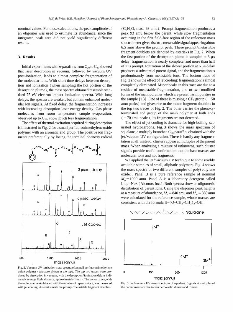
Fig. 2. Vacuum UV ionization mass spectra of a small perfluorotrimethyleneoxide polymer (structure shown at the top). The top two traces were pro-duced by desorption in vacuum, with the desorption/ionization delays indi-cated (average flight distance, approximately 1 mm). The bottom trace, withthe molecular peaks labeled with the number of repeat units n, was measuredwith jet cooling. Asterisks mark the prompt/metastable fragment doublets.
nominal values. For these calculations, the peak amplitude ofan oligomer was used to estimate its abundance, since theintegrated peak area did not yield significantly differentresults.
3. Results
Initial experiments with n-paraffins from C20 to C40 showedthat laser desorption in vacuum, followed by vacuum UVpost-ionization, leads to almost complete fragmentation ofthe molecular ions. With short time delays between desorp-tion and ionization (when sampling the hot portion of thedesorption plume), the mass spectra obtained resemble stan-dard 75 eV electron impact ionization spectra. With longdelays, the spectra are weaker, but contain enhanced molec-ular ion signals. At fixed delay, the fragmentation increaseswith increasing desorption laser energy density. Gas phasemolecules from room temperature sample evaporation,observed up to C22, show much less fragmentation.
The effect of thermal excitation acquired during desorptionis illustrated in Fig. 2 for a small perfluorotrimethylene oxidepolymer with an aromatic end group. The positive ion frag-ments preferentially by losing the terminal phenoxy radical
(C6H5O, mass 93 amu). Prompt fragmentation produces apeak 93 amu below the parent, while slow fragmentationoccurring in the first field-free region of the reflectron massspectrometer gives rise to a metastable signal appearingabout6.5 amu above the prompt peak. These prompt/metastablefragment doublets are denoted by asterisks in Fig. 2. Whenthe fast portion of the desorption plume is sampled at 3 msdelay, fragmentation is nearly complete, and more than halfof it is prompt. Ionization of the slower portion at 6 ms delayproduces a substantial parent signal, and the fragmentation ispredominantly from metastable ions. The bottom trace ofFig. 2 shows the effect of jet cooling: fragmentation is almostcompletely eliminated. Minor peaks in this trace are due to aresidue of metastable fragmentation, and to two modifiedforms of the main polymer which are present as impurities inthe sample [13]. One of these is missing a CF2 group (y50amu peaks) and gives rise to the minor fragment doublets inthe top two traces of Fig. 2. The other carries the phenoxy-terminated end group of the main polymer at both ends(y70 amu peaks); its fragments are not detected.
The effect of jet cooling is dramatic for high-boiling, sat-urated hydrocarbons. Fig. 3 shows the mass spectrum ofsqualane, a multiply branched C30 paraffin, obtained with thejet/vacuum UV configuration. There is hardly any fragmen-tation at all; instead, clusters appear at multiples of the parentmass. When analyzing a mixture of unknowns, such clustersignals provide useful confirmation that the base masses aremolecular ions and not fragments.
We applied the jet/vacuum UV technique to some readilyavailable samples of small, aliphatic polymers. Fig. 4 showsthe mass spectra of two different samples of poly(ethyleneoxide). Panel B is a pure reference sample of nominalMps1000 amu. Panel A is a laboratory detergent calledLiqui-Nox (Alconox Inc.). Both spectra show an oligomericdistribution of parent ions. Using the oligomer peak heightsas a measure of abundance, Mns840 amu and Mws880 amuwere calculated for the reference sample, whose masses areconsistent with the formula H–(O–CH2–CH2)n–OH.

M.S. de Vries, H.E. Hunziker / Journal of Photochemistry and Photobiology A: Chemistry 106 (1997) 31–3634
Journal: JPC (Journal of Photochemistry and Photobiology A: Chemistry) Article: 4653
Fig. 4. Jet/vacuum UV mass spectra of poly(ethylene oxide) samples: (A)Liqui-Nox laboratory detergent; (B) reference sample with nominalMps1000 amu; a fragment series in spectrum B is labeled with asterisks.The number n of ethylene oxide repeat units is given for some peaks (inparentheses for fragment series).
Fig. 5. Jet/vacuum UV mass spectrum of poly(isoprene) with nominalMps1000 amu. The number n of isoprene repeat units is given for somepeaks. I and I2 refer to isoprene monomer and dimer.
Fig. 6. Jet/vacuum UV mass spectra of three samples of poly-(dimethylsiloxane) whose nominal Mp values are indicated in the figure.Numbers of repeat units n are indicated for some of the oligomer peakswhich are all (My15) amu masses.
The spectrum of this sample also shows a distribution oflow-molecular-weight fragments with masses n=44 amuwhose abundance increases monotonically with decreasingweight. In addition, there is a prominent odd-mass fragmentat 71 amu. The polymer in Liqui-Nox is shorter, and its massvalues also conform to the formula n=44 amu. Since this isa detergent, the composition is probably C5H11–(O–CH2–CH2)n–OH.
Fig. 5 shows the mass spectrum of a sample of poly-(isoprene) with nominal Mps1000 amu. It corresponds tothe structure C4H9–(CH2–C(CH3)_CH–CH2)n–H. Fromthe peak heights, we calculate Mns820 amu and Mws870amu. At the low mass end, this spectrum shows peaks for theisoprene monomer (68 amu) and dimer (136 amu).
For two types of polymer, we explored how the techniqueis limited by the molecular weight. The jet/vacuum UV massspectra for three different samples of poly(dimethylsiloxane)with nominal Mw values of 800, 2000 and 6000 amu areshown in Fig. 6. In this case, the masses observed are allfragments missing one methyl group. The molecular ionscorresponding to the structure (CH3)3Si–(O–Si(CH3)2)n–CH3 (Ms88qn=74 amu) are not observed; instead, thespectrum shows the My15 amu ions. We also examined thecyclic tetramer and pentamer (O–Si(CH3)2)m (ms4 and 5)and observed only the My15 amu ions. For the linear poly-mer samples shown in Fig. 6, the Mw (Mn) values determinedfrom the mass spectra of the three samples were 950 (920),1310 (1240) and 2150 (2010) amu respectively. There is
clearly a lack of signals for higher oligomers, increasing withincreasing Mw of the sample, and accompanied by an increas-ing proportion of low mass fragments in the mass spectrum.
The mass spectra of three polystyrene samples character-ized by nominal Mp values of 950, 1300 and 4250 amu are

M.S. de Vries, H.E. Hunziker / Journal of Photochemistry and Photobiology A: Chemistry 106 (1997) 31–36 35
Journal: JPC (Journal of Photochemistry and Photobiology A: Chemistry) Article: 4653
Fig. 7. One-photon (125 nm) and two-photon (193 nm) ionization massspectra of three samples of polystyrene whose nominal Mp values are indi-cated in the figure. Some peaks are labeled with the number n of styrenerepeat units. The low mass peak designated S in the 125 nm ionizationspectra is styrene. Note the vertical scale change (=2) in these traces.
shown in Fig. 7. This is an aromatic polymer which can read-ily be ionized by two-photon ionization at 193 nm, and weused it to compare the ionization efficiencies of the two-photon and one-photon (125 nm) techniques. The samplecomposition corresponds to the structure C4H9–(CH2–CHP)n–H, where P'phenyl. For 193 nm ionization, the val-ues of Mw (Mn) determined from the mass spectra were 910(860), 1340 (1270) and 2120 (1830) amu respectively. For125 nm ionization, the values were 780 (700) and 1220(1150) amu, and the spectrum of the highest molecularweight sample was too weak to be evaluated. As forpoly(dimethylsiloxane), there is a signal deficit for higheroligomers which increases with increasing averagemolecularweight of the sample and is accompanied by an increasingproportion of low mass fragments. The prominent fragmentobserved with 125 nm ionization is styrene; this was alsodetected by Feldmann et al. [15] as a photoablation productof polystyrene using 118.4 nm ionization. A comparison ofthe one-photon and two-photon ionization traces shows thatthe former exhibits a bias towards low mass when comparedwith the latter. Since the method of ionization is the onlydifference between these spectra, it follows that the ionizationefficiency falls off more rapidly with molecular weight forthe one-photon process.
4. Discussion
Our results show that laser desorption, combined with jetcooling and vacuum UV one-photon ionization, is capable ofproducing essentially fragment-free mass spectra of highlybranched paraffins, such as squalane, as well as several typesof small, aliphatic polymers. Cooling is essential; without itextensive fragmentation is observed. This is due to the highinternal energy content of the vaporized molecules. As shownin the classic study by Steiner et al. [22], the photoionizationcross-sections for fragmentation processes near thresholdincrease dramatically with temperature, whereas the cross-section for molecular ionization does not. Thus cooling stopsfragmentation processes especially near the ionizationthreshold.
Cooling can only be effective if the ion has a stable groundstate and fragmentation is thermally activated. This is notalways the case. The complete absence of a molecular ion forall linear and cyclic poly(dimethylsiloxane) moleculesshows that their molecular ions produced by vacuum UVionization are intrinsically unstable. Judging from the ioni-zation potential of (CH3)3Si–O–Si(CH3)3 (9.64"0.01 eV[23]), the excess energy for vacuum UV ionization is smalland similar to that of the paraffins, where cooling suppressesfragmentation completely.
We found that small polymers up to about Mws1000 amuare detected with only minor distortion of their mass distri-butions, whereas the spectra of samples with higher Mw areincreasingly biased towards low mass oligomers. The onsetof severe distortion depends somewhat on the type of poly-
mer. We consider three causes for this effect: (1) preferentialfragmentation of high mass ions; (2) competition betweenvaporization and thermal decomposition in the desorptionstep; (3) reduced ionization efficiency for high mass oligo-mers. Since we have no evidence for less efficient cooling ofhigh mass neutrals, we regard cause (1) as unlikely. Schlagand Levine [24] have presented arguments and some evi-dence for cause (3). They proposed a mass dependence ofthe molecular ionization efficiency of the form exp(yM/M0)
3/2, where M is the molecular weight and M0 is an empir-ical constant. This may account for part of the mass biasobserved, but since the exponential falloff is steepest for thelow masses, it cannot account for the increased distortionobserved for M)1000 amu. This increase is probably due tocause (2) which predominates in this mass range. For highmass molecules, dissociation becomes faster than vaporiza-tion. Desorption changes into ablation, which gives rise to anincreased proportion of small fragments detected for samplesof high average molecular weight.
It should be noted that our use of a graphite substrate and532 nm light for desorption may favor thermal decompositionover vaporization when compared with IR laser-induceddesorption. In the former, but not the latter, case, there is ahigh temperature transient at the substrate–sample interface.IR desorption combined with cationization has been used todetect higher oligomers of poly(ethylene oxide) than thosereported here [4].

M.S. de Vries, H.E. Hunziker / Journal of Photochemistry and Photobiology A: Chemistry 106 (1997) 31–3636
Journal: JPC (Journal of Photochemistry and Photobiology A: Chemistry) Article: 4653
Even for M-1000 amu, there appears to be relativelymoreefficient two-photon than one-photon ionization of the higheroligomers of polystyrene. Schlag et al. [25] reported asimilareffect for small, aromatic peptides. They used identical totalionization energies, whereas in our case comparison is lessdirect because the energies are different (9.9 eV for one-photon ionization; 12.9 eV for two-photon ionization). Thiseffect may be due to the increased oscillator strength (morechromophores) of higher oligomers in the first step of thetwo-photon process.
The poly(ethylene oxide) sample with nominal Mps1000amu shows a fragment series with masses of n=44 amu(nG1), monotonically increasing in intensity with decreas-ing mass (Fig. 4). This series fits none of the products foundor intermediates postulated in thermal degradation studies[26,27], but is consistent with earlier laser desorption workin which the same series was observed in cationized form [4]and attributed to the random scission process
–CH –CH –O–CH –™(–CH –CH qO–CH –)2 2 2 2 2 2
™–CH_CH qHO–CH –2 2
The vinyl-terminated product fits the observed fragmentseries. It must arise from pyrolysis because it is not a typicalether ion fragment [28] and can be detected by cationization[4]. It also does not occur in the spectrum of Fig. 4(A),where lower molecular weight and matrix effects (the pres-ence of other low-molecular-weight mixture components)reduce pyrolysis. The discrepancy with slow thermal degra-dation presumably arises because the vinyl product is formedin a high activation energy unimolecular reaction which pre-dominates under the conditions of laser desorption. A possi-ble mechanism involves a concerted b-C–H hydrogentransfer [29]. In this case, the C–O bond scission shown inparentheses in the above reaction will be simultaneous withhydrogen transfer. The odd mass fragment observed at 71amu may be the radical CH2–CH2–O–CH_CH2 derived fromthe vinyl terminal by a simple C–O bond rupture.
Acknowledgements
We gratefully acknowledge the help of H.R. Wendt, D.Braichotte, K. Reihs and R. Baumann with the constructionand characterization of the vacuum UV source. We also wish
to thank Daikin Industries for a sample of low-molecular-weight poly(perfluorotrimethylene oxide).
References
[1] C.L. Wilkins, D.A. Weil, C.L.C. Yang, C.F. Ijames, Anal. Chem. 57(1985) 520.
[2] D.E. Mattern, D.M. Hercules, Anal. Chem. 57 (1985) 2041.[3] R.S. Brown, D.A. Weil, C.L. Wilkins, Macromolecules 19 (1986)
1255.[4] R.J. Cotter, J.P. Honovich, J.K. Olthoff, R.P. Lattimer,
Macromolecules 19 (1986) 2996.[5] F. Hillenkamp, M. Karas, R.C. Beavis, B.T. Chait, Anal. Chem. 63
(1991) 1193A.[6] U. Bahr, A. Deppe, M. Karas, F. Hillenkamp, Anal. Chem. 64 (1992)
2866.[7] R. Zenobi, Int. J. Mass Spectr. Ion Proc. 145 (1995) 51.[8] K.R. Lykke, P. Wurz, D.H. Parker, M.J. Pellin, Appl. Opt. 32 (1993)
857.[9] H.v. Weyssenhoff, H.L. Selzle, E.W. Schlag, Z. Naturforsch. Teil A
40 (1985) 674.[10] R. Tembreull, D.M. Lubmann, Appl. Spectrosc. 41 (1987) 431.[11] G. Meijer, M.S. de Vries, H.E. Hunziker, H.R. Wendt, Appl. Phys. B
51 (1990) 395.[12] D.A. Lustig, D.M. Lubmann, Int. J. Mass Spectr. Ion Proc. 107 (1991)
265.[13] D.S. Anex, M.S. de Vries, A. Knebelkamp, J. Bargon, H.R. Wendt,
H.E. Hunziker, Int. J. Mass Spectr. Ion Proc. 131 (1994) 319.[14] C. Koster, J. Grotemeyer, Org. Mass Spectr. 27 (1992) 463.[15] D. Feldmann, J. Kutzner, J. Laukemper, S. MacRobert, K.H. Welge,
Appl. Phys. B 44 (1987) 81.[16] J.B. Pallix, U. Schuehle, C.H. Becker, D.L. Huestis, Anal. Chem. 61
(1989) 805.[17] S.E. Van Bramer, M.V. Johnston, Appl. Spectrosc. 46 (1992) 255.[18] R. Hilbig, R. Wallenstein, IEEE J. Quantum Electron. 17 (1981) 1566.[19] R. Hilbig, R. Wallenstein, IEEE J. Quantum Electron. 19 (1983) 1759.[20] M.S. de Vries, D.J. Elloway, H.R. Wendt, H.E. Hunziker, Rev. Sci.
Instrum. 63 (1992) 3321.[21] R.W. Odom, B. Schueler, in: D.N. Lubman (Ed.), Lasers and Mass
Spectrometry, Oxford University Press, New York, 1990, p. 122.[22] B. Steiner, C.F. Giese, M.G. Ingraham, J. Chem. Phys. 34 (1961) 189.[23] S.G. Lias, J.E. Bartmess, J.F. Liebman, J.L. Holmes, R.D. Levine,
W.G. Mallard, J. Phys. Chem. Ref. Data 17 (Suppl. 1) (1988) 280.[24] E.W. Schlag, R.D. Levine, J. Phys. Chem. 96 (1992) 10 608.[25] E.W. Schlag, J. Grotemeyer, R.D. Levine, Chem. Phys. Lett. 190
(1992) 521.[26] N. Grassie, G.A. Perdomo Mendoza, Polym. Degrad. Stab. 9 (1984)
155.[27] E. Bortel, R. Lamot, Makromol. Chem. 178 (1977) 2617.[28] F.W. McLafferty, Interpretation of Mass Spectra, University Science
Books, Mill Valley, CA, 1980, p. 209.[29] G. Allen, S.L. Aggarwal, S. Russo, Comprehensive Polymer Science,
First Supplement, Pergamon, Oxford/New York/Seoul/Tokyo, 1992,p. 234.