urinary tract dr. nasr a. mohammed dr. nasr a. mohammed fibms fibms
TRANSCRIPT
Urinary Tract Urinary Tract
Dr. Nasr A. Mohammed Dr. Nasr A. Mohammed
FIBMSFIBMS
Imaging techniques Imaging techniques Basic examinationsBasic examinations
-Ultrasound.-Ultrasound.
-Intravenous Urography IVU.-Intravenous Urography IVU.
-Computed Tomography CT.-Computed Tomography CT.
-Radionuclide examination.-Radionuclide examination.
Other investigationsOther investigations
investigations limited to selected patients investigations limited to selected patients Magnetic Resonance Imaging MRI.Magnetic Resonance Imaging MRI. Arteriography.Arteriography. Direct puncture of collecting systemDirect puncture of collecting system Catheterization.Catheterization.
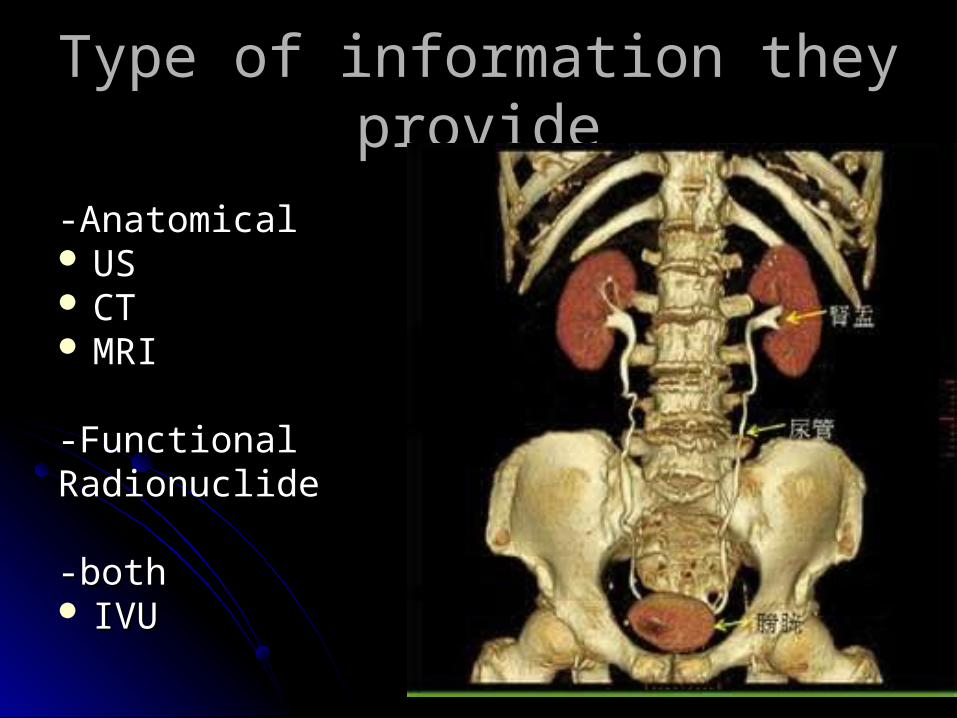
-Anatomical-Anatomical USUS CTCT MRIMRI
-Functional-FunctionalRadionuclide Radionuclide
-both-both IVU IVU
Type of information they provideType of information they provide
ULTRASOUNDULTRASOUND Is the first line investigationIs the first line investigation
Non invasive , no radiation, no contarst…..Non invasive , no radiation, no contarst…..
The main uses of ultrasound : The main uses of ultrasound :
renal symptomsrenal symptoms
renal sizerenal size
hydronephrosishydronephrosis
renal masses renal masses
bladder and prostate bladder and prostate
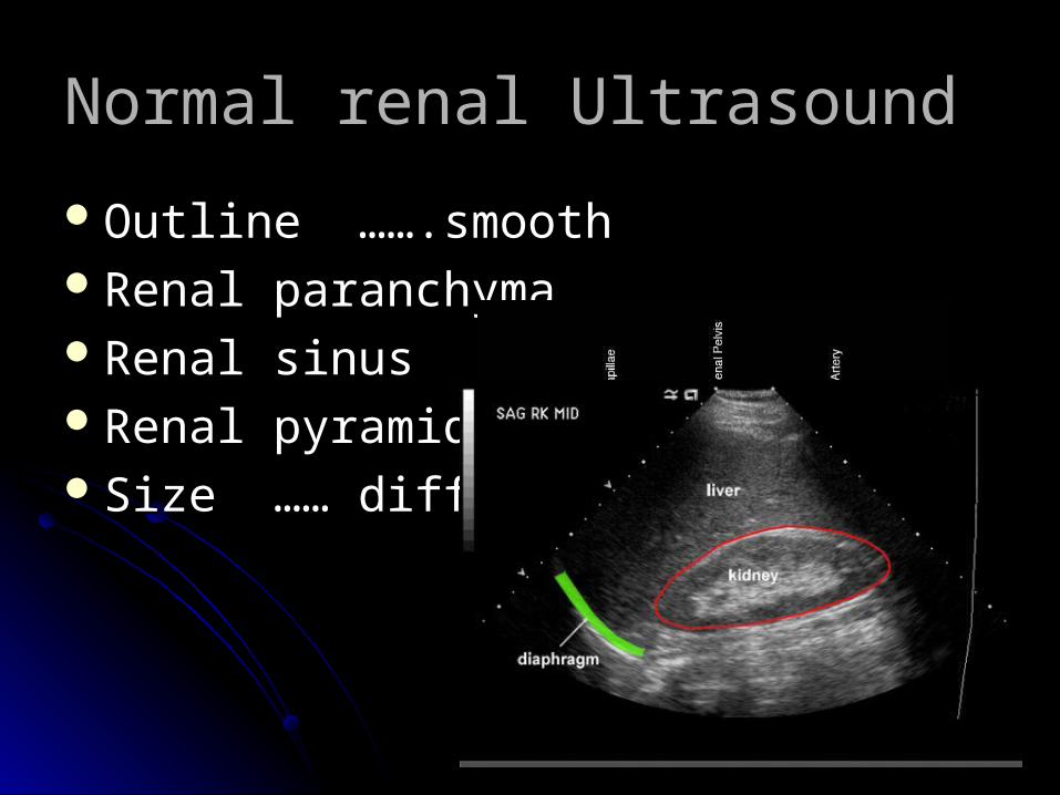
Normal renal Ultrasound Normal renal Ultrasound
Outline …….smooth Outline …….smooth Renal paranchymaRenal paranchymaRenal sinus ..cental partRenal sinus ..cental partRenal pyramidsRenal pyramidsSize …… difference <1.5 cm Size …… difference <1.5 cm
Small kindeysSmall kindeys Unilateral but may be bilateral :Unilateral but may be bilateral :
chronic infection ( including TB) chronic infection ( including TB)
obstructive atrophyobstructive atrophy
RAS RAS
HypoplasticHypoplastic Always bilateral :Always bilateral :
Chronic glomerulonephritisChronic glomerulonephritis
Hypertensive nephropathyHypertensive nephropathy
DM, DM,
collagen vascular diseasecollagen vascular disease
Enlarged kidneysEnlarged kidneys Always unilateral …….compensatory hypertophyAlways unilateral …….compensatory hypertophy May be unilateral or bilateral :May be unilateral or bilateral :
bifid collecting systembifid collecting system
renal mass renal mass
hydronephrosishydronephrosis
lymphoma ( masses or just enlargement)lymphoma ( masses or just enlargement)
renal vein thrombosesrenal vein thromboses Always bilateral :Always bilateral :
PCK (polycystic disease) PCK (polycystic disease)
AGN (acute glomerulonephritis)AGN (acute glomerulonephritis)
amyloidosisamyloidosis
Ureters ……..usually not visualizedUreters ……..usually not visualized
Urinary bladder Urinary bladder
examined in distended state ….thin walls examined in distended state ….thin walls
following micturition….. For residual vol. following micturition….. For residual vol.
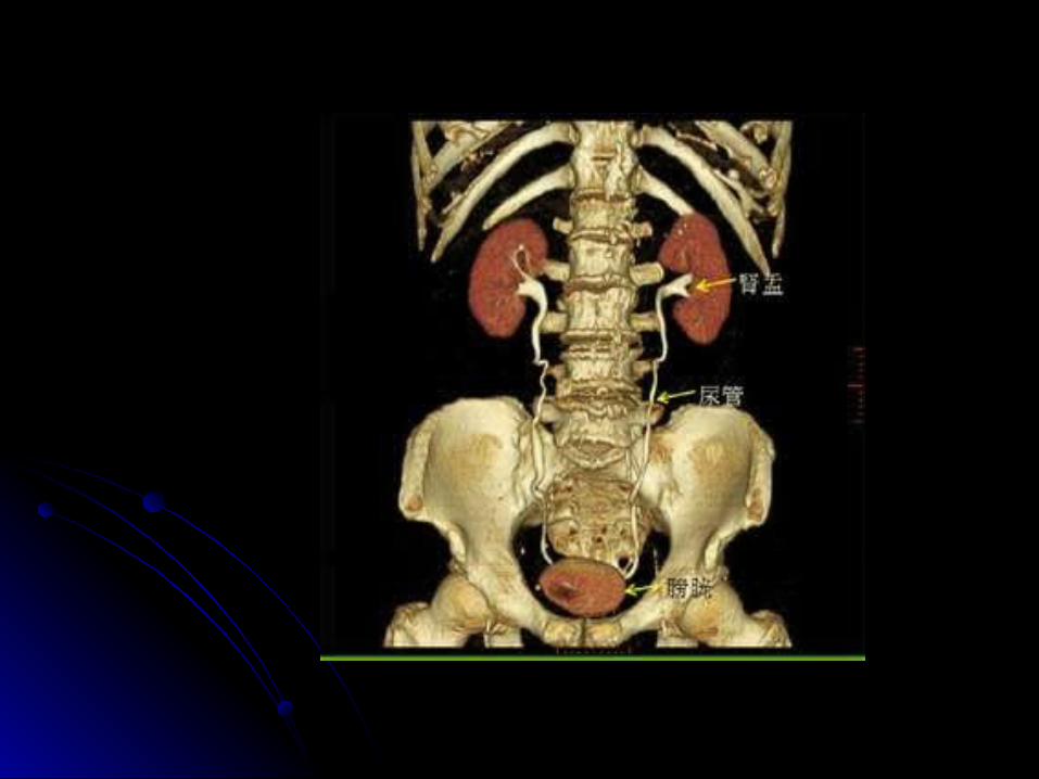
UrographyUrography
Use of IV iodinated contrastUse of IV iodinated contrast
IVUIVU CT UrographyCT UrographyDetails of PCS Renal calculi detection (all types)Details of PCS Renal calculi detection (all types)
Ureteric injury HematuriaUreteric injury Hematuria
Acute ureteric colic Renal massAcute ureteric colic Renal mass
Staging and follow up of CAStaging and follow up of CA
Renal vascular anatomyRenal vascular anatomy
renal trauma renal trauma
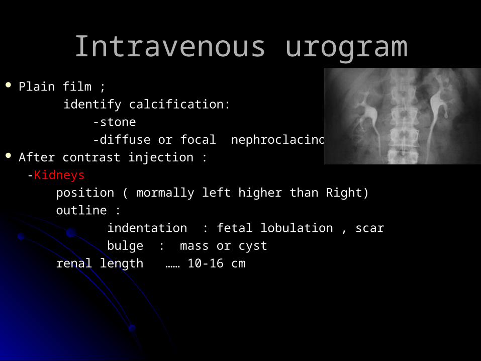
Intravenous urogramIntravenous urogram Plain film ; Plain film ;
identify calcification: identify calcification:
-stone-stone
-diffuse or focal nephroclacinosis -diffuse or focal nephroclacinosis After contrast injection :After contrast injection :
--KidneysKidneys
position ( mormally left higher than Right) position ( mormally left higher than Right)
outline : outline :
indentation : fetal lobulation , scarindentation : fetal lobulation , scar
bulge : mass or cyst bulge : mass or cyst
renal length …… 10-16 cm renal length …… 10-16 cm
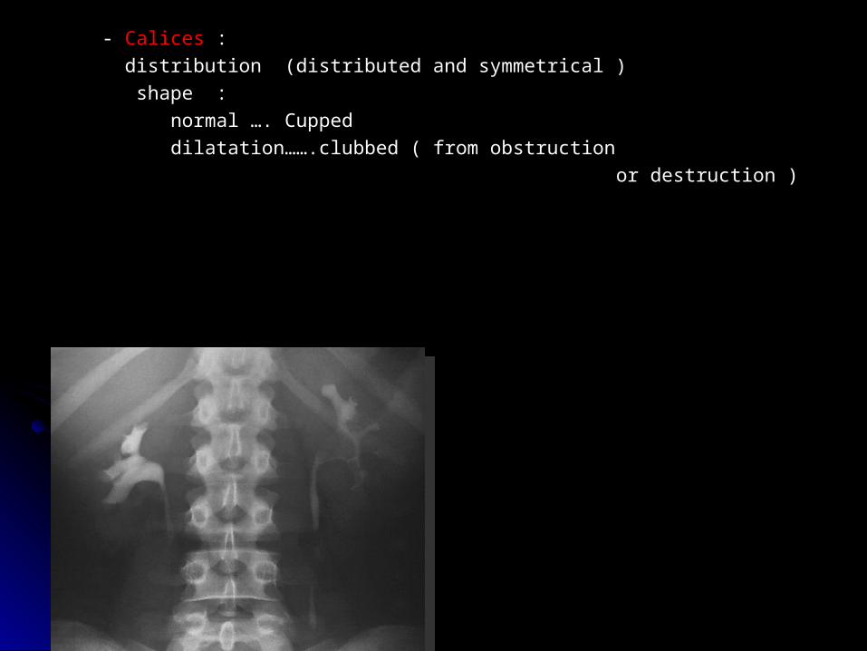
- - CalicesCalices : :
distribution (distributed and symmetrical ) distribution (distributed and symmetrical )
shape :shape :
normal …. Cupped normal …. Cupped
dilatation…….clubbed ( from obstructiondilatation…….clubbed ( from obstruction
or destruction ) or destruction )
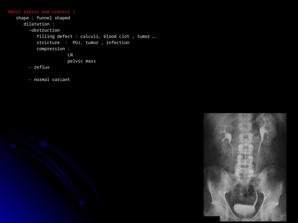
Renal plevis and uretersRenal plevis and ureters : :
shape : funnel shaped shape : funnel shaped
dilatation :dilatation :
-obstruction -obstruction
filling defect : calculi, blood clot , tumor …. filling defect : calculi, blood clot , tumor ….
stricture : PUJ, tumor , infectionstricture : PUJ, tumor , infection
compression : compression :
LN LN
pelvic masspelvic mass
- reflux- reflux
- normal variant - normal variant
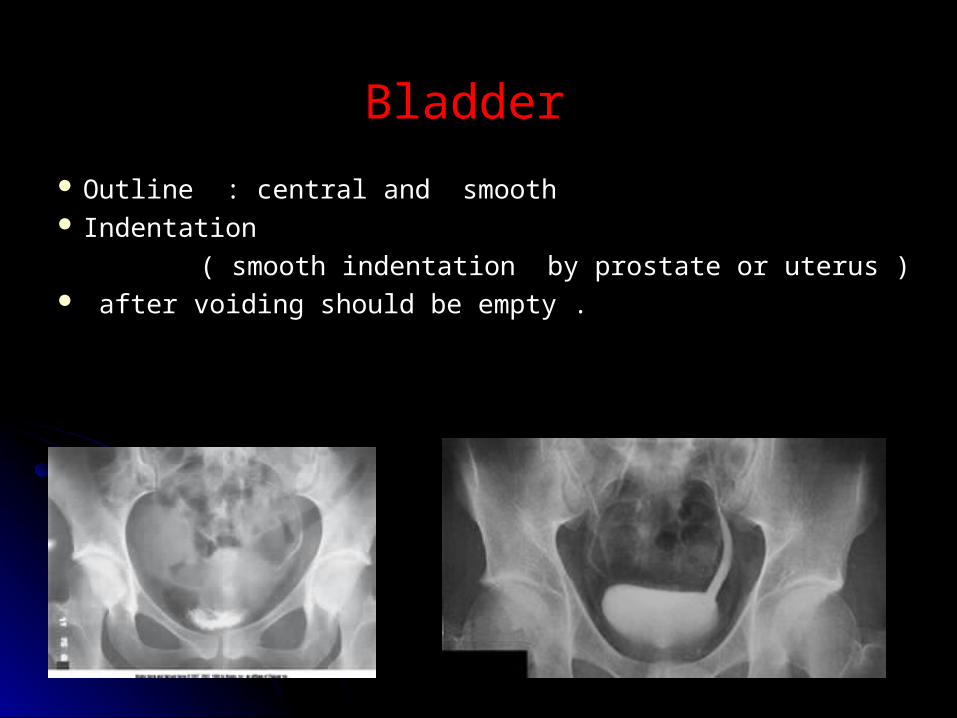
Bladder Bladder
Outline : central and smooth Outline : central and smooth Indentation Indentation
( smooth indentation by prostate or uterus ) ( smooth indentation by prostate or uterus ) after voiding should be empty . after voiding should be empty .
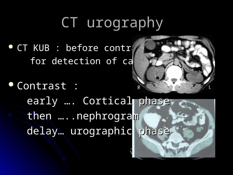
CT urography CT urography
CT KUB : before contrast CT KUB : before contrast
for detection of calcification for detection of calcification
Contrast :Contrast :
early …. Cortical phaseearly …. Cortical phase
then …..nephrogram then …..nephrogram
delay… urographic phasedelay… urographic phase
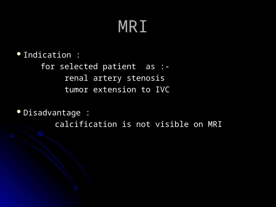
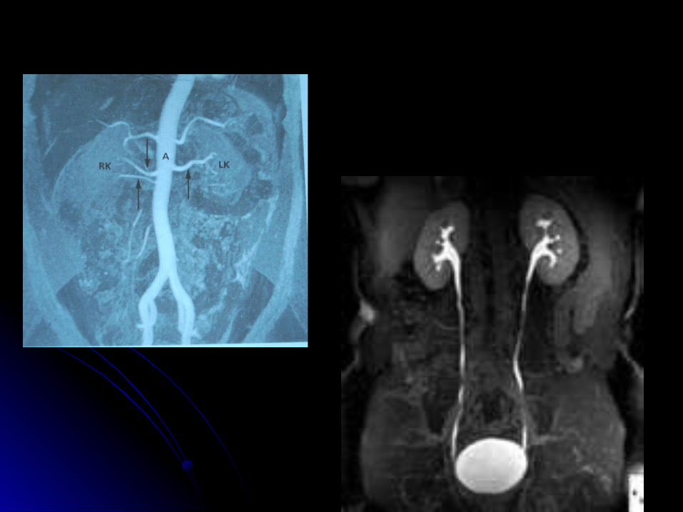
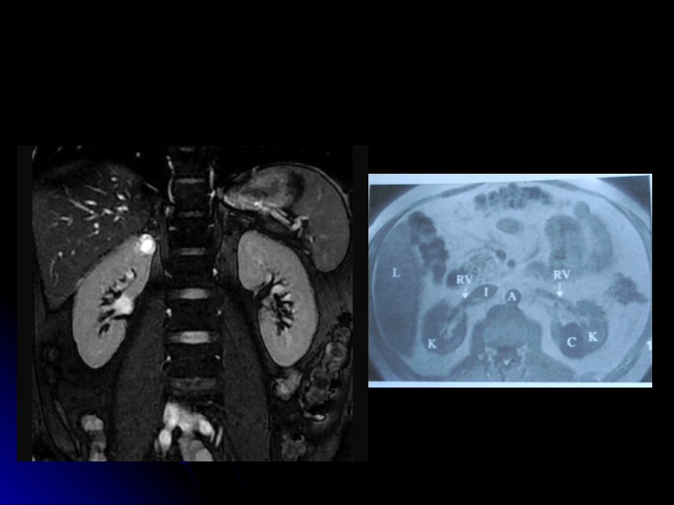
MRI MRI
Indication : Indication :
for selected patient as :- for selected patient as :-
renal artery stenosisrenal artery stenosis
tumor extension to IVC tumor extension to IVC
Disadvantage : Disadvantage :
calcification is not visible on MRIcalcification is not visible on MRI
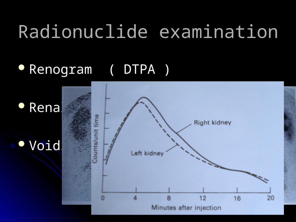
Radionuclide examination Radionuclide examination
Renogram ( DTPA ) Renogram ( DTPA )
Renal morphology (DMSA scan) Renal morphology (DMSA scan)
Voiding cystography Voiding cystography

Special techniques Special techniques
Retrograde and antegrade pyelographyRetrograde and antegrade pyelography
Micturating cystogram Micturating cystogram
UrethrographyUrethrography
Renal arteriographyRenal arteriography

Questions?Questions?