urinalysis and body fluids crg unit 2; session 5 crystals found in the urine microscopic examination...
TRANSCRIPT
Urinalysis and Body Fluids CRg
Unit 2; Session 5
Crystals Found in the Urine Microscopic Examination - Part B, Common Acid Crystals

• Not significant, but have been found in calculi
• Not present in fresh warm urine• Performing all urine tests asap will improve
quality of results.
• Broadly categorized by pH• Sometimes may not obey the rules
Microscopic Sediment – Urine Crystals
Obj. Explain why performing all aspects of the urinalysis as soon as possible will provide the most accurate assessment of macroscopic and microscopic characteristics.Obj. Identify or describe the normal and abnormal constituents that may be seen in the urine microscopic including…….cells… acid, alkaline and pathologically significant crystals….
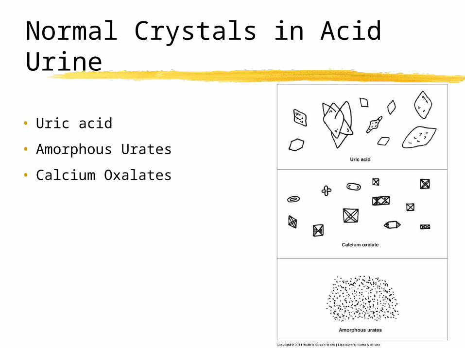
• Uric acid
• Amorphous Urates
• Calcium Oxalates
Normal Crystals in Acid Urine

Normal Crystals in Acid Urine• Amorphous urates
• Amorphous = no shape• Macroscopically
• Urine sediment has pink color due to the pigment uroerythrin attaching on surface of granules
• Microscopically • Red -Yellow-brown granules
• May clump or resemble casts• Soluble in alkali• Dissolve with
gentle heat

• Uric acid • Yellow-brown color. Will polarize. • Pleomorphic
• Diamond shaped, barrels, rhombic / rhomboid, whetstones, wedges, rosettes, needles, etc
• Hexagonal shape may resemble the pathological crystal – cystine
• Generally not significant, • Pathologic only when seen in freshly voided urine• ↑ purines, nucleic acids• Often associated with gout. • Also may see ↑ in leukemia & in patients on
chemotherapy.
Normal Crystals in Acid Urine

Uric Acid Crystals
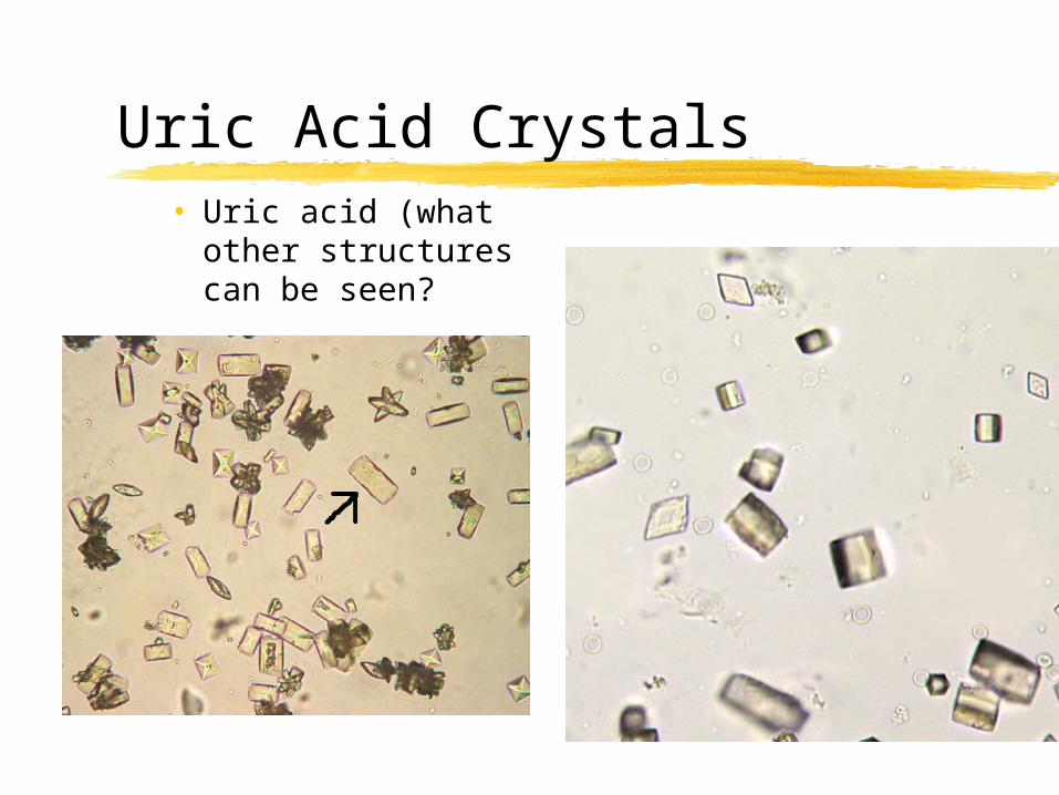
• Uric acid (what other structures can be seen?
Uric Acid Crystals

• Acid and neutral pH• Calcium oxalate is a major
component of renal calculi / lithiasis• @ 75% composed of CaOx
• Also capable of several shapes
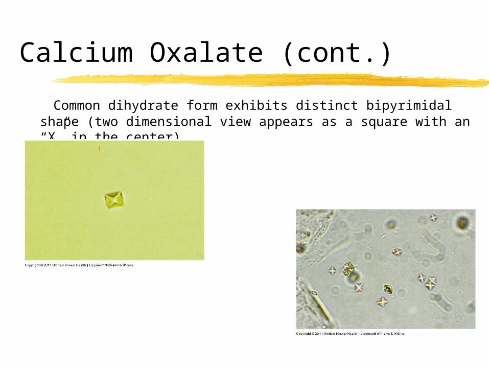
• Dihydrate (2 water molecules) is envelope or two-pyramid–shaped• Most common
Calcium Oxalate Crystals
Calcium Oxalate (cont.)
Common dihydrate form exhibits distinct bipyrimidal shape (two dimensional view appears as a square with an “X” in the center).
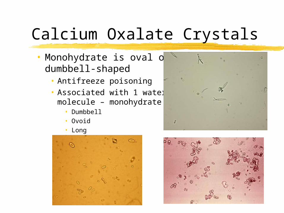
• Monohydrate is oval or dumbbell-shaped• Antifreeze poisoning• Associated with 1 water
molecule – monohydrate• Dumbbell• Ovoid• Long
Calcium Oxalate Crystals
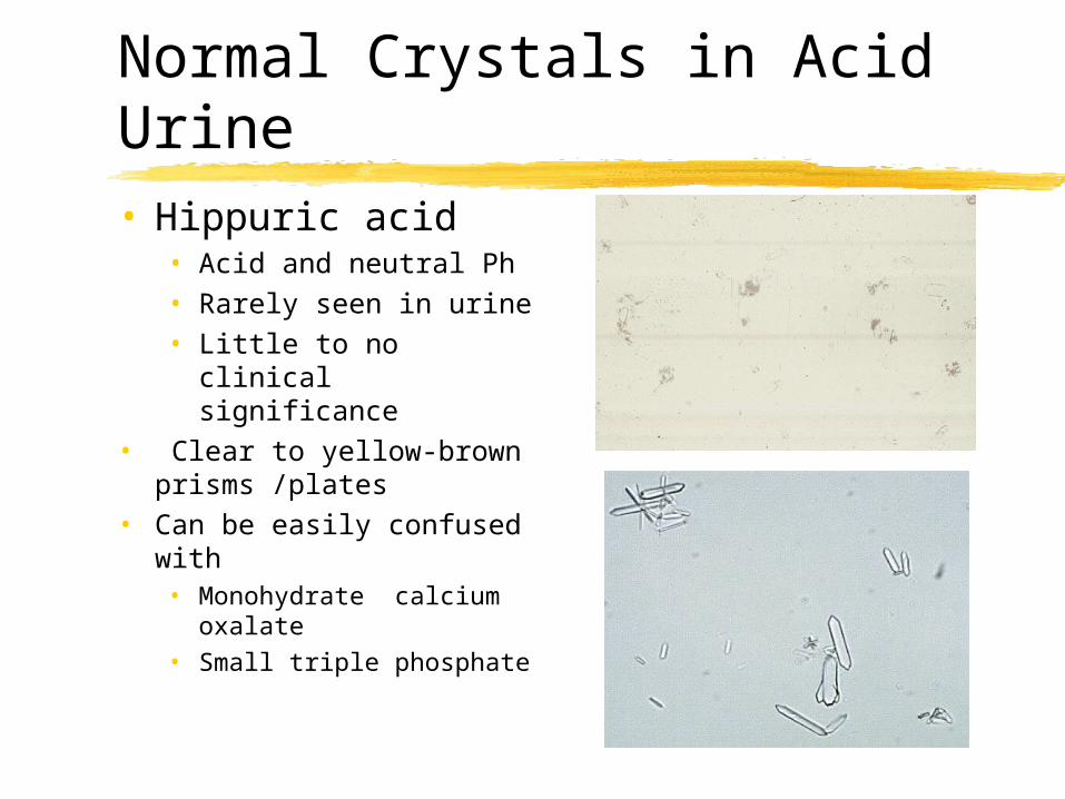
• Hippuric acid • Acid and neutral Ph• Rarely seen in urine• Little to no clinical
significance• Clear to yellow-brown
prisms /plates• Can be easily confused
with • Monohydrate calcium
oxalate• Small triple phosphate
Normal Crystals in Acid Urine
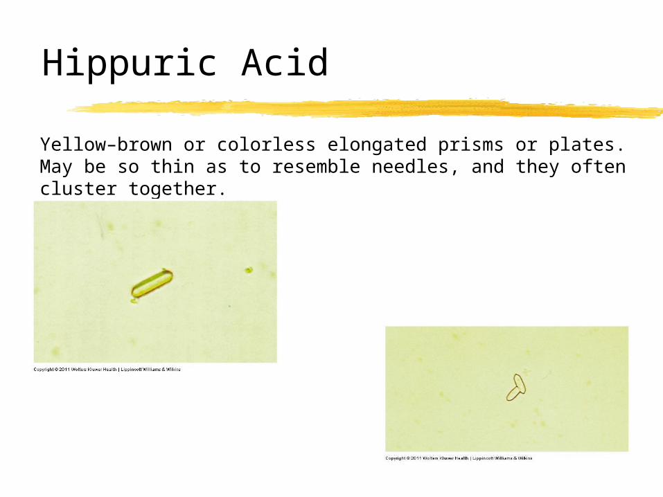
Yellow–brown or colorless elongated prisms or plates. May be so thin as to resemble needles, and they often cluster together.
Hippuric Acid
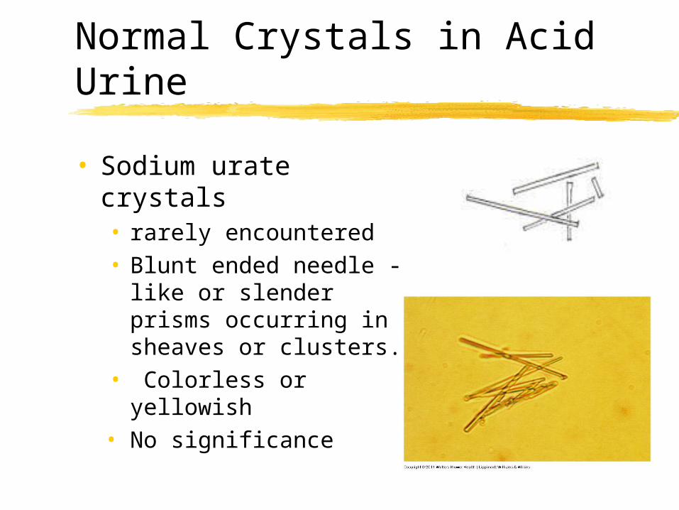
• Sodium urate crystals• rarely encountered• Blunt ended needle - like
or slender prisms occurring in sheaves or clusters.
• Colorless or yellowish• No significance
Normal Crystals in Acid Urine
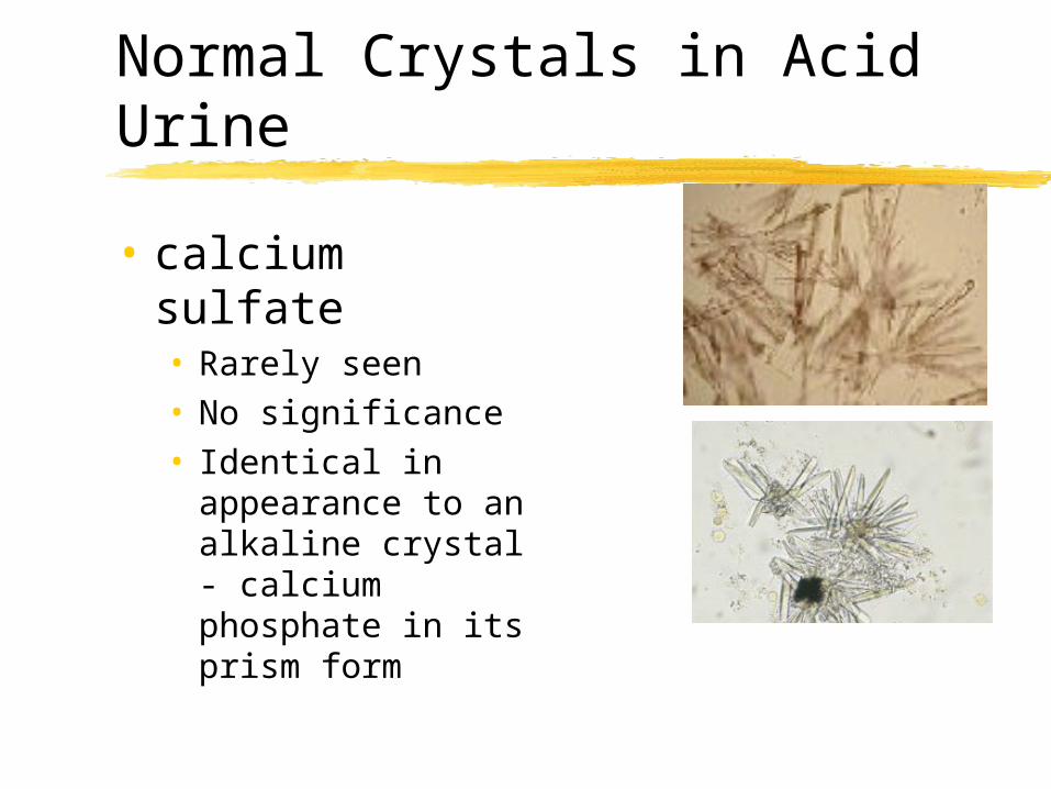
• calcium sulfate• Rarely seen• No significance • Identical in
appearance to an alkaline crystal - calcium phosphate in its prism form
Normal Crystals in Acid Urine

Lillian Mundt & Kristy Shanahan, Graff’s Textbook of Urinalysis and Body Fluids, 2nd Ed.
Susan Strassinger & Marjorie Di Lorenzo, Urinalysis and Body Fluids, 5th Ed.
Meryl Haber, MD, A Primer of Microscopic Urinalysis, 2nd Ed. Nikon Microscopy, The Source for Microscopy Education. Website
http://www.microscopyu.com/articles/polarized/polarizedintro.html
References