university of pennsylvania annual progress report: 2010 ... of... · university of pennsylvania ....
TRANSCRIPT
_____________________________________________________________________________________________Pennsylvania Department of Health – 2012-2013 Annual C.U.R.E. Report
University of Pennsylvania – 2010 Formula Grant – Page 1
University of Pennsylvania Annual Progress Report: 2010 Formula Grant Reporting Period July 1, 2012 – June 30, 2013 Formula Grant Overview The University of Pennsylvania received $8,236,620 in formula funds for the grant award period January 1, 2011 through December 31, 2014. Accomplishments for the reporting period are described below. Research Infrastructure Project 1: Project Title and Purpose Research Infrastructure: Renovation for Laboratory Space for Biological Chemistry - The purpose of this project is to upgrade and renovate 4186 square feet of laboratory space to standards appropriate for a 21st century laboratory performing research at the interface between chemistry and biology. The renovations include outfitting the laboratory space with modern fume hoods, electric, air-handling as well as the creation of a room for radioactive work and a dark room for work with light sensitive nucleic acids. The renovated space will be occupied by researchers from the Chemistry Department whose research includes work at the frontiers ofgene regulation, magnetic resonance imaging, and mechanistic studies of anesthesia. Renovations will also include the creation of a shared Biochemistry Instrumentation Core Facility in the Department of Chemistry. Duration of Project 1/1/2011 - 6/30/2012 Summary of Research Completed This project ended during a prior state fiscal year. For additional information, please refer to the Commonwealth Universal Research Enhancement Annual C.U.R.E. Reports on the Department's Tobacco Settlement/Act 77 web page at http://www.health.state.pa.us/cure. Research Infrastructure Project 2: Project Title and Purpose Enhancing Cognitive Neuroscience and Neuroimaging Research at Penn – Research Infrastructure - In order to support interdisciplinary lines of inquiry related to the areas of cognition, cognitive neurology, brain imaging, and cognitive neuroscience, the Centers for Cognitive Neuroscience (CCN) and Functional Neuroimaging (CFN) will be coalesced within 40,000 square feet of newly renovated space at Penn. This multi-phase project – approximately 20,000 sf for Phase II, which is described in this strategic plan – will unite members of the CCN
_____________________________________________________________________________________________Pennsylvania Department of Health – 2012-2013 Annual C.U.R.E. Report
University of Pennsylvania – 2010 Formula Grant – Page 2
who study cognitive effects and defects with members of the CFN who develop the machine and mathematical interfaces for imaging. The resultant facility will provide access to emerging technologies and shared resources and dramatically promote collaborative science within the neurosciences. This facility will also foster the recruitment and development of new faculty and programs. Anticipated Duration of Project 3/1/2011 – 12/31/2014 Project Overview Cognitive neuroscience is a rapidly evolving field with broad consequences for human health and society. It includes research on normal and abnormal brain development, normal behavior and the effects of brain injury and disease. It integrates concepts from the study of basic human cognition, broadly construed to include emotion, with new noninvasive imaging modalities and other methods to study brain function in healthy subjects and patients with neurological and psychiatric disorders. Penn has 16 core faculty scientists working on cognitive neuroscience and neuroimaging in eight different locations scattered across its campus. This multi-phase project will support the renovation of approximately 40,000 square feet of centrally located space to support their interdisciplinary research programs and facilitate interaction. An initial phase which is already underway involves the renovation of two floors (10,000 square feet) of the Goddard Building for the Center of Cognitive Neurosciences (CCN). This is scheduled for completion by early 2011. In Phase II, supported by this project, completion of an additional 20,000 square feet will enable us to consolidate all CCN components and to integrate faculty from the Center for Functional Neuroimaging (CFN) in collaborative research efforts. The long-term goal and the future project phase of the proposed space will also house key instrumentation including MRI, EEG/ERP, TMS, testing, and advanced computing, and will dramatically improve the quality of lab space for many of the faculty who currently carry out their research under substandard conditions. The University of Pennsylvania is one of the nation’s premier biomedical research institutions and neuroscience has been a strategic focus of Penn’s interdisciplinary research efforts for over five decades. This new Center for Cognitive Neuroscience and Neuroimaging will unite Penn’s talented faculty in a highly collaborative, multi-method research facility enabling them to explore the emerging frontiers of cognitive neuroscience as it relates to human health and disease. This project will also create a geographical focal point on the medical campus for clinical researchers to access collaboration and training in cognitive neuroscience and increase energy efficiency and reduce environmental impact of a national landmark research building complex.
_____________________________________________________________________________________________Pennsylvania Department of Health – 2012-2013 Annual C.U.R.E. Report
University of Pennsylvania – 2010 Formula Grant – Page 3
Principal Investigator Glen N. Gaulton, PhD Executive Vice Dean and Chief Scientific Officer University of Pennsylvania School of Medicine 240 John Morgan Building 3620 Hamilton Walk Philadelphia PA 19104-6160 Other Participating Researchers John A. Detre, MD, H. Branch Coslett, MD – employed by University of Pennsylvania Expected Research Outcomes and Benefits This project will support the renovation of approximately 20,000 square feet of centrally located space to coalesce and expand the interdisciplinary research programs of the Center for Cognitive Neuroscience (CCN) and the Center for Functional Neuroimaging (CFN). Over the years these two complementary Centers have grown in faculty number, and flourished in terms of research productivity, external funding, and intellectual leadership. However, they have flourished despite suboptimal research facilities and geographic fragmentation that has made collaboration challenging and has limited further programmatic growth. To fully benefit from Penn’s extensive intellectual resources in cognitive neuroscience, as well as to attract the best new faculty and trainees, Penn seeks to create an environment that more effectively supports this essential multidisciplinary collaboration. An exceptional opportunity to realize this synthesis of scientific team, research target, and methodological technique now presents itself. With strong support of both University and School of Medicine leadership, the proposed project will integrate the faculty, research laboratories, and infrastructure of the CCN and the CFN to form a world-class neurobehavioral research institute with multidisciplinary expertise in an extremely interactive and collaborative environment. The major need is for contiguous space to accommodate 16 principal faculty and their associates including graduate students, postdoctoral fellows, research faculty and administrative staff, along with seminar space and associated dry lab facilities for testing neurological and psychiatric patients. Evaluation of this project will be based on two sets of criteria: (1) the successful construction of research space in support of the mission and goals of the Penn Center for Cognitive Neuroscience and Neuroimaging; and (2) the number of extramural sponsored projects awarded to Center investigators. Summary of Research Completed Over the last year, the team procured services of a construction manager to work with the architect, EYP, in developing construction documents and detailed estimates for the Phase II
_____________________________________________________________________________________________Pennsylvania Department of Health – 2012-2013 Annual C.U.R.E. Report
University of Pennsylvania – 2010 Formula Grant – Page 4
project. The team is in the process of creating mockups for interior and exterior elements of the building. Construction will occur while half of the building is still occupied; therefore, the team is developing strategies for completing renovation around operational areas. Research Project 3: Project Title and Purpose
Control of Somatic Stem Cell Potency and Tumorigenesis by Musashi RNA Binding Proteins - Mammalian genetic studies have suggested an interconnection between mechanisms governing the symmetry of cell division, somatic stem cell potency, and cancer progression. The Msi RNA binding proteins govern asymmetric cell division and have been implicated in stem cell potency and tumorigenesis in the hematopoietic system, providing a potential link between these processes. In the intestine, Msi proteins are expressed in putative stem cell compartments where asymmetric cell division is observed and in colorectal cancers. We propose to determine the functional contribution of Msi proteins to the progression of colorectal cancer. Gain and loss of function approaches for Msi proteins in a murine model of colorectal cancer will provide a foundation for determining the clinical value of Msi proteins as diagnostic markers and therapeutic targets.
Anticipated Duration of Project 1/1/2011 - 12/31/2014 Project Overview The drosophila RNA binding protein musashi and its mammalian orthologs Msi1 and Msi2 have been shown to govern asymmetric cell division as well as stem cell potency and tumorigenesis in the hematopoietic system, potentially providing a common link between these three processes. In the intestine, Msi proteins are expressed in putative stem cell compartments where asymmetric cell division is observed in homeostatic tissue. Activating mutations in the Wnt signaling pathway initiate the progression of colorectal cancer and abrogate the observed asymmetric mitoses. Here we propose to determine the functional contribution of Msi proteins to the onset and progression of colorectal cancer. Both gain and loss of function approaches for Msi1 and Msi2 will be used in a murine model of colorectal cancer driven by activating Wnt mutations in intestinal stem cells. Drug-inducible ectopic Msi1 and Msi2 expression in the intestine will determine the oncogenic capacity of Msi proteins, and conditional deletion of Msi1 and Msi2 both broadly and in tumor-initiating stem cells at the onset and during the progression of colorectal cancer will precisely determine the contribution of these genes in this stem cell-driven disease. Our findings will provide novel and valuable insight into the mechanisms by which regulators of asymmetric cell division impact both epithelial stem cell function and oncogenic transformation and will provide a foundation for determining the clinical value of Msi proteins as diagnostic markers and therapeutic targets. Specific Aims: Aim 1 - Identification of Msi-expressing cells in APCmin-driven cancer Aim 2 - Determining potential oncogenicity of Msi proteins in the intestine Aim 3 - Effects of Msi inactivation during APCmin-driven tumorigenesis
_____________________________________________________________________________________________Pennsylvania Department of Health – 2012-2013 Annual C.U.R.E. Report
University of Pennsylvania – 2010 Formula Grant – Page 5
Principal Investigator Christopher J. Lengner, PhD Assistant Professor University of Pennsylvania School of Veterinary Medicine 3800 Spruce Street Philadelphia, PA 19104 Other Participating Researchers Ning Li, – employed by University of Pennsylvania Ryan Cedeno- employed by University of Pennsylvania Expected Research Outcomes and Benefits Based on our prior knowledge that Msi is a critical regulator of asymmetric cell division and neuronal stem cell fate in Drosophila melanogaster coupled with our published findings in the hematopoietic system where we demonstrate that Msi2 is a potent cooperative oncogene that promotes a stem cell-like phenotype in both human and mouse models of leukemia, we believe that there is a high likelihood that Msi proteins play an important role in the ontogeny of human colorectal cancers. Our preliminary results demonstrate that, when ectopically expressed in normal intestinal epithelium, Msi1 rapidly activates powerful oncogenic pathways known to contribute to colorectal cancer progression. Further, the observation of functional redundancy of the two Msi proteins in neural progenitors couple with our observations that both family members are coexpressed in the intestinal crypt strongly suggest that these proteins play a critical role in the biology of the intestinal stem cell compartment. These observations provided the impetus for generation of the dual-Msi conditional mouse models that we will employ in this project. The fidelity of the APCmin/+ model in mimicking progression of the human disease also gives us confidence that any functional requirement uncovered for Msi proteins in colorectal oncogenesis will be applicable to the human disease. These proposed studies will therefore begin to determine the potential use of Msi proteins as diagnostic and/or therapeutic targets and the mouse models developed for this project will provide a powerful platform for potential testing of diagnostic and therapeutic agents in the future. Summary of Research Completed Here we summarize the progress on the project during its second full year of funding (July 1, 2012- June 30, 2013). We have made significant progress on all three aims. Aim 1: As indicated in our prior progress report, we have completed Aim1. We have identified Msi1 and Msi2 expression in both the APCmin/+ mouse model of colorectal cancer, in human colorectal cancer patient samples, and in human patient-derived colorectal cancer cell lines. Aim 2: In this year we have nearly completed the goals of specific Aim2. In our last progress report, we presented data on the effects of Msi1 activation in the intestinal epithelium. Msi1 activation is known to occur upon loss of the APC tumor suppressor, so in order to determine the
_____________________________________________________________________________________________Pennsylvania Department of Health – 2012-2013 Annual C.U.R.E. Report
University of Pennsylvania – 2010 Formula Grant – Page 6
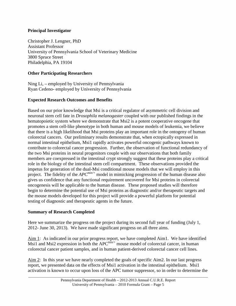
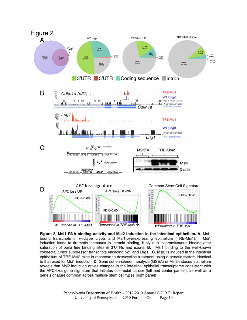
contribution of Msi induction to the APC loss phenotype, we examined the effects of drug-inducible Msi1 activation (TRE-Msi1) in the intestinal epithelium. Briefly, we observed that Msi1 induction alone is sufficient to recapitulate many of the phenotypic and molecular effects of APC loss including crypt fission, increased proliferation in the crypt, a block in differentiation, an activation of the APC-loss transcriptome profile, and robust induction of the AKT-mTorc1 pathway. Msi1 drove these effects independently of APC inactivation or B-catenin activation (the canonical oncogene suppressed by APC). Our findings provided compelling evidence that Msi1 activation lies in a parallel oncogenic pathway to B-catenin activation downstream of APC. In this year, we have expanded on these findings by employing highly sensitive single-cell mRNA profiling techniques using the Fluidigm Biomark microfluidics platform to assess the effects of Msi1 induction on specific stem cell populations in the intestine. This is of great interest as published studies have demonstrated that loss of the APC tumor suppressor has oncogenic consequences only when it occurs in a stem cell, thus suggesting that the cell of origin in colorectal cancer must be a stem cell. The intestinal epithelium contains two functionally distinct stem cell populations: an actively cycling stem cell marked by expression of Lgr5 that is believed to provide the bulk of the daughter cells responsible for homeostatic tissue function, and a second, slow cycling reserve stem cell population marked by Bmi1 that can generate Lgr5+ cycling stem cells in response to tissue damage. We employed Lgr5 and Bmi1 reporter mice in order to determine the effects of Msi1 activation on these intestinal stem cell types. Single stem cells were sorted from Tre-Msi1::Lgr5-eGFP mice or Tre-Msi1::Bmi1-CreER::Lox-Stop-Lox-tdTomato mice (Figure 1A) in the presence or absence of Msi1 induction and profiled on the Fluidigm platform for expression of genes associated with common congenic pathways (Wnt, Notch), proliferation, markers of differentiation, and markers of stem cells. Remarkably, we observed that Msi1 activation specifically affected the quiescent stem cell population marked by activity of the Bmi1 knock-in reporter (Figure 1B). Essentially, Msi1 induction in this reserve stem cell population drove these cells out of their quiescent state, driving expression of pro-mitotic and metabolic genes known to be downstream of the AKT-mTorc1 pathway including Hif1α and H6PD (Figure 1C). The results of this study are currently under consideration for publication. We have now completed the generation of high-quality datasets for both Msi1 and Msi2 RNA binding targets in the intestinal crypts and in transformed epithelium. We have analyzed the Msi1RNA-binding targets and have identified numerous tumor suppressor genes and differentiated-related transcripts bound by Msi1 (Figure 2A-B). This finding is consistent with a role for Msi1 in translational suppression as inactivation of these transcripts accounts for the increased proliferation and differentiation block observed in TRE-Msi1 mice. Currently, the Msi2 dataset is being analyzed and overlayed with the Msi1 dataset in order to determine common vs. distinct RNA binding targets of these family members. In addition to the characterization of Msi2 binding targets, we have nearly completed phenotypic analysis of the effects of Msi2 induction in the intestinal epithelium of TRE-Msi2 mice. Similar to Msi1, Msi2 induction imparts an APC-loss transcriptome profile to the intestinal epithelium, consistent with Msi2 having an analogous role to Msi1 in driving colorectal cancers (Figure 2 C-D). This observation is further consistent with our findings in human clinical colorectal cancer samples
_____________________________________________________________________________________________Pennsylvania Department of Health – 2012-2013 Annual C.U.R.E. Report
University of Pennsylvania – 2010 Formula Grant – Page 7
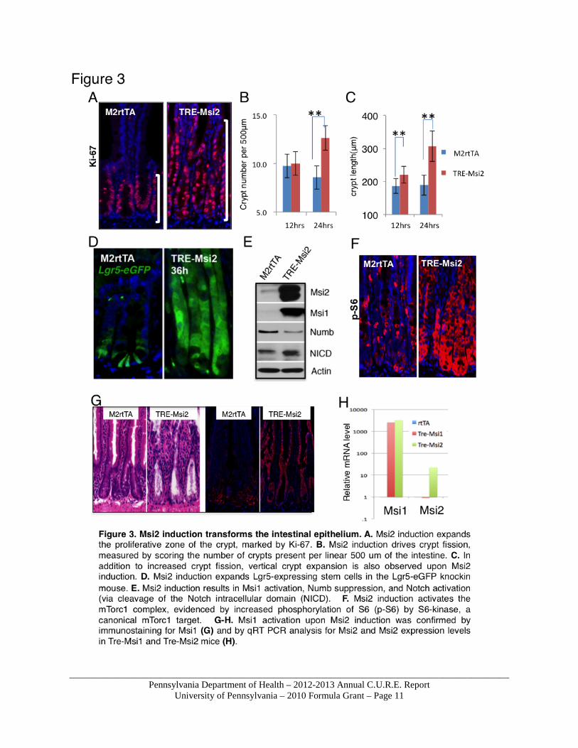
where Msi1 and/or Msi2 are nearly ubiquitously overexpressed. Further, Msi2 drives a common stem cell gene signature in a manner similar to Msi1, suggesting that Msi2 may exert its effects through activation of the quiescent stem cell population as well. The phenotypic consequences of Msi2 induction consist of increased proliferation, crypt height expansion, crypt fission, an increase the frequency of Lgr5-eGFP+ cells in the crypt, a suppression of Numb protein (likely due to translational suppression by Msi), and induction of the mTorc1 pathway (Figure 3 A-F). These phenotypes are all consistent with that of Msi1 induction as well as loss of the APC tumor suppressor. We therefore also examined Msi1 levels in TRE-Msi2 mice and observed a striking activation of Msi1 both at the protein (Figure 3E-F) and RNA (Figure 3H) level. The Msi2-Msi1 crosstalk was not observed in the reciprocal direction. In other words, Msi2 activates Msi1, but Msi1 induction failed to activate Msi2 (Figure 3H). Thus, we cannot formally rule out that the phenotypic consequences of Msi2 induction are not, in fact, due to downstream activation of Msi1. To test this hypothesis, we have bread Tre-Msi2 mice to Msi1flox/flox::VillinCreER mice. This will enable us to delete Msi1 (which we have already established does not affect intestinal homeostasis), and subsequently induce Msi2 and ask whether the phenotype is still fully penetrant. The experimental mice were recently born and these experiments will be carried out shortly. Upon completion of this latter experiment and the comparative analysis of Msi1 and Msi2 RNA-binding datasets, the studies on Msi2 induction will be submitted for publication. Aim 3: In this aim we propose to determine the effects of Msi inactivation during APCmin-driven tumorigenesis. To this end we have generated Msi1flx/flx/Villin-CreER/APCmin/+ and Msi1flx/flx
Msi2flx/flx Villin-CreER APCmin/+ mice. We have deleted the Msi alleles in vivo and maintained mice on high-fat diets to induce tumorigenesis in response to stochastic loss of APC heterozygosity. Our initial results with a cohort of Msi1flx/flx/Villin-CreER/APCmin/+ is that tumorigenesis was not inhibited. This finding indicates a redundant role for Msi2 in tumorigenesis driven by APC loss and further suggests that Msi2 does not rely on Msi1 for its oncogenic properties (as discussed above). Msi1flx/flx Msi2flx/flx Villin-CreER APCmin/+ mice, in which both Msi family members are deleted, are difficult to generate in large numbers and these experiments are ongoing. Preliminary results on a cohort of 6 experimental (Msi1flx/flx Msi2flx/flx
Villin-CreER APCmin/+) and 5 control (APCmin/+) mice indicates an increased lifespan in the absence of both Msi and APC, as APCmin/+ control mice began succumbing to tumor burden after several months of high fat diet. Further, we observed that experimental mice lacking Msi1 had significantly fewer tumors than APCmin/+ control mice, and that most remaining tumors escaped Cre-mediated deletion of Msi1/2. These preliminary findings suggest that Msi activity may, in fact, be required for tumorigenesis driven by APC loss. The requirement for Msi in cancers driven by APC loss is being further explored in larger cohorts of Msi1flx/flx Msi2flx/flx Villin-CreER APCmin/+mice. In addition, we have initiated experiments in a panel of human colorectal cancer cells lines harboring APC inactivating mutations. We have verified that lentiviral delivery of shRNAs targeting Msi abrogates the proliferative potential of these cells in culture, and we are initiating xenograft assays to determine the in vivo oncogenic potential of these cell lines in the absence of Msi. Our progress on the outlined specific aims thus far has revealed that both Msi1 and Msi2 exhibit

_____________________________________________________________________________________________Pennsylvania Department of Health – 2012-2013 Annual C.U.R.E. Report
University of Pennsylvania – 2010 Formula Grant – Page 8
potent oncogenic activity, likely through the proliferative and metabolic activation of slow cycling reserve stem cells of the intestinal epithelium. Further, our preliminary findings indicate that Msi activity may be required for oncogenic transformation of the intestinal epithelium.
_____________________________________________________________________________________________Pennsylvania Department of Health – 2012-2013 Annual C.U.R.E. Report
University of Pennsylvania – 2010 Formula Grant – Page 9
_____________________________________________________________________________________________Pennsylvania Department of Health – 2012-2013 Annual C.U.R.E. Report
University of Pennsylvania – 2010 Formula Grant – Page 10
_____________________________________________________________________________________________Pennsylvania Department of Health – 2012-2013 Annual C.U.R.E. Report
University of Pennsylvania – 2010 Formula Grant – Page 11
_____________________________________________________________________________________________Pennsylvania Department of Health – 2012-2013 Annual C.U.R.E. Report
University of Pennsylvania – 2010 Formula Grant – Page 12
Research Project 4: Project Title and Purpose Translational and Personalized Genomics of Inherited Retinal Degenerations - Inherited retinal degenerations (IRDs) are important causes of blindness. Over 190 different types of IRDs have been identified by clinical and genetic studies. However, we hypothesize that the identified mutations account for only ~50% of patients with these disorders. The purpose of this project is to use next-generation sequencing (NGS) approaches to test the hypothesis that novel IRD disease genes contribute to genetic causes of blindness, and to test the hypothesis that the pathogenesis of retinitis pigmentosa is caused by mutations in RNA splicing factors, with the long term goal of developing genetic therapies for these blinding disorders. Anticipated Duration of Project 1/1/2011 – 12/31/2014 Project Overview The goals of this project are to test the hypothesis that novel unidentified mutations underlie at least 50% of known genetic cases of blindness, and to test the hypothesis that retinitis pigmentosa (RP), a type of IRD, is caused by mutations in genes that encode RNA splicing factors. The specific aims are: Specific Aim 1. Discover novel gene mutations that underlie IRD. The disease genes for ~50% of patients with IRDs remain to be identified. In this Aim we will test the hypothesis that next-generation sequencing (NGS) techniques will facilitate both genetic diagnosis and disease gene discovery in IRD patients. We propose to use selective exon capture and whole exome capture – NGS approaches to identify disease genes in several cohorts of IRD patients. Preliminary experiments using this approach have been successful, with genetic diagnoses achieved for several patients. Specific Aim 2. Identify the role of RNA Splicing Factor in the pathogenesis of RP. The splicing factors affected in the RNA splicing factor forms of RP, pre-mRNA processing factor (PRPF) 3, PRPF8, PRPF31 and SNRNP200 are highly conserved components of the spliceosome, the complex which excises introns from nascent RNA transcripts to generate mature mRNAs. Since RNA splicing is required in all cells, it is not clear how mutations in these ubiquitous proteins lead to retina-specific disease. In this Aim, we will use NGS-based transcriptome analyses to test the hypothesis that mutations in RNA splicing factors cause disease via the generation of aberrant transcripts in the retina and/or retinal pigment epithelium (RPE). We will use three knockin mouse models of the RNA splicing factor forms of RP, and human iPS-derived RPE cells from patients with these disorders for these studies.
_____________________________________________________________________________________________Pennsylvania Department of Health – 2012-2013 Annual C.U.R.E. Report
University of Pennsylvania – 2010 Formula Grant – Page 13
Principal Investigator Junhyong Kim, PhD Co-Director, Penn Genome Frontiers Institute University of Pennsylvania 415 S. University Ave. Philadelphia, PA 19104 Other Participating Researchers Eric Pierce, MD, PhD, James Eberwine, PhD, Jeanne Geskes, BS, Davinder Sandhu, BS, Aaron Goodman, BS, Alex Chekholko, BS, Jean Bennett, MD, PhD, Elizabeth Au, BS, Juan Carlos Perrin, BS – employed by University of Pennsylvania. Xiaowu Gai, PhD – employed by Children’s Hospital of Philadelphia. Expected Research Outcomes and Benefits The completion of this project will help identify novel genetic causes of inherited retinal degenerative (IRD) diseases that lead to blindness. It will also help identify the mechanistic basis of a key subset of retinal degenerative diseases: retinitis pigmentosa. Increasing evidence points to genetic causes of blindness through retinal degeneration. For example, it has been estimated that 75% of macular degeneration, a disease that affects 10 million people a year in the US, has genetic causes. Approximately 190 mutations have been identified that underlie IRD. The results of this study will greatly increase the known mutations that underlie IRD and help develop therapeutic strategies. Retinitis pigmentosa (RP) affects 100,000 people a year in the US. It is currently an incurable disease but its onset and rate of degeneration is highly variable among individuals. While genetic studies have implicated mutations in splicing factors, the precise mechanism by which such ubiquitous molecules play a role in specific eye disease is not known. The results of this study are expected to help identify splicing patterns related to RP that will help determine the specificity mechanism. The results will help dissect pathogenicity mechanism and in the long term aid the development of therapeutic agents. Summary of Research Completed During this period we concentrated on Aim 1 and the following reports our progress on this aim: Aim 1: The disease genes for ~50% of patients with IRDs remain to be identified. We have been testing the hypothesis that NGS techniques will facilitate both genetic diagnosis and disease gene discovery in IRD patients. We are using selective exon capture and whole exome capture – NGS approaches to identify disease genes in several cohorts of IRD patients. In the past year, we have performed genetic analyses for 147 individuals (probands and their close relatives) of families with IRD. In most of these individuals, we first carried out pre-screening of genes associated with clinical diagnosis for which there is a likely disease-causing
_____________________________________________________________________________________________Pennsylvania Department of Health – 2012-2013 Annual C.U.R.E. Report
University of Pennsylvania – 2010 Formula Grant – Page 14
gene. For example, for individuals with choroideremia or retinoschisis, there is a high likelihood that the disease-causing gene is CHM or RS1, respectively. For individuals with Leber’s congenital amaurosis (LCA) and autosomal recessive Stargardt disease, many of the known disease-causing mutations can be identified by Arrayed Primer EXtension (APEX) screening. This screening step identified the disease-causing gene/mutations in approximately one quarter of the families (Table 1). Exon capture and Illumina sequencing was used to screen the remaining probands (and their close relatives, when they were available) for mutations in novel retinal genes. We have also continued to carry out screening on close relatives of probands evaluated in the preceding year (2011-2012) for whom we have not yet made a genetic diagnosis. Results of Exome sequencing for disease gene discovery We have screened for known retinal disease-causing genes as well as using exome sequencing for discovery. Here, we report on our genome-scale efforts. Over the past year, we sequenced exomes from 118 different individuals (probands and close relatives) from families with retinitis pigmentosa (RP) and Leber’s congenital Amaurosis (LCA), Stargardt disease, cone-rod dystrophy, macular degeneration, inherited retinal detachment and several different syndromes, including Heimler Syndrome (macular degeneration, hearing disorder and dental anomalies), a syndrome involving mental retardation and LCA, and Usher’s Syndrome (retinal and cochlear degeneration). We are close to announcing the identification of four additional LCA-causing genes (one of which will be the first X-linked gene correlated with this condition), one new early onset Stargardt disease gene, a form of LCA with mental retardation, and Heimler Syndrome. Conclusive demonstration that these genes/mutations cause the various conditions is currently being established by testing for segregation of the phenotype along with the mutation in additional family members. Where possible, we are also verifying malfunction of the mutant gene in cell culture studies. Examples of the identification of these novel retinal disease-causing /mutations are briefly described as follows: a) Early onset and severe Stargardt disease. A 6yo was evaluated in the clinic for visual symptoms and received the diagnosis of very early onset and severe Stargardt disease (inherited macular degeneration). The pedigree of this family shows autosomal recessive inheritance (Figure 1A). DNA from the probands and his unaffected parents was evaluated by Next Gen sequencing. Rare compound heterozygous mutations were found in a gene that is highly expressed in the retina and associated with cilia formation. There were relatively rare compound heterozygous changes in HEATR5A and rare variants in MT-ATP6, MT-CYB, and HUWE1.
_____________________________________________________________________________________________Pennsylvania Department of Health – 2012-2013 Annual C.U.R.E. Report
University of Pennsylvania – 2010 Formula Grant – Page 15
b) Heimler Syndrome We obtained blood samples from a 13yo boy with Heimler Syndrome and his mother. There were no other affected family members. DNA analyses showed non-synonymous rare variants in two different genes. One of the genes is an intracellular trafficking protein. The other is a homeobox gene thought to be associated with early embryonic development. c) Leber’s Congenital Amaurosis – a Potential new X-Linked Gene Leber’s congenital amaurosis (LCA) is one of the most severe inherited forms of blindness as it is symptomatic in infancy. We studied a 21yo male, JB-17, diagnosed with LCA in infancy. Next Gen sequencing identified a single variant in a gene located on the X chromosome. This protein encoded by this gene is normally expressed in photoreceptors at synaptic termini. JB-17’s visual acuity is approximately 20/1280 and he could not navigate independently even under bright light. We are studying the effects of this new mutation in cell lines using patch-clamping techniques. d) Leber’s Congenital Amaurosis – a Potential new Gene We studied a 23yo female diagnosed with LCA in infancy, JB-148. A screen for mutations in known LCA-causing genes was negative. We obtained a blood sample from her unaffected mother. Genetic analyses of the probands revealed homozygous variants in a retina-specific gene. We are awaiting a sample from her unaffected father to determine whether the gene change segregates with the disease. e) Retinitis pigmentosa (RP) We studied a 94yo male with advanced retinitis pigmentosa. A pedigree was not available upon receipt of his sample. No variants were observed upon screening for known RP-associated genes. DNA analyses revealed a novel hemizygous mutation in the Retinitis Pigmentosa GTPase Regulator (RPGR) gene. The RPGR gene is on the X chromosome, and thus this diagnosis established the inheritance pattern for this individual’s family. Follow-up with his daughter, an obligate carrier, revealed that there were other affected individuals in this family. A pedigree was taken (Figure 1B) and testing for the RPGR mutation was carried out. The affected boy who was identified after this testing was referred to the NEI/NIH, which is planning a gene therapy clinical trial for this condition. Bioinformatics The informatic pipeline to analyze the exon and exome capture data was key to the success of this project and was developed in collaboration with Dr. Xiaowu Gai (Loyola). Candidates identified by bioinformatics are evaluated for the probability that they may be involved in a retinal disease condition and are then screened based on that likelihood. For example, if there is a gene which is thought to play a role in development of cilia or neuronal processes, this is considered to be of greater interest than a gene which might be involved in liver development. Independent sequencing of detected variants has demonstrated a >95% validation rate. The database is continually being annotated as new variants are identified.
_____________________________________________________________________________________________Pennsylvania Department of Health – 2012-2013 Annual C.U.R.E. Report
University of Pennsylvania – 2010 Formula Grant – Page 16
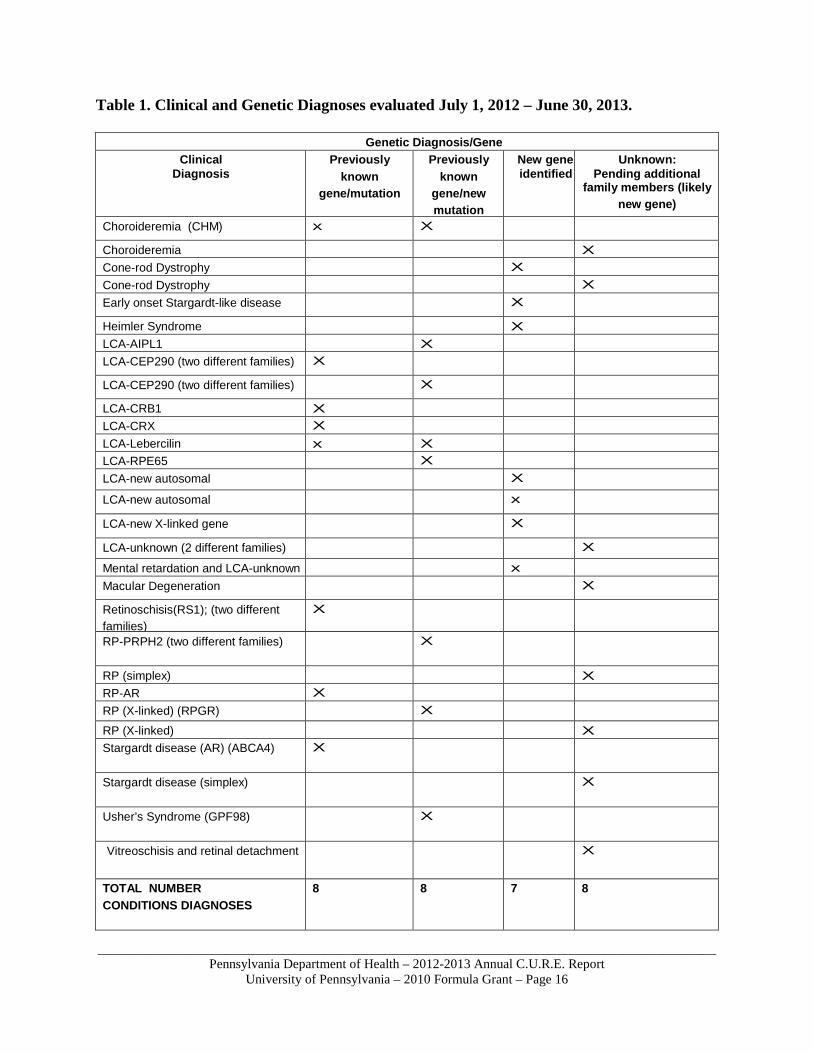
Table 1. Clinical and Genetic Diagnoses evaluated July 1, 2012 – June 30, 2013.
Genetic Diagnosis/Gene Clinical
Diagnosis Previously
known gene/mutation
Previously known
gene/new mutation
New gene identified
Unknown: Pending additional
family members (likely new gene)
Choroideremia (CHM) x X
Choroideremia X Cone-rod Dystrophy X Cone-rod Dystrophy X Early onset Stargardt-like disease X
Heimler Syndrome X LCA-AIPL1 X LCA-CEP290 (two different families) X
LCA-CEP290 (two different families) X
LCA-CRB1 X LCA-CRX X LCA-Lebercilin x X LCA-RPE65 X LCA-new autosomal X
LCA-new autosomal x
LCA-new X-linked gene X
LCA-unknown (2 different families) X Mental retardation and LCA-unknown x Macular Degeneration X
Retinoschisis(RS1); (two different families)
X
RP-PRPH2 (two different families) X
RP (simplex) X RP-AR X RP (X-linked) (RPGR) X
RP (X-linked) X Stargardt disease (AR) (ABCA4) X
Stargardt disease (simplex) X
Usher’s Syndrome (GPF98) X
Vitreoschisis and retinal detachment X
TOTAL NUMBER CONDITIONS DIAGNOSES
8 8 7 8
_____________________________________________________________________________________________Pennsylvania Department of Health – 2012-2013 Annual C.U.R.E. Report
University of Pennsylvania – 2010 Formula Grant – Page 17
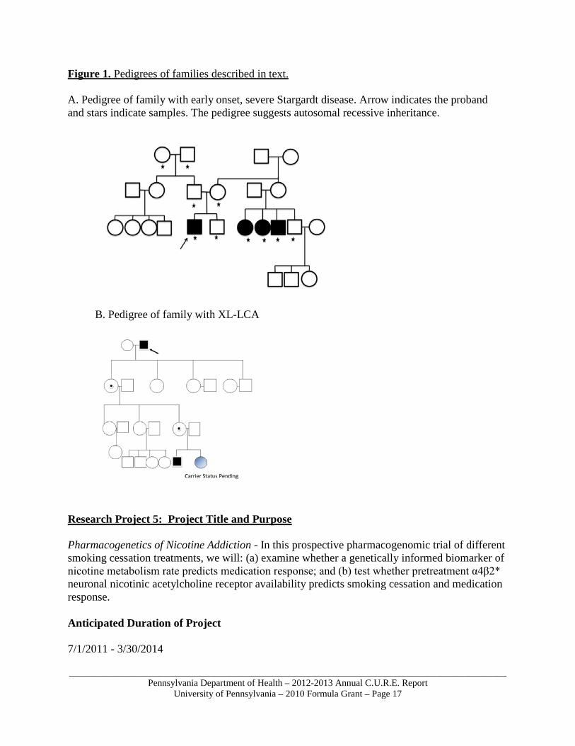
Figure 1. Pedigrees of families described in text. A. Pedigree of family with early onset, severe Stargardt disease. Arrow indicates the proband and stars indicate samples. The pedigree suggests autosomal recessive inheritance.
B. Pedigree of family with XL-LCA
Research Project 5: Project Title and Purpose Pharmacogenetics of Nicotine Addiction - In this prospective pharmacogenomic trial of different smoking cessation treatments, we will: (a) examine whether a genetically informed biomarker of nicotine metabolism rate predicts medication response; and (b) test whether pretreatment α4β2* neuronal nicotinic acetylcholine receptor availability predicts smoking cessation and medication response. Anticipated Duration of Project 7/1/2011 - 3/30/2014
_____________________________________________________________________________________________Pennsylvania Department of Health – 2012-2013 Annual C.U.R.E. Report
University of Pennsylvania – 2010 Formula Grant – Page 18
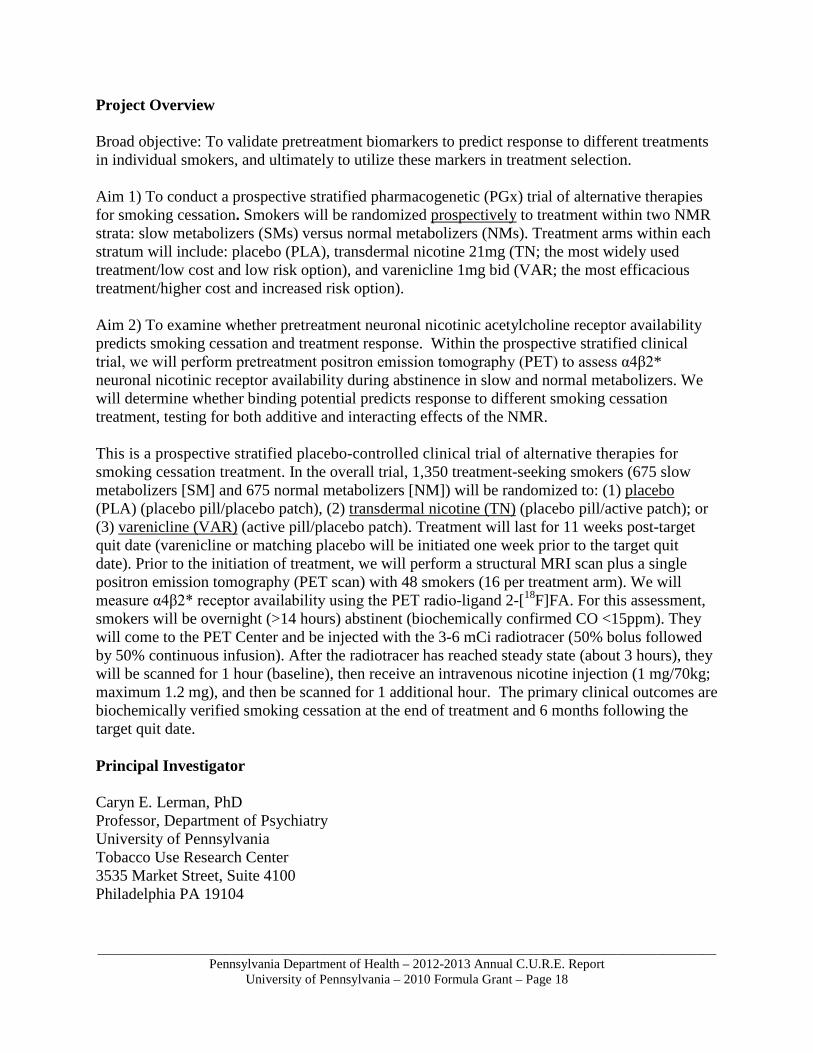
Project Overview Broad objective: To validate pretreatment biomarkers to predict response to different treatments in individual smokers, and ultimately to utilize these markers in treatment selection. Aim 1) To conduct a prospective stratified pharmacogenetic (PGx) trial of alternative therapies for smoking cessation. Smokers will be randomized prospectively to treatment within two NMR strata: slow metabolizers (SMs) versus normal metabolizers (NMs). Treatment arms within each stratum will include: placebo (PLA), transdermal nicotine 21mg (TN; the most widely used treatment/low cost and low risk option), and varenicline 1mg bid (VAR; the most efficacious treatment/higher cost and increased risk option). Aim 2) To examine whether pretreatment neuronal nicotinic acetylcholine receptor availability predicts smoking cessation and treatment response. Within the prospective stratified clinical trial, we will perform pretreatment positron emission tomography (PET) to assess α4β2* neuronal nicotinic receptor availability during abstinence in slow and normal metabolizers. We will determine whether binding potential predicts response to different smoking cessation treatment, testing for both additive and interacting effects of the NMR. This is a prospective stratified placebo-controlled clinical trial of alternative therapies for smoking cessation treatment. In the overall trial, 1,350 treatment-seeking smokers (675 slow metabolizers [SM] and 675 normal metabolizers [NM]) will be randomized to: (1) placebo (PLA) (placebo pill/placebo patch), (2) transdermal nicotine (TN) (placebo pill/active patch); or (3) varenicline (VAR) (active pill/placebo patch). Treatment will last for 11 weeks post-target quit date (varenicline or matching placebo will be initiated one week prior to the target quit date). Prior to the initiation of treatment, we will perform a structural MRI scan plus a single positron emission tomography (PET scan) with 48 smokers (16 per treatment arm). We will measure α4β2* receptor availability using the PET radio-ligand 2-[18F]FA. For this assessment, smokers will be overnight (>14 hours) abstinent (biochemically confirmed CO <15ppm). They will come to the PET Center and be injected with the 3-6 mCi radiotracer (50% bolus followed by 50% continuous infusion). After the radiotracer has reached steady state (about 3 hours), they will be scanned for 1 hour (baseline), then receive an intravenous nicotine injection (1 mg/70kg; maximum 1.2 mg), and then be scanned for 1 additional hour. The primary clinical outcomes are biochemically verified smoking cessation at the end of treatment and 6 months following the target quit date. Principal Investigator Caryn E. Lerman, PhD Professor, Department of Psychiatry University of Pennsylvania Tobacco Use Research Center 3535 Market Street, Suite 4100 Philadelphia PA 19104
_____________________________________________________________________________________________Pennsylvania Department of Health – 2012-2013 Annual C.U.R.E. Report
University of Pennsylvania – 2010 Formula Grant – Page 19
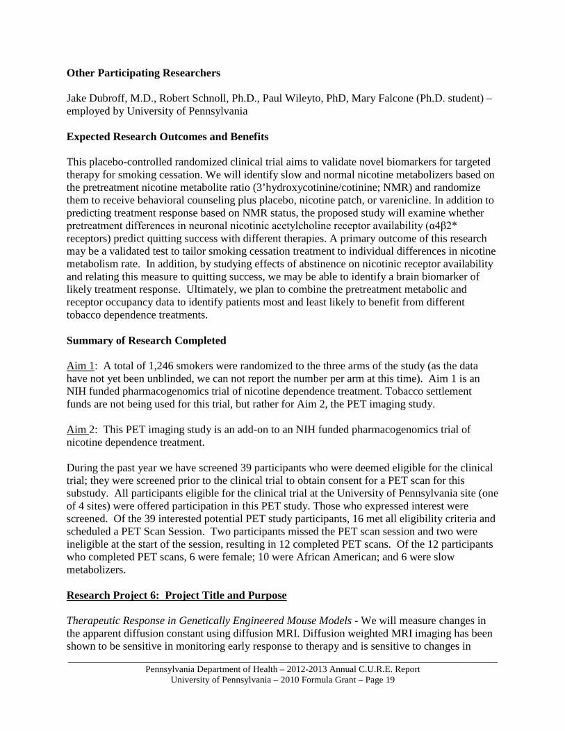
Other Participating Researchers Jake Dubroff, M.D., Robert Schnoll, Ph.D., Paul Wileyto, PhD, Mary Falcone (Ph.D. student) – employed by University of Pennsylvania Expected Research Outcomes and Benefits This placebo-controlled randomized clinical trial aims to validate novel biomarkers for targeted therapy for smoking cessation. We will identify slow and normal nicotine metabolizers based on the pretreatment nicotine metabolite ratio (3’hydroxycotinine/cotinine; NMR) and randomize them to receive behavioral counseling plus placebo, nicotine patch, or varenicline. In addition to predicting treatment response based on NMR status, the proposed study will examine whether pretreatment differences in neuronal nicotinic acetylcholine receptor availability (α4β2* receptors) predict quitting success with different therapies. A primary outcome of this research may be a validated test to tailor smoking cessation treatment to individual differences in nicotine metabolism rate. In addition, by studying effects of abstinence on nicotinic receptor availability and relating this measure to quitting success, we may be able to identify a brain biomarker of likely treatment response. Ultimately, we plan to combine the pretreatment metabolic and receptor occupancy data to identify patients most and least likely to benefit from different tobacco dependence treatments. Summary of Research Completed Aim 1: A total of 1,246 smokers were randomized to the three arms of the study (as the data have not yet been unblinded, we can not report the number per arm at this time). Aim 1 is an NIH funded pharmacogenomics trial of nicotine dependence treatment. Tobacco settlement funds are not being used for this trial, but rather for Aim 2, the PET imaging study. Aim 2: This PET imaging study is an add-on to an NIH funded pharmacogenomics trial of nicotine dependence treatment. During the past year we have screened 39 participants who were deemed eligible for the clinical trial; they were screened prior to the clinical trial to obtain consent for a PET scan for this substudy. All participants eligible for the clinical trial at the University of Pennsylvania site (one of 4 sites) were offered participation in this PET study. Those who expressed interest were screened. Of the 39 interested potential PET study participants, 16 met all eligibility criteria and scheduled a PET Scan Session. Two participants missed the PET scan session and two were ineligible at the start of the session, resulting in 12 completed PET scans. Of the 12 participants who completed PET scans, 6 were female; 10 were African American; and 6 were slow metabolizers. Research Project 6: Project Title and Purpose Therapeutic Response in Genetically Engineered Mouse Models - We will measure changes in the apparent diffusion constant using diffusion MRI. Diffusion weighted MRI imaging has been shown to be sensitive in monitoring early response to therapy and is sensitive to changes in
_____________________________________________________________________________________________Pennsylvania Department of Health – 2012-2013 Annual C.U.R.E. Report
University of Pennsylvania – 2010 Formula Grant – Page 20
extracellular water. In this aim, the apparent diffusion constant (ADC) will be quantified in a panel of mammary tumors arising in a conditional bitransgenic system for mammary tumor development before, and at increasing intervals after, oncogene down-regulation and tumor regression. Anticipated Duration of Project 7/1/2011 - 12/31/2013 Project Overview We have demonstrated that ongogenic Ras mutations or p53 deficiency, frequent genetic alterations in a broad spectrum of human malignancies; produce an increased reliance on the ATR-Chk1 pathway for genome maintenance and long-term cell viability. We have furthermore demonstrated that loss of genome stability following ATR suppression generates an elevated demand for DNA double strand break responses mediated by ATM, which recently has also been shown to be frequently mutated in a variety of human cancers. In this project, we describe experiments to examine the combinatorial effects of these common cancer-associated somatic mutations on their ability to increase demand on the ATR pathway for genome maintenance, to further interrogate the mechanisms underlying these dependencies, and to test whether hypomorphic suppression of the ATR-Chk1 pathway will limit the progression of two murine models of cancers, specifically p53-deficient acute myeloid leukemia (MLL-ENL and p53-/-
AML-ETO) and KrasG12D-driven lung adenocarcinoma. These experiments will determine whether ATR-Chk1 pathway suppression could provide an effective treatment for these cancers in humans and may imply similar treatment approaches for a variety of other malignancies associated with these common somatic mutations. Studies on tumor progression typically necessitate that animals be sacrificed to perform tissue and molecular analysis. This prevents researchers from observing in vivo the natural or perturbed evolution of the processes under study. As a consequence, repetitive studies of tumors using functional, molecular, and morphologic quantitative imaging techniques has the potential to serve as an important tool for providing data about biochemical, genetic and pharmacological processes in the same animal over time in vivo. As such, an additional aim of this project will address this need by determining whether serial MRI can be used to measure tumor response to therapy. Specifically, we will measure changes in the apparent diffusion constant using diffusion MRI. Diffusion weighted MRI imaging has been shown to be sensitive in monitoring early response to therapy and is sensitive to changes in extracellular water. In this aim, the apparent diffusion constant (ADC) will be quantified in a panel of mammary tumors arising in a conditional bitransgenic system for mammary tumor development before, and at increasing intervals after, oncogene down-regulation and tumor regression. Specific Aims: Aim 1: To examine the ability of ATR-Chk1 pathway inhibition to synergistically elevate genomic instability and compromise cell viability when combined with oncogene expression and p53/ATM deficiency. Aim 2: To test the utility of ATR-Chk1 pathway suppression as treatment for chemotherapy-
_____________________________________________________________________________________________Pennsylvania Department of Health – 2012-2013 Annual C.U.R.E. Report
University of Pennsylvania – 2010 Formula Grant – Page 21
resistant acute myeloid leukemias (AML) and p53- or ATM-deficient lung adenocarcinomas. Aim 3: Use diffusion MRI to measure changes in the apparent diffusion constant following blockade of an oncogenic pathway in vivo. Principal Investigator Lewis A. Chodosh, MD, PhD Professor and Chair, Department of Cancer Biology University of Pennsylvania School of Medicine 612 BRB II/III 421 Curie Blvd. Philadelphia, PA 19104-6160 Other Participating Researchers Eric J. Brown, PhD – employed by University of Pennsylvania Expected Research Outcomes and Benefits Although cell culture models can be valuable, validation of therapeutic benefit requires the use of mouse models that emulate the genetic characteristics of human cancers. Our preliminary studies indicate the existence of a therapeutic window for ATR-Chk1 suppression in cancer treatment. Thus, we predict some level of tumor reduction upon ATR pathway suppression and that this effect will not be limited by degeneration of critical tissues. Therefore, these model systems will be useful in identifying the optimal genetic contexts for applying ATR-Chk1 inhibition in personalized therapy and ultimately may be also applied to investigating the compensatory resistance pathways that may affect long-term benefit. The human diseases emulated by these mouse models collectively account for approximately 170,000 deaths per year. In addition, oncogenic mutations in Ras, c-Myc amplification and p53 loss-of-function mutations are prevalent in these cancers and in other malignancies, including colon, pancreatic, breast, and ovarian cancers. Patients suffering these diseases generally have few treatment options. Therefore, the development of effective treatments for even a small fraction of these cases will have a significant impact. Similarly, we anticipate that the application of non-invasive imaging techniques will enable the serial analysis of critical aspects of tumor progression and response to therapy that cannot be provided by longitudinal cross-sectional analysis of tumor-bearing populations. As such, this research project has the potential to provide an important window into the rapid identification of effective molecularly targeted agents, and to thereby facilitate the application of personalized medicine approaches that take advantage of the molecular and genetic heterogeneity of cancers. Summary of Research Completed Aim 1: ATR-Chk1 pathway inhibition and genomic instability Work on Aim 1 was completed and reported on previously.
_____________________________________________________________________________________________Pennsylvania Department of Health – 2012-2013 Annual C.U.R.E. Report
University of Pennsylvania – 2010 Formula Grant – Page 22
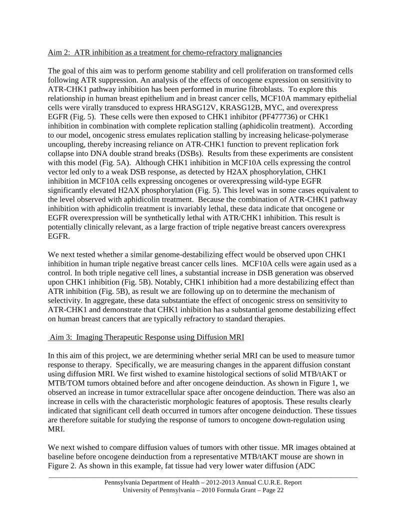
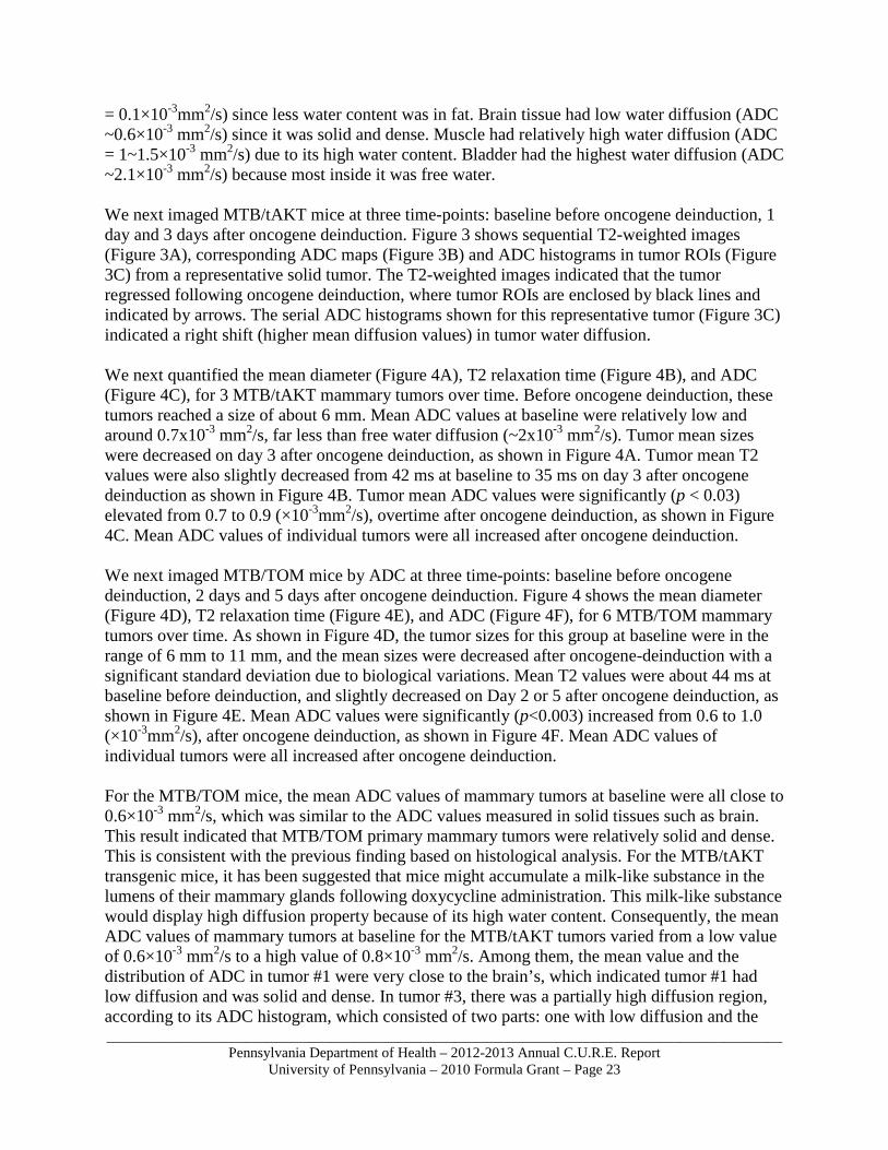
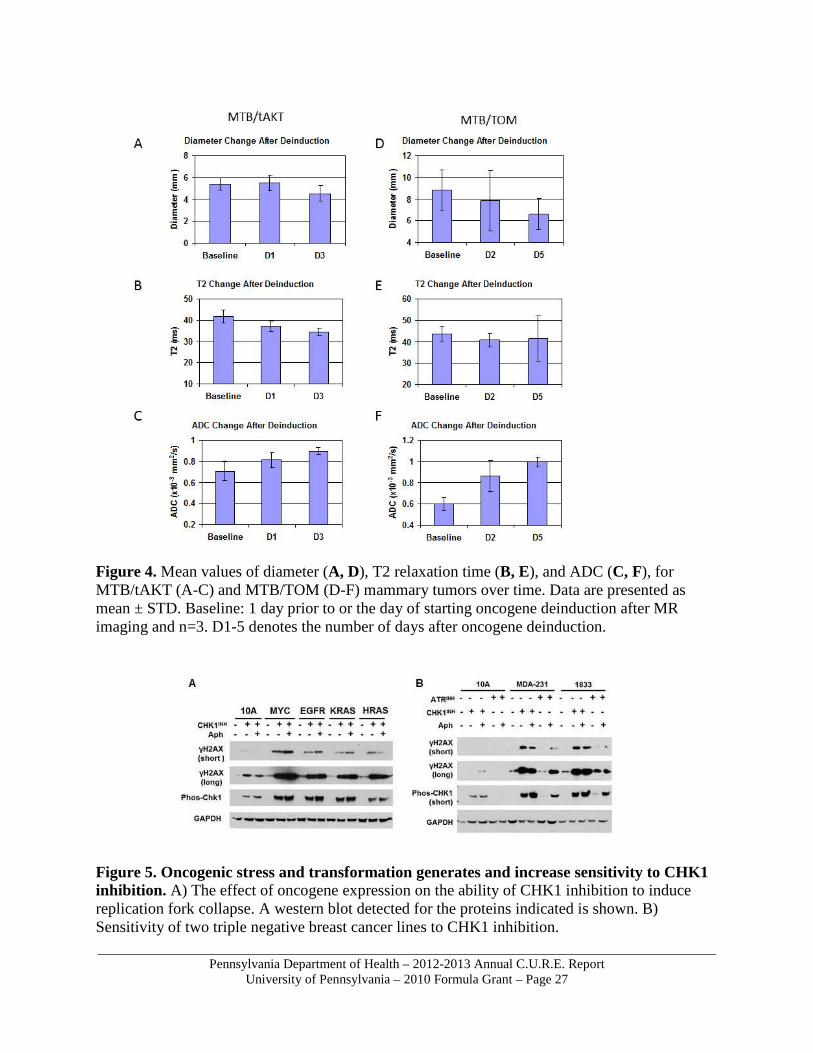
Aim 2: ATR inhibition as a treatment for chemo-refractory malignancies The goal of this aim was to perform genome stability and cell proliferation on transformed cells following ATR suppression. An analysis of the effects of oncogene expression on sensitivity to ATR-CHK1 pathway inhibition has been performed in murine fibroblasts. To explore this relationship in human breast epithelium and in breast cancer cells, MCF10A mammary epithelial cells were virally transduced to express HRASG12V, KRASG12B, MYC, and overexpress EGFR (Fig. 5). These cells were then exposed to CHK1 inhibitor (PF477736) or CHK1 inhibition in combination with complete replication stalling (aphidicolin treatment). According to our model, oncogenic stress emulates replication stalling by increasing helicase-polymerase uncoupling, thereby increasing reliance on ATR-CHK1 function to prevent replication fork collapse into DNA double strand breaks (DSBs). Results from these experiments are consistent with this model (Fig. 5A). Although CHK1 inhibition in MCF10A cells expressing the control vector led only to a weak DSB response, as detected by H2AX phosphorylation, CHK1 inhibition in MCF10A cells expressing oncogenes or overexpressing wild-type EGFR significantly elevated H2AX phosphorylation (Fig. 5). This level was in some cases equivalent to the level observed with aphidicolin treatment. Because the combination of ATR-CHK1 pathway inhibition with aphidicolin treatment is invariably lethal, these data indicate that oncogene or EGFR overexpression will be synthetically lethal with ATR/CHK1 inhibition. This result is potentially clinically relevant, as a large fraction of triple negative breast cancers overexpress EGFR. We next tested whether a similar genome-destabilizing effect would be observed upon CHK1 inhibition in human triple negative breast cancer cells lines. MCF10A cells were again used as a control. In both triple negative cell lines, a substantial increase in DSB generation was observed upon CHK1 inhibition (Fig. 5B). Notably, CHK1 inhibition had a more destabilizing effect than ATR inhibition (Fig. 5B), as result we are following up on to determine the mechanism of selectivity. In aggregate, these data substantiate the effect of oncogenic stress on sensitivity to ATR-CHK1 and demonstrate that CHK1 inhibition has a substantial genome destabilizing effect on human breast cancers that are typically refractory to standard therapies. Aim 3: Imaging Therapeutic Response using Diffusion MRI In this aim of this project, we are determining whether serial MRI can be used to measure tumor response to therapy. Specifically, we are measuring changes in the apparent diffusion constant using diffusion MRI. We first wished to examine histological sections of solid MTB/tAKT or MTB/TOM tumors obtained before and after oncogene deinduction. As shown in Figure 1, we observed an increase in tumor extracellular space after oncogene deinduction. There was also an increase in cells with the characteristic morphologic features of apoptosis. These results clearly indicated that significant cell death occurred in tumors after oncogene deinduction. These tissues are therefore suitable for studying the response of tumors to oncogene down-regulation using MRI. We next wished to compare diffusion values of tumors with other tissue. MR images obtained at baseline before oncogene deinduction from a representative MTB/tAKT mouse are shown in Figure 2. As shown in this example, fat tissue had very lower water diffusion (ADC
_____________________________________________________________________________________________Pennsylvania Department of Health – 2012-2013 Annual C.U.R.E. Report
University of Pennsylvania – 2010 Formula Grant – Page 23
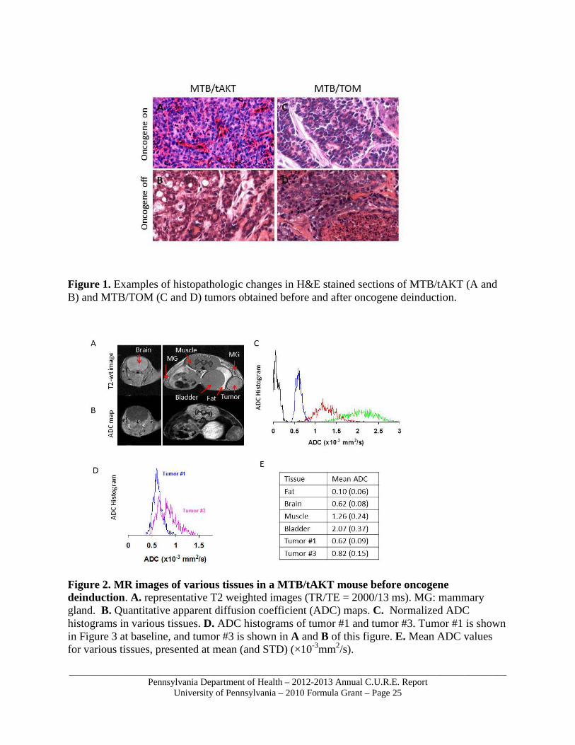
= 0.1×10⁻³mm2/s) since less water content was in fat. Brain tissue had low water diffusion (ADC ~0.6×10⁻³ mm2/s) since it was solid and dense. Muscle had relatively high water diffusion (ADC = 1~1.5×10⁻³ mm2/s) due to its high water content. Bladder had the highest water diffusion (ADC ~2.1×10⁻³ mm2/s) because most inside it was free water. We next imaged MTB/tAKT mice at three time-points: baseline before oncogene deinduction, 1 day and 3 days after oncogene deinduction. Figure 3 shows sequential T2-weighted images (Figure 3A), corresponding ADC maps (Figure 3B) and ADC histograms in tumor ROIs (Figure 3C) from a representative solid tumor. The T2-weighted images indicated that the tumor regressed following oncogene deinduction, where tumor ROIs are enclosed by black lines and indicated by arrows. The serial ADC histograms shown for this representative tumor (Figure 3C) indicated a right shift (higher mean diffusion values) in tumor water diffusion. We next quantified the mean diameter (Figure 4A), T2 relaxation time (Figure 4B), and ADC (Figure 4C), for 3 MTB/tAKT mammary tumors over time. Before oncogene deinduction, these tumors reached a size of about 6 mm. Mean ADC values at baseline were relatively low and around 0.7x10⁻³ mm2/s, far less than free water diffusion (~2x10⁻³ mm2/s). Tumor mean sizes were decreased on day 3 after oncogene deinduction, as shown in Figure 4A. Tumor mean T2 values were also slightly decreased from 42 ms at baseline to 35 ms on day 3 after oncogene deinduction as shown in Figure 4B. Tumor mean ADC values were significantly (p < 0.03) elevated from 0.7 to 0.9 (×10-3mm2/s), overtime after oncogene deinduction, as shown in Figure 4C. Mean ADC values of individual tumors were all increased after oncogene deinduction. We next imaged MTB/TOM mice by ADC at three time-points: baseline before oncogene deinduction, 2 days and 5 days after oncogene deinduction. Figure 4 shows the mean diameter (Figure 4D), T2 relaxation time (Figure 4E), and ADC (Figure 4F), for 6 MTB/TOM mammary tumors over time. As shown in Figure 4D, the tumor sizes for this group at baseline were in the range of 6 mm to 11 mm, and the mean sizes were decreased after oncogene-deinduction with a significant standard deviation due to biological variations. Mean T2 values were about 44 ms at baseline before deinduction, and slightly decreased on Day 2 or 5 after oncogene deinduction, as shown in Figure 4E. Mean ADC values were significantly (p<0.003) increased from 0.6 to 1.0 (×10-3mm2/s), after oncogene deinduction, as shown in Figure 4F. Mean ADC values of individual tumors were all increased after oncogene deinduction. For the MTB/TOM mice, the mean ADC values of mammary tumors at baseline were all close to 0.6×10⁻³ mm2/s, which was similar to the ADC values measured in solid tissues such as brain. This result indicated that MTB/TOM primary mammary tumors were relatively solid and dense. This is consistent with the previous finding based on histological analysis. For the MTB/tAKT transgenic mice, it has been suggested that mice might accumulate a milk-like substance in the lumens of their mammary glands following doxycycline administration. This milk-like substance would display high diffusion property because of its high water content. Consequently, the mean ADC values of mammary tumors at baseline for the MTB/tAKT tumors varied from a low value of 0.6×10⁻³ mm2/s to a high value of 0.8×10⁻³ mm2/s. Among them, the mean value and the distribution of ADC in tumor #1 were very close to the brain’s, which indicated tumor #1 had low diffusion and was solid and dense. In tumor #3, there was a partially high diffusion region, according to its ADC histogram, which consisted of two parts: one with low diffusion and the

_____________________________________________________________________________________________Pennsylvania Department of Health – 2012-2013 Annual C.U.R.E. Report
University of Pennsylvania – 2010 Formula Grant – Page 24
other with high diffusion. This high diffusion part was believed to result from the excreted milk-like substance inside the tumor. For both mouse models, a statistically significant increase of tumor water diffusion after oncogene downregulation in all mammary solid tumors was observed. Average increase was about 30%, compared the mean values of ADC obtained on Day 2 or 3 after oncogene deinduction with the baseline ADC values obtained before oncogene deinduction. A slight decrease of tumor size on Day 2 or 3 after oncogene deinduction was also observed. These results were consistent with the fact that oncogene deinduction resulted in the increase of cell necrosis in oncogene-induced tumors, and consequently the increase of cell death enlarged the extracellular space and membrane permeability, resulting in greater water mobility which displayed higher ADC values by the measurement of diffusion MRI. The significant ADC increase in tumors only 2 or 3 days after oncogene deinduction indicates that anticancer treatment targeting cellular pathways involved in tumor growth regulation could be very effective against cancer. In addition to the ADC increase after oncogene deinduction, a corresponding slight decrease of T2 values in both tumor models was also observed. This result indicated there was some inverse correlation between T2 value and ADC diffusion, as reported in other studies. Compared to the other tissues, the mammary tumors had medium water diffusion and their ADC values were between the ADC values of solid tissues as brain and the ADC values such as in muscle. The sensitivity of diffusion MRI for detection of therapeutic-induced changes depends on the possible overall dynamic range of water diffusion observed by ADC measurements. For example, because of the existing milk-like substance inside, the MTB/tAKT tumor #3 had relatively high mean ADC value at baseline. This would decrease the sensitivity of diffusion MRI for detection of slight ADC changes to therapy. Besides, mammary tumors in mouse models may have central necrosis when they reach a large size (>15 mm in diameter), resulting in some regions of tumors with high extracellular water contents that may elevate the ADC values at baseline and reduce the sensitivity of diffusion MRI for detection of ADC changes to therapy. Therefore, to achieve good detection sensitivity of diffusion MRI in the study of tumor response to oncogene deinduction or targeted therapy in mouse models, the best strategy, as adopted in this study, is to limit tumor size, for example, to not bigger than 10 mm in diameter, to minimize central necrosis. Another potential solution of this problem is to exclude the known regions with necrosis or milk-like substance that will not respond to therapy, and only compare the other regions that are present in both the pre-therapy and post-therapy tumor volumes, but how to segment the overlap regions from the images obtained on different days remains a challenge because mammary tumors can be easily deformed by pressure in experiments. In conclusion, diffusion MRI analysis provides an early biomarker for solid mammary tumor response to oncogene downregulation. The mean values of ADC in mammary tumors is significantly increased within the early days after oncogene deinduction, accompanied by slight decreases of T2 value and tumor size. It clearly indicates that combining the inducible transgenic mouse models for breast cancer and diffusion MRI allows us to visualize and analyze mammary tumor response to anticancer therapy targeted with specific oncogene pathways.
_____________________________________________________________________________________________Pennsylvania Department of Health – 2012-2013 Annual C.U.R.E. Report
University of Pennsylvania – 2010 Formula Grant – Page 25
Figure 1. Examples of histopathologic changes in H&E stained sections of MTB/tAKT (A and B) and MTB/TOM (C and D) tumors obtained before and after oncogene deinduction.
Figure 2. MR images of various tissues in a MTB/tAKT mouse before oncogene deinduction. A. representative T2 weighted images (TR/TE = 2000/13 ms). MG: mammary gland. B. Quantitative apparent diffusion coefficient (ADC) maps. C. Normalized ADC histograms in various tissues. D. ADC histograms of tumor #1 and tumor #3. Tumor #1 is shown in Figure 3 at baseline, and tumor #3 is shown in A and B of this figure. E. Mean ADC values for various tissues, presented at mean (and STD) (×10-3mm2/s).
_____________________________________________________________________________________________Pennsylvania Department of Health – 2012-2013 Annual C.U.R.E. Report
University of Pennsylvania – 2010 Formula Grant – Page 26
Figure 3. ADC imaging of an MTB/tAKT mammary tumor A. Representative T2 weighted images (TR/TE = 2000/13 ms) obtained 1 day before starting oncogene deinduction (Baseline), and 1 day (D1) and 3 days (D3) after oncogene deinduction, respectively. Tumor ROIs were drawn manually as enclosed by black lines and indicated by arrows. The T2 images illustrated the slight tumor shrinkage over time after oncogene deinduction. B. Quantitative apparent diffusion coefficient (ADC) maps at the same time intervals. C. ADC histograms in tumor ROIs at the same time intervals. Vertical axis represents voxel numbers in ROIs. Both the tumor ADC maps and histograms in tumor ROIs revealed increase diffusion or water mobility after oncogene deinduction.
_____________________________________________________________________________________________Pennsylvania Department of Health – 2012-2013 Annual C.U.R.E. Report
University of Pennsylvania – 2010 Formula Grant – Page 27
Figure 4. Mean values of diameter (A, D), T2 relaxation time (B, E), and ADC (C, F), for MTB/tAKT (A-C) and MTB/TOM (D-F) mammary tumors over time. Data are presented as mean ± STD. Baseline: 1 day prior to or the day of starting oncogene deinduction after MR imaging and n=3. D1-5 denotes the number of days after oncogene deinduction.
Figure 5. Oncogenic stress and transformation generates and increase sensitivity to CHK1 inhibition. A) The effect of oncogene expression on the ability of CHK1 inhibition to induce replication fork collapse. A western blot detected for the proteins indicated is shown. B) Sensitivity of two triple negative breast cancer lines to CHK1 inhibition.
_____________________________________________________________________________________________Pennsylvania Department of Health – 2012-2013 Annual C.U.R.E. Report
University of Pennsylvania – 2010 Formula Grant – Page 28
Research Project 7: Project Title and Purpose The Unfolded Protein Response in Cancer - The purpose of this project is to identify novel points within the neoplastic process that can be attached through the development of novel therapeutics. The experiments described will provide proof-of-principle data supporting these novel concepts. Duration of Project 7/1/2011 - 6/30/2012 Summary of Research Completed This project ended during a prior state fiscal year. For additional information, please refer to the Commonwealth Universal Research Enhancement C.U.R.E. Annual Reports on the Department's Tobacco Settlement/Act 77 web page at http://www.health.state.pa.us/cure. Research Project 8: Project Title and Purpose Identifying Genetic Determinants of Mammographic Density - Mammographic density is one of the strongest known predictors of breast cancer risk, but its basis is largely unknown. The realization that mammographic density is primarily determined by genetic factors, rather than reproductive endocrine events, suggests that the genetic pathways that control mammographic density may reflect entirely novel pathways and mechanisms of breast cancer susceptibility. Identifying the genes that control mammographic density would be enabled by a validated animal model for the effect of genetics on mammographic density. The objective of this pilot project is to validate a rat model for the genetic effects of mammographic density on breast cancer risk using in vivo and ex vivo imaging in combination with inbred strains of rats with differing intrinsic susceptibilities to breast cancer. Anticipated Duration of Project 7/1/2011 - 12/31/2014 Project Overview This study will use microCT to radiographically image mammary tissue from different inbred strains of rats that differ in their intrinsic susceptibility to breast cancer. The specific aims of this project are to: 1) develop reproducible in vivo and ex vivo microCT measures of mammographic density in rats; 2) determine intra-animal and inter-animal variability in microCT mammographic density within a rat strain; 3) determine inter-strain variability in mammographic density to identify strains with high versus low density; and 4) identify tissue-based correlates of mammographic density. These validation studies would enable a large-scale genetic analysis of mammographic density among offspring from a backcross between an F1 hybrid of a low-density and a high-density rat strain.
_____________________________________________________________________________________________Pennsylvania Department of Health – 2012-2013 Annual C.U.R.E. Report
University of Pennsylvania – 2010 Formula Grant – Page 29
Principal Investigator Katrina Armstrong, MD, MSCE Professor, Department of Medicine University of Pennsylvania School of Medicine Department of Cancer Biology 1233 Blocklely 416 Curie Blvd. Philadelphia, PA 191904-6021 Other Participating Researchers Lewis A. Chodosh, MD, PhD, Andrew D. Maidment, PhD, Despina Kontos, PhD – employed by University of Pennsylvania Expected Research Outcomes and Benefits Identifying the genes that control mammographic density would be enabled by a validated animal model in which mammographic density can be related to genetic background and breast cancer risk. While animal models have proven to be extraordinarily powerful tools for understanding human disease, there is currently no animal model for mammographic density and no studies in animal models that have attempted to relate variation in mammographic density with differences in breast cancer susceptibility. However, in order to perform such a mapping study, critical proof-of-principle experiments are required to validate the model and quantitative approach. This project is intended to enable those studies. We anticipate that the studies proposed in this project will provide a validated rat model for the genetic determination of mammographic density and breast cancer risk. As such, this research project has the potential to provide a unique and important tool for mapping genes that control breast cancer susceptibility. The identification of such genes, in turn, has the potential to lead to the development of more effective molecularly targeted agents for the treatment or prevention of breast cancer. Summary of Research Completed In this project, we have been exploring the genetic basis for mammographic density using an animal model. Mammographic density is one of the strongest known predictors of breast cancer risk, and it is primarily determined by genetic factors, but the identity of these factors is largely unknown. The objective of this aim was to validate a rat model for the genetic effects of mammographic density on breast cancer risk using in vivo and ex vivo imaging in combination with inbred strains of rats with differing intrinsic susceptibilities to breast cancer. Generation of μCT Images Using methods as described in the previous report, we imaged 44 rat mammary glands bringing the total of glands imaged to date to 89.

_____________________________________________________________________________________________Pennsylvania Department of Health – 2012-2013 Annual C.U.R.E. Report
University of Pennsylvania – 2010 Formula Grant – Page 30
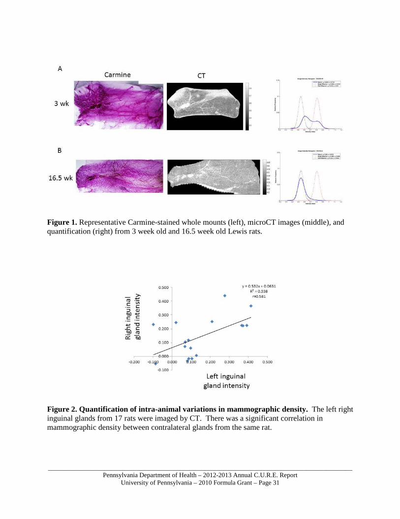
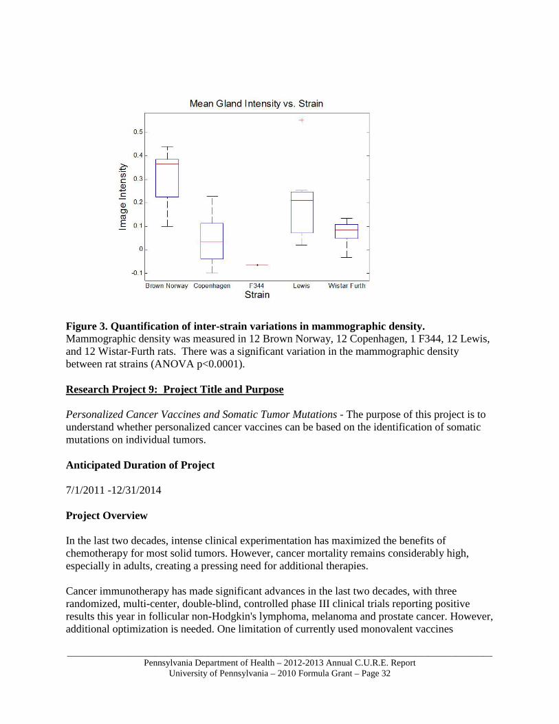
Effect of developmental stage on mammographic density We next asked whether mammographic density varies with developmental stage of the animal. We have removed the inguinal mammary glands from juvenile, adult, pregnant, and lactating Lewis rats. As a control, we also removed three mammary tumors from Lewis rats. Glands will be imaged to determine how density changes with developmental stage. Representative carmine-stained images, CT images, and quantification are shown in Figure 1. Mammographic density in contralateral glands from the same animal We next wished to address whether there was a significant correlation between the measured mammographic densities of contralateral glands taken from the same rat. To accomplish this, we removed the left and right inguinal glands from a number of rats and compared their mammographic density by microCT. We found that there was a statistically significant correlation in mammographic density between glands taken from the same animal, suggesting that this is an intrinsic property of the animal (Figure 2). Inter-strain differences in mammographic density Having developed a method to reproducibly measure mammographic density in rats, we next wished to ask whether the differences in breast cancer susceptibility of different inbred rat strains is reflected by differences in mammographic density. To do this, we removed the inguinal mammary glands from Brown Norway, Copenhagen, F344, Lewis, and Wistar Furth rats. Preliminary results from these studies indicate that there is a significant difference in density between these strains, and this inter-strain difference was observed to be larger than the difference between individual animals of the same strain (ANOVA p<0.0001; Figure 3). Brown Norway rats had the highest mammographic density, while F344 rats had the lowest mammographic density. These studies would enable a large-scale genetic analysis of mammographic density among offspring from a backcross between an F1 hybrid of a low-density and a high-density rat strain.
_____________________________________________________________________________________________Pennsylvania Department of Health – 2012-2013 Annual C.U.R.E. Report
University of Pennsylvania – 2010 Formula Grant – Page 31
Figure 1. Representative Carmine-stained whole mounts (left), microCT images (middle), and quantification (right) from 3 week old and 16.5 week old Lewis rats.
Figure 2. Quantification of intra-animal variations in mammographic density. The left right inguinal glands from 17 rats were imaged by CT. There was a significant correlation in mammographic density between contralateral glands from the same rat.
_____________________________________________________________________________________________Pennsylvania Department of Health – 2012-2013 Annual C.U.R.E. Report
University of Pennsylvania – 2010 Formula Grant – Page 32
Figure 3. Quantification of inter-strain variations in mammographic density. Mammographic density was measured in 12 Brown Norway, 12 Copenhagen, 1 F344, 12 Lewis, and 12 Wistar-Furth rats. There was a significant variation in the mammographic density between rat strains (ANOVA p<0.0001). Research Project 9: Project Title and Purpose Personalized Cancer Vaccines and Somatic Tumor Mutations - The purpose of this project is to understand whether personalized cancer vaccines can be based on the identification of somatic mutations on individual tumors. Anticipated Duration of Project 7/1/2011 -12/31/2014 Project Overview In the last two decades, intense clinical experimentation has maximized the benefits of chemotherapy for most solid tumors. However, cancer mortality remains considerably high, especially in adults, creating a pressing need for additional therapies. Cancer immunotherapy has made significant advances in the last two decades, with three randomized, multi-center, double-blind, controlled phase III clinical trials reporting positive results this year in follicular non-Hodgkin's lymphoma, melanoma and prostate cancer. However, additional optimization is needed. One limitation of currently used monovalent vaccines
_____________________________________________________________________________________________Pennsylvania Department of Health – 2012-2013 Annual C.U.R.E. Report
University of Pennsylvania – 2010 Formula Grant – Page 33
targeting a single cancer protein is that they can select for immune escape tumor variants. Whole tumor antigen vaccines have consistently outperformed single-target immunotherapy in early phase clinical trials, but they carry an overwhelming amount of “self” antigens, which could dampen immunogenicity, or vice versa, markedly increase the risk of autoimmunity, if immune therapy becomes potent enough with the advent of immune modulation. Developing polyvalent tumor vaccines comprised uniquely of cancer-specific proteins should thus maximize cancer vaccine efficacy and safety. However, the specific tumor-rejection antigens remain to date unknown for most solid tumors. Recent deep sequencing analyses have revealed that solid tumors harbor numerous somatic mutations, most of which differ among tumor specimens even within the same tumor type. We hypothesize that somatic missense point mutations acquired by tumor cells result in the expression of aberrant ‘non-self’ protein products, which are tumor rejection antigens. We further hypothesize that effective therapeutic immunization with whole tumor antigen vaccines is mediated in part through a potent response against these unique, mutated proteins. This proposal will test this hypothesis. We proposed the following two aims: Aim 1. Identify and validate somatic missense point mutations in advanced ovarian cancer using next generation high throughput sequencing. Aim 2. Identify immunogenic somatic missense point mutations attacked by vaccine-induced T cells in patients undergoing whole tumor antigen vaccine. Principal Investigator George Coukos, MD, PhD Professor University of Pennsylvania Biomedical Research Building II/III, Rm 1315 421 Curie Blvd. Philadelphia, PA 19104 Other Participating Researchers Annalisa Roberti PhD, Lin Zhang, PhD, Hongzhe Li, PhD, Janos Tanyi, MD, Don Baldwin, PhD – employed by University of Pennsylvania Expected Research Outcomes and Benefits The explosion of genomics information has uncovered numerous candidate targets for cancer molecular therapy but has also revealed the molecular heterogeneity of tumors, highlighting the need for individualized therapy. Although the prospect of targeted personalized therapy has raised high expectations for cancer outcomes, the transition from identifying target candidates to validating these and providing personalized therapy remains a major challenge. Cancer vaccines highlight the major challenges of translating high throughput individual genomic information to delivery of personalized therapy. Given the marked molecular heterogeneity among tumors even
_____________________________________________________________________________________________Pennsylvania Department of Health – 2012-2013 Annual C.U.R.E. Report
University of Pennsylvania – 2010 Formula Grant – Page 34
of the same histotype, and the lack of known tumor antigens, the development of cancer-specific multivalent vaccines must be personalized. This project will test whether missense somatic mutations in tumors are targets for immunotherapy. We expect this to be the case. This result will open the door to develop personalized cancer vaccines based on the identification of unique mutations in patients’ tumors. Summary of Research Completed Aim 1 The subset of the mutations that alters the protein coding sequence creates neoantigens that may elicit cytotoxic T-cell reactivity. From a therapeutic prospective, the definition of the tumor associated epitopes on a patient-specific basis will provide the basis for enhancing T-cell targeting antigen therapy. During this reporting period, we found that of the 838 somatic mutations in coding genes, 460 occurred in expressed genes. The remaining 378 arose in silent or low expressed genes, which mutations cannot be considered as potential neo antigens (Fig.1). RNA sequencing data were further used to analyze the expression level of the mutated genes. As a confirmation, we found that the 460 Somatic Expressed Mutations (SEM) occurred in actively transcribed genes. Although, it has already been confirmed by two independent experiments (RNA sequencing and tumor exome sequencing), we decided to further validate the SEMs by targeted resequencing before being screened for T cell recognition. We generated a multiplexed panel of 384 amplicons specifically targeting each SEMs. Primers were specifically generated to span the exact chromosome position annotated with Illumina sequencing platform. In the primers design we did not include the SNPs that have been already described in Single Nucleotide Polymorphism Database (dbSNP). In order to confirm the somatic origin of the SEMs, for each patient we analyzed both tumor and blood DNA. Sequencing was performed on a PGM sequencer (Ion Torrent). Torrent Suite software (Ion Torrent) was used to align reads to the reference genome, and to generate run metrics, and the variants were identified with Variant Caller software (Ion Torrent). An average of 70% of the mutations were validated by Ion Torrent platform; almost all of them were confirmed to be tumor-specific, only 9 of 471 mutations resulted to be germ line as the mutations were annotated in the matching blood sample (Figure 2). By altering the protein coding sequence, the somatic expressed mutations might generate patient-specific neo antigens. From all of the validated SEMs we will derive sets of mutated peptides and test them for peptides cleavage and MHC binding by using bioinformatics algorithm. During this reporting period we began performing high-resolution HLA genotyping for each patient by using a newly developed next-generation sequencing technology. By providing a deeply HLA typing for Class I and II alleles, this new technology will improve the reliability of the in silico prediction of peptide-MHC binding affinity. In contrast with the high heterogeneity found in the DNA somatic mutations, alterations in RNA editing seem to be a shared mutational mechanism in ovarian cancer. Currently, we are investigating the oncogenic function of two new ovarian cancer correlated editing mutations. We
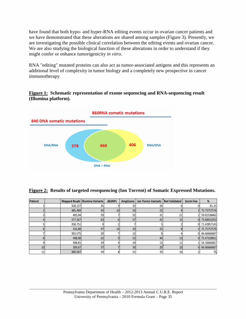
_____________________________________________________________________________________________Pennsylvania Department of Health – 2012-2013 Annual C.U.R.E. Report
University of Pennsylvania – 2010 Formula Grant – Page 35
have found that both hypo- and hyper-RNA editing events occur in ovarian cancer patients and we have demonstrated that these alterations are shared among samples (Figure 3). Presently, we are investigating the possible clinical correlation between the editing events and ovarian cancer. We are also studying the biological function of these alterations in order to understand if they might confer or enhance tumorigenicity in vitro. RNA "editing" mutated proteins can also act as tumor-associated antigens and this represents an additional level of complexity in tumor biology and a completely new prospective in cancer immunotherapy. Figure 1: Schematic representation of exome sequencing and RNA-sequencing result (Illumina platform).
Figure 2: Results of targeted resequencing (Ion Torrent) of Somatic Expressed Mutations.
Patient Mapped Reads Illumina Variants dbSNPs Amplicons Ion Torren Variants Not Validated Germ line %
1 328,127 35 3 32 26 6 0 81.252 385,484 43 10 33 25 8 2 75.757575763 405,04 59 7 52 31 21 1 59.615384624 377,567 63 6 57 42 15 0 73.684210535 358,751 9 2 7 5 2 0 71.428571436 316,88 47 14 33 25 8 3 75.757575767 303,575 20 7 12 8 4 0 66.666666678 498,08 62 9 53 40 13 0 75.471698119 408,65 29 4 24 13 11 1 54.16666667
10 309,67 37 7 30 20 10 0 66.6666666711 389,567 59 8 51 35 16 2 70
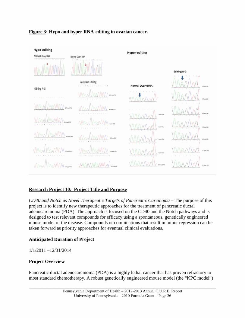
_____________________________________________________________________________________________Pennsylvania Department of Health – 2012-2013 Annual C.U.R.E. Report
University of Pennsylvania – 2010 Formula Grant – Page 36
Figure 3: Hypo and hyper RNA-editing in ovarian cancer.
Research Project 10: Project Title and Purpose CD40 and Notch as Novel Therapeutic Targets of Pancreatic Carcinoma – The purpose of this project is to identify new therapeutic approaches for the treatment of pancreatic ductal adenocarcinoma (PDA). The approach is focused on the CD40 and the Notch pathways and is designed to test relevant compounds for efficacy using a spontaneous, genetically engineered mouse model of the disease. Compounds or combinations that result in tumor regression can be taken forward as priority approaches for eventual clinical evaluations. Anticipated Duration of Project 1/1/2011 –12/31/2014 Project Overview Pancreatic ductal adenocarcinoma (PDA) is a highly lethal cancer that has proven refractory to most standard chemotherapy. A robust genetically engineered mouse model (the “KPC model”)
_____________________________________________________________________________________________Pennsylvania Department of Health – 2012-2013 Annual C.U.R.E. Report
University of Pennsylvania – 2010 Formula Grant – Page 37
faithfully reproduces the clinical and histopathological features of PDA. This model, which is based on the pancreatic specific expression of mutated Kras and p53, is particularly relevant from the standpoint of preclinical testing since – unlike xenografted PDAC cells – KPC animals are also non-responsive to current standard-of-care therapy, such as gemcitabine chemotherapy. We hypothesize that the KPC model represents a superior experimental platform for the design and testing of novel therapies for this disease. For this project we will use the KPC model both to gain mechanistic insight and to improve therapies targeting CD40 and Notch. Our ‘mouse hospital’ approach will be to use ultrasound to identify KPC animals with incipient tumors and administer active compounds (or inert control) to these animals. Our specific aims are: Aim 1: Determine whether the treatment effects of CD40 activation can be amplified by myeloid cell stimulation (with G-CSF) or supplemented by T cell engagement (with CTLA-4 mAb). Determine whether Hedgehog pathway inhibition augments the anti-tumor effect of CD40. This project is based on the hypothesis that CD40 mAb activates tumor infiltrating macrophages but that engagement of additional pathways will optimize the treatment effect. Aim 2: Determine whether persistent Notch signaling is required for PDA growth and whether Hedgehog pathway inhibition augments the anti-tumor effect of GSI. This project is based on the hypothesis that pancreatic tumors require sustained reciprocal signaling between the epithelial (neoplastic) compartment and the stromal compartment and that Notch and Hedgehog comprise two critical components of this signaling network. Principal Investigator Robert H. Vonderheide, MD, DPhil Associate Professor of Medicine University of Pennsylvania School of Medicine 551 BRB II/III 421 Curie Blvd Philadelphia, PA 19104 Other Participating Researchers Ben Stanger, MD, PhD – employed by University of Pennsylvania Expected Research Outcomes and Benefits This study uses a spontaneous, genetically engineering mouse model of pancreatic adenocarcinoma to identify, screen, and prioritize new therapies for the human condition. Our investigations are designed to determine whether the combination of agonist CD40 mAb and G-CSF enhances the anti-tumor effect of CD40 mediated therapy, likely due to enhanced numbers of myeloid cells systemically or in the tumor. Similarly the combination of CD40 mAb and blocking CTLA-4 mAb may enhance the anti-tumor effect by stimulating T cells to infiltrate the tumor tissue. We also intend to demonstrate whether GSI therapy impacts tumor growth and possibly induces regression. Finally, our studies will demonstrate whether or not smoothened
_____________________________________________________________________________________________Pennsylvania Department of Health – 2012-2013 Annual C.U.R.E. Report
University of Pennsylvania – 2010 Formula Grant – Page 38
inhibition abrogates the dense stroma of the PDA lesions and enhances drug delivery and cellular infiltration, and whether this inhibition further enhances treatment effects in this model. The main benefit of our research is the delivery of new compounds with a scientific rationale to the clinic for eventual testing in patients. There is an urgent clinical need for new therapies in pancreatic adenocarcinoma. This project may help to improve health status by offering novel strategies to reduce morbidity and mortality from this common devastating disease. Summary of Research Completed Recent clinical studies show that combining gemcitabine (Gem) with nanoparticle albumin-bound paclitaxel, Abraxane (Abrx), prolongs progression-free and overall survival as compared to Gem alone, but the objective response rate of the combination is only 23%, and nearly every patient eventually progresses. Given that CD40 activation can reverse tumor-induced immune suppression and promote anti-tumor T cell responses, we tested whether CD40 plus chemotherapy might be better starting point than CD40 alone for testing GSI. We therefore administered agonistic CD40 monoclonal antibody (FGK45) with combined Gem/Abrx chemotherapy to mice bearing subcutaneous PDA tumors established using syngeneic tumor cell lines derived from the KrasLSL-G12D/+;Trp53LSL-R172H/+;Pdx-1-Cre genetically engineered mouse model of PDA. We found only rare tumor regressions in mice treated with chemotherapy alone (Gem, Abrx, or Gem/Abrx) or FGK45 alone, whereas more than 50% of mice treated with the combination of Gem/Abrx/FGK45 experienced tumor regressions (p<0.0001). Mechanistic studies revealed that this effect was T cell dependent but macrophage independent. Although the overall CD3+ T cell population in the tumor after therapy did not vary across treatment cohorts, the proportion of FoxP3+CD4+ T regulatory cells was reduced 6.9-fold after treatment with Gem/Abrx/FGK45 (p<0.05, Gem/Abrx/FGK45 treatment versus chemotherapy or FGK45 alone), with a concurrent 1.75-fold increase in activated CD44hiCD4+ T helper cells (p<0.01). As a result, the ratio of CD4 T helper cells or CD8 T effector cells to regulatory T cells was increased intratumorally after treatment with Gem/Abrx/FGK45. In some mice treated with Gem/Abrx/FGK45, tumors were completely rejected within 3 weeks, and the mice remained free of tumor for at least 4 months, suggesting the potential for long-lived T cell memory directed against PDA. These studies not only highlight the clinical potential of adding standard-of-care chemotherapy to CD40 as a novel strategy for PDA, but also underscore the rationale of now adding Notch inhibition to this approach, rather than CD40 alone. A manuscript is being prepared. Also this year we have submitted a manuscript entitled “Hedgehog pathway inactivation suppresses stroma but accelerates progression in pancreatic ductal adenocarcinoma” which is under review. PDA is characterized by a dense desmoplastic stroma which begins to accrue during the formation of precursor PanIN lesions. One of the mediators of the desmoplastic reaction which we believe may be co-regulated with Notch signaling is Sonic hedgehog (Shh), a secreted ligand that is overexpressed from epithelial cells in pancreatic cancer and sends a proliferative signal to nearby stromal cells. Although acute inhibition of the Hedgehog pathway effector Smoothened (Smo) depletes pancreatic stroma, facilitates drug delivery and extends overall survival in KPC mice, Smoothened inhibitors have not exhibited efficacy in Phase I clinical trials, and may even result in a worse clinical outcome. Thus, there is considerable uncertainty regarding the role of this pathway, and its suitability as a target for therapy, in PDA.
_____________________________________________________________________________________________Pennsylvania Department of Health – 2012-2013 Annual C.U.R.E. Report
University of Pennsylvania – 2010 Formula Grant – Page 39
This year, our team used genetically engineered mouse models in combination with an in vivo lineage tracing approach to study the role of Shh in pancreas cancer. We found (i) Tumors develop in the absence of Shh. To test the role of Shh in pancreas cancer, we used our previously described KPC-Y pancreas cancer model and introduced a conditional “floxed” allele of Shh. Such ShhKPC-Y mice had normal pancreas development and developed tumors, (ii) Shh deficient pancreatic tumors lack a desmoplastic stroma. Although they arose at with the same latency as PKCY tumors, ShhKPC-Y tumors had an almost complete depletion of myofibroblastic stroma, (iii) Shh deficient pancreatic tumors progress rapidly. Contrary to our expectations, the stroma-poor pancreatic tumors in ShhKPCY mice progressed rapidly, significantly decreasing overall survival. ShhKPCY tumors were much more proliferative than in KPC-Y mice and they developed more metastases, (iv) Shh deficient pancreatic tumors were poorly differentiated and well vascularized. In contrast with the well- to moderately-differentiated morphology of pancreatic tumors in KPC-Y mice (and humans), the stroma-poor tumors arising in ShhKPC-Y mice were poorly differentiated or undifferentiated. These tumors also had a much higher vascular density. To confirm the relevance of this observation to human PDAC, we found that poorly differentiated human pancreatic tumors (which comprise 5-10% of all PDAC) are well vascularized, (v) Undifferentiated Shh-deficient tumors are sensitive to VEGF inhibition. VEGF inhibitors have minimal impact on KPC-Y tumors (and have failed to show efficacy in human clinical trials). By contrast, ShhKPC-Y tumors – with their increased vascular density and perfusion – exhibit areas of necrosis and a significant reduction in growth following treatment with a VEGF blocking antibody. These results challenge the view that the desmoplastic stroma acts solely as a tumor promoter, and suggest how tumor and stromal histology (e.g. differentiation status and vascular density) could be used to stratify patients for different types of therapy (e.g. VEGF inhibition). Shh inhibition with CD40 is now a lower priority. Research Project 11: Project Title and Purpose Customization of Advanced Non-small Cell Lung Cancer Treatment Based on Emerging Histologic and Molecular Markers - The main purpose of this project is to show that non-empiric therapy that is scientifically chosen on the basis of existing clinical and molecular markers, including EGFR and KRAS mutation and EML4/ALK translocation, can result in improved progression-free and overall survival (PFS and OS) in good performance status patients with advanced non-squamous Non-small Cell Lung Cancer (NSCLC), compared to historic controls and contemporaneous subjects who are treated empirically. Anticipated Duration of Project 7/1/2011 – 12/31/2014 Project Overview Major objectives include (1) the collection of tumor tissue and serum to further elucidate the molecular typing of patients with advanced NSCLC; (2) the development of a molecular pathology platform that incorporates simultaneous reflex testing of multiple candidate

_____________________________________________________________________________________________Pennsylvania Department of Health – 2012-2013 Annual C.U.R.E. Report
University of Pennsylvania – 2010 Formula Grant – Page 40
biomarkers; (3) evaluation of thymidylate synthetase status in tumor specimens as both a prognostic and predictive marker in patients receiving pemetrexed; (4) identification of circulating tumor cells in serum specimens collected at the start of treatment and at first radiographic evaluation, both to predict outcome and to determine or predict sensitivity to therapy based on molecular testing; and (5) collection of baseline smoking assessment surveys to elucidate any linkage between current smoking status / total lifetime tobacco exposure and both therapeutic outcome and toxicity. We anticipate the enrollment of 200+ patients, who will consent to biospecimen collection for genetic-environmental interaction (GEI) and whose status will be tracked once treatment starts, both on and off protocol. We will gauge toxicity, response, progression-free and overall survival. Principal Investigator Corey J Langer, MD Professor of Medicine University of Pennsylvania Abramson Cancer Center 3400 Civic Center Blvd Philadelphia, PA 19104 Other Participating Researchers Steven Albelda, MD, Anil Vachani, MD, Tracey Evans, MD, Roger Cohen, MD, James Stevenson, MD, Daniel Schwed, MD, Charu Deshpande, MD, Vivianna Vandeerlin, MD, Chris Watt, MD, Lynn Werner, RN, Guanjun Cheng, MD, PhD – employed by University of Pennsylvania Expected Research Outcomes and Benefits In a global sense, we expect this study to show that “customized” therapy based on molecular correlates, immunohistochemical correlates and clinical factors will lead to better care and improved outcomes compared to historic, empiric treatment in patients with advanced NSCLC. If we are able to do this, patients will directly benefit by receiving treatments that will enhance response rates to treatment, delay tumor progression, increase survival, and show less toxicity. More specifically, we expect that 1) By improving tissue and serum collection approaches (i.e. by starting to do “reflex testing”),
we will be able to apply current and developing predictive tests in a more useful fashion. We expect that advances in analysis of circulating tumor cells may be an important way in which we can obtain molecular correlates in patients without readily accessible tissue for biopsy, thus giving them access to these new tests and obviating the need for additional painful and expensive biopsy tests.
2) Thymidilate synthase staining test will be especially useful in helping us to decide which patients should receive pemetrexed therapy.
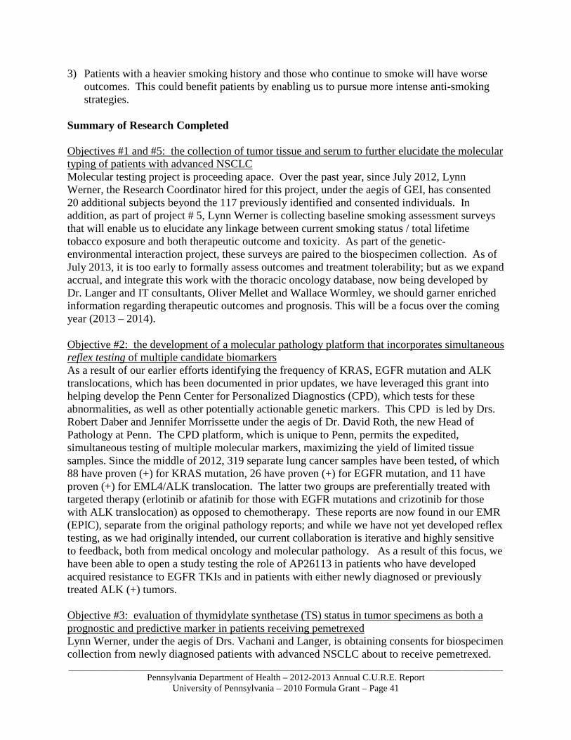
_____________________________________________________________________________________________Pennsylvania Department of Health – 2012-2013 Annual C.U.R.E. Report
University of Pennsylvania – 2010 Formula Grant – Page 41
3) Patients with a heavier smoking history and those who continue to smoke will have worse outcomes. This could benefit patients by enabling us to pursue more intense anti-smoking strategies.
Summary of Research Completed Objectives #1 and #5: the collection of tumor tissue and serum to further elucidate the molecular typing of patients with advanced NSCLC Molecular testing project is proceeding apace. Over the past year, since July 2012, Lynn Werner, the Research Coordinator hired for this project, under the aegis of GEI, has consented 20 additional subjects beyond the 117 previously identified and consented individuals. In addition, as part of project # 5, Lynn Werner is collecting baseline smoking assessment surveys that will enable us to elucidate any linkage between current smoking status / total lifetime tobacco exposure and both therapeutic outcome and toxicity. As part of the genetic-environmental interaction project, these surveys are paired to the biospecimen collection. As of July 2013, it is too early to formally assess outcomes and treatment tolerability; but as we expand accrual, and integrate this work with the thoracic oncology database, now being developed by Dr. Langer and IT consultants, Oliver Mellet and Wallace Wormley, we should garner enriched information regarding therapeutic outcomes and prognosis. This will be a focus over the coming year (2013 – 2014). Objective #2: the development of a molecular pathology platform that incorporates simultaneous reflex testing of multiple candidate biomarkers As a result of our earlier efforts identifying the frequency of KRAS, EGFR mutation and ALK translocations, which has been documented in prior updates, we have leveraged this grant into helping develop the Penn Center for Personalized Diagnostics (CPD), which tests for these abnormalities, as well as other potentially actionable genetic markers. This CPD is led by Drs. Robert Daber and Jennifer Morrissette under the aegis of Dr. David Roth, the new Head of Pathology at Penn. The CPD platform, which is unique to Penn, permits the expedited, simultaneous testing of multiple molecular markers, maximizing the yield of limited tissue samples. Since the middle of 2012, 319 separate lung cancer samples have been tested, of which 88 have proven (+) for KRAS mutation, 26 have proven (+) for EGFR mutation, and 11 have proven (+) for EML4/ALK translocation. The latter two groups are preferentially treated with targeted therapy (erlotinib or afatinib for those with EGFR mutations and crizotinib for those with ALK translocation) as opposed to chemotherapy. These reports are now found in our EMR (EPIC), separate from the original pathology reports; and while we have not yet developed reflex testing, as we had originally intended, our current collaboration is iterative and highly sensitive to feedback, both from medical oncology and molecular pathology. As a result of this focus, we have been able to open a study testing the role of AP26113 in patients who have developed acquired resistance to EGFR TKIs and in patients with either newly diagnosed or previously treated ALK (+) tumors. Objective #3: evaluation of thymidylate synthetase (TS) status in tumor specimens as both a prognostic and predictive marker in patients receiving pemetrexed Lynn Werner, under the aegis of Drs. Vachani and Langer, is obtaining consents for biospecimen collection from newly diagnosed patients with advanced NSCLC about to receive pemetrexed.
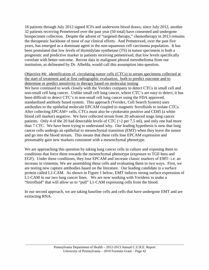
_____________________________________________________________________________________________Pennsylvania Department of Health – 2012-2013 Annual C.U.R.E. Report
University of Pennsylvania – 2010 Formula Grant – Page 42
18 patients through July 2012 signed ICFs and underwent blood draws; since July 2012, another 32 patients receiving Pemetrexed over the past year (50 total) have consented and undergone biospecimen collection. Despite the advent of “targeted therapy,” chemotherapy in 2013 remains the therapeutic backbone of most of our clinical efforts. And Pemetrexed, over the past five years, has emerged as a dominant agent in the non-squamous cell carcinoma population. It has been postulated that low levels of thymidylate synthetase (TS) in tumor specimens is both a prognostic and predictive marker in patients receiving pemetrexed, that low levels specifically correlate with better outcome. Recent data in malignant pleural mesothelioma from our institution, as delineated by Dr. Albelda, would call this assumption into question. Objective #4: identification of circulating tumor cells (CTCs) in serum specimens collected at the start of treatment and at first radiographic evaluation, both to predict outcome and to determine or predict sensitivity to therapy based on molecular testing We have continued to work closely with the Veridex company to detect CTCs in small cell and non-small cell lung cancer. Unlike small cell lung cancer, where CTC’s are easy to detect, it has been difficult to detect CTC’s in non-small cell lung cancer using the FDA approved, standardized antibody based system. This approach (Veridex, Cell Search System) uses antibodies to the epithelial molecule EPCAM coupled to magnetic ferrofluids to isolate CTCs. After collecting EPCAM+ cells, CTCs must also be cytokeratin positive and CD45 (a white blood cell marker) negative. We have collected serum from 20 advanced stage lung cancer patients. Only 4 of the 20 had detectable levels of CTC (>2 per 7.5 ml), and only one had more than 7 CTC. We have been trying to understand why. Our leading hypothesis is now that lung cancer cells undergo an epithelial to mesenchymal transition (EMT) when they leave the tumor and go into the blood stream. This means that these cells lose EPCAM expression and presumably gain new markers consistent with a mesenchymal phenotype. We are approaching this question by taking lung cancer cells in culture and exposing them to conditions that force them towards the mesenchymal phenotype (exposure to TGF-beta and EGF). Under these conditions, they lose EPCAM and increase classic markers of EMT- i.e. an increase in vimentin. We are assembling these cells and evaluating them in two ways. First, we are testing new capture antibodies based on the literature. Our leading candidate is a surface protein called L1-CAM. As shown in Figure 1 below, EMT induces strong surface expression of L1-CAM in our two lung cancer lines. We are now working with Veridenx to make a “ferrofluid” that will allow us to “pull” L1-CAM expressing cells from the blood. In our second approach, we are taking baseline cells and cells that have undergone EMT and are extracting RNA.
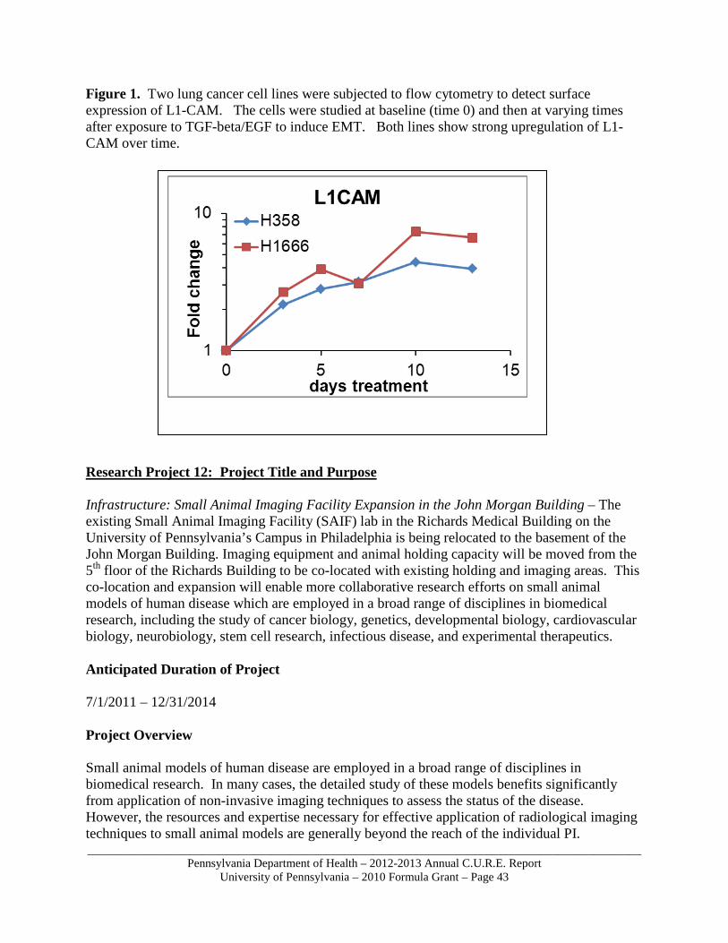
_____________________________________________________________________________________________Pennsylvania Department of Health – 2012-2013 Annual C.U.R.E. Report
University of Pennsylvania – 2010 Formula Grant – Page 43
Figure 1. Two lung cancer cell lines were subjected to flow cytometry to detect surface expression of L1-CAM. The cells were studied at baseline (time 0) and then at varying times after exposure to TGF-beta/EGF to induce EMT. Both lines show strong upregulation of L1-CAM over time. Research Project 12: Project Title and Purpose Infrastructure: Small Animal Imaging Facility Expansion in the John Morgan Building – The existing Small Animal Imaging Facility (SAIF) lab in the Richards Medical Building on the University of Pennsylvania’s Campus in Philadelphia is being relocated to the basement of the John Morgan Building. Imaging equipment and animal holding capacity will be moved from the 5th floor of the Richards Building to be co-located with existing holding and imaging areas. This co-location and expansion will enable more collaborative research efforts on small animal models of human disease which are employed in a broad range of disciplines in biomedical research, including the study of cancer biology, genetics, developmental biology, cardiovascular biology, neurobiology, stem cell research, infectious disease, and experimental therapeutics. Anticipated Duration of Project 7/1/2011 – 12/31/2014 Project Overview Small animal models of human disease are employed in a broad range of disciplines in biomedical research. In many cases, the detailed study of these models benefits significantly from application of non-invasive imaging techniques to assess the status of the disease. However, the resources and expertise necessary for effective application of radiological imaging techniques to small animal models are generally beyond the reach of the individual PI.

_____________________________________________________________________________________________Pennsylvania Department of Health – 2012-2013 Annual C.U.R.E. Report
University of Pennsylvania – 2010 Formula Grant – Page 44
The Small Animal Imaging Facility was formed by the University of Pennsylvania in order to address this need for researchers within the university and surrounding institutions. The facility provides instrumentation and support staff for performing magnetic resonance imaging (MRI), positron emission tomography (PET), computed tomography (CT), single photon emission computed tomography (SPECT), ultrasound imaging (US) and bioluminescence imaging (to detect luciferase and green fluorescent protein) in small animal models. In addition, the SAIF provides limited animal housing for rats and mice that allows multiple imaging sessions to be performed on a particular animal over an extended period (i.e., longitudinal studies) without risking infection of the general population of animals. The facility also provides support for data transfer and analysis. This project will renovate approximately 2,000 square feet of space on the basement level of the John Morgan Building to expand and consolidate the Small Animal Imaging Facility. Imaging equipment and animal holding capacity will be moved from the 5th floor of the Richards Building to be co-located with existing holding and imaging areas. The specific aims of the project are to:
1) fully renovate existing, aging research space to expand and consolidate the Small Animal Imaging Facility
2) create a centralized core facility to serve the School of Medicine research programs Principal Investigator Glen N. Gaulton, PhD Executive Vice Dean/Chief Scientific Officer University of Pennsylvania Perelman School of Medicine 240 John Morgan Building 3620 Hamilton Walk Philadelphia, PA 19104-6055 Other Participating Researchers Chandra Sehgal, PhD, Jerry Glickson, PhD, Stephen Pickup, PhD, Joel Karp, PhD, Edward J. Delikatny, PhD, Mitch Schnall, MD, PhD – employed by University of Pennsylvania Expected Research Outcomes and Benefits The Small Animal Imaging Facility enables cancer investigators to perform longitudinal structural and functional imaging of a wide range of molecular and cellular processes in small animals, tissue specimens, and cell cultures of biologically derived fluids. The resource is divided into four sub-cores based on imaging modality, which are Magnetic Resonance Imaging (MRI) and Spectroscopy, Nuclear Medical Imaging (NMI), Optical/Bioluminescence Imaging, and Small Animal Ultrasound Imaging. This project will renovate spaces on the basement level of the John Morgan Building to expand

_____________________________________________________________________________________________Pennsylvania Department of Health – 2012-2013 Annual C.U.R.E. Report
University of Pennsylvania – 2010 Formula Grant – Page 45
and consolidate the Small Animal Imaging Facility. Imaging equipment and animal holding capacity will be moved from the 5th floor of the Richards Building to be co-located with existing holding and imaging areas. The program includes: Renovation of approximately 1,040 gsf for new imaging lab, including:
o Micro CT, SPECT and PET Scanner o Control Room o Holding Room for radioactive rodents o Existing Vertical Bore Magnet to remain
Conversion of existing 312 gsf to a new office Conversion of approximately 475 sf of existing office and hallway area into animal
holding rooms and support, including: o Two (2) new rodent holding rooms o Expanded workroom o Storage o Cage Wash
Summary of Research Completed Documentation of the John Morgan Building basement project to expand and consolidate the Small Animal Imaging Facility (SAIF) started last year. Construction began in April 2013. It was determined that the existing Vertical Bore Magnet would be relocated to the Founders Building, freeing up program space within the expansion area. In addition, the cage wash area and its equipment have been removed from the project allowing for an increase in the size of the two new rodent holding rooms and the adjacent support area. There is an existing cage wash on the 3rd floor of the building that will be utilized to support the facility.