understanding your blood work results - mpn advocates · understanding your blood work results...
TRANSCRIPT

Understanding Your Blood Work Results
Carlos Besses, MD, PhD
Hematology Department
Hospital del Mar - IMIM, Barcelona

Carlos Besses – Disclosures
Novartis Honorarium Speaker
Shire Honorarium Speaker
Galena Honorarium Advisory Board

Abnormal blood results
Diagnostic process
History
taking Physical
examination
Additional
tests
Bone marrow
examination
Genetic
tests

Blood Results Leading to Suspicion of MPN
─ Thrombocytosis ( platelets)
─ Erythrocytosis ( red blood cells)
─ Leukocytosis ( white blood cells)
─ Anemia ( red blood cells)
─ Thrombocytopenia ( platelets)
─ Any combination of the above
PV
PMF
ET

Work-up for a Patient with Thrombocytosis
Platalet count ≥450x109/L
Secondary thrombocytosis Persistent unexplained thrombocytosis
History taking Physical
examination
Usually normal
Additional
tests
• Iron deficiency • Infection • Inflammation • Surgery • Cancer • Splenectomy • ……
• Any relative with MPN or excess of blood cells?
• Microvascular symptoms
Genetic
tests
Bone marrow
examination

Normal

ET

Workflow for Molecular Diagnosis of ET
ET Suspicion
JAK2 V617F
Positive 50-60% Negative
CALR
Positive 15-35% Negative
MPL
Positive 3-5% Negative
BCR-ABL1
Triple-Negative
5-25%
Negative Positive CML
Essential Thrombocythemia

ET Clinical Case, bone marrow biopsy

ET

A
C
B
Megakaryocyte nuclei:
A. Hyperlobulation: staghorn-like
B. Hypolobulation: bulbous/cloud-like
C. Hyperchromatic
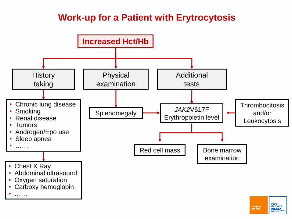
Work-up for a Patient with Erytrocytosis
Increased Hct/Hb
History
taking
Physical
examination
Additional
tests
Splenomegaly JAK2V617F
Erythropoietin level
Thrombocitosis
and/or
Leukocytosis
Red cell mass Bone marrow
examination
• Chronic lung disease • Smoking • Renal disease • Tumors • Androgen/Epo use • Sleep apnea • ……
• Chest X Ray • Abdominal ultrasound • Oxygen saturation • Carboxy hemoglobin • ……

Normal

PV

JAK2 JAK2
NUCLEUS
CYTOPLASM
Type I cytokine receptors
EPOR
TPOR
G-CSF
EPO
TPO

STAT STAT
JAK2 JAK2
P P
NUCLEUS
CYTOPLASM
Type I cytokine receptors
EPOR
TPOR
G-CSF
Gene transcription
EPO
TPO
P
P
P P
P
P
P P

STAT STAT
JAK2 JAK2
P P
NUCLEUS
CYTOPLASM
Gene transcription
EPO
TPO
P
P
P P
P
P
P P
G>T
GTC TTC
Val (V) Phe (F)
Type I cytokine receptors
EPOR
TPOR
G-CSF

PV

Increased leukocyte count
History
taking
Physical
examination Additional
tests
Splenomegaly
and/or
lymphadenopathy
Blood
film
Secondary
leukocytosis
Myeloid cells Lymphoid cells
• Chromosome and/or
genetic studies
• Cell flow cytometry
• CT scans
• PET, MRI
• ……
• Infections
• Acute
inflammation
• Corticosteroids
• Smoking
• ……
Work-up for a Patient with Leukocytosis

What are the Cells Involved in Myeloid Diseases?

PV
CLL

CML

Work-up for a Patient with Suspicion of Myelofibrosis
Anemia +/-
Other cytopenia +/-
WBC/platelets
Genetic test Peripheral blood Bone marrow biopsy +/-
Cytogenetic study
Mandatory Staining for:
Hematoxylin-eosin
Reticulin
Collagen ASXL1,
SRSF2, EZH2,
IDH1/2
Splenomegaly plus
Optional
JAK2V617F,
CALR, MPL
+/-
BCR-ABL1
Blood film
+/-
Cytogenetic study

PMF

Myelofibrosis: an heterogeneous disease
ET
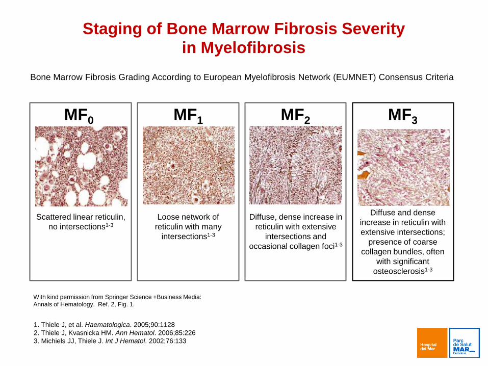
Staging of Bone Marrow Fibrosis Severity in Myelofibrosis
1. Thiele J, et al. Haematologica. 2005;90:1128
2. Thiele J, Kvasnicka HM. Ann Hematol. 2006;85:226
3. Michiels JJ, Thiele J. Int J Hematol. 2002;76:133
MF0 MF1 MF2 MF3
Scattered linear reticulin,
no intersections1-3
Loose network of
reticulin with many
intersections1-3
Diffuse, dense increase in
reticulin with extensive
intersections and
occasional collagen foci1-3
Diffuse and dense
increase in reticulin with
extensive intersections;
presence of coarse
collagen bundles, often
with significant
osteosclerosis1-3
Bone Marrow Fibrosis Grading According to European Myelofibrosis Network (EUMNET) Consensus Criteria
With kind permission from Springer Science +Business Media:
Annals of Hematology. Ref. 2, Fig. 1.

MF0
MF2 MF3
MF1
Reticulin fibrosis

PMF

Thiele et al, Blood 2011;117(21):5710-5718
WHO-Classification

A
C
B
Megakaryocyte nuclei:
A. Hyperlobulation: staghorn-like
B. Hypolobulation: bulbous/cloud-like
C. Hyperchromatic

Survival and Disease Complications
in ET vs. Early/Prefibrotic PMF
Outcome ET
15-Year Cumulative
Incidence (%)
Early/Prefibrotic PMF 15-Year Cumulative
Incidence (%)
Survival 75% 44%
Leukemic transformation 2% 12%
Transformation to overt myelofibrosis 9% 17%
Thrombosis 22% 25%
Results from 1104 patients; 891 (81%) True ET; 180 (16%) early/prefibrotic PMF; 33 (3%) not evaluable
Median follow-up: 6.2 years for ET; 7 years for early/prefibrotic PMF
Barbui et al, J Clin Oncol 2011;29(23):3179-3184

PV Suspicion ET Suspicion PMF Suspicion
Essential Thrombocythemia
Positive
Primary Myelofibrosis
Positive Negative Negative
Polycythemia Vera
Positive
JAK2 exon 12
Positive
Negative
JAK2V617F JAK2V617F JAK2V617F
Discard BCR-ABL
Negative
MPL
Positive
Negative
MPL
Positive
Positive
CALR
Positive
CALR
Negative
Negative Negative

1. Blood work results are important information but the
diagnosis relies on the whole picture
2. Genetic studies & Bone marrow examination are
essential components of the diagnosis
3. Common-sense and step-based tests are the best
way to reach a well-grounded diagnosis
Please, trust in your doctor!
Conclusions

Thank you for your attention