ultrasound safety - kth · diagnostic ultrasound, physics and equipment, chapter 12 (edited by...
TRANSCRIPT
Ultrasound Safety
Mats Olsson
Medtek konsult, Huddinge 2015
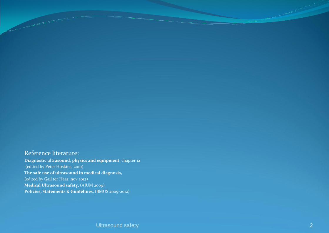
Reference literature: Diagnostic ultrasound, physics and equipment, chapter 12
(edited by Peter Hoskins, 2010)
The safe use of ultrasound in medical diagnosis,
(edited by Gail ter Haar, nov 2012)
Medical Ultrasound safety, (AIUM 2009)
Policies, Statements & Guidelines, (BMUS 2009-2012)
Ultrasound safety 2

Are there any risks with ultrasound scanning?
”Ultrasound is widely used because it provides many clinical benefits to the patient and has an
outstanding safety record. No patient injury has ever been reported from diagnostic ultrasound”
Clinicians are performing over 1 miljon examinations worldwide every year in cardiology, radiology,
obstetrics, gynecology, vascular ultrasound among others and the number continues to grow
Hundreds of thousends ultrasound scanners are in use every day
Only in England are 2,5 milj obstetric scans performed every year without any reports of injuries,
many of them were performed with new generations of scanners with potential of significantly
higher outputs than earlier
Ultrasound safety
(AIUM 2009 and Dept of Health in England 2012)
3
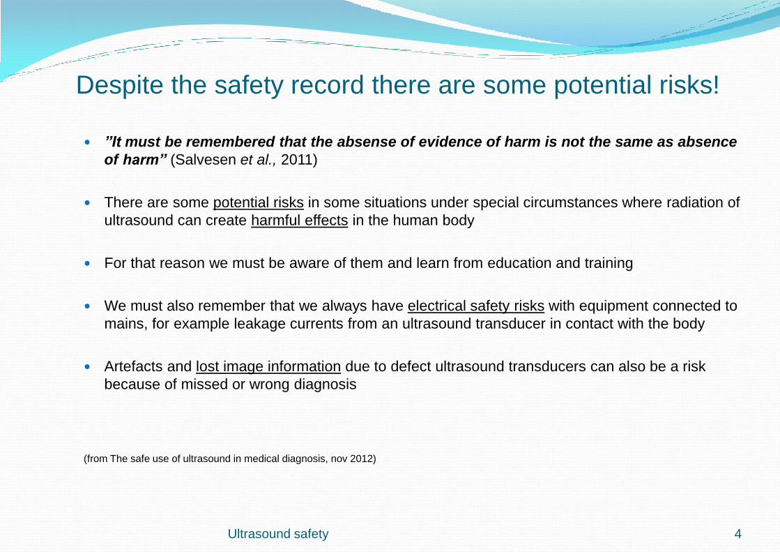
Despite the safety record there are some potential risks!
”It must be remembered that the absense of evidence of harm is not the same as absence
of harm” (Salvesen et al., 2011)
There are some potential risks in some situations under special circumstances where radiation of
ultrasound can create harmful effects in the human body
For that reason we must be aware of them and learn from education and training
We must also remember that we always have electrical safety risks with equipment connected to
mains, for example leakage currents from an ultrasound transducer in contact with the body
Artefacts and lost image information due to defect ultrasound transducers can also be a risk
because of missed or wrong diagnosis
(from The safe use of ultrasound in medical diagnosis, nov 2012)
Ultrasound safety 4

Education and training of users are very important
Ultrasound examinations shall only be performed by competent medical staff who have
deep knowledge of those specific risk situations. There is a strong need for continuing
education to ensure that appropriate risk/benefit assessments are made. The ultrasound
safety record depend on that
This is particularly important these days since more responsibility for the safe
management is passed over to the user by the standard societies
Several societies and organisations around the world, so called ”bodies”, have given
ultrasound safety special attention and education and training is one very important part
of it
Ultrasound safety
(From The safe use of ultrasound in medical diagnosis, nov 2012)
5

Standards and specifications regarding diagnostic ultrasound
Standard institutes as IEC (International Electrotechnical Commision) is the main body all over the world
regarding definition of measurement standards for all electrically connected equipment. For diagnostic
ultrasound they have published IEC60601 Part 2-37 (2001) ”to define display of safety indices and to
limit transducer surface temperature” and IEC62359 (2006) ”to define safety indices”
Another important standard institute, especially in Europé, is MDD, Medical Device Directive.
MDD is intended to harmonise the laws relating to medical devices within the European Union,
Counsil Directive 93/42 EEC 1993
FDA (Food and Drugs Administration) an American body which is responsible for compliance documents
in the American ultrasound industrial sector detailing permitted description of an ultrasound machine,
testing methods and limits to acoustic outputs. An important document is 510(k), FDA 2008. FDA has a
great influence over the whole world.
AIUM/NEMA ( American Institute of Ultrasound in Medicine / National Electrical Manufacturers
Association), has defined methods for calculation and display of safety indices
(From British Medical Ultrasound Society, BMUS, nov 2010 and
Diagnostic Ultrasound, physics and equipment, Hoskins 2010)
Ultrasound safety 6

Policies, statements and guidelines regarding diagnostic ultrasound
AIUM ( American Institute of Ultrasound in Medicine) in US, their role is to be a learned society
who is defining policies, guidelines and statements for end-users about safety. They have also
education programs for its members
BMUS ( Brittish Medical Ultrasound Society) has the same role in UK as AIUM
WFUMB ( World Federation of Ultrasound in Medicine and Biology) The umbrella group for
regional ultrasound societies. They produces safety statements and recommendations
EFSUMB ( European Federation of Ultrasound in Medicine and Biology) has the same role as
WFUMB but nationally. The umbrella group for regional ultrasound societies. They produces
safety statements and recommendations
Ultrasound safety
(From British Medical Ultrasound Society, BMUS, nov 2010)
7
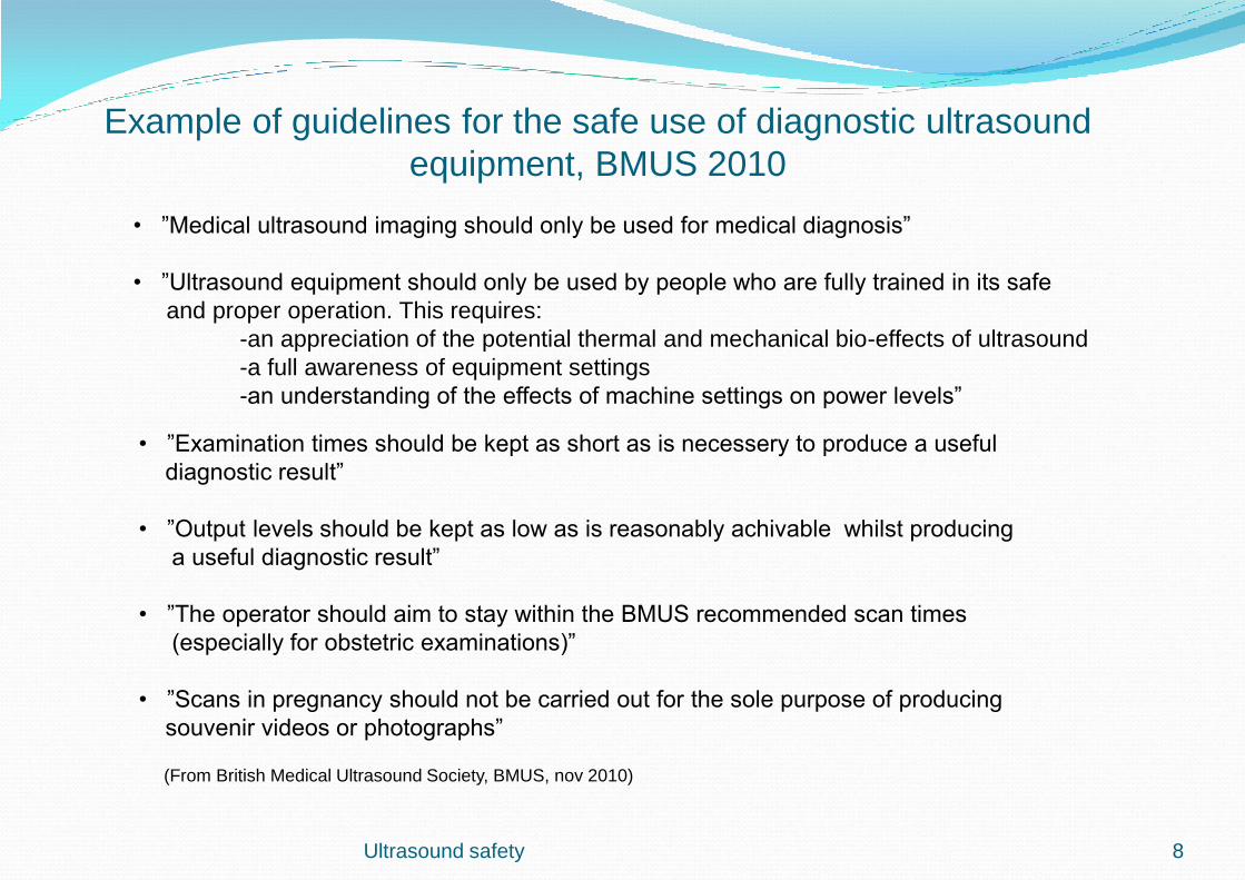
Example of guidelines for the safe use of diagnostic ultrasound
equipment, BMUS 2010
Ultrasound safety
• ”Medical ultrasound imaging should only be used for medical diagnosis”
• ”Ultrasound equipment should only be used by people who are fully trained in its safe
and proper operation. This requires:
-an appreciation of the potential thermal and mechanical bio-effects of ultrasound
-a full awareness of equipment settings
-an understanding of the effects of machine settings on power levels”
• ”Examination times should be kept as short as is necessery to produce a useful
diagnostic result”
• ”Output levels should be kept as low as is reasonably achivable whilst producing
a useful diagnostic result”
• ”The operator should aim to stay within the BMUS recommended scan times
(especially for obstetric examinations)”
• ”Scans in pregnancy should not be carried out for the sole purpose of producing
souvenir videos or photographs”
(From British Medical Ultrasound Society, BMUS, nov 2010)
8
Statement about prudent use from AIUM 2009
” The AIUM advocates the responsible use of diagnostic ultrasound and strongly
discourages the nonmedical use of ultrasound for entertainment purposes”
”The use of ultrasound without a medical indication to view the fetus, obtain a picture of
the fetus or determine the fetal gender is inappropriate and contrary to responsible
medical practice”
”Ultrasound should be used by qualified health professionals to provide medical benefit to
the patient”
Ultrasound safety 9
How You can minimize any eventual risk?
Apply a simple principle, ALARA!
ALARA stand for ”As Low As Reasonably Achievable” and means that always perform
an examination with as low ultrasound exposition as possible without to loose
diagnostic information
Always think of prudent use when examing a patient with ultrasound!
(From Medical Ultrasound Safety, AIUM 2009)
Ultrasound safety 10
Which adjustments can users do?
The most important parameter to minimize is the Intensity or Power because with that
one You can directly make adjustments on the output power from the ultrasound
transmitter (page 41)
By choosing an application you will automatically influence output power since it is
connected to chosen application, f ex ”peripherial vessel, cardiac, carotid, fetal
scanning etc”
Thereafter you can change adjustments on those functions that indirectly influence
output power, f ex system mode as 2D, M-mode and Doppler. Think also of pulse
repetition frequency (depth), focus, puls length, sample volume and choice of
transducer (see also page 40)
A basic rule is always to adjust ”receiver gain” or TGC to begin with, thereafter make
adjustments according to the rules above.
Ultrasound safety 11

Scanned och unscanned modes
In unscanned modes, as PW-doppler, CW-doppler och M-mode are the energy
concentrated along a thin layer in the patient which means over a much smaller volume
compared with 2D. In those modes are the highest temperatures to be found between
the body surface and focus
When scanned modes are used f ex 2D and color doppler is the ultrasound field spred
out over a large volume why the highest temperatures is to be found at the body
surface
(Medical ultrasound safety, AIUM 2009)
Ultrasound safety 12

Acoustic power and intensity
Acoustic power from the transmitter in the scanner is measured in Watts (W) and can
be seen on the screen as a certain value in - dB or % in dB of full power
Intensity( I ) = Power / Area, I = W/m², W/cm² or mW/cm² where the area is the
transducer head area
Ultrasound safety 13
Ultrasound safety
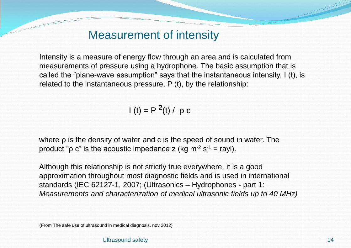
Measurement of intensity
Intensity is a measure of energy flow through an area and is calculated from
measurements of pressure using a hydrophone. The basic assumption that is
called the ”plane-wave assumption” says that the instantaneous intensity, I (t), is
related to the instantaneous pressure, P (t), by the relationship:
I (t) = P ²(t) / ρ c
where ρ is the density of water and c is the speed of sound in water. The
product ”ρ c” is the acoustic impedance z (kg m-2 s-1 = rayl).
Although this relationship is not strictly true everywhere, it is a good
approximation throughout most diagnostic fields and is used in international
standards (IEC 62127-1, 2007; (Ultrasonics – Hydrophones - part 1:
Measurements and characterization of medical ultrasonic fields up to 40 MHz)
(From The safe use of ultrasound in medical diagnosis, nov 2012)
14

Risks for high intensity in the ultrasound field
There is a potential risk for too high energy radiation from the ultrasound
equipment via the transducers to the patient
The risks can be of two kind :
- Thermal, risk for heating of soft tissue or bone
- Non thermal, for example mechanical phenomena like cavitation
Ultrasound safety 15
Thermal risks in general
Temperature increase is caused by absorption of acoustic energy in tissue
Since absorption coefficient is high in bone tissue, temperature can increase very fast
there and in the surrounding tissue
Frequency has an influence of absorption so that higher frequency gives higher
absorption and accordingly higher temperature, which is true specially in superficial
tissues
The highest temperatures will otherwise arise in the focus area when the wave front are
passing by the tissue
The examination time has a critical roll since temperature will increase with the scanning
time
(from The safe use of ultrasound in medical diagnosis, nov 2012)
Ultrasound safety 16
Particularly thermal sensitive tissues
• Reproductive cells
• Embryo and fetus
• The central nervous system
• The eyes
(AIUM 2007 and BMUS 2009)
Ultrasound safety 17

Special risks during embryo and fetus examinations
A very sensitive situation is when you are performing an ultrasound examination on a fetus during the
pregnancy month 4th and 6th, when the bone growing process is taking place (2nd trimester), especially
if the mothers belly wall is thin. Energy absorption in amniotic is very small why almost all absorption
take place in the bone tissue of the fetus
You must be aware of where you have focus so you can avoid exposures over long time near bone areas
”It seems most likely that the greatest potential risk in ultrasound diagnosis is with fetal spectral doppler
studies during first trimester. These studies involve potentially high-output intensities with stationary
geometry and a presumably more temperature sensitive fetus. Pulsed wave doppler or colour flow
imaging should not be used routinely”
Developing tissues of the embryo and fetus are particularly susceptible to damage by heating and the effects
can have serious consequences
The developing fetus is especially sensitive to hyperthermia during the period of neural tube closure
The induction of teratogenic effects (production of malformed fetus) depends on a combination of the elevation
above normal physiological temperature and the duration for which the increased temperature is maintained
(from BMUS Safety Group 2010 and EFSUMB/AIUM 2011)
Ultrasound safety 18
Temperature limits and other guidelines
An increase of 4°C above normal body temperature in 0,5 minutes may be hazardous to
embryonic and fetal development. Exposures longer than 5 min involve significant risk of harm
A temperature increase in fetal tissue to 41ºC and more is considered as dangerous while
temperature increases < 1ºC in general is considered not to be any biological risk
1,5°C above normal physiological temperature (37°C) does not appear to present a risk from
thermal effects in humans for an imaging session of less than 30 min
Ultrasound scanning of febrile obstetric patients requires particular care
A 30-fold increase in absorption coefficient has been reported as the fetal bone matures and have
become ossified, important to think of when scanning a fetus during 2nd and 3rd trimester
Simple 2D greyscale exposures are not capable of producing harmful temperature increases in
tissue
Ultrasound safety
(from The safe use of ultrasound in medical diagnosis, nov 2012)
19
Animal studies in laboratory
Lung haemorrhage in animal models has been observed as a result of
ultrasound exposure (rat, rabbit, mouse and pig)
Diagnostic frequencies used, about 3-5 MHz and exposure times from 10
seconds to 3 minutes
Other organs in which effects in small animals have been seen:
Bone; ”vascular damage near developing bone”
Intestine; ”activation from ultrasound”
Heart; ” radiation force can reduce the strength of contraction”
Ultrasound safety
(From The safe use of ultrasound in medical diagnosis, nov 2012)
20
Selfheating of the transducer
During use of endocavity transducers f ex vaginal transducers, rectal transducers and esophagus
transducers must attention be taken to the selfheating of the transducer. The temperature indices
system can therefore underestimate the temperature rise within about 5 mm of the transducer
Esofagus (TEE) transducers have f ex always a temperature sensing device at the tip
Temperature can together with the patients body temperature (37 ºC or more at fever) rise to
more than 40 ºC. In this situation there is no possibility for the heat energy in an endocavity
transducer to reach the surroundings, so the transducer can not be cooled down properly.
The problem can arise f ex with use of vaginal transducers during checks of pregnancy
Ultrasound safety 21
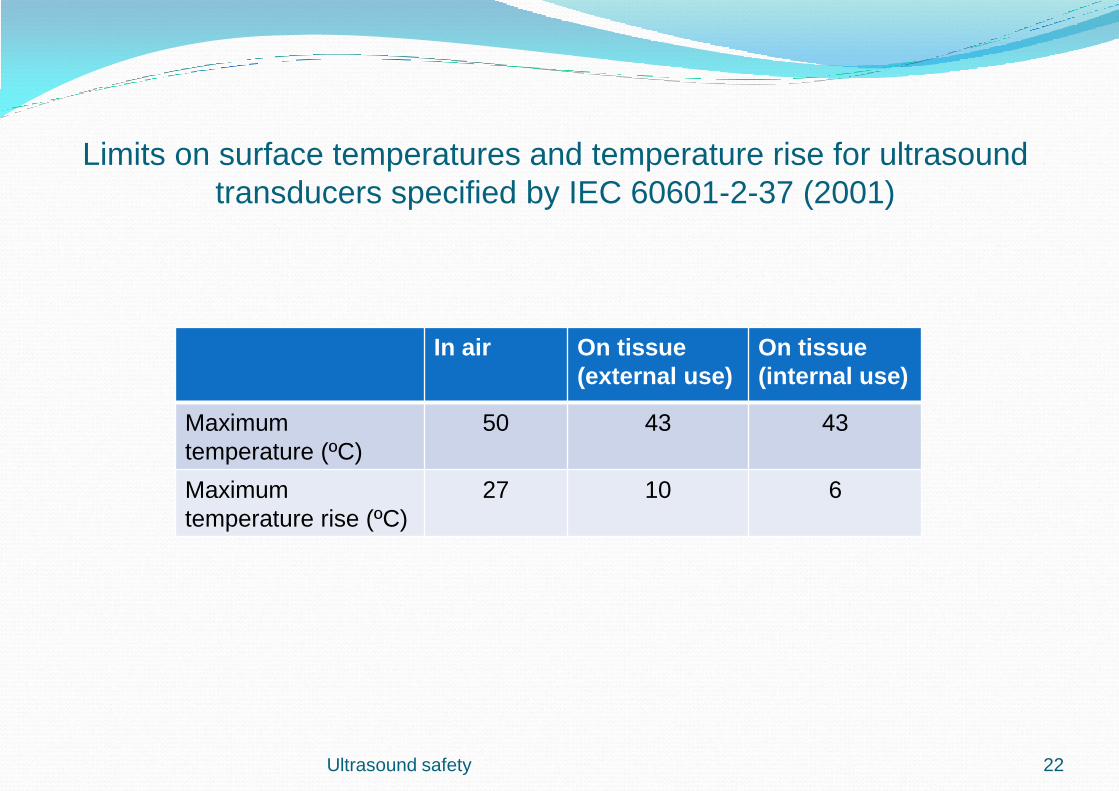
Limits on surface temperatures and temperature rise for ultrasound
transducers specified by IEC 60601-2-37 (2001)
Ultrasound safety
In air On tissue
(external use)
On tissue
(internal use)
Maximum
temperature (ºC)
50 43 43
Maximum
temperature rise (ºC)
27 10 6
22
Ultrasound safety
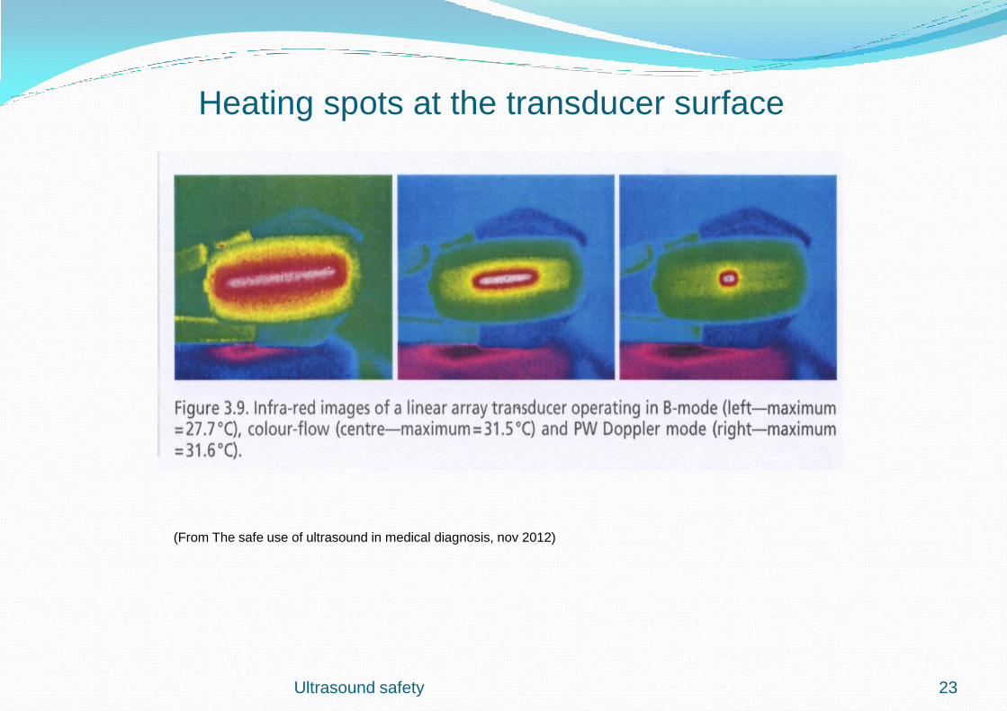
Heating spots at the transducer surface
(From The safe use of ultrasound in medical diagnosis, nov 2012)
23
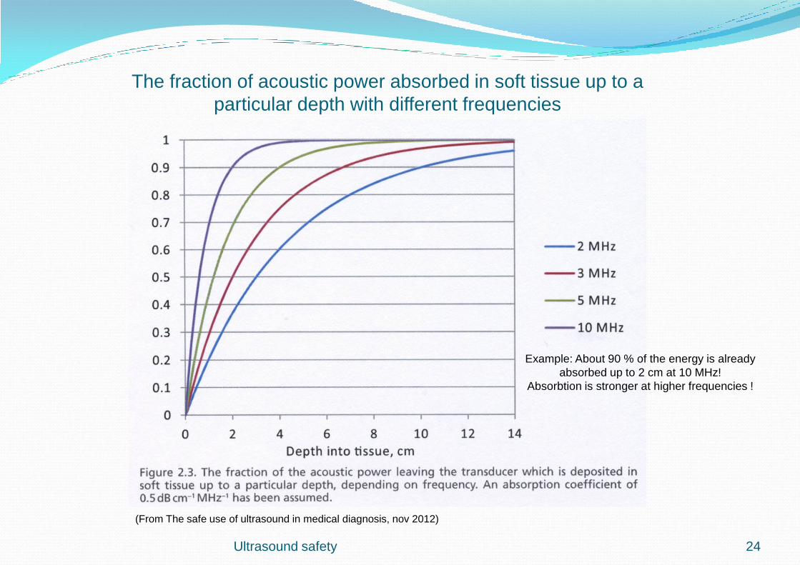
The fraction of acoustic power absorbed in soft tissue up to a
particular depth with different frequencies
(From The safe use of ultrasound in medical diagnosis, nov 2012)
Example: About 90 % of the energy is already
absorbed up to 2 cm at 10 MHz!
Absorbtion is stronger at higher frequencies !
Ultrasound safety 24
Ultrasound safety
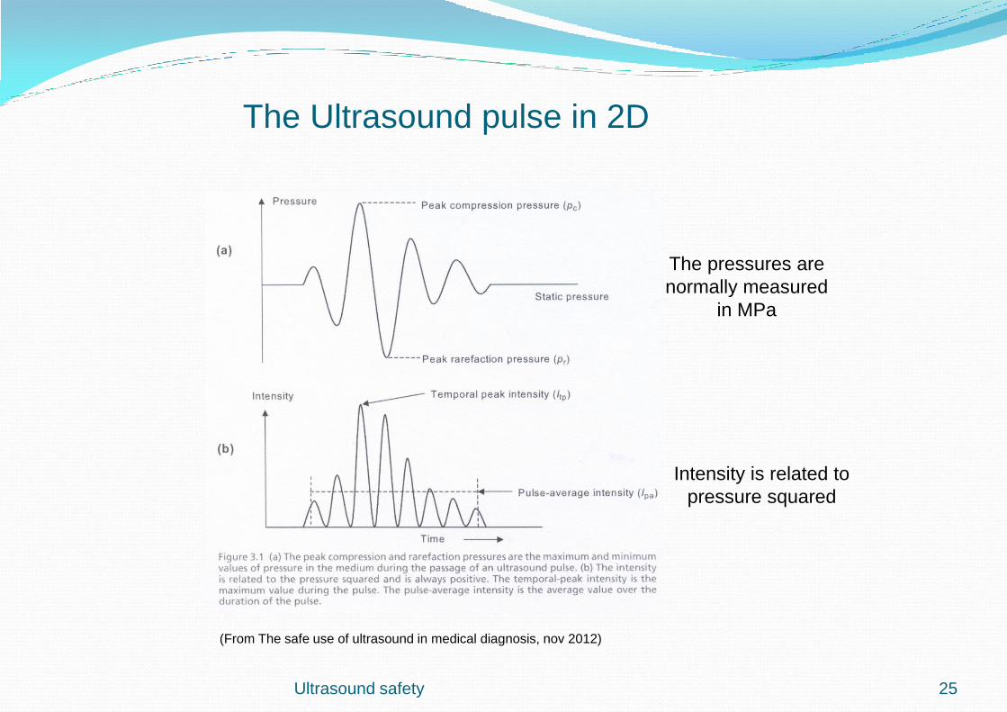
The Ultrasound pulse in 2D
The pressures are
normally measured
in MPa
(From The safe use of ultrasound in medical diagnosis, nov 2012)
25
Intensity is related to
pressure squared

Ultrasound safety
I-sppa
I-spta
Quantities often used by FDA among others
(mW/cm²)
(W/cm²)
26
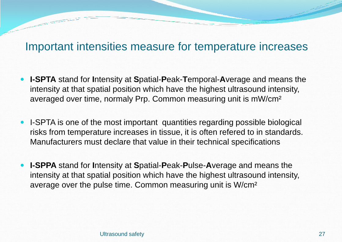
Important intensities measure for temperature increases
I-SPTA stand for Intensity at Spatial-Peak-Temporal-Average and means the
intensity at that spatial position which have the highest ultrasound intensity,
averaged over time, normaly Prp. Common measuring unit is mW/cm²
I-SPTA is one of the most important quantities regarding possible biological
risks from temperature increases in tissue, it is often refered to in standards.
Manufacturers must declare that value in their technical specifications
I-SPPA stand for Intensity at Spatial-Peak-Pulse-Average and means the
intensity at that spatial position which have the highest ultrasound intensity,
average over the pulse time. Common measuring unit is W/cm²
Ultrasound safety 27
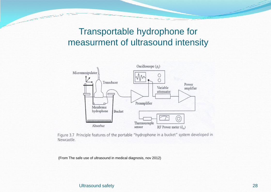
Ultrasound safety
Transportable hydrophone for
measurment of ultrasound intensity
(From The safe use of ultrasound in medical diagnosis, nov 2012)
28
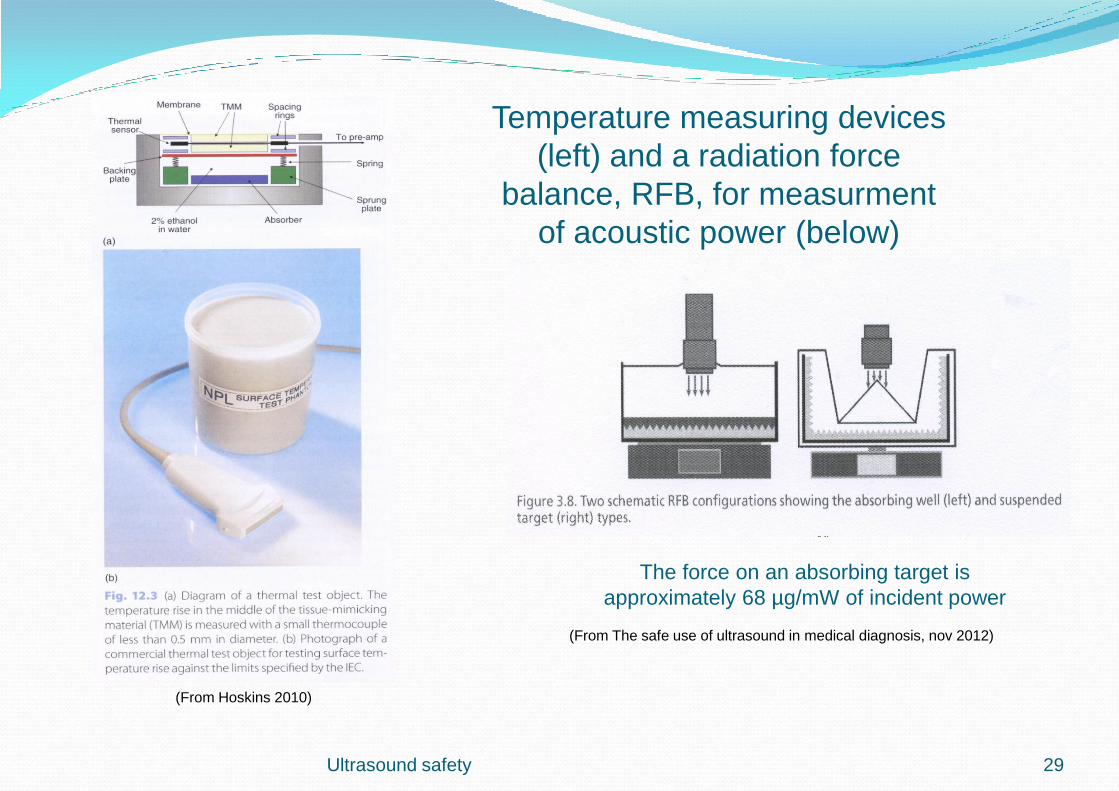
Temperature measuring devices
(left) and a radiation force
balance, RFB, for measurment
of acoustic power (below)
The force on an absorbing target is
approximately 68 µg/mW of incident power
(From The safe use of ultrasound in medical diagnosis, nov 2012)
(From Hoskins 2010)
Ultrasound safety 29

Ultrasound safety 30
Acoustic Intensity Measurement System
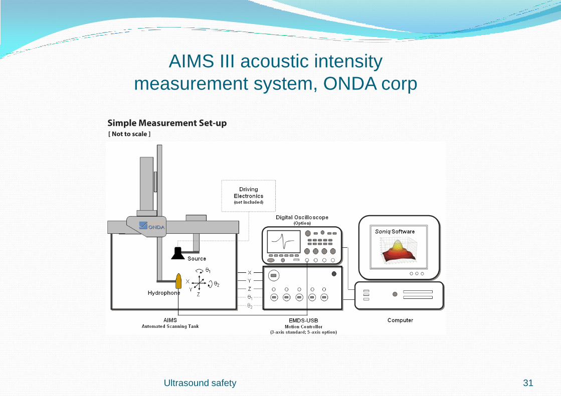
AIMS III acoustic intensity
measurement system, ONDA corp
Ultrasound safety 31

Ultrasound safety
Membrane and needle hydrophones
32

Non-thermal risks
Ultrasound pulses consists of both positive and negative pressures
The largest negative pressure, s c peak rarefactional pressure, ( pr.3
), together with the center
frequency may influence certain gas microbubbles in the tissue
This behavior is called acoustic cavitation and can be stable or unstable
Stable or non-inertial cavitation can arise at low pressures and refers to a pulsating or breathing
motion of gas bubbles which can exist in some tissues. The diameter follows the pressure
variation in the ultrasonic wave (next page)
If the rarefactional pressure rise too high, there is a risk for unstable inertial cavitation which
occurs at higher and short peakpressure pulses where small bubbles undergoes very large size
variations and can be brought to burst and cause damage in the surrounding tissue
(From Hoskins 2010)
Ultrasound safety 33
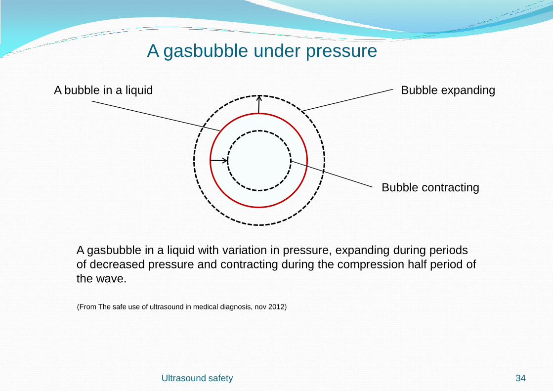
Ultrasound safety
A bubble in a liquid Bubble expanding
Bubble contracting
A gasbubble in a liquid with variation in pressure, expanding during periods
of decreased pressure and contracting during the compression half period of
the wave.
(From The safe use of ultrasound in medical diagnosis, nov 2012)
34
A gasbubble under pressure
Mechanical Index
The risk of acoustic cavitation depends primarily on the ultrasound pulse itself, peak
rarefactional pressure, pr.3
, and transducer frequency. fc. Also grade of focusing of the
ultrasound field, pulsed or continues wave, standing waves etc is of importance. The
Mechanical index, MI, may be used as a risk indicator for cavitation and is written as;
Cavitation is a threshold phenomena which means that it can only happen above a
certain MI value
The highest risk for cavitation is during use of ultrasound together with contrast agents
but can also happen in other gasfilled tissues , f ex at the surface of lung tissues or in
gasfilled intestine tissues
MI = pr.3
/ √ fc with no units
Ultrasound safety 35
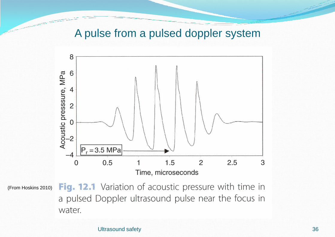
(From Hoskins 2010)
36
A pulse from a pulsed doppler system
Ultrasound safety
Output Display Standard
Authorities, organisations such as AIUM/NEMA and IEC have agreed about an
Output Display Standard, ODS, (1998, 2001 and 2006) which mean that
ultrasound scanners must have a built-in display system so the ultrasound
users can see what parameters are active in a certain examination
From that display shall two types of s c index be shown, which have strong
connection to output power, frequency and tissue pressure
Those are:
Thermal Index or TI, which is related to temperature increase and
Mechanical Index or MI, which has connection to non-thermal biological
effects f ex cavitation
Ultrasound safety 37
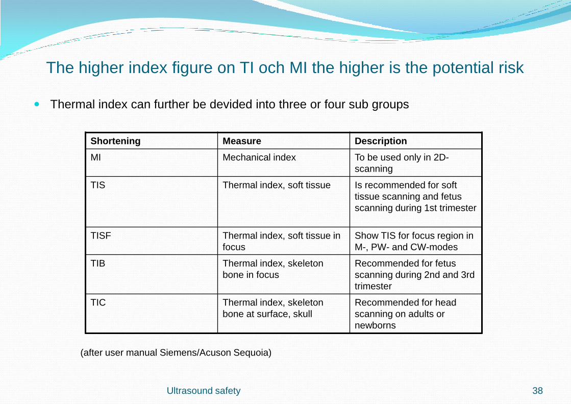
The higher index figure on TI och MI the higher is the potential risk
Thermal index can further be devided into three or four sub groups
Shortening Measure Description
MI Mechanical index To be used only in 2D-
scanning
TIS Thermal index, soft tissue Is recommended for soft
tissue scanning and fetus
scanning during 1st trimester
TISF Thermal index, soft tissue in
focus
Show TIS for focus region in
M-, PW- and CW-modes
TIB Thermal index, skeleton
bone in focus
Recommended for fetus
scanning during 2nd and 3rd
trimester
TIC Thermal index, skeleton
bone at surface, skull
Recommended for head
scanning on adults or
newborns
(after user manual Siemens/Acuson Sequoia)
Ultrasound safety 38
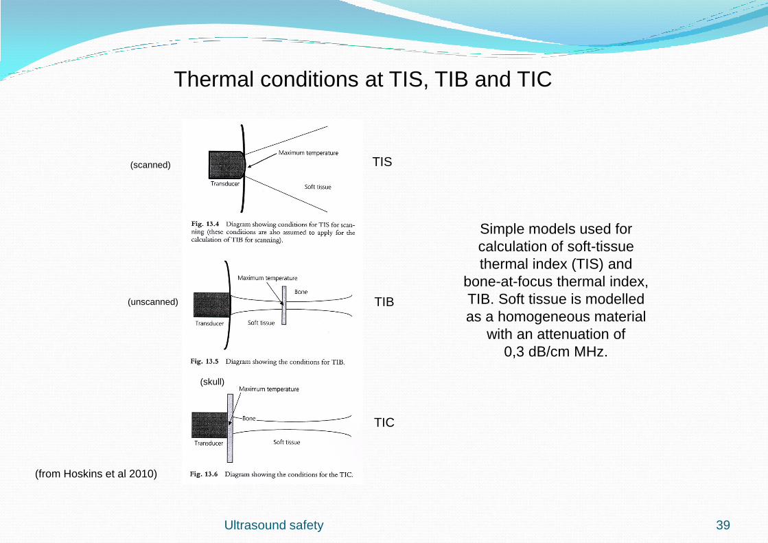
(from Hoskins et al 2010)
TIS
TIB
TIC
Simple models used for
calculation of soft-tissue
thermal index (TIS) and
bone-at-focus thermal index,
TIB. Soft tissue is modelled
as a homogeneous material
with an attenuation of
0,3 dB/cm MHz.
(scanned)
(unscanned)
(skull)
Ultrasound safety 39
Thermal conditions at TIS, TIB and TIC

Ultrasound safety
Temperature measurements on transducer
surface when changing settings
(From The safe use of ultrasound in medical diagnosis, nov 2012)
(Opportunity for useful staff training of awareness)
40
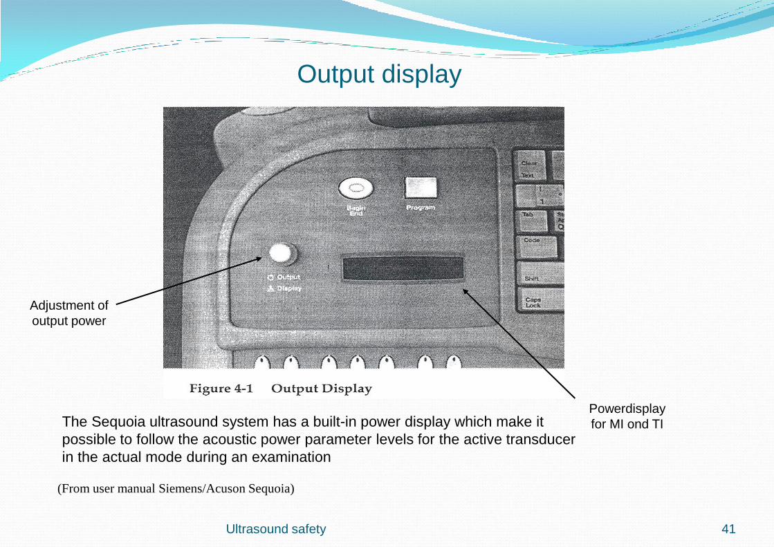
Ultrasound safety
(From user manual Siemens/Acuson Sequoia)
The Sequoia ultrasound system has a built-in power display which make it
possible to follow the acoustic power parameter levels for the active transducer
in the actual mode during an examination
Adjustment of
output power
Powerdisplay
for MI ond TI
41
Output display

Ultrasound safety
(From user manual GE
Healthcare Vivid 7)
42
Acoustic Power Indication
Color-coded TI levels
and recommended
exposure times
Vivid 7 has an internal
limit of 4.0 on TI
MI and TIS
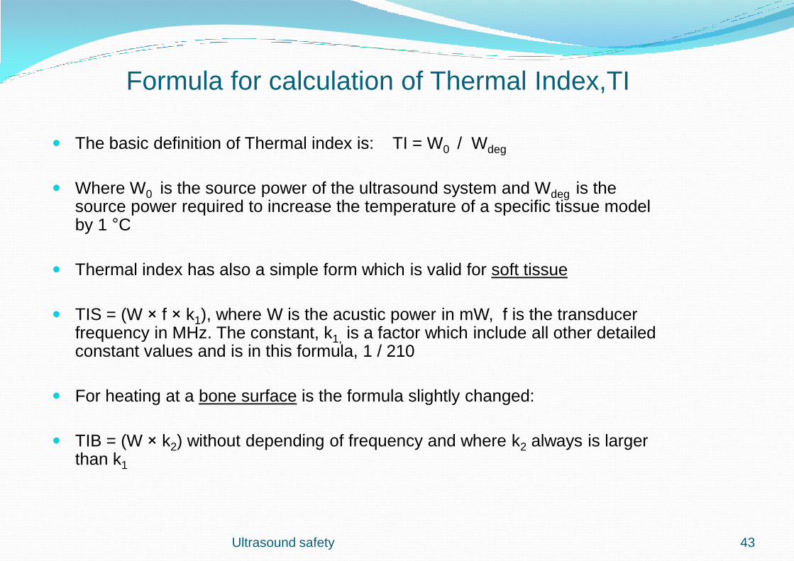
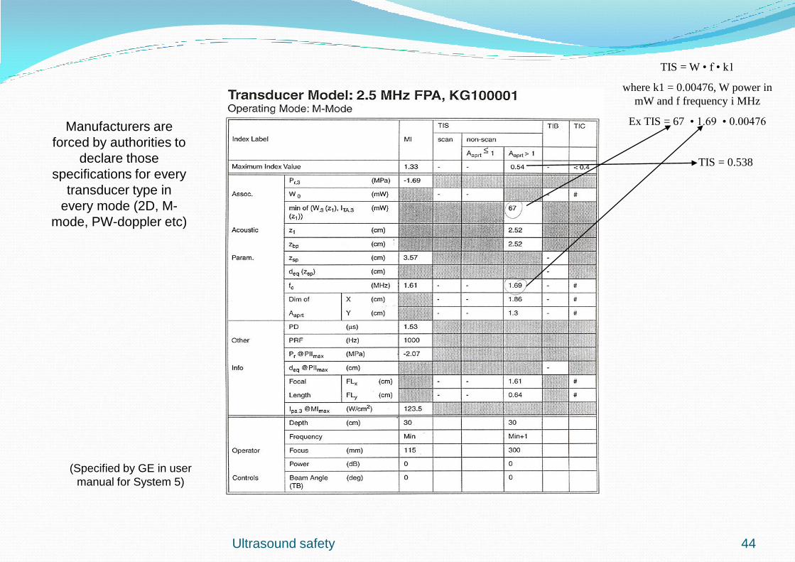
Formula for calculation of Thermal Index,TI
The basic definition of Thermal index is: TI = W0 / Wdeg
Where W0 is the source power of the ultrasound system and Wdeg is the source power required to increase the temperature of a specific tissue model by 1 °C
Thermal index has also a simple form which is valid for soft tissue
TIS = (W × f × k1), where W is the acustic power in mW, f is the transducer frequency in MHz. The constant, k1, is a factor which include all other detailed constant values and is in this formula, 1 / 210
For heating at a bone surface is the formula slightly changed:
TIB = (W × k2) without depending of frequency and where k2 always is larger than k1
Ultrasound safety 43
Ultrasound safety
TIS = W • f • k1
where k1 = 0.00476, W power in
mW and f frequency i MHz
Ex TIS = 67 • 1.69 • 0.00476
TIS = 0.538
(Specified by GE in user
manual for System 5)
Manufacturers are
forced by authorities to
declare those
specifications for every
transducer type in
every mode (2D, M-
mode, PW-doppler etc)
44
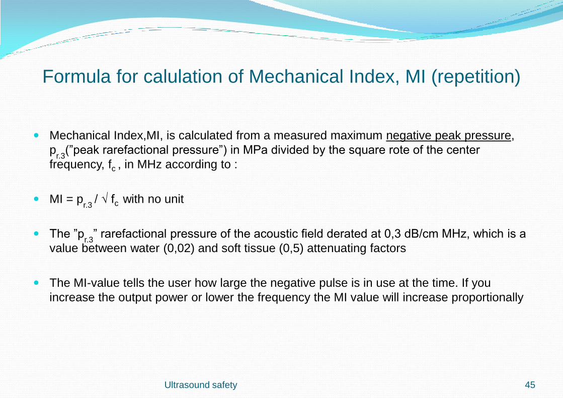
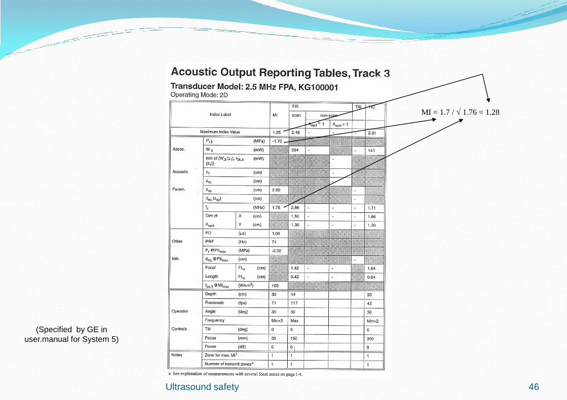
Formula for calulation of Mechanical Index, MI (repetition)
Mechanical Index,MI, is calculated from a measured maximum negative peak pressure,
pr.3
(”peak rarefactional pressure”) in MPa divided by the square rote of the center
frequency, fc , in MHz according to :
MI = pr.3
/ √ fc with no unit
The ”pr.3
” rarefactional pressure of the acoustic field derated at 0,3 dB/cm MHz, which is a
value between water (0,02) and soft tissue (0,5) attenuating factors
The MI-value tells the user how large the negative pulse is in use at the time. If you
increase the output power or lower the frequency the MI value will increase proportionally
Ultrasound safety 45
Ultrasound safety
MI = 1.7 / 1.76 = 1.28
(Specified by GE in
user.manual for System 5)
46
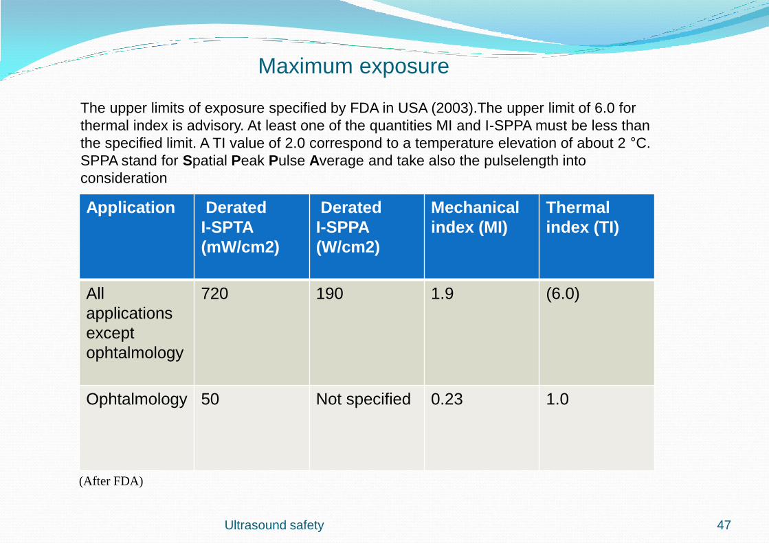
Application
Derated
I-SPTA
(mW/cm2)
Derated
I-SPPA
(W/cm2)
Mechanical
index (MI)
Thermal
index (TI)
All
applications
except
ophtalmology
720 190 1.9 (6.0)
Ophtalmology 50 Not specified 0.23 1.0
The upper limits of exposure specified by FDA in USA (2003).The upper limit of 6.0 for
thermal index is advisory. At least one of the quantities MI and I-SPPA must be less than
the specified limit. A TI value of 2.0 correspond to a temperature elevation of about 2 °C.
SPPA stand for Spatial Peak Pulse Average and take also the pulselength into
consideration
(After FDA)
Ultrasound safety 47
Maximum exposure
Ultrasound safety
(From, Hoskins
2010)
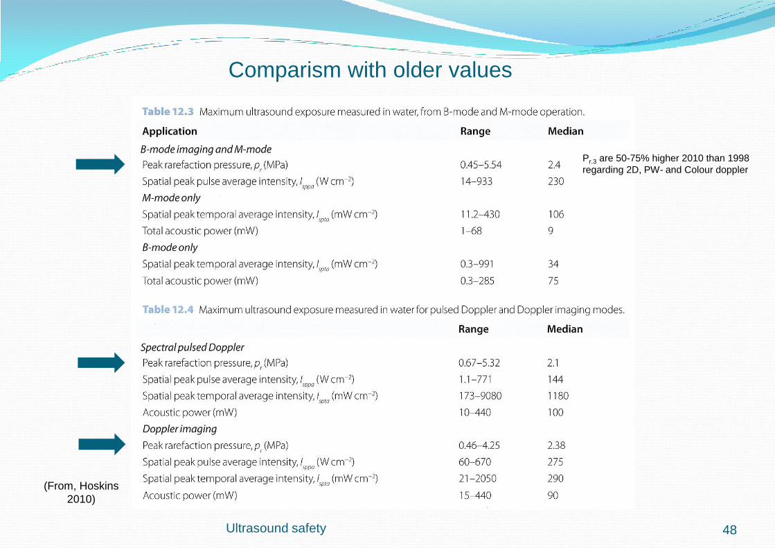
Pr.3 are 50-75% higher 2010 than 1998
regarding 2D, PW- and Colour doppler
48
Comparism with older values
( from Safety guidelines BMUS nov 2010)
Ultrasound safety 49
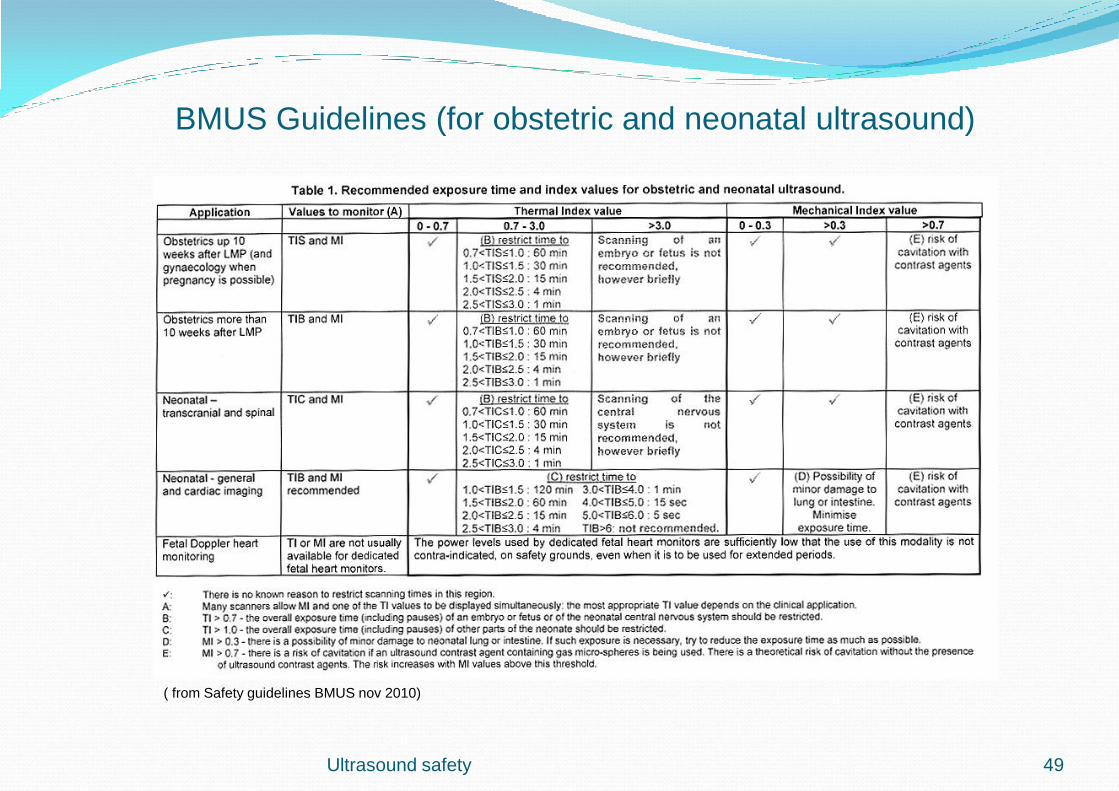
BMUS Guidelines (for obstetric and neonatal ultrasound)

(from Safety guidelines BMUS nov 2009 )
Ultrasound safety 50
BMUS Guidelines (for non-obstetric and non-neonatal ultrasound)

Leakage currents
According to SS-EN 60601-1, Electromedical equipment, general requirements for safety
Following protection grades are used :
Type B (Body), means low leakage currents but you have no limitation from currents floating from equipment through the patient. This can be a dangerous situation for the patient if he come in contact with mains voltage from a broken equipment near him
Type BF (Body; Floating), means the same low leakage currents as type B but has also a high resistance against currents from eqiupment to the patient. In this case is it limited to 5 mA which can protect the patient from dangerous currents. Normal protection grade for an ultrasound scanner with probes
Type CF (Cardiac; Floating), means better isolation and lower leakage currents than type BF and the maximum leakage current is here limited to 50 μA. Ultrasound transducers for internal use have this protection grade, f ex esofagus transducers
Ultrasound safety 51
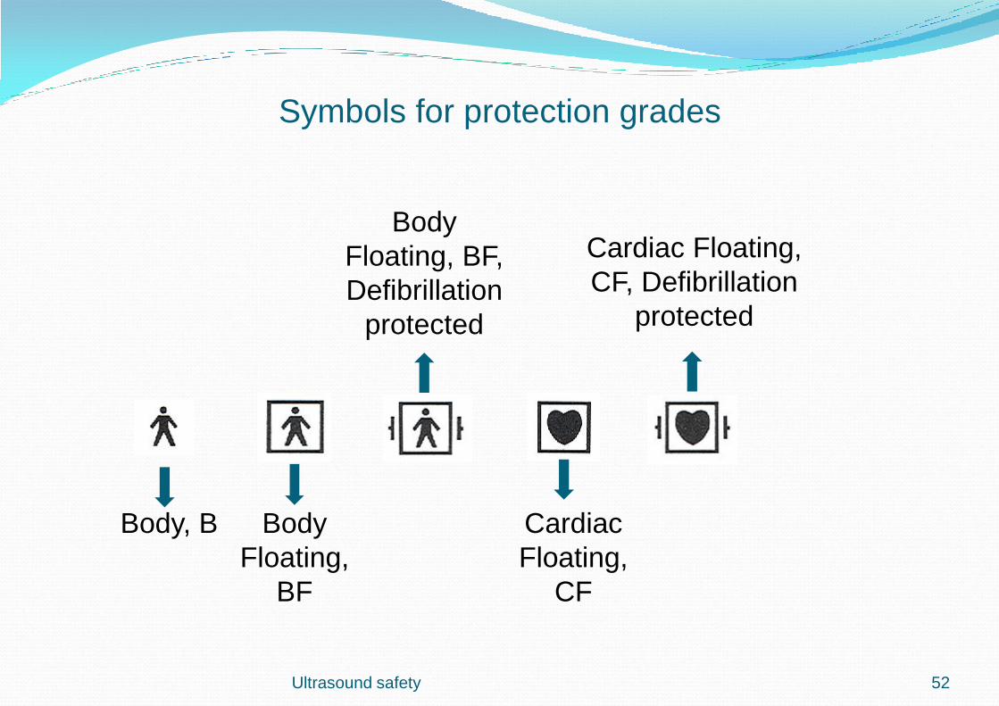
Body, B Body
Floating,
BF
Cardiac
Floating,
CF
Body
Floating, BF,
Defibrillation
protected
Cardiac Floating,
CF, Defibrillation
protected
Symbols for protection grades
Ultrasound safety 52

Checking of leakage currents from an ultrasound scanner
Very important to do this
tests because there are
often broken lenses at
transducer surfaces. A
broken lens can result in
higher leakage currents to
the patient
Ultrasound safety 53

TEE probes must be handled with care
Trans Esophageal Echocardiography probes are tested for electrical failures
before each patient examination because of the sensitive scanning situation.
Special bite-guards are used.
Ultrasound safety 54
Analyzer for
checking of
electrical safety
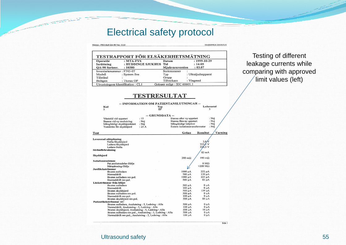
Ultrasound safety 55
Electrical safety protocol
Testing of different
leakage currents while
comparing with approved
limit values (left)
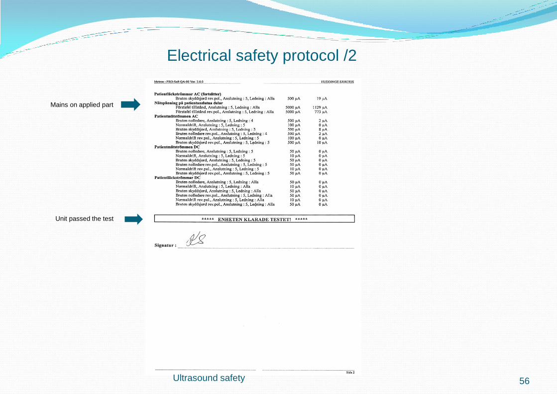
Unit passed the test
Mains on applied part
Ultrasound safety 56
Electrical safety protocol /2
Sensitivity
0
0,2
0,4
0,6
0,8
1
1 11 21 31 41 51 61 71 81 91 101 111 121
Elements
Vo
lts p
-p
Sensitivity
0
0,2
0,4
0,6
0,8
1
1 11 21 31 41 51 61 71 81 91 101 111 121
Elements
Vo
lts p
-p
New transducer Older transducer with many elements defect
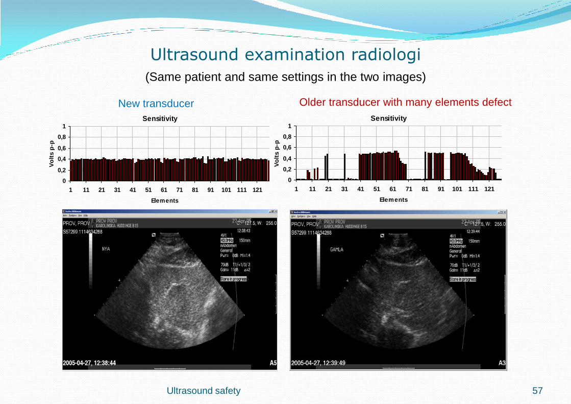
Ultrasound examination radiologi
(Same patient and same settings in the two images)
Ultrasound safety 57