ultrasound guided steroids injection for carpal...
TRANSCRIPT
International Journal of Medical Imaging 2015; 3(4): 75-81
Published online June 13, 2015 (http://www.sciencepublishinggroup.com/j/ijmi)
doi: 10.11648/j.ijmi.20150304.12
ISSN: 2330-8303 (Print); ISSN: 2330-832X (Online)
Ultrasound Guided Steroids Injection for Carpal Tunnel Syndrome
Naglaa Dabees1, Hanan El-Saadany
2, Aly El-Barbary
1, Al-Shimaa Ammar
1
1Radiodiagnosis Departments, Faculty of Medicine, Tanta University, Tanta, Egypt 2Physical Medicine Departments, Faculty of Medicine, Tanta University, Tanta, Egypt
Email address: [email protected] (N. Dabees), [email protected] (H. El-Saadany), [email protected] (Al-Shimaa A.),
[email protected] (A. El-Barbary)
To cite this article: Naglaa Dabees, Hanan El-Saadany, Aly El-Barbary, Al-Shimaa Ammar. Ultrasound Guided Steroids Injection for Carpal Tunnel Syndrome.
International Journal of Medical Imaging. Vol. 3, No. 4, 2015, pp. 75-81. doi: 10.11648/j.ijmi.20150304.12
Abstract: Objective: The aim of this work was to evaluate the role of ultrasound-guided steroids injection for carpal tunnel
syndrome (CTS). Study design: This is a prospective study done on thirty patients with carpal tunnel syndrome with mild to
moderate severity. Patients were divided into two groups; group I of fifteen patients `wrists injected by steroids using
ultrasound guidance and group II of another fifteen patients injected blindly- as control- with the same material .Results: There
was significant improvement in symptoms and ultrasound findings, after steroid injection, in both groups, being more in group
I than group II, with shorter average time to symptom relief, in group I than group II. Moreover, the complications were
significantly lower in group I than group II. Conclusions: Ultrasound guided steroids injection is more effective in reducing the
symptoms and improving function of CTS with lower risk of nerve injury than blind local steroid injection.
Keywords: Ultrasound, Steroids Injection, Carpal Tunnel Syndrome
1. Introduction
Carpal tunnel syndrome (CTS) is one of the most common
entrapment neuropathy syndromes of the upper limb (1,2)
.
Its incidence is 139/100,000 per year for men and
506/100,000 per year for women. (3)
Carpal tunnel syndrome (CTS) occurs at a rate of 5.2 per
10,000 full time workers, and this syndrome requires the
longest re-occupational period of all conditions that result in
lost work days, with a median of 30 days lost. (4)
It arises from entrapment of the median nerve at the wrist;
typically the disorder is characterized by numbness or
tingling in the sensory distribution of the nerve in the hand,
which may be accompanied by pain and or weakness of the
muscles of the thumb (5)
.
Women between the ages of 45–60 years suffer from CTS
more than men with a ratio of 3:1(6)
.
Generally, accepted treatments include: steroids injected
locally, splinting, and surgical release of the transverse carpal
ligament (7)
. Steroid injection into the carpal tunnel is
frequently used and is known to be effective in the treatment
of CTS. (8, 9, 10)
In daily clinical practice, this intervention is often
performed blindly, with risk of damaging the median nerve
and the surrounding vulnerable structures such as tendons
and vessels. (11, 12, 13, 14)
To improve accuracy of the intervention, image-guided
injections are required. Because ultrasound is none ionizing,
readily available, relatively oflow cost, and easily accepted
by patients, it has become the first choice to guide carpal
tunnel injections. Moreover, ultrasound provides a real-time
anatomic high-resolution image of the median nerve, the
carpal tunnel, and the neighboring structures and assists the
diagnosis as well as the suggestion of an etiology (13)
.
The real-time capability of ultrasound provides a clear
advantage in guiding carpal tunnel injections because it
enables visualizing the needle tip continuously and ensures
that the needle is placed precisely in the desired location,
avoiding the risk for inadvertent nerve and surrounding
structure damage (13, 14)
.
2. Aim of the Work
The aim of this work was to evaluate the role of
ultrasound-guided steroids injection for treating carpal tunnel
syndrome.
76 Naglaa Dabees et al.: Ultrasound Guided Steroids Injection for Carpal Tunnel Syndrome
3. Patients and Methods
3.1. Patients
This study was conducted on thirty patients with idiopathic
CTS diagnosed by clinical examination, electrophysiological
study, and ultrasound. Patients were referred from the
departments of physical medicine and rheumatology, to the
department of Diagnostic Radiology and Medical Imaging, at
Tanta University Hospital, during the period from 12/2013 to
11/2014.
They were twenty six females (86.7%) and four males
(13.3 %) with age range from 20 - 60 years and a mean age
of 40 years.
3.1.1. Inclusion Criteria
Patients with mild to moderate symptoms of carpal tunnel
syndrome, who failed responding to other methods of
treatment and / or those refused surgery.
3.1.2. Exclusion Criteria
Male and female Patients with Parkinsonism, traumatic
nerve injury, previous steroids injection in the same wrist and
other causes of neuropathy rather than CTS.
3.1.3. Study Design
It is a prospective study done on thirty patients with carpal
tunnel syndrome with mild to moderate severity. Patients
were divided into two groups; group I of fifteen patients
whose wrists were injected with steroids under ultrasound
guidance and group II of another fifteen patients injected
blindly- as control- with the same material
All patients were subjected to full history taking, clinical
examination and laboratory investigations.
Electrophysiological studies were done pre and post (after
6 weeks of injection) to all patients in both groups. Studies
included motor and sensory nerve conduction as well as F
wave for both median and ulnar nerves.
Assessment of all patients in both groups pre and post
injection (after 6 weeks of injection) was then done by
Boston Carpal tunnel questionnaire (BCTQ). Also,
Ultrasound examination was done to all patients pre and post
(6 weeks after injection) injection in both group, to assess the
median nerve and flexor retinaculum.
All patients were examined at Radiology Department,
Tanta university hospital, using “biomedical P-K” US
machine. The status of CTS was evaluated pre and post
steroid injection, considering the following parameters: cross
surface area (CSA) of median nerve, ratio between major and
minor axis of median nerve at distal carpal tunnel, and finally
thickness and bowing of flexor retinaculum (FR).
3.2. Methods
3.2.1. US- Guided Steroid Injection Technique
Before injection, the patient was seated, with the forearm
supinated and the wrist slightly dorsiflexed. Ultrasound-guided
steroids injection of carpal tunnel syndrome was done using 26
gauge needle to minimize discomfort. After obtaining skin
anesthesia with a vapocoolant spray, the needle was passed
into the skin on the ulnar side of the proximal carpal tunnel at
the level of the distal wrist crease. The needle was passed
superficial to the ulnar nerve and artery, penetrating the FR on
the ulnar side of the carpal tunnel.
With ultrasound guidance, the needle was directed to the
superficial ulnar side of the median nerve, after which a
portion of the injectate was delivered. As the injectate was
delivered, the median nerve typically peeled off the
undersurface of the FR, allowing the needle to be advanced
between the nerve and the overlying retinaculum.
The needle was then withdrawn to the ulnar side of the
median nerve and redirected to the deep side of the ulnar
portion of the nerve. Under real-time visualization, additional
injectate was delivered, separating the nerve from the
underlying sub synovial connective tissue.
In this manner, the nerve was completely surrounded with
the injectate and mechanically separated from the potentially
constricting surrounding connective tissues. Finally, the needle
was directed yet deeper into the sub synovial connective tissue,
where the remainder of the injectate was delivered.
A typical injectate consists of 1 ml of 40-mg/mL
methylprednisolone acetate (Depo- Medrol 40mg/ml) and 1
mL of 1% lidocaine, delivered in equal portions above the
nerve, below the nerve, and into the subsynovial connective
tissue. After completion of the injection, the distal carpal
tunnel is scanned to ensure injectate distribution throughout
the distal extent of the carpal tunnel.
3.2.2. The Blind Injection Technique
The Blind Injection Technique was conducted using the
ulnar approach; the wrist was positioned in 30 degrees
dorsiflexion resting on a towel roll. The injection sites were
either the proximal wrist crease or ulnar side marked by
Palmaris longus tendon or wrist midline if Palmaris longus is
absent.
Under aseptic measures, needle was inserted and aimed
30-45 degrees distally toward middle-ring finger. Insertion
was done for 1-2 cm until no resistance was encountered.
Injection was stopped once parasthesia developed. The
injected material used in the blind injection technique was
the same as in the ultrasound- guided technique.
Local compression at the site of injection was applied to all
patients in both groups to allow proper absorption of the
injected steroid and to minimize any possibility for occurrence
of hematoma. Analgesics was given to relieve the pain.
3.2.3. Follow Up
Follow up was done at the base line and after 6 weeks by:
I. clinical evaluation using BCTQ score, the latter
evaluates symptomatic severity and function
recovery with a score of 0-5 according to severity of
symptoms or difficulty in a certain activity, the worse
the symptoms, the higher the score.
II. Ultrasound of volar wrist, evaluating:
a. Cross surface area of median nerve ( N up to 10 mm2)
b. Ratio between major to minor axis of median nerve
( N up to 3)
International Journal of Medical Imaging 2015; 3(4): 75-81 77
c. Flexor retinaculum thickening ( N less than 4 mm)
III. Electrophysiological studies including, motor and
sensory nerve conduction and F wave of median and
ulnar nerves.
3.3. Statistics Analysis
Statistical analysis was done, using the mean, standard
error, student t- test, Chi-square and Wilcoxon Signed Ranks
Test, tests by SPSS V17.
4. Results
This study was conducted on thirty patients with carpal
tunnel syndrome with mild to moderate severity. Patients
were divided into two groups; group I of fifteen patients
whose ‘wrists were injected with steroids under ultrasound
guidance and group II of another fifteen patients injected
blindly- as control- with the same material.
They were twenty six females (86.7%) and four males
(13.3 %) with age range from 20 - 60 years and mean age of
40 years.
In both groups, pain was the commonest symptom (100%),
followed by parasthesia, hyposethesia, hand weakness and
pain awakening patient from sleep. After steroid injection
symptoms improvement was encountered in both groups.
Pain improved in 80% of group I, four days after injection
compared to 63.33% of group II, seven days after injection
with statistically significant difference. Again, pain that
awakened patient from sleep, was improved in 83.3% of
group I vs. 52% of group II. Hand weakness was surprisingly
improved in 100% of group I vs. 66.6 in group II. Parasthesia
as well as hypoesthesia, had also improved in both groups,
more with US guided injection, than with blind injection.
Table (1) shows significant improvement (p = 0.002)of
severity of symptoms and function by BCTQ after steroid
injection in group I. Mild to moderate and moderate severity
of the fifteen cases, before injection, became normal in 8
cases and mild in 5cases, while severity persists as mild to
moderate in one case ,and moderate in one case, with total
improvement of 86.66%. However, in group II severity
persists as mild to moderate in 2 cases and moderate in 6
cases with total improvement of 53.33% (p=0.252).
Table (1). The improvement of symptomatic severity and functional evaluation by BCTQ in both groups before and after steroids injection.
Normal Mild Mild to moderate moderate
Total P. value % of improvement N % N % N % N %
G I Pre - - - - 8 53.33 7 46.67 15
0.002* 86.66% Post 8 53.33 5 33.33 1 6.67 1 6.67 15
G II Pre - - - - 7 46.67 8 53.33 15
0.252 53.33% Post 2 13.33 5 33.33 2 13.33 6 40.01 15
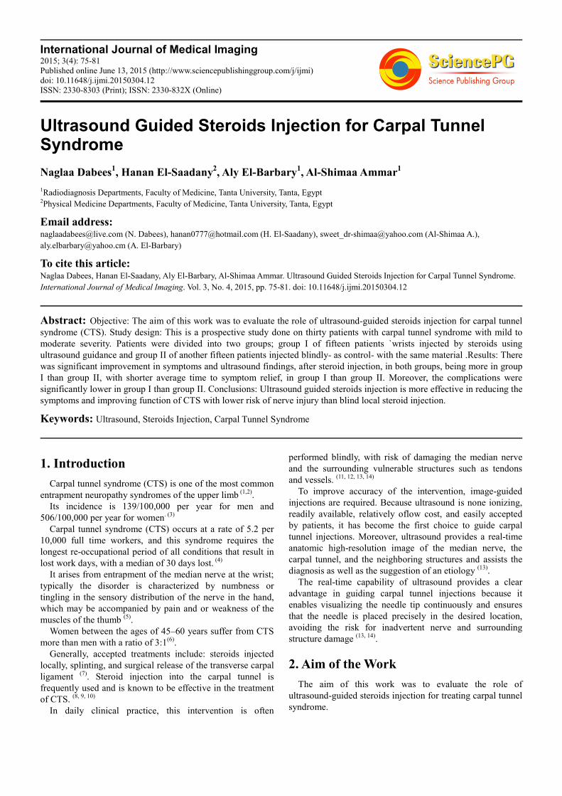
Fig. 1. a) US image of a patients wrist with mild to moderate CTS showing swelling of the median nerve (CSA=12mm2),b) US image of carpal tunnel injection,
the needle is seen as a white line. C) US image after 6 weeks follow up showing decreased swelling of the median nerve (CSA=7mm2).
78 Naglaa Dabees et al.: Ultrasound Guided Steroids Injection for Carpal Tunnel Syndrome
The ultrasound findings of the two studied groups before
steroids injection (table 2) included 1) Swelling of the
median nerve <10 mm2 (86.67%) in group I vs. (66.67%) in
group II, at the proximal carpal tunnel (commonest),
followed by 2) Hypoechgenic appearance of the median
nerve (80%) in group I vs. (46.67%) in group II 3)Flattening
of the median nerve at the distal carpal tunnel (20%) in group
I vs. (26.6%) in group II 4)Thickening and bowing of the FR
(40%) in group I vs. (53.33%) in group II. The ultrasound
findings of the diseased wrists, before injection, (table 2), had
improved significantly, after steroids injection (table 3). The
median nerve cross surface area (CSA), had dramatically
been reduced, in 92 % of group I, and 69.77 % in group II.
Moreover, the median nerve regained its normal
ecchogenicity, in 100 % of group I vs. 84 % of group II. The
flattening of the median nerve had been relieved in 66 % of
group I, and in 57 % of group II. The thickness of FR, had
returned to normal in 83 % of group I, vs. 57.5 of group II.
Table (2). The US findings of the studied wrists of both groups before steroids injection.
US finding of carpal tunnel syndrome Group I Group II Total
N % N % N %
Swelling of the median nerve <10 mm2 at the proximal carpal tunnel 13 86.67 10 66.67 23 76.67
Hypoechgenic appearance of median nerve 12 80.00 7 46.67 19 63.33
Flattening of the median nerve at the distal carpal tunnel 3 20.00 4 26.67 7 23.33
Thickening and bowing of the FR 6 40.00 8 53.33 14 46.67
(NB: Some patient's writs showed more than one US finding)
Table (3). The ultrasound findings in group I and II wrists after steroids injection.
Post Group I Group II Total Chi-square
N % N % N % X2 P-value
Reduced diameter of median nerve less than 10 mm2 12/13. 92.31 4/10. 40.00 16/23. 69.57 7.304 0.007*
Normal echogencity of median nerve 12/12. 100.00 4/7. 57.14 16/19. 84.21 6.107 0.013*
Relieved flattening and return to normal axis 2/3. 66.67 2/4. 50.00 4/7. 57.14 0.194 0.659
Returned of normal thickness of FR 5/6. 83.33 3/8. 37.50 8/14 57.14 3.13 0.044*
(NB: Some patient show more than one US finding)
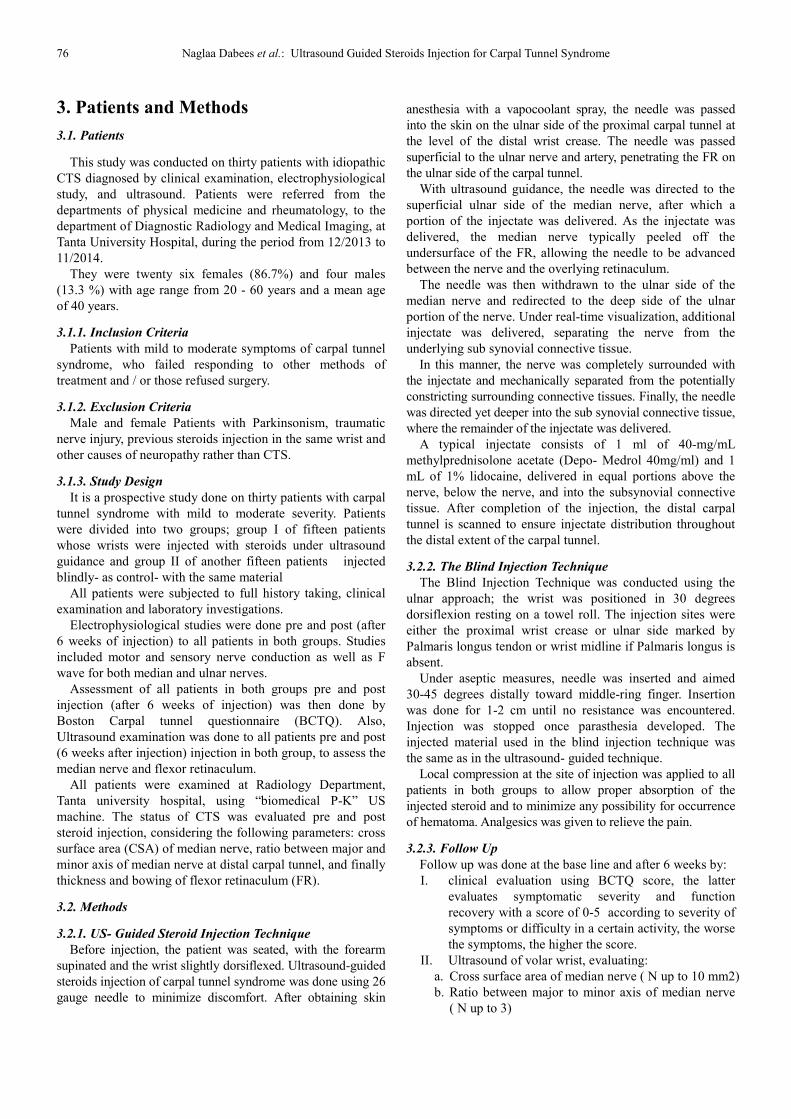
Fig. 2. a) US image of a female patients wrist with moderate CTS showing swelling of the median nerve (CSA=13mm2),b) US image of carpal tunnel injection,
the needle is seen as a white line. C) after 6 weeks follow up US image shows decreased swelling of the median nerve ( CSA=8mm2).
International Journal of Medical Imaging 2015; 3(4): 75-81 79
The electrophysiological findings of the thirty wrists
before steroids injection in both groups ranged from mild to
moderate. After steroids injection (table 4), the following
electrophysiological changes were encountered ; In group I,
out of the seven cases with moderate CTS, 2 cases became
normal, 4 cases became mild, and one case showed no
changes. While, out of 8 cases with mild CTS, 7cases
became normal, and one case didn’t change.
In group II, out of 8 cases with moderate CTS, one case
became normal, four case became mild, and 3 cases remained
unchanged. There were 7 cases in group II suffering from
mild to CTS, out of these, 2 cases became normal, and 5
cases didn’t respond.
Post injection complication in both blind and ultrasound
guided technique were summarized in (table 5). Pain, oedema
and tingling, although had occurred in both groups, however,
they were significally lower in group I compared to group II.
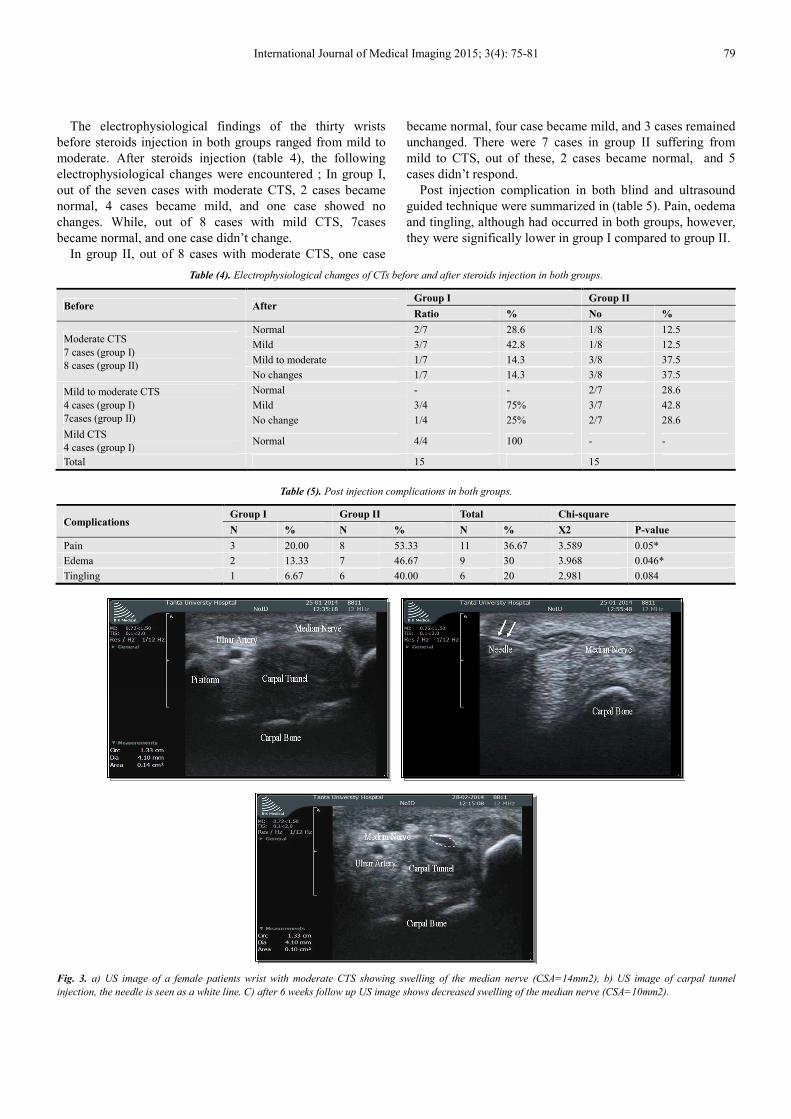
Table (4). Electrophysiological changes of CTs before and after steroids injection in both groups.
Before After Group I Group II
Ratio % No %
Moderate CTS
7 cases (group I)
8 cases (group II)
Normal 2/7 28.6 1/8 12.5
Mild 3/7 42.8 1/8 12.5
Mild to moderate 1/7 14.3 3/8 37.5
No changes 1/7 14.3 3/8 37.5
Mild to moderate CTS
4 cases (group I)
7cases (group II)
Normal - - 2/7 28.6
Mild 3/4 75% 3/7 42.8
No change 1/4 25% 2/7 28.6
Mild CTS
4 cases (group I) Normal 4/4 100 - -
Total 15 15
Table (5). Post injection complications in both groups.
Complications Group I Group II Total Chi-square
N % N % N % X2 P-value
Pain 3 20.00 8 53.33 11 36.67 3.589 0.05*
Edema 2 13.33 7 46.67 9 30 3.968 0.046*
Tingling 1 6.67 6 40.00 6 20 2.981 0.084
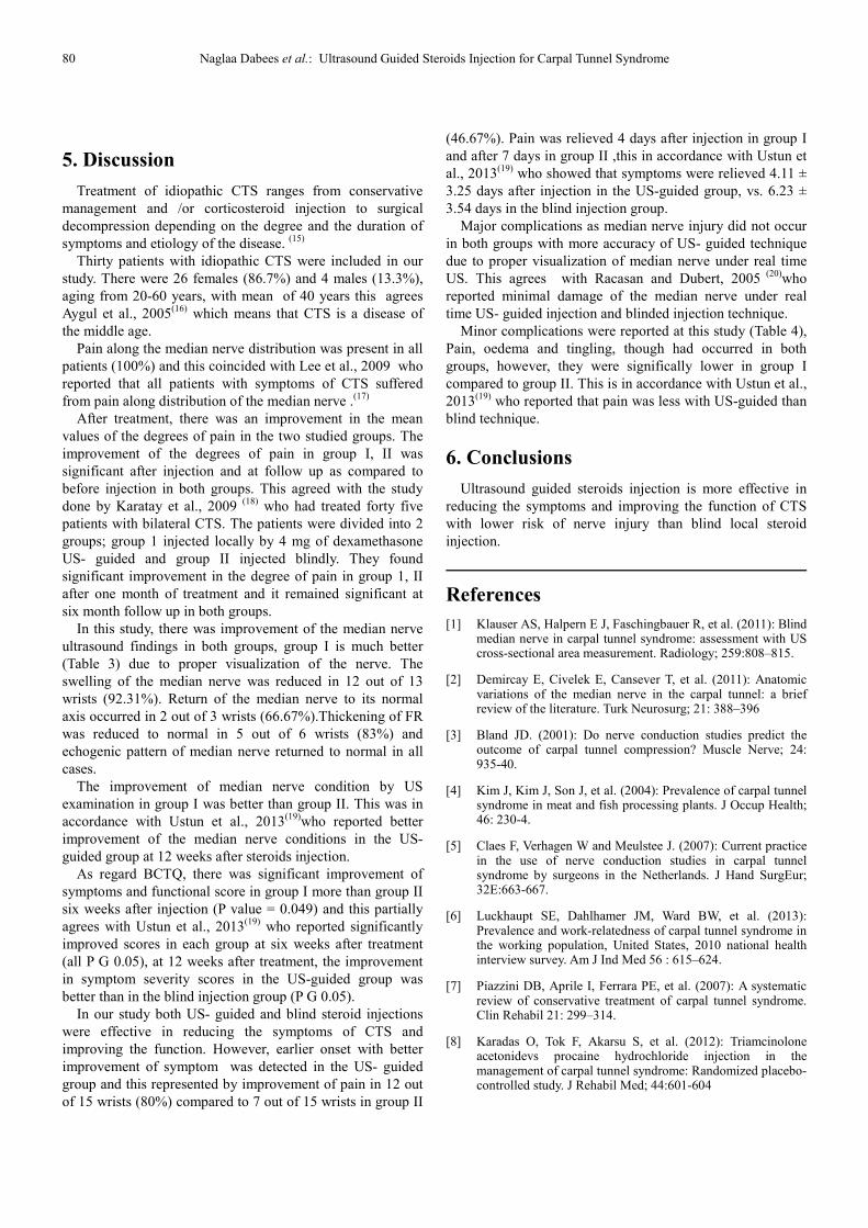
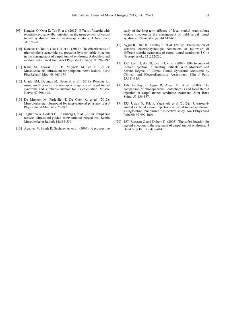
Fig. 3. a) US image of a female patients wrist with moderate CTS showing swelling of the median nerve (CSA=14mm2), b) US image of carpal tunnel
injection, the needle is seen as a white line. C) after 6 weeks follow up US image shows decreased swelling of the median nerve (CSA=10mm2).
80 Naglaa Dabees et al.: Ultrasound Guided Steroids Injection for Carpal Tunnel Syndrome
5. Discussion
Treatment of idiopathic CTS ranges from conservative
management and /or corticosteroid injection to surgical
decompression depending on the degree and the duration of
symptoms and etiology of the disease. (15)
Thirty patients with idiopathic CTS were included in our
study. There were 26 females (86.7%) and 4 males (13.3%),
aging from 20-60 years, with mean of 40 years this agrees
Aygul et al., 2005(16)
which means that CTS is a disease of
the middle age.
Pain along the median nerve distribution was present in all
patients (100%) and this coincided with Lee et al., 2009 who
reported that all patients with symptoms of CTS suffered
from pain along distribution of the median nerve .(17)
After treatment, there was an improvement in the mean
values of the degrees of pain in the two studied groups. The
improvement of the degrees of pain in group I, II was
significant after injection and at follow up as compared to
before injection in both groups. This agreed with the study
done by Karatay et al., 2009 (18)
who had treated forty five
patients with bilateral CTS. The patients were divided into 2
groups; group 1 injected locally by 4 mg of dexamethasone
US- guided and group II injected blindly. They found
significant improvement in the degree of pain in group 1, II
after one month of treatment and it remained significant at
six month follow up in both groups.
In this study, there was improvement of the median nerve
ultrasound findings in both groups, group I is much better
(Table 3) due to proper visualization of the nerve. The
swelling of the median nerve was reduced in 12 out of 13
wrists (92.31%). Return of the median nerve to its normal
axis occurred in 2 out of 3 wrists (66.67%).Thickening of FR
was reduced to normal in 5 out of 6 wrists (83%) and
echogenic pattern of median nerve returned to normal in all
cases.
The improvement of median nerve condition by US
examination in group I was better than group II. This was in
accordance with Ustun et al., 2013(19)
who reported better
improvement of the median nerve conditions in the US-
guided group at 12 weeks after steroids injection.
As regard BCTQ, there was significant improvement of
symptoms and functional score in group I more than group II
six weeks after injection (P value = 0.049) and this partially
agrees with Ustun et al., 2013(19)
who reported significantly
improved scores in each group at six weeks after treatment
(all P G 0.05), at 12 weeks after treatment, the improvement
in symptom severity scores in the US-guided group was
better than in the blind injection group (P G 0.05).
In our study both US- guided and blind steroid injections
were effective in reducing the symptoms of CTS and
improving the function. However, earlier onset with better
improvement of symptom was detected in the US- guided
group and this represented by improvement of pain in 12 out
of 15 wrists (80%) compared to 7 out of 15 wrists in group II
(46.67%). Pain was relieved 4 days after injection in group I
and after 7 days in group II ,this in accordance with Ustun et
al., 2013(19)
who showed that symptoms were relieved 4.11 ±
3.25 days after injection in the US-guided group, vs. 6.23 ±
3.54 days in the blind injection group.
Major complications as median nerve injury did not occur
in both groups with more accuracy of US- guided technique
due to proper visualization of median nerve under real time
US. This agrees with Racasan and Dubert, 2005 (20)
who
reported minimal damage of the median nerve under real
time US- guided injection and blinded injection technique.
Minor complications were reported at this study (Table 4),
Pain, oedema and tingling, though had occurred in both
groups, however, they were significally lower in group I
compared to group II. This is in accordance with Ustun et al.,
2013(19)
who reported that pain was less with US-guided than
blind technique.
6. Conclusions
Ultrasound guided steroids injection is more effective in
reducing the symptoms and improving the function of CTS
with lower risk of nerve injury than blind local steroid
injection.
References
[1] Klauser AS, Halpern E J, Faschingbauer R, et al. (2011): Blind median nerve in carpal tunnel syndrome: assessment with US cross-sectional area measurement. Radiology; 259:808–815.
[2] Demircay E, Civelek E, Cansever T, et al. (2011): Anatomic variations of the median nerve in the carpal tunnel: a brief review of the literature. Turk Neurosurg; 21: 388–396
[3] Bland JD. (2001): Do nerve conduction studies predict the outcome of carpal tunnel compression? Muscle Nerve; 24: 935-40.
[4] Kim J, Kim J, Son J, et al. (2004): Prevalence of carpal tunnel syndrome in meat and fish processing plants. J Occup Health; 46: 230-4.
[5] Claes F, Verhagen W and Meulstee J. (2007): Current practice in the use of nerve conduction studies in carpal tunnel syndrome by surgeons in the Netherlands. J Hand SurgEur; 32E:663-667.
[6] Luckhaupt SE, Dahlhamer JM, Ward BW, et al. (2013): Prevalence and work-relatedness of carpal tunnel syndrome in the working population, United States, 2010 national health interview survey. Am J Ind Med 56 : 615–624.
[7] Piazzini DB, Aprile I, Ferrara PE, et al. (2007): A systematic review of conservative treatment of carpal tunnel syndrome. Clin Rehabil 21: 299–314.
[8] Karadas O, Tok F, Akarsu S, et al. (2012): Triamcinolone acetonidevs procaine hydrochloride injection in the management of carpal tunnel syndrome: Randomized placebo-controlled study. J Rehabil Med; 44:601-604
International Journal of Medical Imaging 2015; 3(4): 75-81 81
[9] Karadas O, Oma K, Tok F, et al (2012): Effects of steroid with repetitive procaine HCl injection in the management of carpal tunnel syndrome: An ultrasonographic study. J NeurolSci; 316:76-78
[10] Karadas O, Tok F, Ulas UH, et al. (2011): The effectiveness of triamcinolone acetonide vs. procaine hydrochloride injection in the management of carpal tunnel syndrome: A double-blind randomized clinical trial. Am J Phys Med Rehabil; 90:287-292
[11] Kara M, zoakar L, De Muynck M, et al (2012): Musculoskeletal ultrasound for peripheral nerve lesions. Eur J PhysRehabil Med; 48:665-674
[12] Ulasli AM, Duymus M, Nacir B, et al. (2013): Reasons for using swelling ratio in sonographic diagnosis of carpal tunnel syndrome and a reliable method for its calculation. Muscle Nerve; 47:396-402.
[13] De Muynck M, Parlevliet T, De Cock K, et al. (2012): Musculoskeletal ultrasound for interventional physiatry. Eur J Phys Rehabil Med; 48:675-687.
[14] Tagliafico A, Bodner G, Rosenberg I, et al. (2010): Peripheral nerves: Ultrasound-guided interventional procedures. Semin Musculoskelet Radiol; 14:514-559.
[15] Aganval V, Singh R, Sachdev A, et al. (2005): A prospective
study of the long-term efficacy of local methyl prednisolone acetate injection hi the management of mild carpal tunnel syndrome. Rheumatology; 44:647-650.
[16] Aygul R, Ulvi H, Karatay S, et al. (2005): Determination of sensitive electrophysiologic parameters at follow-up of different steroid treatments of carpal tunnel syndrome. J Clin Neurophysiol.; 22 :222-230.
[17] 152. Lee JH, An JH, Lee SH, et al. (2009): Effectiveness of Steroid Injection in Treating Patients With Moderate and Severe Degree of Carpal Tunnel Syndrome Measured by Clinical and Electrodiagnostic Assessment. Clin J Pain; 25:111-115.
[18] 156. Karatay S, Aygul R, Alkan M, et al. (2009): The comparison of phonophoresis, iontophoresis and local steroid injection in carpal tunnel syndrome treatment. Joint Bone Spine; 55:156-157.
[19] 135. Ustun N, Tok F, Yagız AE et al (2013): Ultrasound-guided vs. blind steroid injections in carpal tunnel syndrome: a single-blind randomized prospective study. Am J Phys Med Rehabil; 92:999-1004.
[20] 157. Racasan O and Dubert T. (2005): The safest location for steroid injection in the treatment of carpal tunnel syndrome. J Hand Surg Br: 30; 412–414.