type i collagen promotes the malignant phenotype of...
TRANSCRIPT
Type I Collagen Promotes the Malignant Phenotype of PancreaticDuctal Adenocarcinoma
Thomas Armstrong,1 Graham Packham,2
Lindsay B. Murphy,1 Adrian C. Bateman,1
John A. Conti,1 David R. Fine,1
Colin D. Johnson,2 R. Christopher Benyon,1 andJohn P. Iredale1
Divisions of 1Tissue Remodelling and Repair and 2Cancer Sciences,University of Southampton, Southampton General Hospital,Southampton, United Kingdom
ABSTRACTPurpose: The purpose of this study was to determine the
role of functional interactions between pancreatic cancercells and pancreatic stellate cells (PSCs) in the formation ofthe desmoplastic reaction (DR) in pancreatic cancer and tocharacterize the effect of type I collagen (the predominantcomponent of the DR) on pancreatic cancer cell phenotype.
Experimental Design: PSCs and type I collagen wereidentified in sections of pancreatic cancer using immunohis-tochemistry, and their anatomic relationship was studied.Interactions among pancreatic cancer cell lines (MIAPaCa-2, Panc-1, and AsPC-1), primary cultures of humanPSCs, and type I collagen were investigated in a series oftissue culture models.
Results: In vivo, the DR causes gross distortion of nor-mal pancreas, bringing cancer cells into close contact withnumerous PSCs and abundant type I collagen. In tissueculture models of pancreatic cancer, conditioned mediafrom each cell line increased PSC [3H]thymidine incorpora-tion up to 6.3-fold that of controls, and AsPC-1 cells alsoincreased PSC collagen synthesis 1.3-fold. Type I collagenwas observed to increase long-term survival of pancreaticcancer cells treated with 5-fluorouracil, by up to 62% inclonogenic assays. This was because type I collagen in-creased the proliferation of cancer cells ([3H]thymidine in-corporation was up to 2.8-fold that of cells cultured on tissue
culture plastic) and reduced apoptosis of AsPC-1 cells inresponse to 5-fluorouracil (by regulating mcl-1).
Conclusions: These experiments elucidate a mechanismby which the DR in pancreatic cancer may form and, via thecollagen within it, promote the malignant phenotype of pan-creatic cancer cells, suggesting significant detriment to thehost.
INTRODUCTIONPancreatic ductal adenocarcinoma (PDAC) is characterized
by an intense fibrotic reaction associated with the tumor, knownas the desmoplastic reaction (DR; ref. 1). This manifests itself asbands of fibrous stroma surrounding malignant cells and resultsin a 3-fold increase in collagen compared with the normalpancreas (2–4). This is predominantly composed of collagentype I and, to a lesser degree, collagen type III, both of which arefibrillar collagens (3). Fibrillar collagens normally contribute tothe structural integrity of epithelial organs by providing tensilestrength to the interstitial matrix, which is separated from epi-thelial cells by basement membranes (5). In common with otherepithelial malignancies, a key and prognostic event in pancreaticcancer is loss of basement membrane integrity and invasion ofmalignant cells into the interstitial matrix. This exposes malig-nant cells to interstitial collagens type I collagen of the DR (3,6, 7).
Numerous myofibroblastic cells have been identifiedthroughout the DR in pancreatic cancer and are often intimatelyrelated to malignant glands (8). Current evidence indicates thatthese are stellate cells, which are mesenchymal cells found inthe liver and pancreas (9–11). They are characterized by theirability to transdifferentiate from a “quiescent” retinoid storingphenotype in the normal pancreas to an “activated” myofibro-blastic phenotype in disease, indicated by �-smooth muscleactin (�SMA) expression (10, 11). Myofibroblastic PSCs se-crete extracellular matrix proteins, principally type I collagen,and express matrix metalloproteinases (MMPs) and tissue in-hibitors of MMPs (TIMPs), which regulate matrix turnover(10–13). It is emerging that PSCs play a central role in chronicpancreatitis, in which the pathological accumulation of fibrillarcollagen is central to the disease process (14, 15). Analysis ofcollagen in chronic pancreatitis and PDAC demonstratesmarked homology in composition between the two, implicatinga common cellular source (4). Although extracellular matrix isknown to regulate phenotype, proliferation, and survival ofmultiple cell types, very little is known about the functionalinteractions among malignant pancreatic cells, PSCs, and type Icollagen and their effects on the malignant phenotype of pan-creatic cancer (3, 16, 17).
In this study, we present novel evidence for interplaybetween pancreatic cancer cells and PSCs. We show that pan-creatic cancer cells promote proliferation and collagen synthesisby PSCs. We also demonstrate that the major product of acti-
Received 12/30/03; revised 5/17/04; accepted 7/28/04.Grant support: A Wellcome Trust Research Training Fellowship anda Royal College of Surgeons of Edinburgh pump-priming grant awardedto T. Armstrong. G. Packham is supported by Cancer Research UK. J.Iredale and R. Benyon are members of a Medical Research CouncilCOGG, and J. Iredale is a Medical Research Council Senior ClinicalFellow.The costs of publication of this article were defrayed in part by thepayment of page charges. This article must therefore be hereby markedadvertisement in accordance with 18 U.S.C. Section 1734 solely toindicate this fact.Requests for reprints: Thomas Armstrong, University Department ofMedicine, Mailpoint 811, Southampton General Hospital, TremonaRoad, Southampton, SO16 6YD, United Kingdom. Phone: 44-23-8079-5099; Fax: 44-23-8079-8159, E-mail: [email protected].
©2004 American Association for Cancer Research.
7427Vol. 10, 7427–7437, November 1, 2004 Clinical Cancer Research
Research. on July 8, 2018. © 2004 American Association for Cancerclincancerres.aacrjournals.org Downloaded from

vated PSCs, type I collagen, confers a survival advantage to thepancreatic cancer cells by regulating proliferation and apoptosis.
MATERIALS AND METHODSCell Culture. Pancreatic cancer cell lines MIA PaCa-2
and Panc-1 (derived from primary tumors) were purchased fromEuropean Collection of Cell Cultures (Wiltshire, United King-dom), and AsPC-1 (derived from metastasis) was purchasedfrom American Type Culture Collection (Manassas, VA). Theywere cultured at 37°C with 5% CO2 in Dulbecco’s modifiedEagle’s medium (DMEM; Invitrogen, Carlsbad, CA) supple-mented with L-glutamine, penicillin, streptomycin, and genta-micin and 10% (v/v) heat-inactivated fetal calf serum (FCS;Invitrogen). All cells were passaged with 0.25% (v/v) trypsin(Biomedia) and used for a maximum of 20 passages. Cellviability was routinely checked after passage by trypan blueexclusion and was consistently �95%.
Isolation of Pancreatic Stellate Cells. PSCs were iso-lated from normal pancreas taken from specimens removedfrom patients undergoing surgical resection for malignancy orfrom tissue removed in the course of surgical treatment ofchronic pancreatitis according to a method originally describedby Apte et al. (11) with minor modifications (12). Briefly, thepancreas was digested using a solution of bacterial Pronase,collagenase, and DNase. After centrifugation, the cell pellet wasresuspended in Optiprep (Sigma, Poole, Dorset, United King-dom) at a concentration of 12% (v/v) in Hanks’ balanced saltsolution (HBSS), with HBSS layered on top before densitycentrifugation. Cells were harvested from the density interfaceand cultured in DMEM supplemented with 16% FCS and anti-biotics. All experiments were performed using three or moreseparate isolates of human PSCs, between passages 2 and 5.
Immunohistochemistry. Six tissue blocks were identi-fied from patients who had recently undergone pancreatic re-section for PDAC and three sections of normal pancreas wereobtained after Whipple’s operation for duodenal neoplasia.Paraffin-fixed sections (5 �m) were immunostained with anti-bodies to �SMA (clone 1A4; Sigma) and type I collagen (cloneCOL-1; Abcam, Cambridge, United Kingdom) by using stand-ard laboratory methods (12).
Conditioned Media. Conditioned supernatant was ob-tained by culturing subconfluent layers of MIA PaCa-2, Panc-1,and AsPC-1 cells in DMEM containing 0.5% FCS or 0.01%bovine serum albumin (BSA; Sigma) for 24 hours. The super-natant was centrifuged (500 � g for 5 minutes) and filtersterilized (0.2 �m) and then diluted in equal quantities withfresh 0.5% FCS or 0.01% BSA (v/v) DMEM before use inexperiments. In all experiments using conditioned media, cellswere initially cultured in normal medium for 24 hours; normalmedium was removed, and the cells were washed twice withHBSS before addition of conditioned medium or appropriatecontrol.
Measurement of [3H]Thymidine Incorporation. Theeffect of supernatant transfer and collagen on proliferation ofcancer cells and PSCs was measured using [3H] thymidineincorporation. Experiments were conducted in triplicate in 24-well plates. Before the addition of 1 �Ci of [methyl-3H]thymi-dine (Amersham Biosciences, Buckinghamshire, United King-
dom), cells were incubated with medium containing 0.5% FCSfor 6 hours. Cells were then cultured for an additional 16 hours(with conditioned medium or on collagen) in the presence of[3H]thymidine, washed with HBSS, and fixed in ice-cold meth-anol. The fixed cells were solubilized in 0.25 mol/L sodiumhydroxide/0.2% (w/v) SDS (Sigma) and neutralized with 5mol/L hydrochloric acid, the lysates were added to OptiPhaseHiSafe 3 scintillation fluid (Wallac, Turku, Finland), and scin-tillation was counted using a MicroBeta 1450 ScintillationCounter (Elkay). When this technique was used to compareproliferation of cancer cells on tissue culture plastic (TCP)versus collagen, potential variation in adhesion to each matrixwas controlled by quantifying total DNA.
Quantification of DNA. Total DNA was quantified inassays, where indicated, to control for changes in cell prolifer-ation or adhesion in the case of experiments on collagen. DNAwas quantified using PicoGreen Reagent (Molecular Probes,Eugene, OR), either directly from the cell monolayer or fromparallel duplicate wells in experiments using [3H] thymidine/hydroxyproline, by methods described previously (18).
Quantification of Stellate Cell Procollagen I RNA byTaqMan Quantitative Real-Time Polymerase Chain Reac-tion. RNA was extracted using Qiagen RNAeasy RNA ex-traction kit. RNA integrity was routinely verified by demon-strating undegraded 28S and 18S ribosomal bands in RNA,separated on a 1% agarose gel. Subsequently, cDNA was pre-pared from 1 �g of RNA using a proprietary kit (ReverseTranscription System; Promega, Madison, WI), and equal vol-umes of first-strand cDNA were subjected to TaqMan analysis.The sequences of primers (used at 0.5 �mol/L) and probes (usedat 1 �mol/L) used for procollagen I in the TaqMan were as follows:sense, 5�-caagaggaaggccaagtcgagg-3�; antisense, 5�-cgttgtcgcagac-gcagat-3� (Oswel Laboratories); and probe, 5�-cctcaggtaccatgacc-gagacgtgtggaaacc-3�. Glyceraldehyde-3-phosphate dehydrogenase(GAPDH) sequences were as follows: sense, 5�-gaaggtgaaggtcg-gagtc-3�; antisense, 5�-gaagatggtgatgggatttc-3�; and probe, 5�-caagcttcccgttctcagcc-3� (Applied Biosystems, Foster City, CA).The conditions for the reaction (after initial hold at 50°C for 2minutes and 95°C for 10 minutes) were 95°C for 15 seconds(denaturing) then 60°C for 60 seconds (annealing). All reactionswere performed in triplicate, and the results obtained for pro-collagen I were normalized to those obtained in parallel TaqManquantification of mRNA for the housekeeping gene GAPDHaccording to Pfaffl (19).
Quantification of Stellate Cell Collagen Synthesis by[3H]Hydroxyproline Incorporation. Stellate cells weregrown to subconfluent monolayers in duplicate wells of 12-wellplates, at which point media were replaced with cancer cellconditioned media [made with DMEM containing 0.01% (w/v)BSA by the method above], supplemented with 25 �g/mLascorbic acid. After 24 hours of culture, 1�Ci of L-[2,3,4,5-3H]hydroxyproline (Amersham Biosciences) was added to thecells, allowing incorporation into newly synthesized collagen.After an additional 24 hours of incubation, cells were processedusing the method described previously, and results are expressedas cpm per microgram of cellular DNA (measured using Pico-Green; ref. 20).
7428 Type I Collagen in Pancreatic Cancer
Research. on July 8, 2018. © 2004 American Association for Cancerclincancerres.aacrjournals.org Downloaded from
Transforming Growth Factor �1 Enzyme-Linked Im-munosorbent Assay. Total TGF-�1 secreted in cancer cellconditioned media was measured by activating latent TGF-�1with hydrochloric acid and completing the protocol of a com-mercial enzyme-linked immunosorbent assay (ELISA) exactlyaccording to the manufacturer’s instructions (Bio-Rad, Hercu-les, CA). TGF-�1 concentration was derived from a standardcurve generated with recombinant TGF-�1 (as supplied).
MMP-2 Activity Assay and TIMP-1 ELISA. A total of2.5 � 104 PSCs or cancer cells were cultured in 12-well platesfor 24 hours in normal medium. After 24 hours, the medium wasreplaced with 500 �L of 0.5% FCS (v/v) DMEM and incubatedfor an additional 24 hours. MMP-2 and TIMP-1 were subse-quently measured in the conditioned medium using a proprietaryMMP-2 Biotrak Activity Assay System or TIMP-1 BiotrakELISA System (Amersham Biosciences) exactly following themanufacturer’s instructions. The MMP-2 bound to anti–MMP-2antibody-coated 96-well plates. The captured MMP-2 wasactivated using p-aminophenylmercuric acetate. The activatedMMP-2, in turn, catalyzed activation of a urokinase detectionenzyme that degraded a colorimetric substrate, the absorbanceof which is detected at 405 nm. Results were normalized to totalDNA measured using PicoGreen to account for different rates ofcell proliferation.
Culture of Cells on Collagen. TCP (Greiner, Fricken-hausen, Germany) was coated with 15 �g/cm2 type I collagen(Sigma, Gillingham, United Kingdom) dissolved in 0.1 mol/Lacetic acid. The collagen was left to adhere overnight at 4°C, theacid was washed off with HBSS, and the surface was blockedwith 0.1% (w/v) BSA/DMEM for 1 hour at room temperature,which was removed by washing with HBSS. Control TCP wellswere also blocked with BSA. In all experiments using collagen,the cancer cells were cultured in normal medium supplementedwith 0.1 mmol/L Mn2� and allowed to adhere to the matrix for24 hours. These measures were taken to prevent nonspecificadhesion and promote integrin binding in these experiments(21).
Clonogenic Assay. Cells were cultured in a 25-cm2 flaskon TCP or type I collagen in 0.5% FCS or 0.5% FCS containing5-fluorouracil (5-FU) for 24 hours. Cells from each treated anduntreated pair were trypsinized and seeded in quadruplicate at200 cells per well and left for 8 to 14 days until colonies hadformed. Colonies were visualized with Giemsa stain (Sigma)and counted. The number of colonies (�16 cells) formed bycells exposed to 5-FU was divided by the number of colonies inthe untreated pair, giving an index of viability derived from eachsubstratum.
Fluorescence-Activated Cell-Sorting Analysis. Pancre-atic cancer cells (2.4 � 106) were subjected to a 1-hour pulse of25 mmol/L bromodeoxyuridine (BrdUrd; Becton Dickinson,San Jose, CA) while being cultured on TCP or type I collagenin 75-cm2 cell culture flasks. Cells were harvested and fixed atthis stage (1 hour time point), or the BrdUrd was removed andreplaced with fresh culture medium, and cells were harvested atincreasing time intervals. Cells were harvested by trypsiniza-tion, fixed in cold EtOH, and then prepared for fluorescence-activated cell-sorting (FACS) analysis following the protocol
published on the Cancer Research United Kingdom website.3
Briefly, DNA was denatured using 2 mol/L hydrochloric acidand BrdUrd labeled primarily with anti-BrdUrd antibodies andthen with fluorescein isothiocyanate (FITC)-conjugated F(ab�)2
secondary antibodies (both from DAKO, United Kingdom). Thecells were counterstained with 5 mg/mL propidium iodide (PI)in PBS containing 2 mg/mL RNase A (both from Sigma, Gill-ingham, United Kingdom). FACS analysis was performed usingstandard protocols on a Becton Dickinson flow cytometer, and10,000 events were routinely acquired. Doublets were excludedfrom further analysis by gating. The remaining events wereplotted according to DNA content (PI staining, channel FL-2)and FITC labeling (channel FL-1 log scale), making it possibleto identify cells in G1, S, and G2M phases (Fig. 9B).
Western Analysis. A total of 8 � 105 cancer cells wereseeded in 25-cm2 culture flasks on TCP or type I collagenprepared as described above. After 24 hours, the medium wasreplaced with 0 to 100 �g/mL 5-FU (F. H. Faulding & Co. Ltd.)and incubated for an additional 72 hours. Stellate cells werecultured under standard conditions. Western analysis was per-formed according to Brimmell et al. (22) using the followingantibodies: poly(ADP-ribose) polymerase [PARP (clone C2–10;R&D Systems, Minneapolis, MN)], �SMA (clone 1A4; Sigma),bak (clone N20; Santa Cruz Biotechnology, Santa Cruz, CA),bclXL (clone AF800; R&D Systems), bax (clone N20; SantaCruz Biotechnology), mcl-1 (clone S19; Santa Cruz Biotechnol-ogy), and �-actin (clone AC-15; Sigma). These antibodies wereapplied in 5% milk diluted in PBS, Tris-buffered saline, or0.05% Tween (Sigma) v/v tris buffered saline, followed byappropriate horseradish peroxidase-conjugated secondary anti-body (all from Amersham Biosciences; ref. 22). Reactive bandswere identified by chemiluminescent substrate (Super Signal;Pierce, Rockford, IL), imaged, and analyzed using a Flour SMultimager and software (Bio-Rad).
Cell Counting Experiments. Cells were prepared in 25-cm2 flasks using an identical method described for Westernanalysis and treated with 5-FU (25 �g/mL). Detached andadherent cells (collected after trypsinization) were suspended inequal volumes of PBS and counted using a hemocytometer. Themean cell count from four grids was determined in triplicate.Detached and adherent cells were stained with acridine orange(1 �g/mL; Sigma), allowing identification of the morphologicchanges of apoptosis, and subjected to Western analysis forPARP.
Statistical Analysis. Statistical differences betweengroups in these assays were calculated using a Student’s two-tailed t test. Significance was defined as P � 0.05.
RESULTSHistologic Changes in Type I Collagen Expression and
PSC Distribution in Pancreatic Cancer. The DR in PDAChas previously been reported to contain an abundance of type Icollagen and numerous �SMA-positive PSCs (2, 3, 8). This wasconfirmed by immunostaining sections from six randomly se-
3 www.icnet.uk.
7429Clinical Cancer Research
Research. on July 8, 2018. © 2004 American Association for Cancerclincancerres.aacrjournals.org Downloaded from
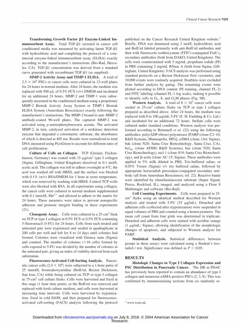
lected patients who had undergone pancreaticoduodenectomyfor pancreatic cancer at Southampton General Hospital. In thenormal pancreas (removed for duodenal neoplasia), occasional�SMA-expressing stellate cells were identified in interlobularsepta and the connective tissue supporting pancreatic ductules(Fig. 1A). In the neoplastic tissue, however, there was a markedincrease in stellate cell number in the fibrotic bands, particularlyaround malignant glands (Fig. 1B). Similarly, in the normalpancreas, type I collagen was confined to the connective tissuesurrounding larger pancreatic ducts (Fig. 1C), in contrast tolarge tracts that surround the malignant epithelium in PDAC(Fig. 1D). These anatomic studies illustrate the close spatialrelationship that exists among malignant epithelial cells, PSCs,and type I collagen in PDAC. Secondly, they demonstrate thedramatic increase in the proportion of stromal elements ofPDAC compared with the normal pancreas.
Effect of Cancer Cell Conditioned Medium on Prolif-eration and Collagen Secretion in PSCs. It was hypothe-sized that the pancreatic cancer cells may stimulate PSC growthand collagen secretion in PDAC, thus promoting the develop-
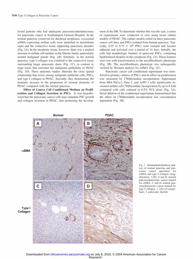
ment of the DR. To determine whether this was the case, a seriesof experiments were conducted in vitro using tissue culturemodels of PDAC. The culture models relied on three pancreaticcancer cell lines and PSCs isolated from human pancreas. Typ-ically, 0.25 to 0.75 � 106 PSCs were isolated and becameadherent and activated over a period of 14 days. Initially, thecells had morphologic features of quiescent PSCs, containinglipid/retinoid droplets in the cytoplasm (Fig. 2A). These featureswere lost with transformation to the myofibroblastic phenotype(Fig. 2B). The myofibroblastic phenotype was subsequentlyverified by Western analysis for �SMA (Fig. 2C).
Pancreatic cancer cell conditioned supernatant was trans-ferred to primary cultures of PSCs, and its effect on proliferationwas measured by [3H]thymidine incorporation. Supernatantfrom MIA PaCa-2, Panc-1, and AsPC-1 cells significantly in-creased stellate cell [3H]thymidine incorporation by up to 6-foldcompared with cells cultured in 0.5% FCS alone (Fig. 3A).Serial dilution of the conditioned supernatant demonstrated thatthe effect on [3H]thymidine incorporation was concentrationdependent (Fig. 3B).
Fig. 1 Immunohistochemical anal-ysis of normal pancreas and pan-creatic cancer specimens for�SMA and type I collagen (mag-nification, �20). A and B, normalpancreas/pancreatic cancer stainedfor �SMA. C and D, normal pan-creas/pancreatic cancer stained fortype I collagen. 1, islet of Langer-hans; 2, pancreatic ductule.
7430 Type I Collagen in Pancreatic Cancer
Research. on July 8, 2018. © 2004 American Association for Cancerclincancerres.aacrjournals.org Downloaded from
PSCs are known to secrete collagen and type I collagen inparticular (23). Experiments were therefore undertaken to de-termine whether conditioned supernatant from pancreatic cancercells also regulated type I collagen synthesis. PSC procollagenI mRNA was measured using TaqMan reverse transcription-polymerase chain reaction. Procollagen I mRNA was readilydetectable in the PSCs with a mean number of PCR cycles tofluorescent threshold of 20.7, compared with 26.0 for GAPDHin cells cultured in 0.5% FCS. After 24 hours of incubation withPanc-1 and AsPC-1 conditioned medium, there appeared to be amodest, nonsignificant increase in procollagen I mRNA expres-sion by PSCs (Fig. 4A). The effect of cancer cell conditioned
medium on collagen synthesis was also assayed at protein levelby measuring tritiated hydroxyproline incorporation (24). Al-though not specific to type I collagen, the results obtained usingthis technique were important because it accounted for theextensive posttranscriptional regulation that collagen synthesisis subject to (24). These experiments demonstrated that AsPC-1conditioned medium increased PSC collagen synthesis com-pared with controls (Fig. 4B), whereas Mia PaCa-2 and Panc-1conditioned media did not appear to stimulate PSC collagensynthesis. Recombinant TGF-�1 (used as a positive control)consistently increased collagen expression at RNA and proteinlevel by PSCs. TGF-�1 was also detected in conditioned super-natants of all three cell lines by ELISA, at significantly greaterlevels than controls [0.5% FCS (v/v) DMEM; Fig. 5]. Collagensynthesis by cancer cells was measured using the tritiated hy-droxyproline assay and found to be approximately 5-fold lessthan that by PSCs (Fig. 4B).
MMPs and TIMPs regulate turnover of secreted collagen,and therefore MMP-2 and TIMP-1 were measured in the con-ditioned media of pancreatic cancer cells and PSCs (Fig. 6).This demonstrated that PSCs express MMP-2 at between 7.2-fold and 28.4-fold that of the pancreatic cancer cell lines tested(Fig. 6A). Furthermore, PSCs expressed TIMP-1 at between 8.1-
Fig. 2 Isolated human PSCs. A, photomicrograph of freshly isolatedhuman PSCs after 4 days of culture on TCP, demonstrating numerouscytoplasmic lipid droplets (magnification, �400). B, photomicrographof isolated human PSCs cultured on TCP for 21 days (magnification,�200). C, Western analysis for �SMA expression in three separateprimary cultures of human PSCs after two passages, demonstrating themyofibroblastic phenotype of the activated PSCs. Lane 1, MIA PaCa-2cells (negative control); Lane 2, human hepatic stellate cell line (positivecontrol); Lanes 3–5, PSCs.
Fig. 3 Effect of supernatant transfer from pancreatic cancer cells toPSCs. A. PSCs (1.5 � 104 cells) were cultured with 0.5% FCS, 5% FCS(positive control), and cancer cell conditioned medium from each cellline for 16 hours, during which time [3H]thymidine incorporation wasmeasured. Bars represent the mean SE of six independent experi-ments. B, the effect of conditioned media from AsPC-1 cells, seriallydiluted with fresh 0.5% FCS on PSC [3H]thymidine incorporation over16 hours. Bars represent the mean SE of three independent experi-ments (similar results were obtained using conditioned media from MIAPaCa-2 and Panc-1 cells). Data are presented as a percentage of control(0.5% FCS), which was given an arbitrary value of 100%. *, P � 0.05;**, P � 0.01 (with respect to 0.5% FCS control).
7431Clinical Cancer Research
Research. on July 8, 2018. © 2004 American Association for Cancerclincancerres.aacrjournals.org Downloaded from
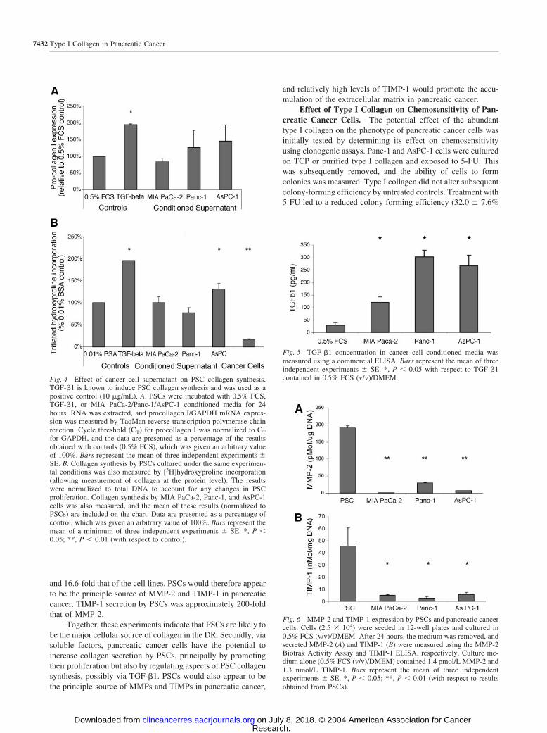
and 16.6-fold that of the cell lines. PSCs would therefore appearto be the principle source of MMP-2 and TIMP-1 in pancreaticcancer. TIMP-1 secretion by PSCs was approximately 200-foldthat of MMP-2.
Together, these experiments indicate that PSCs are likely tobe the major cellular source of collagen in the DR. Secondly, viasoluble factors, pancreatic cancer cells have the potential toincrease collagen secretion by PSCs, principally by promotingtheir proliferation but also by regulating aspects of PSC collagensynthesis, possibly via TGF-�1. PSCs would also appear to bethe principle source of MMPs and TIMPs in pancreatic cancer,
and relatively high levels of TIMP-1 would promote the accu-mulation of the extracellular matrix in pancreatic cancer.
Effect of Type I Collagen on Chemosensitivity of Pan-creatic Cancer Cells. The potential effect of the abundanttype I collagen on the phenotype of pancreatic cancer cells wasinitially tested by determining its effect on chemosensitivityusing clonogenic assays. Panc-1 and AsPC-1 cells were culturedon TCP or purified type I collagen and exposed to 5-FU. Thiswas subsequently removed, and the ability of cells to formcolonies was measured. Type I collagen did not alter subsequentcolony-forming efficiency by untreated controls. Treatment with5-FU led to a reduced colony forming efficiency (32.0 7.6%
Fig. 5 TGF-�1 concentration in cancer cell conditioned media wasmeasured using a commercial ELISA. Bars represent the mean of threeindependent experiments SE. *, P � 0.05 with respect to TGF-�1contained in 0.5% FCS (v/v)/DMEM.
Fig. 6 MMP-2 and TIMP-1 expression by PSCs and pancreatic cancercells. Cells (2.5 � 104) were seeded in 12-well plates and cultured in0.5% FCS (v/v)/DMEM. After 24 hours, the medium was removed, andsecreted MMP-2 (A) and TIMP-1 (B) were measured using the MMP-2Biotrak Activity Assay and TIMP-1 ELISA, respectively. Culture me-dium alone (0.5% FCS (v/v)/DMEM) contained 1.4 pmol/L MMP-2 and1.3 nmol/L TIMP-1. Bars represent the mean of three independentexperiments SE. *, P � 0.05; **, P � 0.01 (with respect to resultsobtained from PSCs).
Fig. 4 Effect of cancer cell supernatant on PSC collagen synthesis.TGF-�1 is known to induce PSC collagen synthesis and was used as apositive control (10 �g/mL). A. PSCs were incubated with 0.5% FCS,TGF-�1, or MIA PaCa-2/Panc-1/AsPC-1 conditioned media for 24hours. RNA was extracted, and procollagen I/GAPDH mRNA expres-sion was measured by TaqMan reverse transcription-polymerase chainreaction. Cycle threshold (CT) for procollagen I was normalized to CT
for GAPDH, and the data are presented as a percentage of the resultsobtained with controls (0.5% FCS), which was given an arbitrary valueof 100%. Bars represent the mean of three independent experiments SE. B. Collagen synthesis by PSCs cultured under the same experimen-tal conditions was also measured by [3H]hydroxyproline incorporation(allowing measurement of collagen at the protein level). The resultswere normalized to total DNA to account for any changes in PSCproliferation. Collagen synthesis by MIA PaCa-2, Panc-1, and AsPC-1cells was also measured, and the mean of these results (normalized toPSCs) are included on the chart. Data are presented as a percentage ofcontrol, which was given an arbitrary value of 100%. Bars represent themean of a minimum of three independent experiments SE. *, P �0.05; **, P � 0.01 (with respect to control).
7432 Type I Collagen in Pancreatic Cancer
Research. on July 8, 2018. © 2004 American Association for Cancerclincancerres.aacrjournals.org Downloaded from
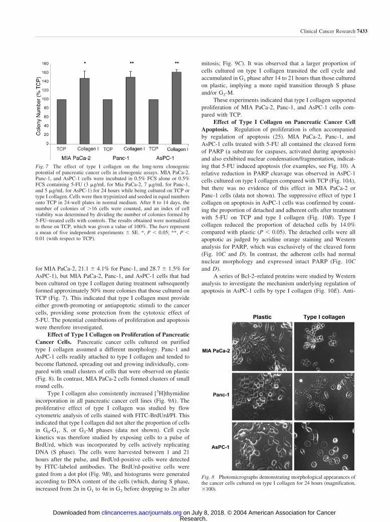
for MIA PaCa-2, 21.1 4.1% for Panc-1, and 28.7 1.5% forAsPC-1), but MIA PaCa-2, Panc-1, and AsPC-1 cells that hadbeen cultured on type I collagen during treatment subsequentlyformed approximately 50% more colonies that those cultured onTCP (Fig. 7). This indicated that type I collagen must provideeither growth-promoting or antiapoptotic stimuli to the cancercells, providing some protection from the cytotoxic effect of5-FU. The potential contributions of proliferation and apoptosiswere therefore investigated.
Effect of Type I Collagen on Proliferation of PancreaticCancer Cells. Pancreatic cancer cells cultured on purifiedtype I collagen assumed a different morphology. Panc-1 andAsPC-1 cells readily attached to type I collagen and tended tobecome flattened, spreading out and growing individually, com-pared with small clusters of cells that were observed on plastic(Fig. 8). In contrast, MIA PaCa-2 cells formed clusters of smallround cells.
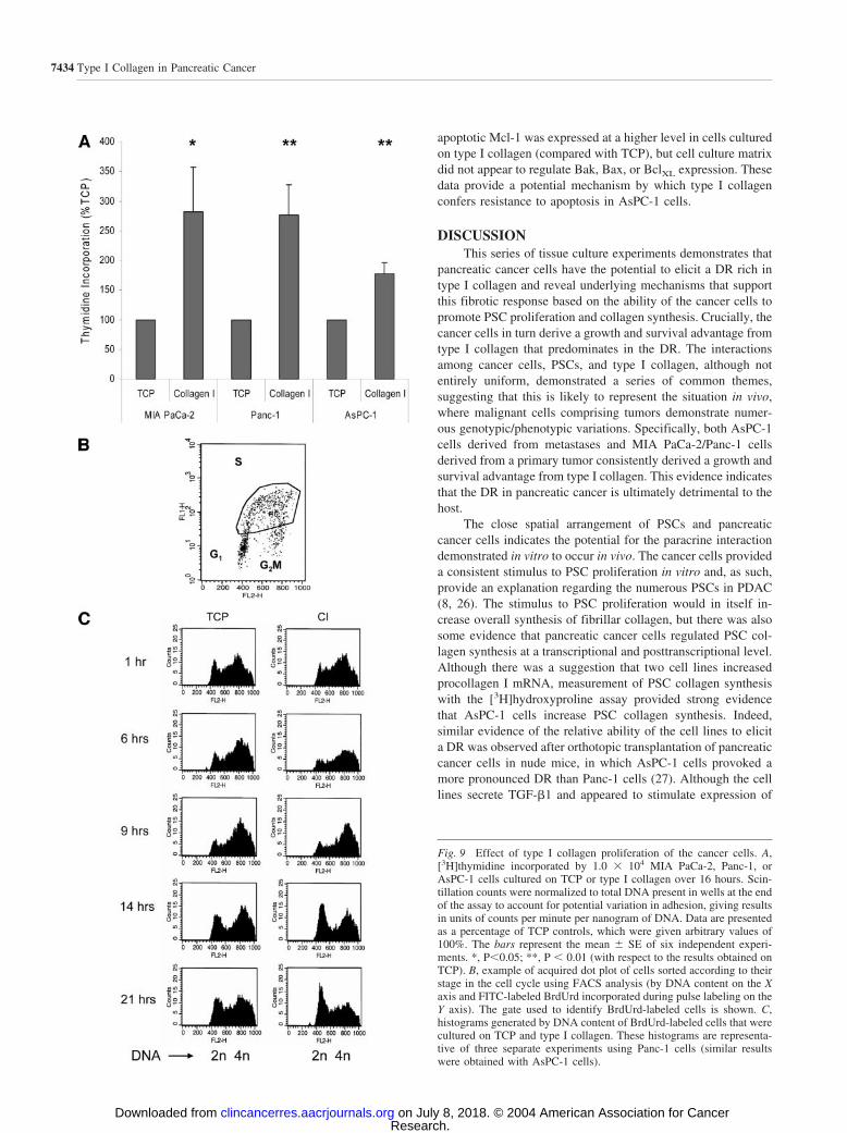
Type I collagen also consistently increased [3H]thymidineincorporation in all pancreatic cancer cell lines (Fig. 9A). Theproliferative effect of type I collagen was studied by flowcytometric analysis of cells stained with FITC-BrdUrd/PI. Thisindicated that type I collagen did not alter the proportion of cellsin G0-G1, S, or G2-M phases (data not shown). Cell cyclekinetics was therefore studied by exposing cells to a pulse ofBrdUrd, which was incorporated by cells actively replicatingDNA (S phase). The cells were harvested between 1 and 21hours after the pulse, and BrdUrd-positive cells were detectedby FITC-labeled antibodies. The BrdUrd-positive cells weregated from a dot plot (Fig. 9B), and histograms were generatedaccording to DNA content of the cells (which, during S phase,increased from 2n in G1 to 4n in G2 before dropping to 2n after
mitosis; Fig. 9C). It was observed that a larger proportion ofcells cultured on type I collagen transited the cell cycle andaccumulated in G1 phase after 14 to 21 hours than those culturedon plastic, implying a more rapid transition through S phaseand/or G2-M.
These experiments indicated that type I collagen supportedproliferation of MIA PaCa-2, Panc-1, and AsPC-1 cells com-pared with TCP.
Effect of Type I Collagen on Pancreatic Cancer CellApoptosis. Regulation of proliferation is often accompaniedby regulation of apoptosis (25). MIA PaCa-2, Panc-1, andAsPC-1 cells treated with 5-FU all contained the cleaved formof PARP (a substrate for caspases, activated during apoptosis)and also exhibited nuclear condensation/fragmentation, indicat-ing that 5-FU induced apoptosis (for examples, see Fig. 10). Arelative reduction in PARP cleavage was observed in AsPC-1cells cultured on type I collagen compared with TCP (Fig. 10A),but there was no evidence of this effect in MIA PaCa-2 orPanc-1 cells (data not shown). The suppressive effect of type Icollagen on apoptosis in AsPC-1 cells was confirmed by count-ing the proportion of detached and adherent cells after treatmentwith 5-FU on TCP and type I collagen (Fig. 10B). Type Icollagen reduced the proportion of detached cells by 14.0%compared with plastic (P � 0.05). The detached cells were allapoptotic as judged by acridine orange staining and Westernanalysis for PARP, which was exclusively of the cleaved form(Fig. 10C and D). In contrast, the adherent cells had normalnuclear morphology and expressed intact PARP (Fig. 10Cand D).
A series of Bcl-2–related proteins were studied by Westernanalysis to investigate the mechanism underlying regulation ofapoptosis in AsPC-1 cells by type I collagen (Fig. 10E). Anti-
Fig. 8 Photomicrographs demonstrating morphological appearances ofthe cancer cells cultured on type I collagen for 24 hours (magnification,�100).
Fig. 7 The effect of type I collagen on the long-term clonogenicpotential of pancreatic cancer cells in clonogenic assays. MIA PaCa-2,Panc-1, and AsPC-1 cells were incubated in 0.5% FCS alone or 0.5%FCS containing 5-FU (3 �g/mL for Mia PaCa-2, 7 �g/mL for Panc-1,and 5 �g/mL for AsPC-1) for 24 hours while being cultured on TCP ortype I collagen. Cells were then trypsinized and seeded in equal numbersonto TCP in 24-well plates in normal medium. After 8 to 14 days, thenumber of colonies of �16 cells were counted, and an index of cellviability was determined by dividing the number of colonies formed by5-FU–treated cells with controls. The results obtained were normalizedto those on TCP, which was given a value of 100%. The bars representa mean of five independent experiments SE. *, P � 0.05; **, P �0.01 (with respect to TCP).
7433Clinical Cancer Research
Research. on July 8, 2018. © 2004 American Association for Cancerclincancerres.aacrjournals.org Downloaded from
apoptotic Mcl-1 was expressed at a higher level in cells culturedon type I collagen (compared with TCP), but cell culture matrixdid not appear to regulate Bak, Bax, or BclXL expression. Thesedata provide a potential mechanism by which type I collagenconfers resistance to apoptosis in AsPC-1 cells.
DISCUSSIONThis series of tissue culture experiments demonstrates that
pancreatic cancer cells have the potential to elicit a DR rich intype I collagen and reveal underlying mechanisms that supportthis fibrotic response based on the ability of the cancer cells topromote PSC proliferation and collagen synthesis. Crucially, thecancer cells in turn derive a growth and survival advantage fromtype I collagen that predominates in the DR. The interactionsamong cancer cells, PSCs, and type I collagen, although notentirely uniform, demonstrated a series of common themes,suggesting that this is likely to represent the situation in vivo,where malignant cells comprising tumors demonstrate numer-ous genotypic/phenotypic variations. Specifically, both AsPC-1cells derived from metastases and MIA PaCa-2/Panc-1 cellsderived from a primary tumor consistently derived a growth andsurvival advantage from type I collagen. This evidence indicatesthat the DR in pancreatic cancer is ultimately detrimental to thehost.
The close spatial arrangement of PSCs and pancreaticcancer cells indicates the potential for the paracrine interactiondemonstrated in vitro to occur in vivo. The cancer cells provideda consistent stimulus to PSC proliferation in vitro and, as such,provide an explanation regarding the numerous PSCs in PDAC(8, 26). The stimulus to PSC proliferation would in itself in-crease overall synthesis of fibrillar collagen, but there was alsosome evidence that pancreatic cancer cells regulated PSC col-lagen synthesis at a transcriptional and posttranscriptional level.Although there was a suggestion that two cell lines increasedprocollagen I mRNA, measurement of PSC collagen synthesiswith the [3H]hydroxyproline assay provided strong evidencethat AsPC-1 cells increase PSC collagen synthesis. Indeed,similar evidence of the relative ability of the cell lines to elicita DR was observed after orthotopic transplantation of pancreaticcancer cells in nude mice, in which AsPC-1 cells provoked amore pronounced DR than Panc-1 cells (27). Although the celllines secrete TGF-�1 and appeared to stimulate expression of
Fig. 9 Effect of type I collagen proliferation of the cancer cells. A,[3H]thymidine incorporated by 1.0 � 104 MIA PaCa-2, Panc-1, orAsPC-1 cells cultured on TCP or type I collagen over 16 hours. Scin-tillation counts were normalized to total DNA present in wells at the endof the assay to account for potential variation in adhesion, giving resultsin units of counts per minute per nanogram of DNA. Data are presentedas a percentage of TCP controls, which were given arbitrary values of100%. The bars represent the mean SE of six independent experi-ments. *, P�0.05; **, P � 0.01 (with respect to the results obtained onTCP). B, example of acquired dot plot of cells sorted according to theirstage in the cell cycle using FACS analysis (by DNA content on the Xaxis and FITC-labeled BrdUrd incorporated during pulse labeling on theY axis). The gate used to identify BrdUrd-labeled cells is shown. C,histograms generated by DNA content of BrdUrd-labeled cells that werecultured on TCP and type I collagen. These histograms are representa-tive of three separate experiments using Panc-1 cells (similar resultswere obtained with AsPC-1 cells).
7434 Type I Collagen in Pancreatic Cancer
Research. on July 8, 2018. © 2004 American Association for Cancerclincancerres.aacrjournals.org Downloaded from

procollagen I mRNA, the process of matrix deposition is com-plex and also regulated by proteases. MMP-2 is consistentlyexpressed in pancreatic cancer and correlates with a worseprognosis (28–30). Likewise, TIMP-1 is widely expressed inpancreatic cancer and inhibits most MMPs (31). MMP-2 andTIMP-1 were therefore studied as representatives of MMPs andTIMPs to determine their relative expression in pancreatic can-cer and how this may impact on the DR. In vitro experimentsindicate that PSCs rather than pancreatic cancer cells are quan-titatively the main source of MMP-2 and TIMP-1. Furthermore,
the relative abundance of TIMP-1 over MMP-2 would alsopromote the accumulation of extracellular matrix in pancreaticcancer. Cancer cells and PSCs do not necessarily occur in equalnumbers; neoplasia and malignant cells may therefore play amore significant role in tumor matrix turnover in vivo than theseexperiments suggest.
The effect of type I collagen on the growth and survival ofpancreatic cancer cells in vivo remains unknown. The role ofstromal reaction associated with malignancy has long been amatter of debate (32). The clonogenic assays demonstrated that
Fig. 10 Effect of type I collagen on apoptosis of AsPC-1 cells exposed to 5-FU. A, Western analysis of PARP (an enzyme cleaved by caspases duringapoptosis yielding an 85-kDa band). AsPC-1 cells underwent dose-dependent apoptosis in response to 5-FU, but a relative reduction in PARP cleavagewas observed in cells treated with 25 �g/mL 5-FU on type I collagen compared with TCP. This relative protection disappeared at higherconcentrations of 5-FU (the image is representative of three separate experiments). B. This protection from apoptosis was observed in AsPC-1 cellstreated with 5-FU while being cultured on type I collagen. The relative number of adherent cells (u) was counted and compared with those that hadundergone apoptosis and become detached (�). Bars represent the mean of five independent experiments SE. *, P � 0.05 with respect to resultsobtained on TCP. C. Detached cells were apoptotic as judged by nuclear morphology after staining with acridine orange. D. Western analysis of PARPindicated that it was uncleaved in the adherent cells but cleaved in the detached cells, confirming the apoptotic nature of the detached cells. E. Westernanalysis of protein regulators of mitochondrial cytochrome c release during apoptosis revealed up-regulation of antiapoptotic mcl-1 proteins by typeI collagen, consistent with changes observed in apoptosis (the image is representative of three separate experiments). �-Actin was used as a loadingcontrol. F. Densitometric analysis was conducted on three separate Western blots, and protein expression on type I collagen is shown as a percentageof that obtained in cells cultured on TCP (**, P � 0.01).
7435Clinical Cancer Research
Research. on July 8, 2018. © 2004 American Association for Cancerclincancerres.aacrjournals.org Downloaded from
type I collagen confers a survival advantage to MIA PaCa-2,Panc-1, and AsPC-1 cells. The clonogenic assay is analogous toa cycle of chemotherapy with surviving cells repopulating thetumor, thus selecting chemoresistant clones. Additional studiesindicated that type I collagen influenced clonogenicity by reg-ulating proliferation and apoptosis of the pancreatic cancer cells.Each cell line derived a marked proliferative advantage fromtype I collagen, the dominant matrix component of the stromalreaction in pancreatic cancer. Type I collagen has also beenshown to promote proliferation of prostate cancer cells bysignaling via mitogen-activated protein kinase and phosphati-dylinositol 3�-kinase, culminating in increased cyclin D1 ex-pression and cell cycle progression (33, 34).
AsPC-1 cells demonstrated a less marked proliferative re-sponse but became resistant to apoptosis when cultured on typeI collagen. Mcl-1, a bcl-related protein that inhibits the releaseof cytochrome c from mitochondria during apoptosis, was sub-ject to regulation, consistent with the changes in apoptosisobserved on type I collagen. Bcl-2–related proteins have alsobeen shown to be regulated by mitogen-activated protein/extra-cellular signal-regulated kinase pathways in pancreatic cancercells, and manipulation of this pathway has shown that it isimportant in regulating 5-FU–induced apoptosis (35, 36). Al-though the precise signaling pathways and molecular controlunderlying the growth-promoting effects of type I collagen inPDAC have yet to be determined, their combination givespancreatic cancer cells a potentially real survival advantage inthe clinical setting.
As well as the effects on proliferation and apoptosis, fibril-lar collagen has also been implicated in migration and invasionof pancreatic cancer cells (37). Type I collagen reduces E-cadherin expression via a Src-dependent mechanism, which isconsistent with the observation that pancreatic cancer cellscultured in type I collagen gels form only loose aggregates (37,38). In contrast, type IV collagen and other basement membranecomponents including laminin and certain proteoglycans inhib-ited proliferation of pancreatic cancer cells and promoted red-ifferentiation to a more regulated cellular phenotype (38–41).Type IV collagen has also been shown to inhibit proliferationand migration and reduce angiogenesis and MMP expression inother tumor cells (42–45). Hence, the shift in expression ofmatrix in the DR, with type I collagen predominating, poten-tially promotes a number of phenotypic qualities that enhancetumor development, growth, and spread of PDAC.
We conclude that there is a potentially deleterious tissueresponse in pancreatic cancer, in which pancreatic cancer cellselicit the formation of a DR, which has the potential to promotea number of phenotypic qualities that would ultimately bedetrimental to the host. These findings complement growingrecognition of the numerous, important roles that tumor stromaplays in angiogenesis, local invasion, metastasis, chemosensi-tivity, and tumorigenesis in neoplasia (16, 46–51).
REFERENCES1. Cruickshank AH. Solid carcinomas of the exocrine pancreas. In:Pathology of the pancreas. Springer-Verlag, London, UK; 1986. p.155–77.2. Linder S, Castanos-Velez E, von Rosen A, Biberfeld P. Immunohis-tochemical expression of extracellular matrix proteins and adhesion
molecules in pancreatic carcinoma. Hepatogastroenterology 2001;48:1321–7.
3. Mollenhauer J, Roether I, Kern HF. Distribution of extracellularmatrix proteins in pancreatic ductal adenocarcinoma and its influence ontumor cell proliferation in vitro. Pancreas 1987;2:14–24.
4. Imamura T, Iguchi H, Manabe T, et al. Quantitative analysis ofcollagen and collagen subtypes I, III, and V in human pancreatic cancer,tumor-associated chronic pancreatitis, and alcoholic chronic pancreati-tis. Pancreas 1995;11:357–64.5. Alberts B, Bray D, Lewis J, Raft M, Roberts K, and Watson JD.Molecular biology of the cell. Garland Publishing; 1994.6. Haglund C, Roberts PJ, Nordling S, Ekblom P. Expression of lamininin pancreatic neoplasms and in chronic pancreatitis. Am J Surg Pathol1984;8:669–76.7. Lee CS, Montebello J, Georgiou T, Rode J. Distribution of type IVcollagen in pancreatic adenocarcinoma and chronic pancreatitis. Int JExp Pathol 1994;75:79–83.8. Yen TW, Aardal NP, Bronner MP, et al. Myofibroblasts are respon-sible for the desmoplastic reaction surrounding human pancreatic car-cinomas. Surgery 2002;131:129–34.9. Berk PD, Friedman SL. The hepatic stellate cell. Semin Liver Dis2001;21:307–461.10. Bachem MG, Schneider E, Gross H, et al. Identification, culture,and characterization of pancreatic stellate cells in rats and humans.Gastroenterology 1998;115:421–32.11. Apte MV, Haber PS, Applegate TL, et al. Periacinar stellate shapedcells in rat pancreas: identification, isolation, and culture. Gut 1998;43:128–33.12. Shek FW, Benyon RC, Walker FM, et al. Expression of transform-ing growth factor-beta 1 by pancreatic stellate cells and its implicationsfor matrix secretion and turnover in chronic pancreatitis. Am J Pathol2002;160:1787–98.13. Phillips PA, Wu MJ, Kumar RK, et al. Cell migration: a novelaspect of pancreatic stellate cell biology. Gut 2003;52:677–82.14. Haber PS, Keogh GW, Apte MV, et al. Activation of pancreaticstellate cells in human and experimental pancreatic fibrosis. Am J Pathol1999;155:1087–95.15. Yokota T, Denham W, Murayama K, et al. Pancreatic stellate cellactivation and MMP production in experimental pancreatic fibrosis.J Surg Res 2002;104:106–11.16. Sethi T, Rintoul RC, Moore SM, et al. Extracellular matrix proteinsprotect small cell lung cancer cells against apoptosis: a mechanism forsmall cell lung cancer growth and drug resistance in vivo. Nat Med1999;5:662–8.17. Uhm JH, Dooley NP, Kyritsis AP, Rao JS, Gladson CL. Vitronec-tin, a glioma-derived extracellular matrix protein, protects tumor cellsfrom apoptotic death. Clin Cancer Res 1999;5:1587–94.18. Murphy FR, Issa R, Zhou X, et al. Inhibition of apoptosis ofactivated hepatic stellate cells by tissue inhibitor of metalloproteinase-1is mediated via effects on matrix metalloproteinase inhibition: implica-tions for reversibility of liver fibrosis. J Biol Chem 2002;277:11069–76.19. Pfaffl MW. A new mathematical model for relative quantification inreal-time RT-PCR. Nucleic Acids Res 2001;29:e45.20. Williams EJ, Benyon RC, Trim N, et al. Relaxin inhibits effectivecollagen deposition by cultured hepatic stellate cells and decreases ratliver fibrosis in vivo. Gut 2001;49:577–83.21. Bazzoni G, Shih DT, Buck CA, Hemler ME. Monoclonal antibody9EG7 defines a novel beta 1 integrin epitope induced by soluble ligandand manganese, but inhibited by calcium. J Biol Chem 1995;270:25570–7.22. Brimmell M, Mendiola R, Mangion J, Packham G. BAX frameshiftmutations in cell lines derived from human haemopoietic malignanciesare associated with resistance to apoptosis and microsatellite instability.Oncogene 1998;16:1803–12.23. Bachem MG, Schmid-kostas A, Gross HJ, Schneider E, Menke A,Weidenbach H, Adler G, Siech M, Berger H, Grunert A. Pancreaticstellate cells and their role in human pancreatic fibrogenesis. In: Buchler
7436 Type I Collagen in Pancreatic Cancer
Research. on July 8, 2018. © 2004 American Association for Cancerclincancerres.aacrjournals.org Downloaded from

MW, Friess H, Uhl W, Malfertheiner P, editors. Chronic pancreatitis:novel concepts in biology and therapy. Blackwell Publishing, Oxford,UK; 2002. p. 134–47.24. Lindquist JN, Marzluff WF, Stefanovic B. Fibrogenesis. III. Post-transcriptional regulation of type I collagen. Am J Physiol GastrointestLiver Physiol 2000;279:G471–6.25. Evan GI, Vousden KH. Proliferation, cell cycle and apoptosis incancer. Nature (Lond) 2001;411:342–8.26. Bachem MG, Schmid-Kotsas A, Fink M, et al. The increaseddeposition of connective tissue in pancreas carcinoma is the result of aparacrine stimulation of pancreatic stellate cells by cancer cells. Pan-creas 2000;21:431.27. Lohr M, Schmidt C, Ringel J, et al. Transforming growth factor-beta1 induces desmoplasia in an experimental model of human pancre-atic carcinoma. Cancer Res 2001;61:550–5.28. Ellenrieder V, Alber B, Lacher U, et al. Role of MT-MMPs andMMP-2 in pancreatic cancer progression. Int J Cancer 2000;85:14–20.29. Matsuyama Y, Takao S, Aikou T. Comparison of matrix metallo-proteinase expression between primary tumors with or without livermetastasis in pancreatic and colorectal carcinomas. J Surg Oncol 2002;80:105–10.30. Nagakawa Y, Aoki T, Kasuya K, Tsuchida A, Koyanagi Y. Histo-logic features of venous invasion, expression of vascular endothelialgrowth factor and matrix metalloproteinase-2 and matrix metallopro-teinase-9, and the relation with liver metastasis in pancreatic cancer.Pancreas 2002;24:169–78.31. Bramhall SR, Neoptolemos JP, Stamp GW, Lemoine NR. Imbal-ance of expression of matrix metalloproteinases (MMPs) and tissueinhibitors of the matrix metalloproteinases (TIMPs) in human pancreaticcarcinoma. J Pathol 1997;182:347–55.32. Seemayer TA, Schurch W, Lagace R. The myofibroblast anddefense against neoplasia: a hypothesis. Surv Immunol Res 1982;1:268–73.33. Kiefer JA, Farach-Carson MC. Type I collagen-mediated prolifer-ation of PC3 prostate carcinoma cell line: implications for enhancedgrowth in the bone microenvironment. Matrix Biol 2001;20:429–37.34. Schwartz MA, Assoian RK. Integrins and cell proliferation: regu-lation of cyclin-dependent kinases via cytoplasmic signaling pathways.J Cell Sci 2001;114:2553–60.35. Boucher MJ, Morisset J, Vachon PH, et al. MEK/ERK signalingpathway regulates the expression of Bcl-2, Bcl-XL, and Mcl-1 andpromotes survival of human pancreatic cancer cells. J Cell Biochem2000;79:355–69.36. Shi X, Liu S, Kleeff J, Friess H, Buchler MW. Acquired resistanceof pancreatic cancer cells towards 5-fluorouracil and gemcitabine is
associated with altered expression of apoptosis-regulating genes. On-cology (Basel) 2002;62:354–62.
37. Menke A, Philippi C, Vogelmann R, et al. Down-regulation ofE-cadherin gene expression by collagen type I and type III in pancreaticcancer cell lines. Cancer Res 2001;61:3508–17.
38. Yamanari H, Suganuma T, Iwamura T, et al. Extracellular matrixcomponents regulating glandular differentiation and the formation ofbasal lamina of a human pancreatic cancer cell line in vitro. Exp CellRes 1994;211:175–82.
39. Paddenberg R, Flocke K, Elsasser HP, et al. Phenotypical changes ofa human pancreatic adenocarcinoma cell line after selection on laminin-1/nidogen (LM/Ng) substratum. Eur J Cell Biol 1998;76: 251–64.
40. Zucker S, Wieman JM, Lysik RM, et al. Enrichment of collagen andgelatin degrading activities in the plasma membranes of human cancercells. Cancer Res 1987;47:1608–14.
41. Weber CK, Sommer G, Michl P, et al. Biglycan is overexpressed inpancreatic cancer and induces G1-arrest in pancreatic cancer cell lines.Gastroenterology 2001;121:657–67.
42. Pasco S, Han J, Gillery P, et al. A specific sequence of the noncol-lagenous domain of the alpha3(IV) chain of type IV collagen inhibitsexpression and activation of matrix metalloproteinases by tumor cells.Cancer Res 2000;60:467–73.43. Petitclerc E, Boutaud A, Prestayko A, et al. New functions fornon-collagenous domains of human collagen type IV. Novel integrinligands inhibiting angiogenesis and tumor growth in vivo. J Biol Chem2000;275:8051–61.44. Takeda S, Nakao A, Miyoshi K, Takagi H. Gene therapy forpancreatic cancer. Semin Surg Oncol 1998;15:57–61.45. Martinella-Catusse C, Polette M, Noel A, et al. Down-regulation ofMT1-MMP expression by the alpha3 chain of type IV collagen inhibitsbronchial tumor cell line invasion. Lab Investig 2001;81:167–75.46. Liotta LA, Kohn EC. The microenvironment of the tumour-hostinterface. Nature (Lond) 2001;411:375–9.47. Seljelid R, Jozefowski S, Sveinbjornsson B. Tumor stroma. Anti-cancer Res 1999;19:4809–22.48. Tuxhorn JA, Ayala GE, Rowley DR. Reactive stroma in prostatecancer progression. J Urol 2001;166:2472–83.49. Noel A, Foidart JM. The role of stroma in breast carcinoma growthin vivo. J Mammary Gland Biol Neoplasia 1998;3:215–25.50. Tlsty TD, Hein PW. Know thy neighbor: stromal cells can contrib-ute oncogenic signals. Curr Opin Genet Dev 2001;11:54–9.51. Netti PA, Berk DA, Swartz MA, Grodzinsky AJ, Jain RK. Role ofextracellular matrix assembly in interstitial transport in solid tumors.Cancer Res 2000;60:2497–503.
7437Clinical Cancer Research
Research. on July 8, 2018. © 2004 American Association for Cancerclincancerres.aacrjournals.org Downloaded from

2004;10:7427-7437. Clin Cancer Res Thomas Armstrong, Graham Packham, Lindsay B. Murphy, et al. Pancreatic Ductal AdenocarcinomaType I Collagen Promotes the Malignant Phenotype of
Updated version
http://clincancerres.aacrjournals.org/content/10/21/7427
Access the most recent version of this article at:
Cited articles
http://clincancerres.aacrjournals.org/content/10/21/7427.full#ref-list-1
This article cites 46 articles, 12 of which you can access for free at:
Citing articles
http://clincancerres.aacrjournals.org/content/10/21/7427.full#related-urls
This article has been cited by 19 HighWire-hosted articles. Access the articles at:
E-mail alerts related to this article or journal.Sign up to receive free email-alerts
SubscriptionsReprints and
To order reprints of this article or to subscribe to the journal, contact the AACR Publications
Permissions
Rightslink site. (CCC)Click on "Request Permissions" which will take you to the Copyright Clearance Center's
.http://clincancerres.aacrjournals.org/content/10/21/7427To request permission to re-use all or part of this article, use this link
Research. on July 8, 2018. © 2004 American Association for Cancerclincancerres.aacrjournals.org Downloaded from