two cases of synovitis, acne, pustulosis, hyperostosis, · two cases of synovitis, acne,...
TRANSCRIPT
Trifon Spyridonidis,
Costas Giannakenas,
Nikos Papandrianos,
Panagiota Barla,
Dimitris J. Apostolopoulos
University of PatrasMedical School, Department of Nuclear Medicine, Regional,University Hospital of Patras,Patras, Greece
✬✬✬
Keywords: SAPHO syndrome –Bone scan imaging – Differentialdiagnosis
Correspondence address: Trifon Spyridonidis, MDUniversity of Patras, Medical School, Department of Nuclear Medicine, Regional University Hospital of Patras, Rion, Patras, PC 26500, Greece Tel: +30-2610-999210Fax: +30-2610-994470e-mail : [email protected]
Received:
22 March 2007
Accepted revised:
25 April 2007
Two cases of synovitis, acne, pustulosis, hyperostosis,osteitis - SAPHO syndrome
www.nuclmed.gr Hellenic Journal of Nuclear Medicine ñ May - August 2007
AbstractSynovitis, acne, pustulosis, hyperostosis, osteitis (SAPHO) syndrome is rare with yet unknown preva-lence. The difficulty in recognizing the syndrome is due to the very wide diversity of its signs andsymptoms, the lack of skin manifestations in many cases and to confusion in medical terminology indescribing this syndrome. In this paper, we present two cases with characteristic bone lesions in bonescan and in radiology images that are considered to be SAPHO syndrome. In the first case the char-acteristic bone single photon emission tomography scan findings in a patient with spine involvementsupposed by bone biopsy but were not followed by characteristic skin manifestations. The point of in-terest of this case lies on the significant improvement of both symptoms and scintigraphic findings af-ter treatment with biphosphonates. In the second case the diagnosis was also based on the charac-teristic bone scan findings, although the patient referred to us for staging of prostate cancer. Detailedhistory and clinical examination revealed skin manifestations of the syndrome.
Hell J Nucl Med 2007; 10(2): 109-112
Introduction
Observations connecting synovitis, acne, pustulosis, hyperostosis, osteitis (SAPHO)syndrome to chronic cutaneous pustular lesions, such as palmar and plantar pustu-losis, severe acne, pustular psoriasis, with bone lesions including a variety of os-
teoarticular manifestations from synovitis or/and arthritis to hyperostosis and aseptic osteitis,have been reported since early 60’s [1-3]. The entity of chronic recurrent multifocal os-teomyelitis (CRMO) was reported in 1972 [4] and an association between CRMO and pal-moplantar pustulosis was described in 1978 [5]. Later, in 1987 Chamot et al proposed theterm: SAPHO (synovitis, acne, pustulosis, hyperostosis, osteitis) as including many signs ofthis syndrome [6]. Kahn and Kahn [7] in 1994 determined the diagnostic criteria for theSAPHO syndrome. The syndrome is of unclarified aetiology, and by some rheumatologistsis considered as a form of seronegative spodylarthropathy [8], while others relate this syn-drome to the low virulence infectious agent propiobacterium acnes [9]. In adults, the mostcommon sites of involvement are the joints of the anterior chest wall, followed by involve-ment of the axial skeleton, most frequently thoracic spine. Mandible is frequently involved[10].
Due to the diagnostic significance and the rarity of this syndrome, we report two caseswith SAPHO syndrome, studied in our department during the last seven years.
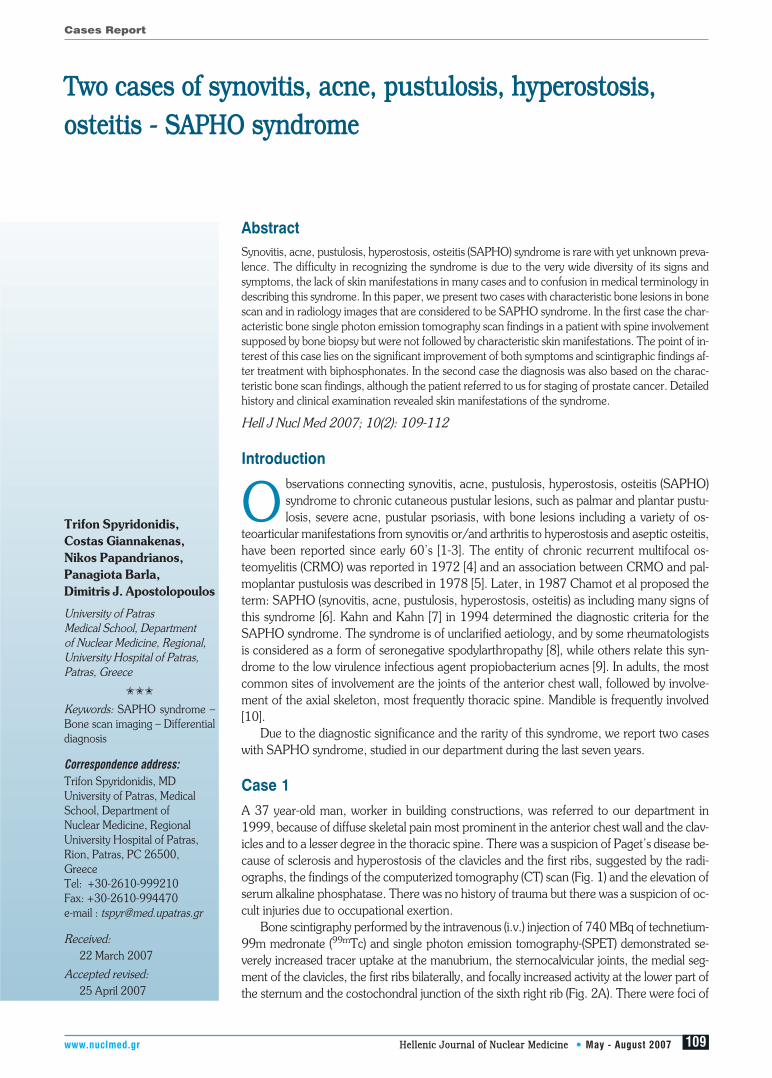
Case 1A 37 year-old man, worker in building constructions, was referred to our department in1999, because of diffuse skeletal pain most prominent in the anterior chest wall and the clav-icles and to a lesser degree in the thoracic spine. There was a suspicion of Paget’s disease be-cause of sclerosis and hyperostosis of the clavicles and the first ribs, suggested by the radi-ographs, the findings of the computerized tomography (CT) scan (Fig. 1) and the elevation ofserum alkaline phosphatase. There was no history of trauma but there was a suspicion of oc-cult injuries due to occupational exertion.
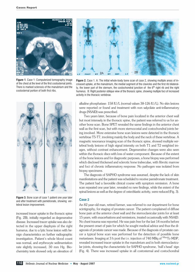
Bone scintigraphy performed by the intravenous (i.v.) injection of 740 MBq of technetium-99m medronate (99mTc) and single photon emission tomography-(SPET) demonstrated se-verely increased tracer uptake at the manubrium, the sternocalvicular joints, the medial seg-ment of the clavicles, the first ribs bilaterally, and focally increased activity at the lower part ofthe sternum and the costochondral junction of the sixth right rib (Fig. 2A). There were foci of
Cases Report
CM
YK
CM
YK
C MYK C MYK
C MYK C MYK
109
109
Hellenic Journal of Nuclear Medicine ñ May - August 2007 www.nuclmed.gr110
increased tracer uptake in the thoracic spine(Fig. 2B), initially regarded as degenerativedisease. Increased tracer uptake was also de-tected in the upper diaphysis of the righthumerus, due to a lytic bone lesion with be-nign characteristics on further radiographicinvestigation. Patient’s whole blood countwas normal, and erythrocyte sedimentationrate slightly increased, 30 mm Hg. Bio-chemistry tests showed only an elevation of
alkaline phosphatase: 158 IU/L (normal values 38-126 IU/L). No skin lesionswere reported or found and treatment with non salycilate anti-inflammatorydrugs (NSAID) was prescribed.
Two years later, because of bone pain localized in the anterior chest wallbut most intensely in the thoracic spine, the patient was referred to us for an-other bone scan. Bone SPET revealed the same findings in the anterior chestwall as the first scan, but with more sternocostal and costochondral joints be-ing involved. More extensive bone scan lesions were detected in the thoracicvertebrae T5-T7, involving mainly the body and the neck of these vertebrae. Amagnetic resonance imaging scan of the thoracic spine, showed multiple ver-tebral body lesions of high signal intensity on both T1 and T2 weighted im-ages, without contrast enhancement. Degenerative changes were also seenwithin the thoracic discs with loss of water component. Because of the extentof the bone lesions and for diagnostic purposes, a bone biopsy was performedwhich disclosed thickened and sclerotic bone trabeculae, with fibrotic marrowand foci of chronic inflammatory response. No pathogen was isolated frombiopsy specimens.
The diagnosis of SAPHO syndrome was assumed, despite the lack of skinmanifestations and the patient was scheduled to receive pamidronate treatment.The patient had a favorable clinical course with symptom remission. A bonescan repeated one year later, revealed no new findings, while the extent of thespinal lesions as well as the degree of osteoblastic activity, were reduced (Fig. 3).
Case 2
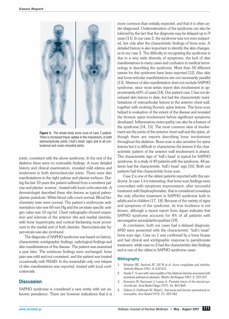
An 82 year-old man, retired farmer, was referred to our department for bonescintigraphy, for staging of prostate cancer. The patient complained of diffusebone pain at the anterior chest wall and the sternoclavicular joints for at least15 years, with exacerbations and remissions, treated occasionally with NSAID.No chest trauma was reported. He was pain free for the last 14 months beforethe present onset of pain for which he sought medical advice and thus the di-agnosis of prostate cancer was made. Because of the diagnosis of prostate can-cer a typical bone scan was performed for the detection of possible bonemetastases. Imaging at 3 h post the i.v. injection of 740 MBq of 99mTc-MDP,revealed increased tracer uptake in the manubrium and in both sternoclavicu-lar joints, showing the characteristic for SAPHO syndrome, ‘bull’s head’ sign(Fig. 4). There was increased uptake in all costosternal and costochondral
Cases Report
C MYKC
MY
KC
MY
KC MYK
C MYK C MYK
110
Figure 2. Case 1. ∞: The initial whole-body bone scan of case 2, showing multiple areas of in-creased uptake, at the manubrium, the medial segment of the clavicles and the first rib bilateral-ly, the lower part of the sternum, the costochondral junction of the 6th right rib and the righthumerus. µ: Right posterior oblique view of the thoracic spine, showing multiple foci of increasedactivity in the thoracic vertebrae.
Figure 1. Case 1: Computerized tomography imageof the chest at the level of the first costosternal joints.There is marked sclerosis of the manubrium and thecostosternal portion of both first ribs.
Figure 3. Bone scan of case 1 patient one year laterand after treatment with pamidronate, showing ver-tebral lesion improvement.
www.nuclmed.gr Hellenic Journal of Nuclear Medicine ñ May - August 2007
joints, consistent with the above syndrome. In the rest of theskeleton there were no noticeable findings. A more detailedhistory and clinical examination, revealed mild edema andtenderness in both sternoclavicular joints. There were skinmanifestations in the right palmar and plantar surfaces. Dur-ing the last 10 years the patient suffered from a remittent pal-mar and plantar ‘eczema’, treated with local corticosteroids. Adermatologist described these skin lesions as typical palmo-plantar pustulosis. White blood cells count normal. Blood bio-chemistry tests were normal. The patient’s erythrocyte sedi-mentation rate was 60 mm Hg and the prostate specific anti-gen value was 10 ng/ml. Chest radiographs showed expan-sion and sclerosis of the anterior ribs and medial clavicles,with bone hypertrophy and cortical thickening most promi-nent in the medial end of both clavicles. Sternoclavicular hy-perostosis was also profound.
The diagnosis of SAPHO syndrome was based on history,characteristic scintigraphic findings, radiological findings andskin manifestations of the disease. The patient was examineda year later. The scintiscan findings were unchanged, bonepain was mild and not consistent, and the patient was treatedoccasionally with NSAID. In the meanwhile only one relapseof skin manifestations was reported, treated with local corti-costeroids.
DiscussionSAPHO syndrome is considered a rare entity with yet un-known prevalence. There are however indications that it is
more common than initially expected, and that it is often un-der-diagnosed. Underestimation of the syndrome can also beinferred by the fact that the diagnosis may be delayed up to 9years [11]. In our case 2, the syndrome was not even suspect-ed, but only after the characteristic findings of bone scan. Adetailed history is also important to identify the skin changes,as in our case 2. The difficulty in recognizing the syndrome isdue to a very wide diversity of symptoms, the lack of skinmanifestations in many cases and confusion in medical termi-nology in describing the syndrome. More than 50 differentnames for this syndrome have been reported [12]. Also skinand bone-articular manifestations are not necessarily parallel[13]. Absence of skin manifestation does not exclude SAPHOsyndrome, since most series report skin involvement in ap-proximately 60% of cases [14]. Our patient case 1 has not de-veloped skin lesions to date, but had the characteristic mani-festations of osteoarticular lesions in the anterior chest wall,together with evolving thoracic spine lesions. The bone scanhelped in evaluation of the extent of the disease and revealedthe thoracic spine involvement before significant symptomsdeveloped. Inflammatory enteropathy can also be a feature ofthe syndrome [14, 15]. The most common sites of involve-ment are the joints of the anterior chest wall and the spine, al-though there are reports describing bone involvementthroughout the skeleton. Bone scan is also sensitive for spinelesions but it is difficult to characterize the lesions if the char-acteristic pattern of the anterior wall involvement is absent.The characteristic sign of ‘bull’s head’ is typical for SAPHOsyndrome. In a study of 49 patients with the syndrome, 44 pa-tients had the characteristic ‘bull’s head’ sign [16]. Both ourpatients had this characteristic bone scan.
Case 2 is one of the oldest patients reported with this syn-drome. In case 1 it is interesting, that bone scan findings wereconcordant with symptoms improvement, after successfultreatment with bisphosphonates, that is considered nowadaysthe only effective treatment in SAPHO syndrome both inadults and in children [17, 18]. Because of the variety of signsand symptoms of the syndrome, its true incidence is notknown, although a recent report from Japan indicates thatSAPHO syndrome accounts for 4% of all patients withseronegative spondylarthropathies [19].
In conclusion, both our cases had a delayed diagnosis,AND were presented with the characteristic “bull’s head”bone scan sign. Case no 1 was confirmed by a bone biopsyand had clinical and scintigraphic response to pamidronatetreatment, while case no 2 had the characteristic skin findingsand is one of the oldest in SAPHO syndrome cases.
Bibliography1. Windom RE, Sanford JP, Ziff M et al. Acne conglobata and arthritis.
Arthritis Rheum 1961; 4: 632-651.
2. Sasaki T. A case with osteomyelitis of the bilateral clavicles associated withpustulosis palmaris et plantaris. Rinsho Sei Keigara 1967; 2: 333-337.
3. Kormano M, Karvonen J, Lassus A. Psoriatic lesion of the sternal syn-chondrosis. Acta Radiol Diagn 1975; 16: 463-468.
4. Gideon A, Holthusen W, Masel L. Sub-acute and chronic symmetrical os-teomyelitis. Ann Radiol 1972; 15: 329-342.
Cases Report
CM
YK
CM
YK
C MYK C MYK
C MYK C MYK
111
111
Figure 4. The whole-body bone scan of case 2 patient.There is increased tracer uptake in the manubrium, in bothsternoclavicular joints (‘bull’s head’ sign) and in all cos-tosternal and costo-chondral joints.

Hellenic Journal of Nuclear Medicine ñ May - August 2007 www.nuclmed.gr112
5. Bjorksten B, Gustavson KH, Erikson B et al. Chronic recurrent multifocal os-teomyelitis and pustulosis palmoplantaris. J Pediatr 1978; 932: 227-231.
6. Chamot AM, Benhamou CL, Kahn MF et al. Le syndrome acne pustu-lose hyperostose osteite (SAPHO). Results d’ une unquete nationale. 85observations. Rev Rhum 1987; 54: 187-196.
7. Kahn MF, Kahn MA. The SAPHO syndrome. Baillieres Clin Rheumatol1994; 8: 333-362.
8. Steinoff J, Kilursu A, Falasa G et al. A study of musculoskeletal manifes-tations in 12 patients with SAPHO syndrome. J Clin Rheumatology2002; 8: 13-22.
9. Kirchhoff T, Merkesdal S, Rosenthal H et al. Diagnostic management ofpatients with SAPHO syndrome: use of MRI to guide bone biopsy at CTfor microbiological and histological work-up. Eur Radiol 2003; 13: 2304-2308.
10. Suei Y, Tagushi A, Tanimoto K. Diagnostic points and possible origin ofosteomyelitis in synovitis, acne, pustulosis, hyperostosis, and osteitis(SAPHO) syndrome.a radiographic study of 77 mandibular osteomyelitiscases. Rheumatology 2003; 42: 1398-1403.
11. Hayem G, Bouchaud-Chabot A, Benali K et al. SAPHO syndrome: alongterm follow up of 120 cases. Semin Arthritis Rheum 1999; 293: 159-171.
12. Chamot AM, Kahn MF. Das SAPHO syndrom. Z Rheumatol 1994; 53:234-242.
13. Boutin RD, Resnik D. The SAPHO syndrome: an evolving concept forunifying several idiopathic disorders of bone and skin. AJR 1998; 170:585-591..
14. Kahn MF, Bouvier MB, Palazzo E et al. Sternoclavicular pustulotic osteitis(SAPHO): 20 year interval between skin and bone lesions. J Rheumat1991; 18: 1104-1108.
15. Skoura E, Giannopoulou C, Zaromytidou E et al. A case of non-malig-nant multifocal lesions on the methyl diphosphonate technetium-99mbone scan with enteropathy. SAPHO syndrome. Hell J Nucl Med 2007;10: 26-28.
16. Freyschmidt J, Sternberg A. The bullhead sign: scintigraphic pattern ofsternoclavicular hyperostosis and pustulotic arthroosteitis. Eur Radiol1998; 8: 807-812.
17. Amital H, Applbaum Y, Aamar S et al. SAPHO syndrome treated withpamidronate: an open-label study of 10 patients. Rheumatology 2004;43: 658-661.
18. Kerrison C, Davidson A, Cleary A et al. Pamidronate in the treatment ofchildhood SAPHO syndrome. Rheumatology 2004; 43: 1246-1251.
19. Hukuda S, Minami M, Saito T et al. Spondylarthropathies in Japan: na-tionwide questionnaire survey performed by the Japan AnkylosingSpondylitis Society. J Rheumatol 2001;28:555-559.
[
Cases Report
C MYKC
MY
KC
MY
KC MYK
C MYK C MYK
112