tuberculosis...tuberculosis pathophysiology and transmission june 16, 2016 tuberculosis clinical...
TRANSCRIPT
Paul K. Drain, MD, MPH, FACP
Assistant Professor Depts. of Global Health, Medicine (Infectious Diseases), Epidemiology
University of Washington
Tuberculosis Pathophysiology and Transmission
June 16, 2016 Tuberculosis Clinical Intensive
The following planner/speaker has reported a relevant financial relationship with a commercial interest: - None.
DISCLOSURE
Outline
• Historical Context of Tuberculosis (TB)
• Mycobacterium spp. and M. tuberculosis
• TB Pathophysiology
• TB Transmission
• Summary
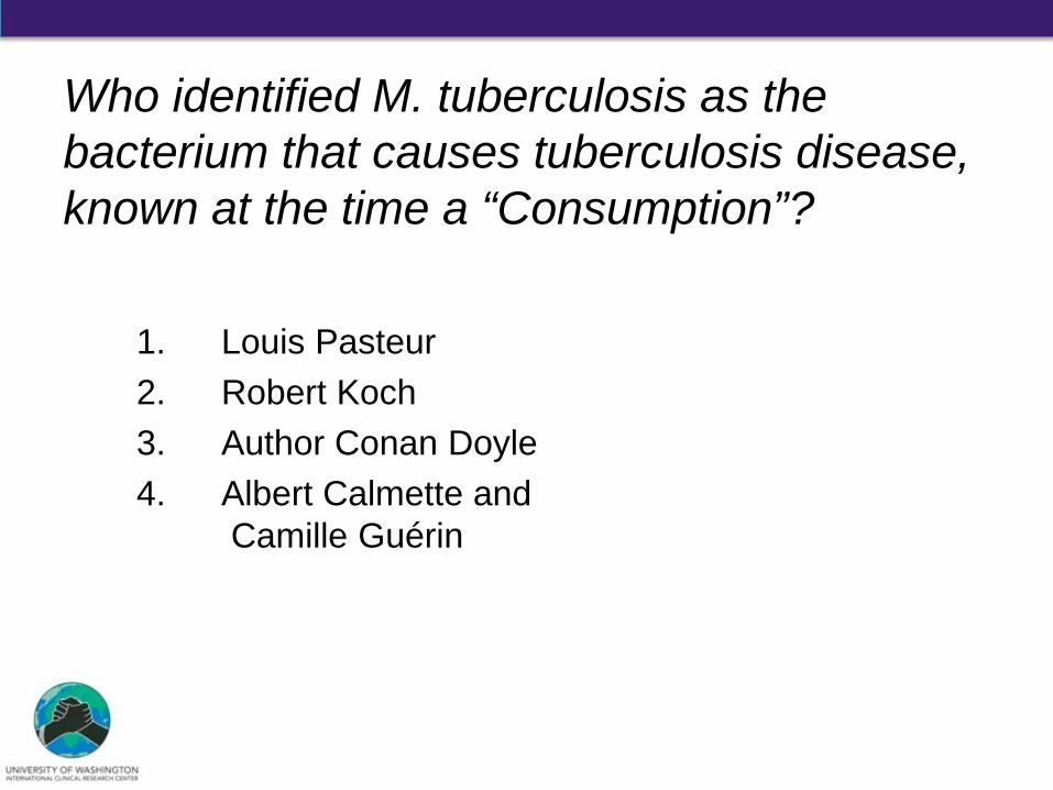
Who identified M. tuberculosis as the bacterium that causes tuberculosis disease, known at the time a “Consumption”?
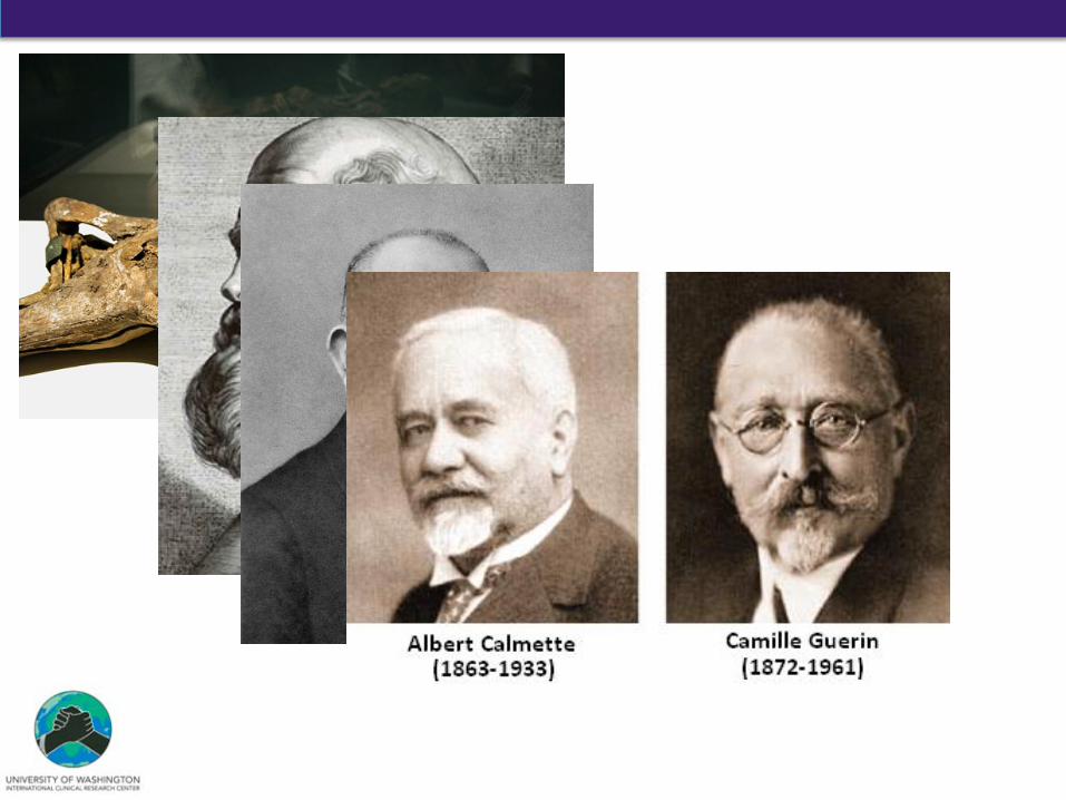
1. Louis Pasteur 2. Robert Koch 3. Author Conan Doyle 4. Albert Calmette and
Camille Guérin

Influence of TB on Medicine
1821 – Laennec invented stethoscope and described utility in diagnosing TB
1882 – Koch presented TB bacilli as the infectious agent of TB on March 24
1895 – Roentgen invented chest X-ray and used to track TB progression
1890s – Franz Ziehl/Friedrich Neelson developed acid-fast stain for TB
1908 – Mantoux developed tuberculin skin test for latent TB
1936 – Solid culture introduced to grow and identify TB Robert Koch, Nobel Prize in 1905
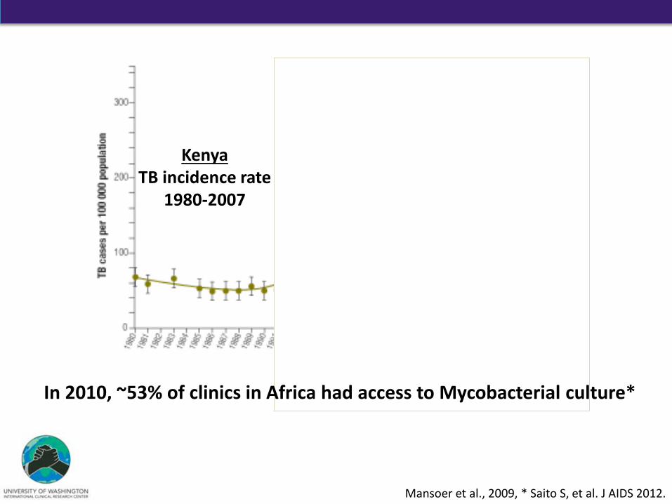
Mansoer et al., 2009, * Saito S, et al. J AIDS 2012.
Kenya TB incidence rate
1980-2007
In 2010, ~53% of clinics in Africa had access to Mycobacterial culture*
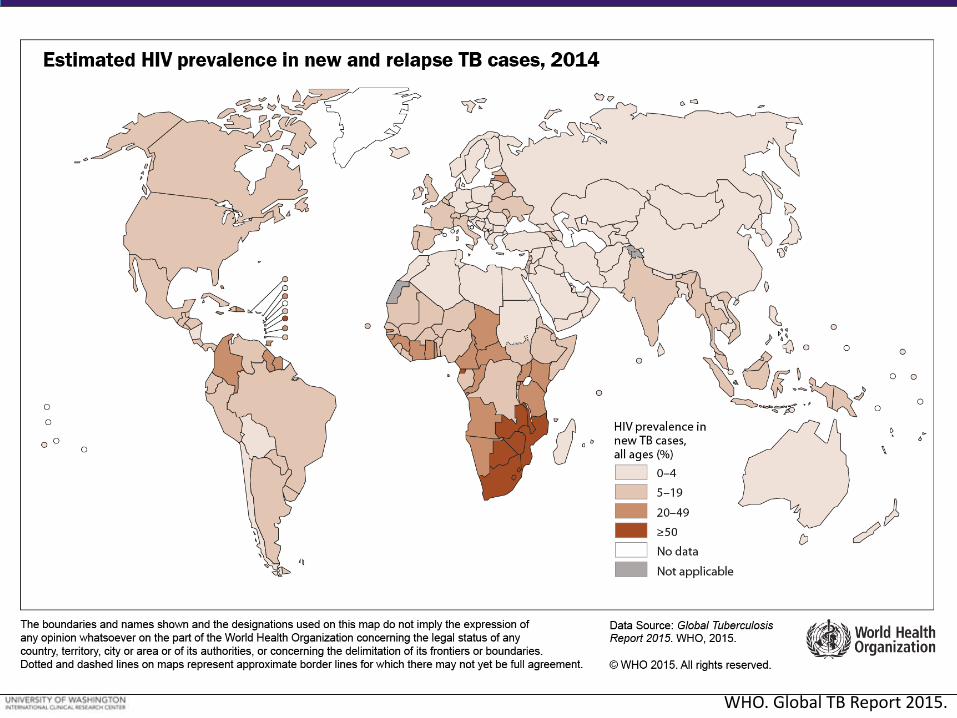
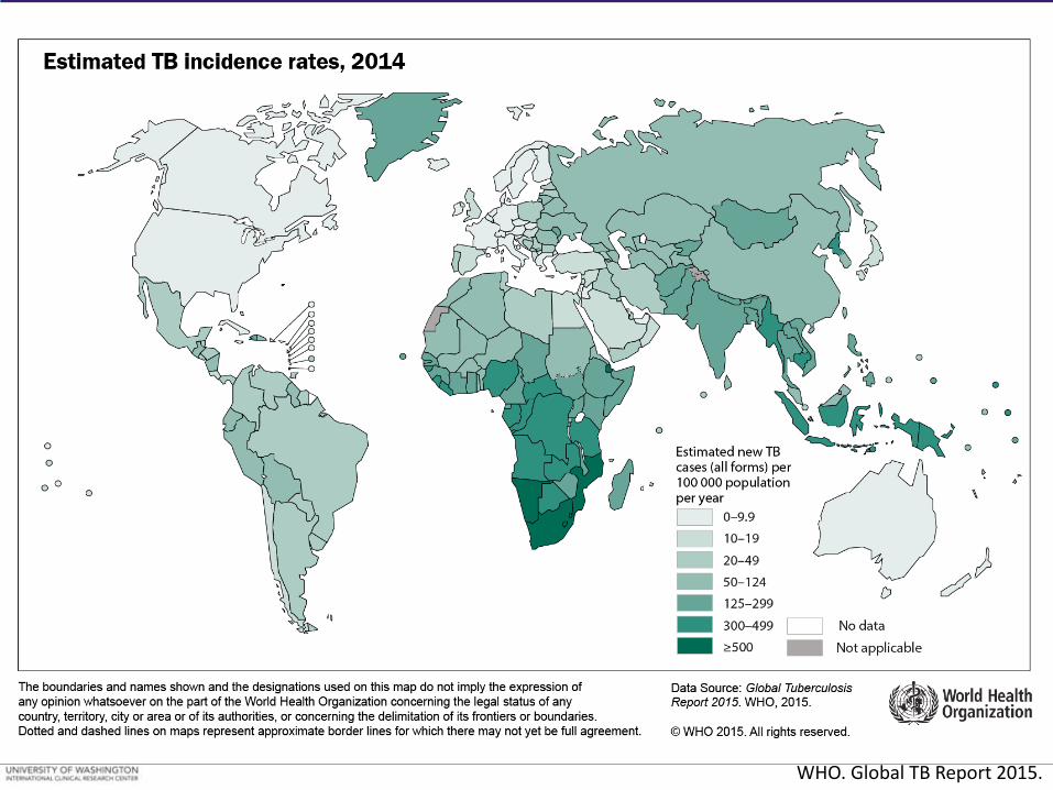
WHO. Global TB Report 2015.
WHO. Global TB Report 2015.
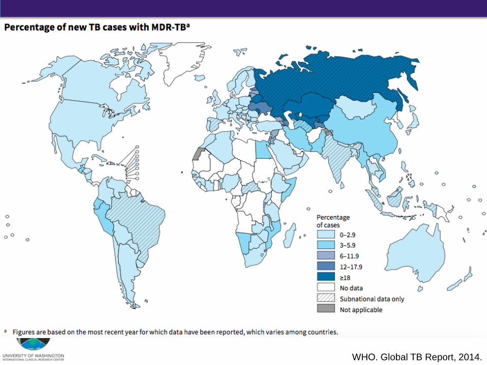
WHO. Global TB Report, 2014.
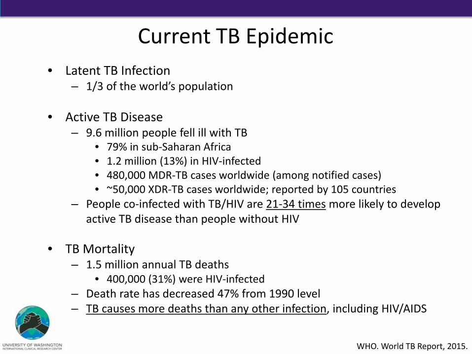
• Latent TB Infection – 1/3 of the world’s population
• Active TB Disease
– 9.6 million people fell ill with TB • 79% in sub-Saharan Africa • 1.2 million (13%) in HIV-infected • 480,000 MDR-TB cases worldwide (among notified cases) • ~50,000 XDR-TB cases worldwide; reported by 105 countries
– People co-infected with TB/HIV are 21-34 times more likely to develop active TB disease than people without HIV
• TB Mortality
– 1.5 million annual TB deaths • 400,000 (31%) were HIV-infected
– Death rate has decreased 47% from 1990 level – TB causes more deaths than any other infection, including HIV/AIDS
Current TB Epidemic
WHO. World TB Report, 2015.
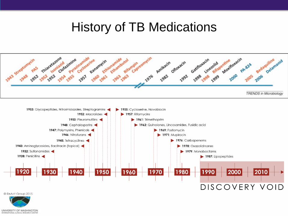
History of TB Medications
Outline
• Historical Context of Tuberculosis (TB)
• Mycobacterium spp. and M. tuberculosis
• TB Pathophysiology
• TB Transmission
• Summary
How many species of Mycobacterium tuberculosis complex cause disease in humans?
1. 1 2. 4 3. 7 4. 10
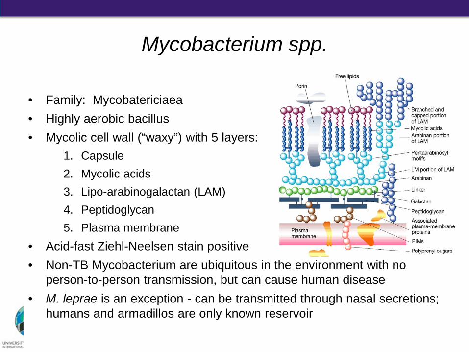
• Family: Mycobatericiaea • Highly aerobic bacillus • Mycolic cell wall (“waxy”) with 5 layers:
1. Capsule 2. Mycolic acids 3. Lipo-arabinogalactan (LAM) 4. Peptidoglycan 5. Plasma membrane
• Acid-fast Ziehl-Neelsen stain positive • Non-TB Mycobacterium are ubiquitous in the environment with no
person-to-person transmission, but can cause human disease • M. leprae is an exception - can be transmitted through nasal secretions;
humans and armadillos are only known reservoir
Mycobacterium spp.
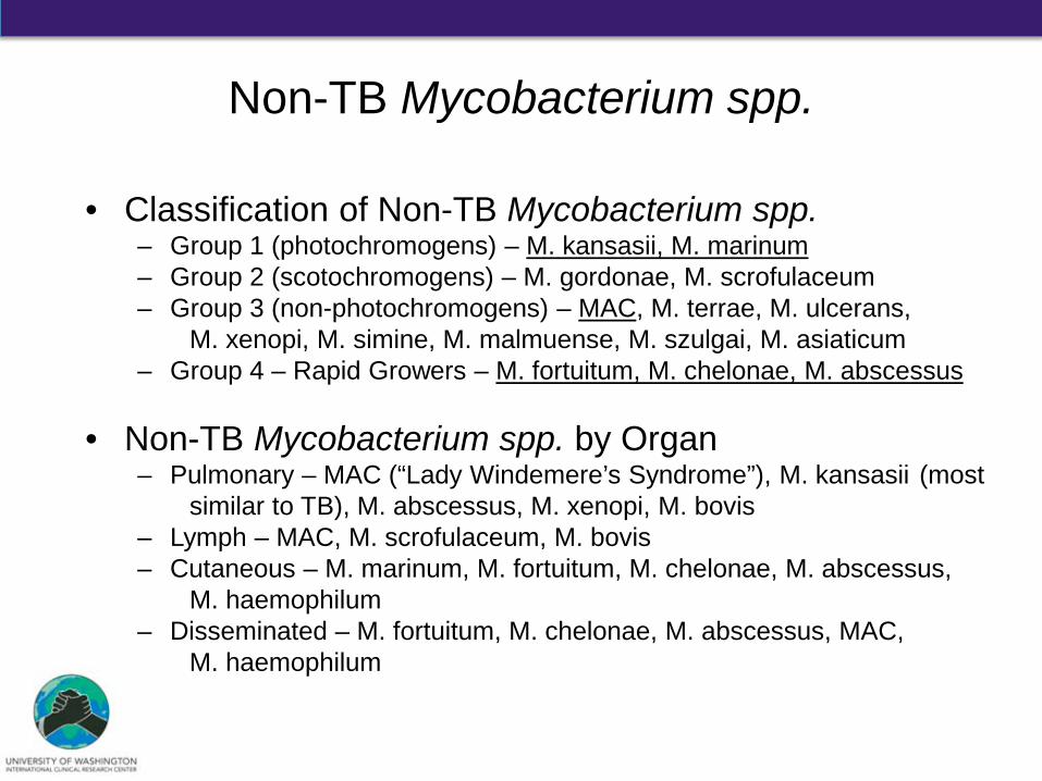
Non-TB Mycobacterium spp.
• Classification of Non-TB Mycobacterium spp. – Group 1 (photochromogens) – M. kansasii, M. marinum – Group 2 (scotochromogens) – M. gordonae, M. scrofulaceum – Group 3 (non-photochromogens) – MAC, M. terrae, M. ulcerans,
M. xenopi, M. simine, M. malmuense, M. szulgai, M. asiaticum – Group 4 – Rapid Growers – M. fortuitum, M. chelonae, M. abscessus
• Non-TB Mycobacterium spp. by Organ – Pulmonary – MAC (“Lady Windemere’s Syndrome”), M. kansasii (most
similar to TB), M. abscessus, M. xenopi, M. bovis – Lymph – MAC, M. scrofulaceum, M. bovis – Cutaneous – M. marinum, M. fortuitum, M. chelonae, M. abscessus,
M. haemophilum – Disseminated – M. fortuitum, M. chelonae, M. abscessus, MAC,
M. haemophilum
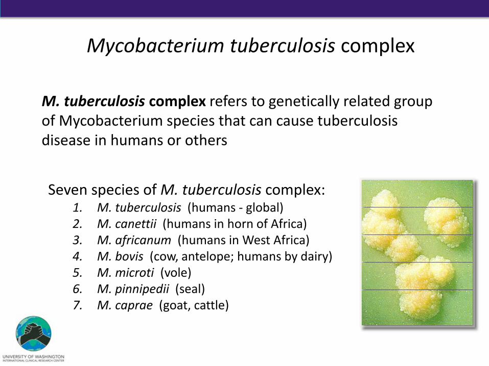
Mycobacterium tuberculosis complex
M. tuberculosis complex refers to genetically related group of Mycobacterium species that can cause tuberculosis disease in humans or others
Seven species of M. tuberculosis complex: 1. M. tuberculosis (humans - global) 2. M. canettii (humans in horn of Africa) 3. M. africanum (humans in West Africa) 4. M. bovis (cow, antelope; humans by dairy) 5. M. microti (vole) 6. M. pinnipedii (seal) 7. M. caprae (goat, cattle)
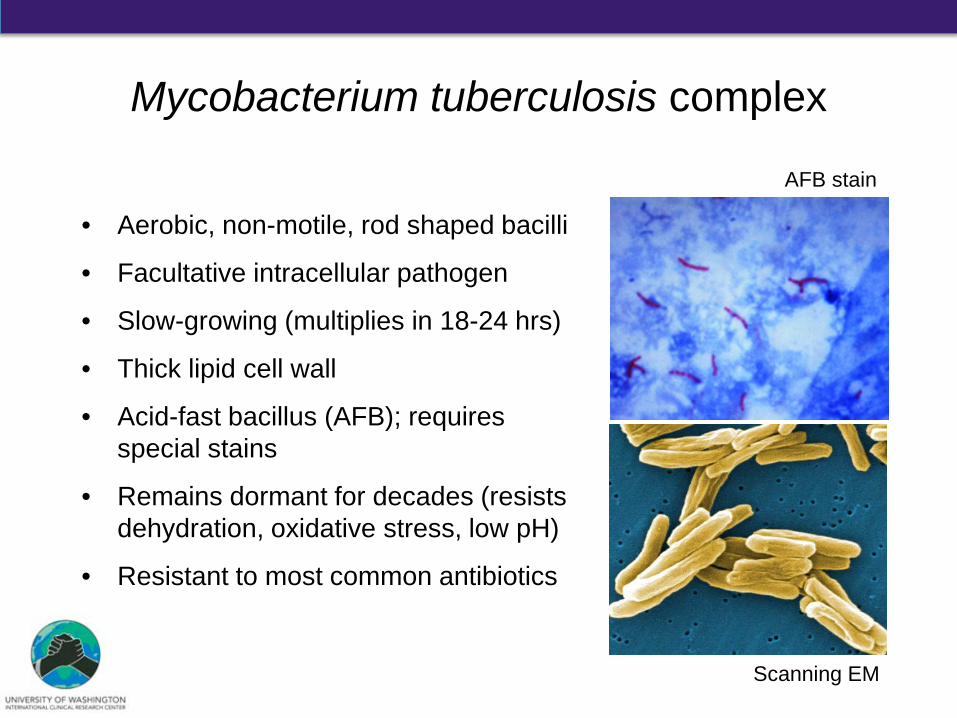
• Aerobic, non-motile, rod shaped bacilli
• Facultative intracellular pathogen
• Slow-growing (multiplies in 18-24 hrs)
• Thick lipid cell wall
• Acid-fast bacillus (AFB); requires special stains
• Remains dormant for decades (resists dehydration, oxidative stress, low pH)
• Resistant to most common antibiotics
Scanning EM
AFB stain
Mycobacterium tuberculosis complex

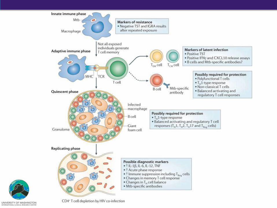
Latent TB Infection
• Asymptomatic people • Mantoux PPD skin test (TST) or
interferon-gamma release assay (IGRA)
• Risk factors for exposure: – High local TB prevalence – Close household contact – Institutional settings (hospitals,
prisons, shelters) – Social contact (public transit) – Urbanization – Age – Low socioeconomic status
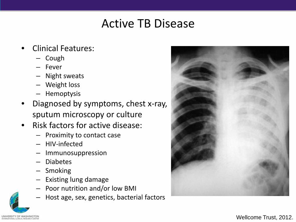
Active TB Disease
• Clinical Features: – Cough – Fever – Night sweats – Weight loss – Hemoptysis
• Diagnosed by symptoms, chest x-ray, sputum microscopy or culture
• Risk factors for active disease: – Proximity to contact case – HIV-infected – Immunosuppression – Diabetes – Smoking – Existing lung damage – Poor nutrition and/or low BMI – Host age, sex, genetics, bacterial factors
Wellcome Trust, 2012.
Outline
• Historical Context of Tuberculosis (TB)
• Mycobacterium spp. and M. tuberculosis
• TB Pathophysiology
• TB Transmission
• Summary
What percentage of new TB infections lead to a primary active TB disease?
1. 5% 2. 20% 3. 40% 4. 60%
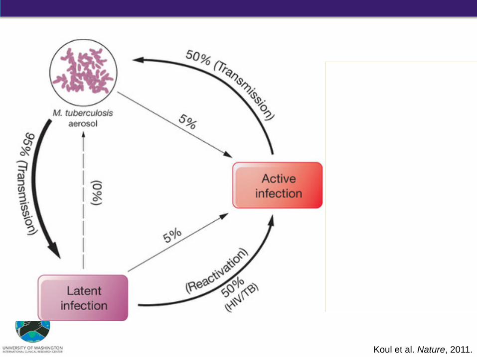
Koul et al. Nature, 2011.
Without treatment
With completed treatment
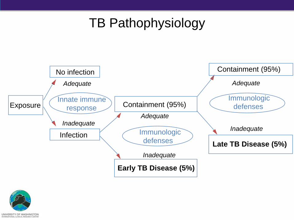
Exposure
Containment (95%) No infection Adequate
Innate immune response
Inadequate
Infection
Adequate
Immunologic defenses
Inadequate
Containment (95%)
Early TB Disease (5%)
Late TB Disease (5%)
Adequate
Immunologic
defenses
Inadequate
TB Pathophysiology
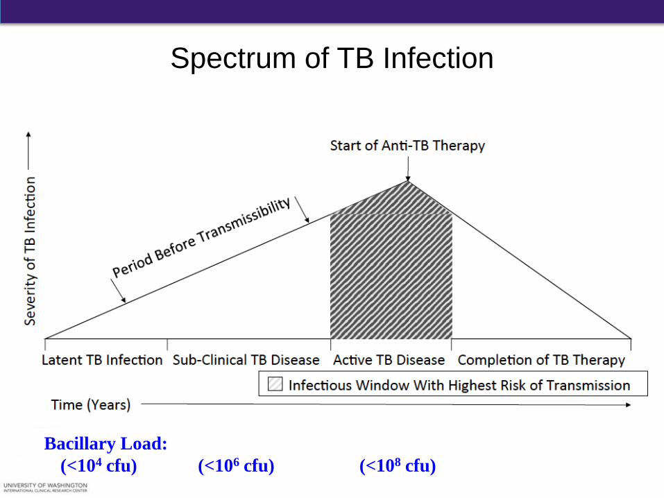
Spectrum of TB Infection
Bacillary Load: (<104 cfu) (<106 cfu) (<108 cfu)
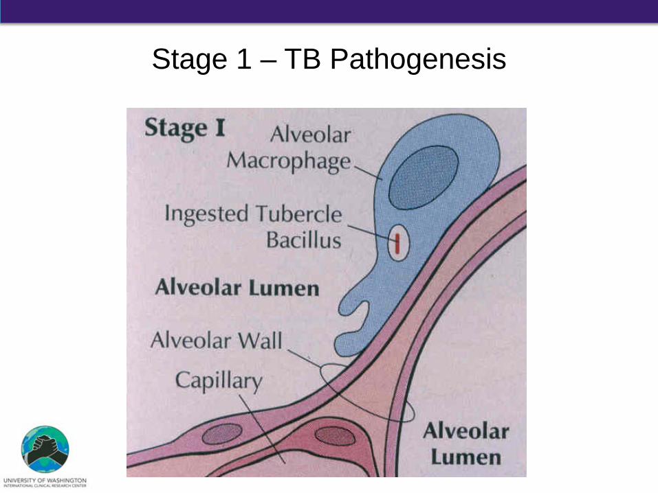
Stage 1 – TB Pathogenesis
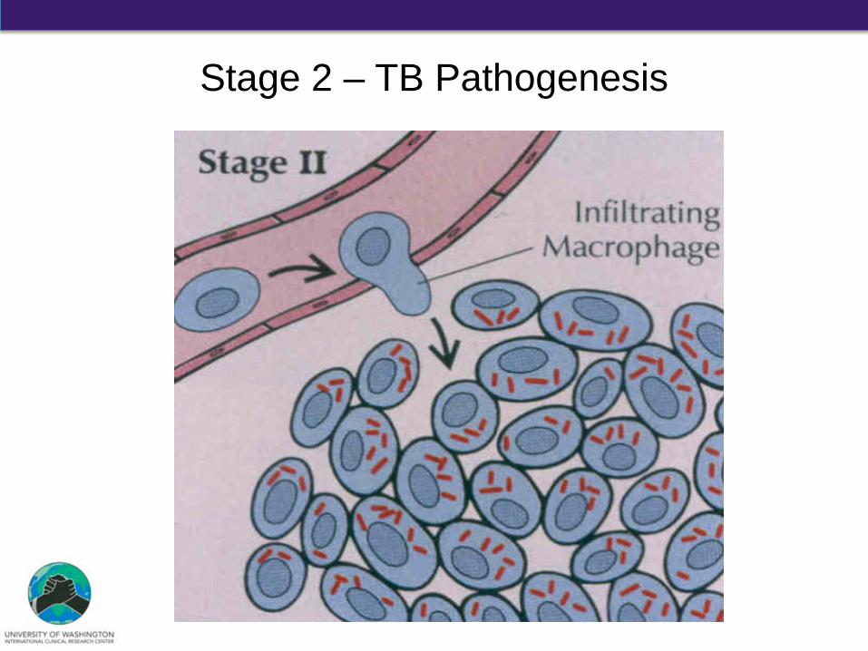
Stage 2 – TB Pathogenesis
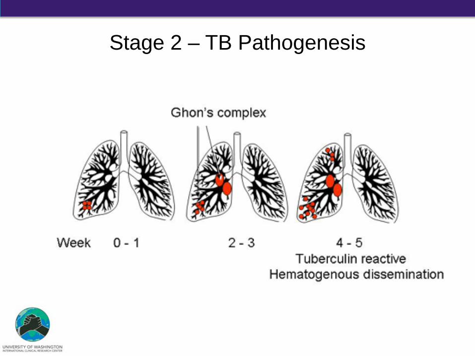
Stage 2 – TB Pathogenesis
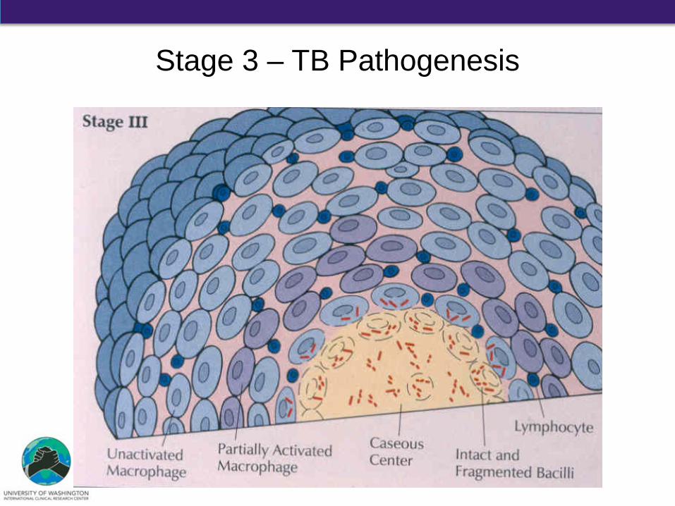
Stage 3 – TB Pathogenesis
D. G. Russell et al., Science 328, 852-856 (2010)
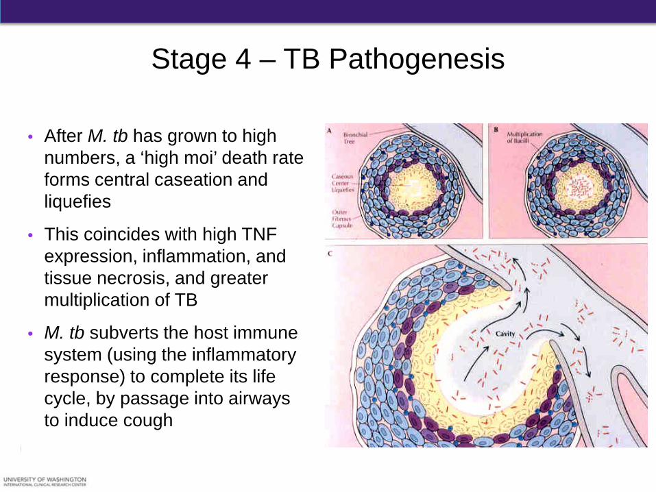
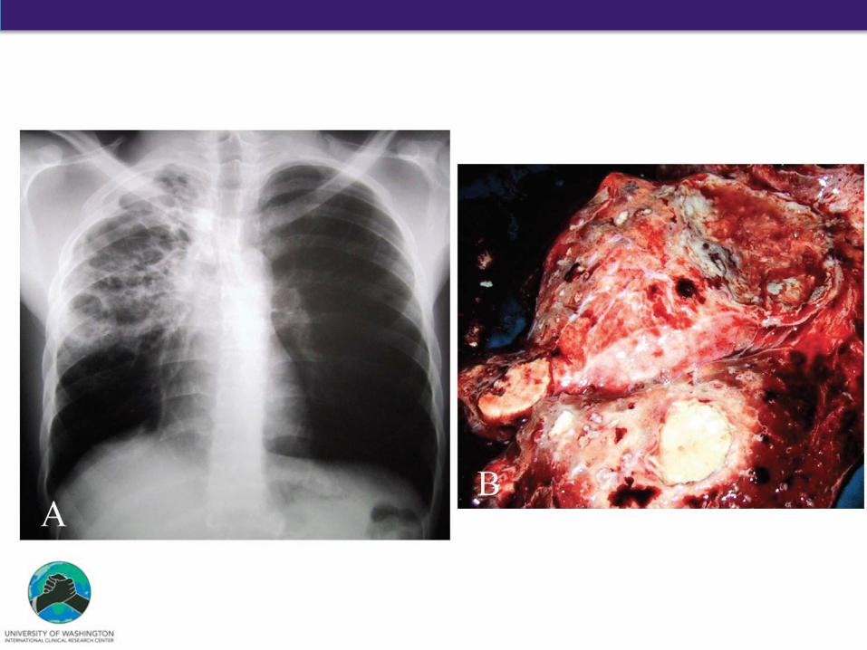
Stage 4 – TB Pathogenesis
• After M. tb has grown to high numbers, a ‘high moi’ death rate forms central caseation and liquefies
• This coincides with high TNF expression, inflammation, and tissue necrosis, and greater multiplication of TB
• M. tb subverts the host immune system (using the inflammatory response) to complete its life cycle, by passage into airways to induce cough
NIAID, 2012. Ulrichs & Kaumann. Front Biosci. 2002.
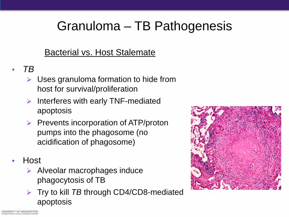
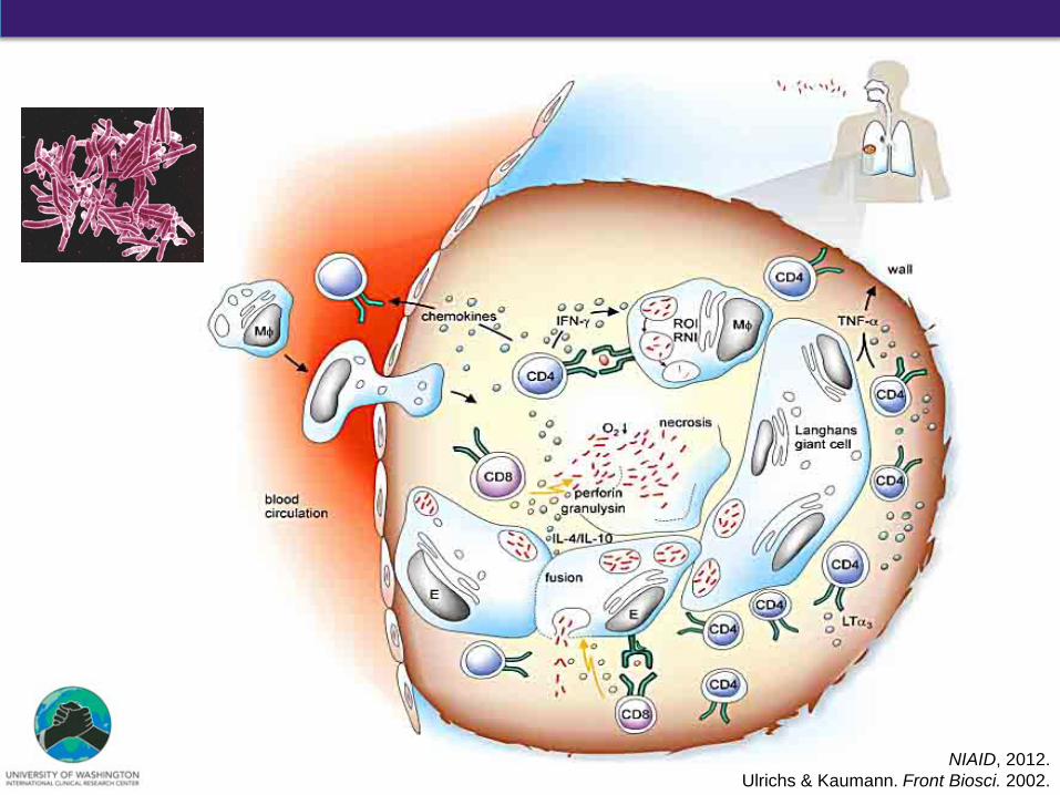
Bacterial vs. Host Stalemate
• TB Uses granuloma formation to hide from
host for survival/proliferation Interferes with early TNF-mediated
apoptosis Prevents incorporation of ATP/proton
pumps into the phagosome (no acidification of phagosome)
• Host Alveolar macrophages induce
phagocytosis of TB Try to kill TB through CD4/CD8-mediated
apoptosis D. G. Russell et al., Science 328, 852-856 (2010)
Granuloma – TB Pathogenesis
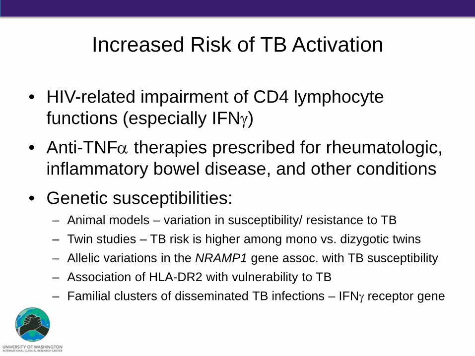
Increased Risk of TB Activation
• HIV-related impairment of CD4 lymphocyte functions (especially IFNγ)
• Anti-TNFα therapies prescribed for rheumatologic, inflammatory bowel disease, and other conditions
• Genetic susceptibilities: – Animal models – variation in susceptibility/ resistance to TB – Twin studies – TB risk is higher among mono vs. dizygotic twins – Allelic variations in the NRAMP1 gene assoc. with TB susceptibility – Association of HLA-DR2 with vulnerability to TB – Familial clusters of disseminated TB infections – IFNγ receptor gene
Outline
• Historical Context of Tuberculosis (TB)
• Mycobacterium spp. and M. tuberculosis
• TB Pathophysiology
• TB Transmission
• Summary
How is TB transmitted between humans?
1. Fecal-oral contamination
2. Skin-to-skin contact
3. Aerosolized droplet nuclei
4. Blood-borne exposure
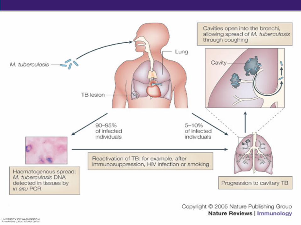
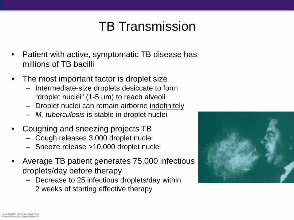
• Patient with active, symptomatic TB disease has millions of TB bacilli
• The most important factor is droplet size – Intermediate-size droplets desiccate to form
“droplet nuclei” (1-5 µm) to reach alveoli – Droplet nuclei can remain airborne indefinitely – M. tuberculosis is stable in droplet nuclei
• Coughing and sneezing projects TB – Cough releases 3,000 droplet nuclei – Sneeze release >10,000 droplet nuclei
• Average TB patient generates 75,000 infectious droplets/day before therapy – Decrease to 25 infectious droplets/day within
2 weeks of starting effective therapy
TB Transmission
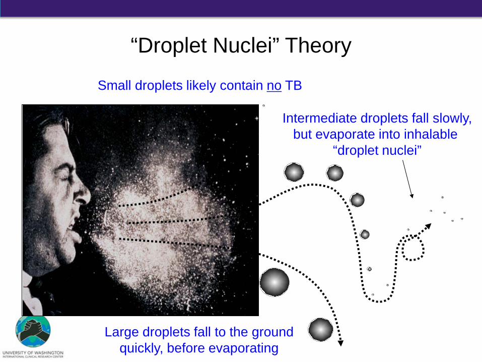
“Droplet Nuclei” Theory
Intermediate droplets fall slowly, but evaporate into inhalable
“droplet nuclei”
Large droplets fall to the ground quickly, before evaporating
Small droplets likely contain no TB
TB Transmission
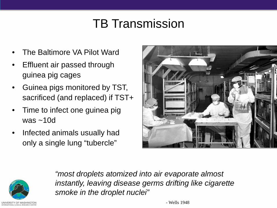
• The Baltimore VA Pilot Ward • Effluent air passed through
guinea pig cages • Guinea pigs monitored by TST,
sacrificed (and replaced) if TST+ • Time to infect one guinea pig
was ~10d • Infected animals usually had
only a single lung “tubercle”
- Riley and Wells
“most droplets atomized into air evaporate almost instantly, leaving disease germs drifting like cigarette smoke in the droplet nuclei” - Wells 1948
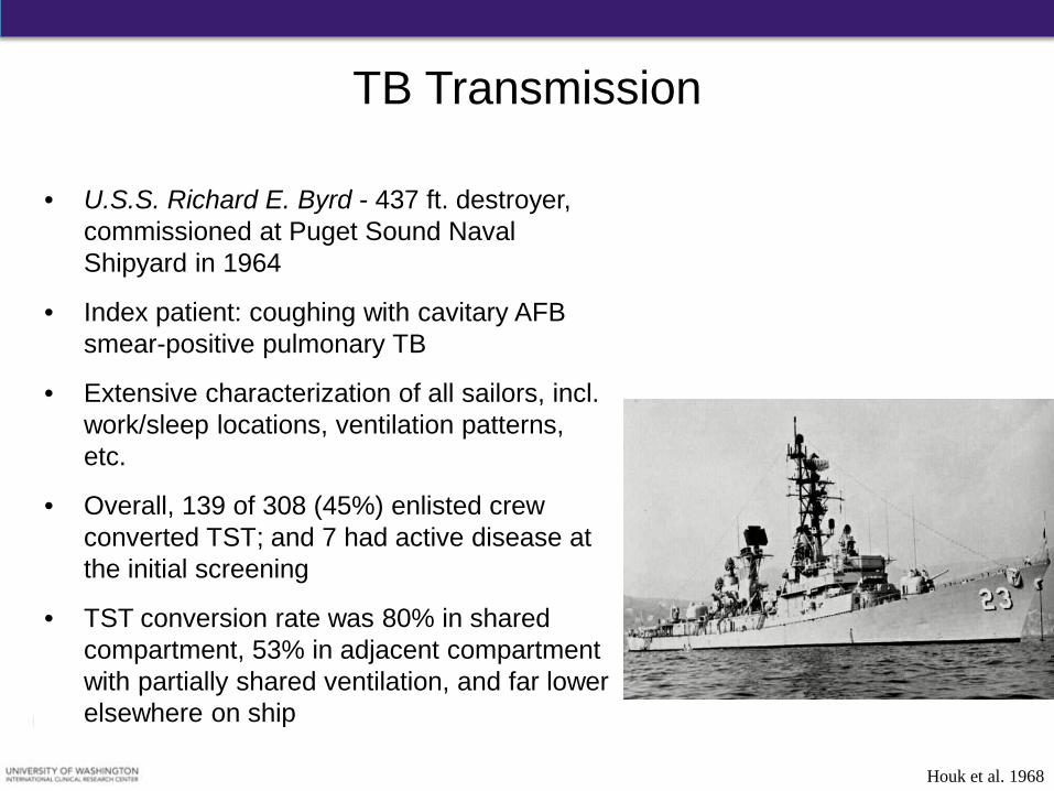
• U.S.S. Richard E. Byrd - 437 ft. destroyer, commissioned at Puget Sound Naval Shipyard in 1964
• Index patient: coughing with cavitary AFB smear-positive pulmonary TB
• Extensive characterization of all sailors, incl. work/sleep locations, ventilation patterns, etc.
• Overall, 139 of 308 (45%) enlisted crew converted TST; and 7 had active disease at the initial screening
• TST conversion rate was 80% in shared compartment, 53% in adjacent compartment with partially shared ventilation, and far lower elsewhere on ship
TB Transmission
Houk et al. 1968
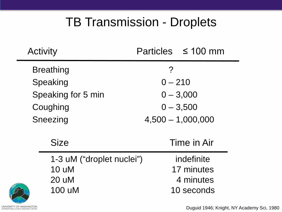
TB Transmission - Droplets
Activity Particles ≤ 100 mm
Breathing ? Speaking 0 – 210 Speaking for 5 min 0 – 3,000 Coughing 0 – 3,500 Sneezing 4,500 – 1,000,000
Duguid 1946; Knight, NY Academy Sci, 1980
Size Time in Air
1-3 uM (“droplet nuclei”) indefinite 10 uM 17 minutes 20 uM 4 minutes 100 uM 10 seconds
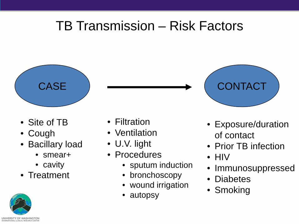
TB Transmission – Risk Factors
CASE CONTACT
• Site of TB • Cough • Bacillary load
• smear+ • cavity
• Treatment
• Exposure/duration of contact
• Prior TB infection • HIV • Immunosuppressed • Diabetes • Smoking
• Filtration • Ventilation • U.V. light • Procedures
• sputum induction • bronchoscopy • wound irrigation • autopsy
Environment
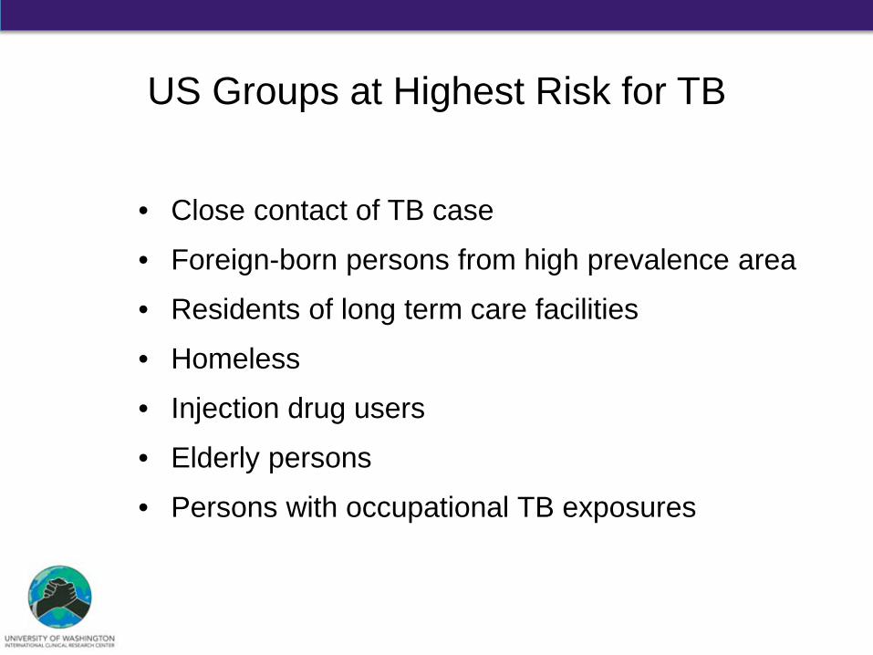
• Close contact of TB case
• Foreign-born persons from high prevalence area
• Residents of long term care facilities
• Homeless
• Injection drug users
• Elderly persons
• Persons with occupational TB exposures
US Groups at Highest Risk for TB
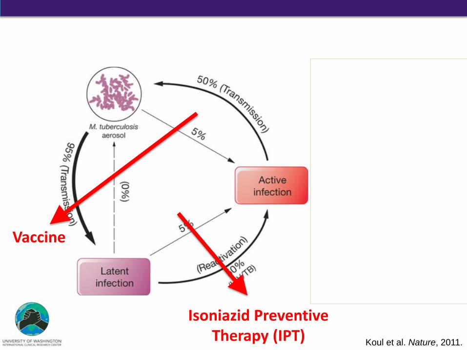
Koul et al. Nature, 2011.
Without treatment
With completed treatment
Isoniazid Preventive Therapy (IPT)
Vaccine
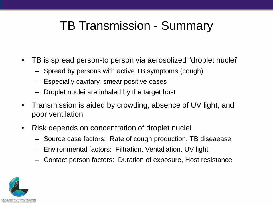
TB Transmission - Summary
• TB is spread person-to person via aerosolized “droplet nuclei” – Spread by persons with active TB symptoms (cough) – Especially cavitary, smear positive cases – Droplet nuclei are inhaled by the target host
• Transmission is aided by crowding, absence of UV light, and poor ventilation
• Risk depends on concentration of droplet nuclei – Source case factors: Rate of cough production, TB diseaease – Environmental factors: Filtration, Ventaliation, UV light – Contact person factors: Duration of exposure, Host resistance
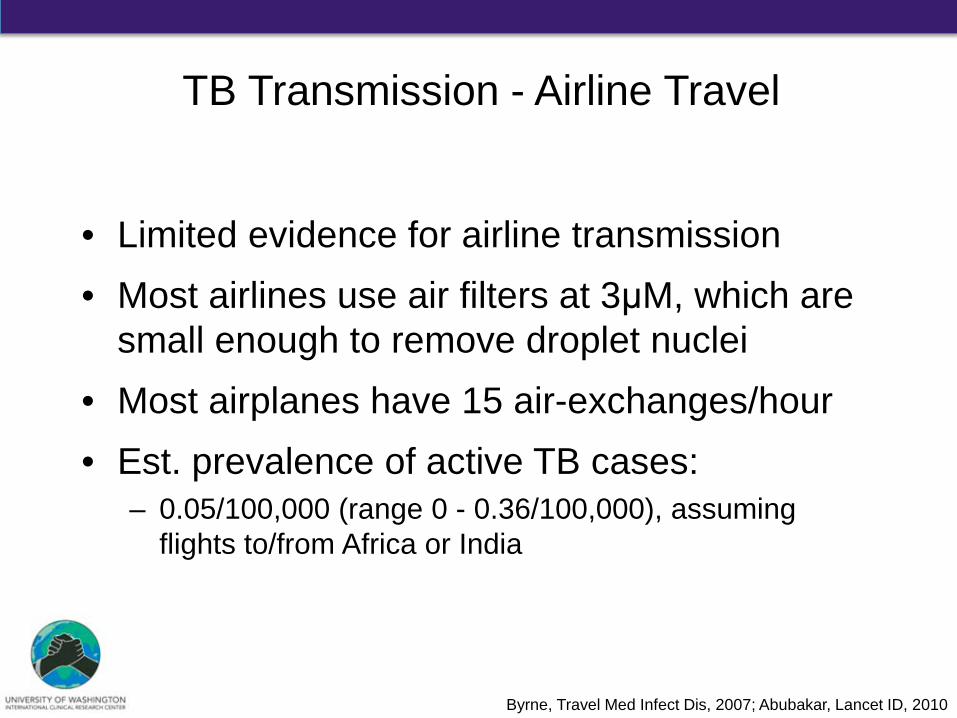
• Limited evidence for airline transmission • Most airlines use air filters at 3μM, which are
small enough to remove droplet nuclei • Most airplanes have 15 air-exchanges/hour • Est. prevalence of active TB cases:
– 0.05/100,000 (range 0 - 0.36/100,000), assuming flights to/from Africa or India
TB Transmission - Airline Travel
Byrne, Travel Med Infect Dis, 2007; Abubakar, Lancet ID, 2010
Outline
• Historical Context of Tuberculosis (TB)
• Mycobacterium spp. and M. tuberculosis
• TB Pathophysiology
• TB Transmission
• Summary
Summary
• The global burden of TB is severe – TB causes more deaths than any other infection
• Global TB incidence/deaths is decreasing – But, not fast enough
• Pathogenesis of TB is complicated • Transmission remains a major problem
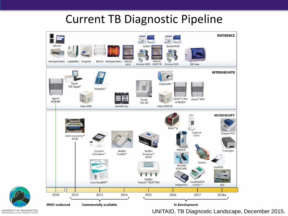
UNITAID, TB Diagnostic Landscape, December 2015.
Current TB Diagnostic Pipeline
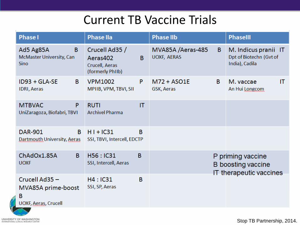
Current TB Vaccine Trials
Stop TB Partnership, 2014.