traumatic diseases of oral cavity. infectious diseases of oral cavity. etiology, clinical picture,...
TRANSCRIPT
Traumatic diseases of oral cavity. Infectious diseases of oral cavity.
Etiology, clinical picture, diagnostics, treatment and prevention.

Oral ulcers
How to diagnose?
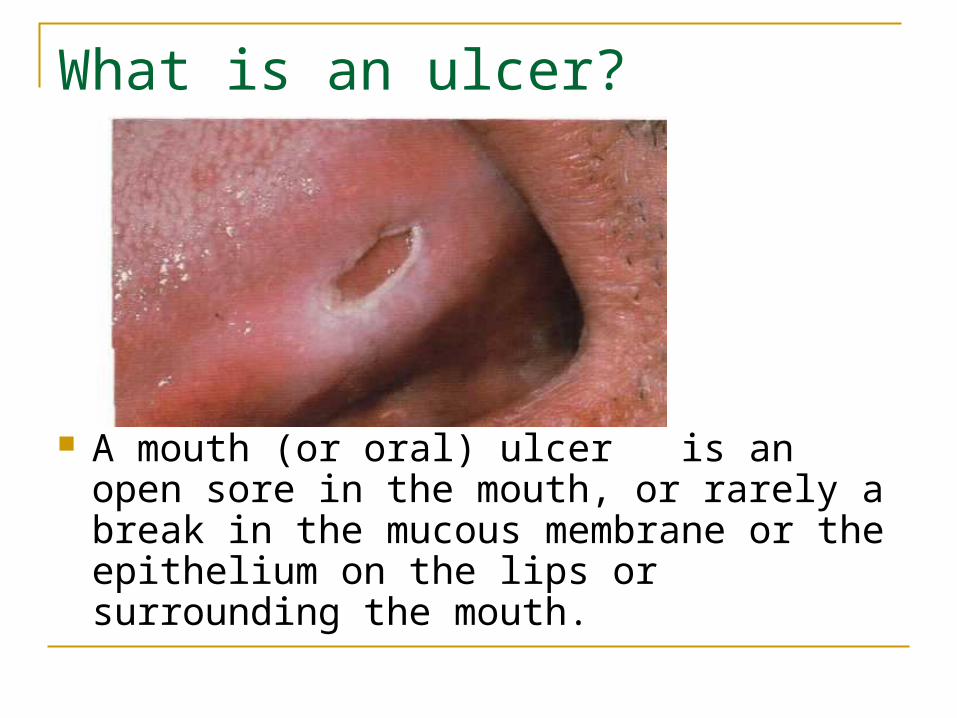
What is an ulcer?
A mouth (or oral) ulcer is an open sore in the mouth, or rarely a break in the mucous membrane or the epithelium on the lips or surrounding the mouth.

Epidemiology and frequency
Mouth ulcer is a very common oral lesion. Epidemiological studies show an average prevalence between 15% and 30%. Mouth ulcers tend to be more common in women and those under 45.
The frequency of mouth ulcers varies from fewer than 4 episodes per year (85% of all cases) to more than one episode per month (10% of all cases) including people suffering from continuous recurrent aphthous stomatitis
What cause ulcers?
Trauma (physical injuries) Chemical injuries Smoking Infection - viral - bacterial - fungal - protozoans Immunodeficiency Autoimmunity Allergy Dietary

Tools for diagnosis:
History Examination Further investigation Additional diagnostic methods – physical
(VELscope, Visilite, Orascoptic DK) Biopsy
Further investigation – questions you should ask: How many ulcers do you have? How long have you had that ulcer? Is it painful? Can you relate them to any trauma, hot food
or another factors? Is it the first time or you had them before? Where are they located in the mouth? Do they start as ulcer or as vesicle/bulla? Do you get them anywhere else on the body?
Step by step guide to performing an oral cavity examination
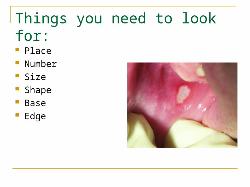
Things you need to look for:
Place Number Size Shape Base Edge
Differential diagnostic, based on history How long have you had the ulcer?
Long time – chronic1. Chronic trauma (single, can identify the cause,
and should improve after removal) 2. Malignancy (single, painless)3. TB (single ulcer in the tongue/palate,
associated symptoms – chronic cough)4. Mucous membrane pemphigoid (multiple ulcers
affect mainly gingiva, blood filled blisters) 5. Primary or tetriary syphilis

Recent – acute
1. Acute trauma (single, can identify the cause, and should improve after removal)
2. Viral infection (multiple, associated symptoms – fever)
3. Immune mediated disease (Erythema multiforme, RAS)

How many ulcers do you have? Single
1. Traumatic
2. Primary or tetriary syphilis
3. TB
4. Malignancy Multiple
1. Viral infection
2. Immune mediated disease
Is it painful?
Yes
Acute causes (trauma, viral, immune mediate) No
Chronic causes (trauma, TB)

Can you relate them to trauma or hot food? Yes
1. Confirm traumatic ulcer
2. Remove the cause
3. Review after 1 week No
Look for other causes
Do you get them anywhere else on the body? No
Think or oral conditions Yes – where?
1. Skin – mucocutanous disease (lichen planus, pemphigus)
So, just a few simple questions can help you to put diagnosis even before examination

Traumatic lesions
Can be divided to: Mechanical trauma (acute or chronic) Chemical injury (acid, alcali) Physical injury (thermal injury, electrical
injury)

1. Traumatic lesion after teeth brush2. White lesion caused by trauma.
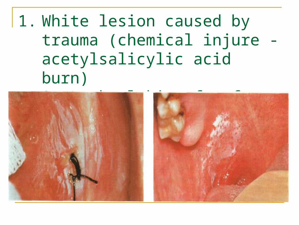
1. White lesion caused by trauma (chemical injure - acetylsalicylic acid burn)2. Leukoplakia of soft palatine
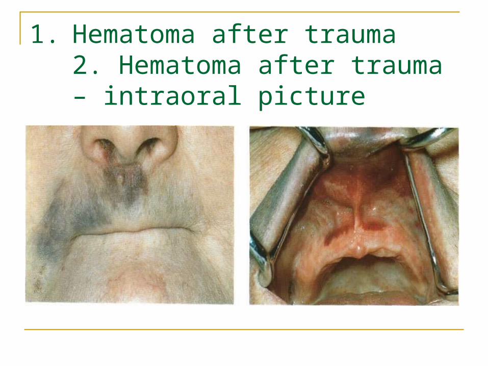
1. Hematoma after trauma2. Hematoma after trauma – intraoral picture

Oral ulcers after long chemotherapy

Dental braces
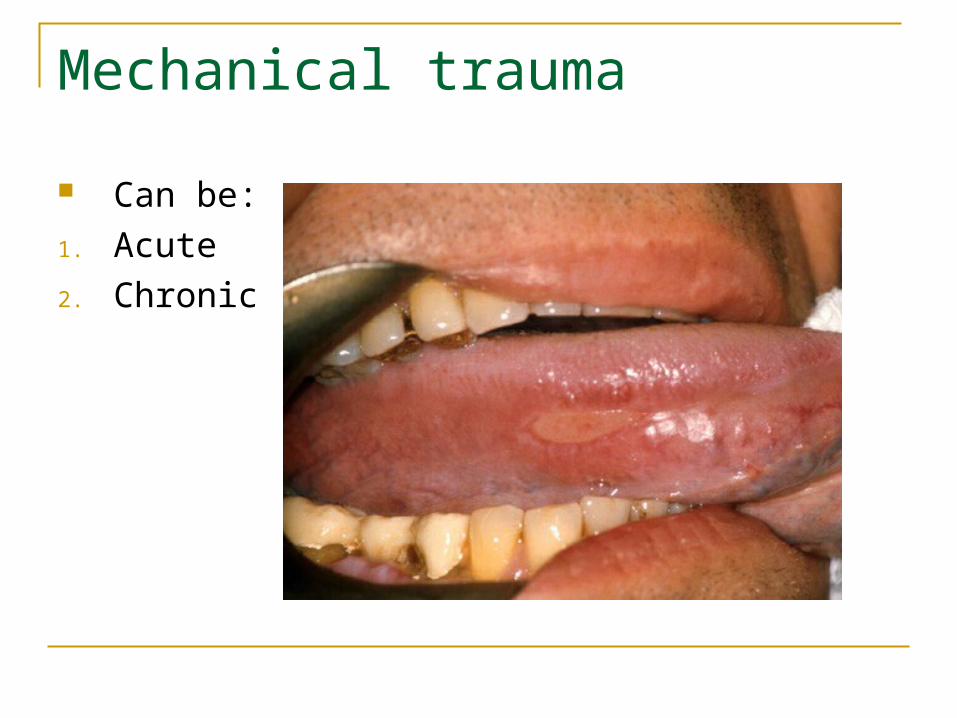
Mechanical trauma
Can be:
1. Acute
2. Chronic

And caused by:
1. A sharp edge of a tooth
2. Accidental biting3. Sharp, abrasive,or
excessively salty food 4. Poorly fitting dentures 5. Dental braces 6. Trauma from a tooth
brush 7. Patients bad habits

Acute trauma
Single, Can identify the
cause Should improve after
removal
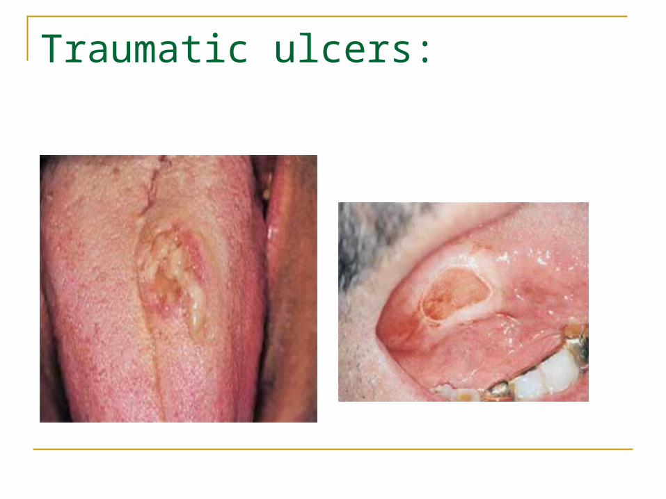
Traumatic ulcers:
Clinical featuresof traumatic ulcers: •They are clinically diverse, but usually appear as a single,
painful ulcer with a smooth red or whitish-yellow surface and a thin erythematous halo.
•They are usually soft on palpation, and heal without scarring within 6–10 days, spontaneously or after removal of the cause.
•However, chronic traumatic ulcers may clinically mimic a carcinoma.
•The tongue, lip, and buccal mucosa are the sites of predilection. •The diagnosis is based on the history and clinical features. •However, if an ulcer persists over 10–12 days a biopsy must be
taken to rule out cancer.
Treatment
1. Removing factors, caused trauma
2. Good hygiene of oral cavity
3. Antiseptic for 7-10 days
4. Analgetics if it is necessary
5. Topical steroids may be used for a short time.
6. Biopsy
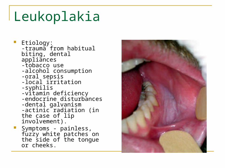
Leukoplakia
Etiology: - trauma from habitual biting, dental appliances- tobacco use- alcohol consumption -oral sepsis- local irritation -syphilis -vitamin deficiency- endocrine disturbances -dental galvanism -actinic radiation (in the case of lip involvement).
Symptoms - painless, fuzzy white patches on the side of the tongue or cheeks.

Clinical picture
- located on the tongue, mandibular alveolar ridge and buccal mucosa in ~50%.- palate, maxillary alveolar ridge, lower lip, floor of the mouth and the retromolar regions are somewhat less frequently involved.- may vary from nonpalpable, faintly translucent white areas to thick, fissured, papillomatous, indurated lesions.- surface is often wrinkled or shriveled in appearance and may feel rough on palpation.- color may be white, gray, yellowish-white, or even brownish-gray in patients with heavy tobacco use.
Physical:- lesion cannot be wiped away with a gauze

Differential diagnosis:
1. Candidiasis and aspirin burn - can be wiped away with a gauze
2. Erythroplakia (a red plaque that does not rub off) is a dysplastic lesion (or worse) in 90 percent of cases.
Treatment
Treat dental causes such as rough teeth, irregular denture surface, or fillings as soon as possible.
Stopping tobacco or/and alcohol. Removal of leukoplakic patches with
using a scalpel, a laser or an extremely cold probe that freezes and destroys cancer cells (cryoprobe).
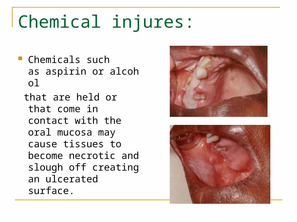
Chemical injures:
Chemicals such as aspirin or alcohol
that are held or that come in contact with the oral mucosa may cause tissues to become necrotic and slough off creating an ulcerated surface.
Treatment:
1. Wash a mouth with lot of water
2. Analgetics (for 3-5 days )
3. Antiseptics ( for 7 – 10 days)
4. Keratolytics (after 5-7 days)
5. Keratoplastics (after 7-10 days)
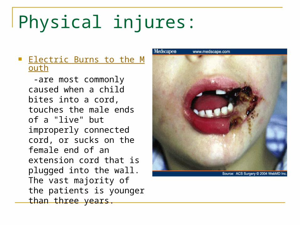
Physical injures:
Electric Burns to the Mouth -are most commonly caused when a child bites into a cord, touches the male ends of a "live" but improperly connected cord, or sucks on the female end of an extension cord that is plugged into the wall. The vast majority of the patients is younger than three years.
Treatment:
1. Conservative Antiseptics Antibiotics Analgetic
2. Surgical (after the healing was completed and after the degree of functional and/or aesthetic deformity was established (usually 6 months post-injury)).

Stomatitis
Description
Stomatitis is an inflammation of the lining of any of the soft-tissue structures of the mouth. Stomatitis is usually a painful condition, associated with redness, swelling, and occasional bleeding from the affected area. Bad breath (halitosis) may also accompany the condition. Stomatitis affects all age groups, from the infant to the elderly.
Definition
Inflammation of the mucous lining of any of the structures in the mouth, which may involve the cheeks, gums, tongue, lips, and roof or floor of the mouth. The word "stomatitis" literally means inflammation of the mouth. The inflammation can be caused by conditions in the mouth itself, such as poor oral hygiene, poorly fitted dentures, or from mouth burns from hot food or drinks, or by conditions that affect the entire body, such as medications, allergic reactions, or infections.
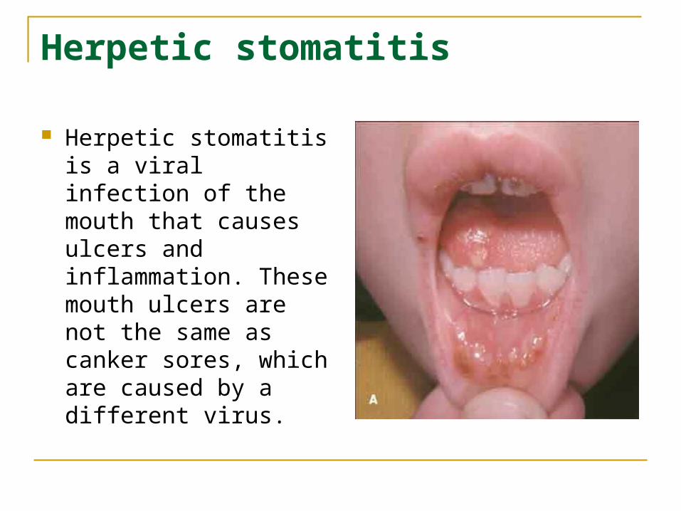
Herpetic stomatitis
Herpetic stomatitis is a viral infection of the mouth that causes ulcers and inflammation. These mouth ulcers are not the same as canker sores, which are caused by a different virus.
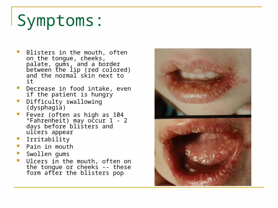
Symptoms:
Blisters in the mouth, often on the tongue, cheeks, palate, gums, and a border between the lip (red colored) and the normal skin next to it
Decrease in food intake, even if the patient is hungry
Difficulty swallowing (dysphagia) Fever (often as high as 104
°Fahrenheit) may occur 1 - 2 days before blisters and ulcers appear
Irritability Pain in mouth Swollen gums Ulcers in the mouth, often on the
tongue or cheeks -- these form after the blisters pop
Causes
Herpetic stomatitis is a contagious viral illness caused by Herpes virus hominis (also herpes simplex virus, HSV). It is seen mainly in young children. This condition is probably a child's first exposure to the herpes virus.
An adult member of the family may have a cold sore at the time the child develops herpetic stomatitis. More likely, no source for the infection will be discovered.

Herpetic stomatitis – intraoral and extraoral picture

1. Herpes recurrent2. Herpes recurrent in AIDS patient
Differential diagnostics:
Streptococcal pharyngitis –does not involve the lips or perioral tissues and vesicles do not precede the ulcers
Erythema multiforme – ulcers are larger, usually without a vesicular stage and are less likely to affect the gingiva.
Aphthous stomatitis – the multiply lesions, palatal and gingival location are indicative to herpesvirus infection. Additionally, aphthae usually localized only on nonceratinized mucoza, such as the floor of the mouth, alveolar mucosa and buccal mucosae.

Treatment:
Good oral hygiene Acyclovir family of antiviral medications – non
later than 48 hours! Analgetic ( or oral topical anesthetic ) Diet (no hot or pepper food) Antiseptics
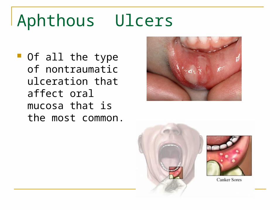
Aphthous Ulcers
Of all the type of nontraumatic ulceration that affect oral mucosa that is the most common.

Aphthous ulcers are also known as canker sores. They are painful, temporary sores that may occur anywhere in the mouth. Usually, they show up in several places:
On the inside of the lips Inside the cheeks On the tongue At the base of the gums

Canker sores on tongue:
Etiology:
1. Immune system disorders2. Other conditions cause similar sores: Blood and immune system diseases,
including HIV Vitamin and mineral deficiencies Allergies
Trauma Crohn's disease Lupus
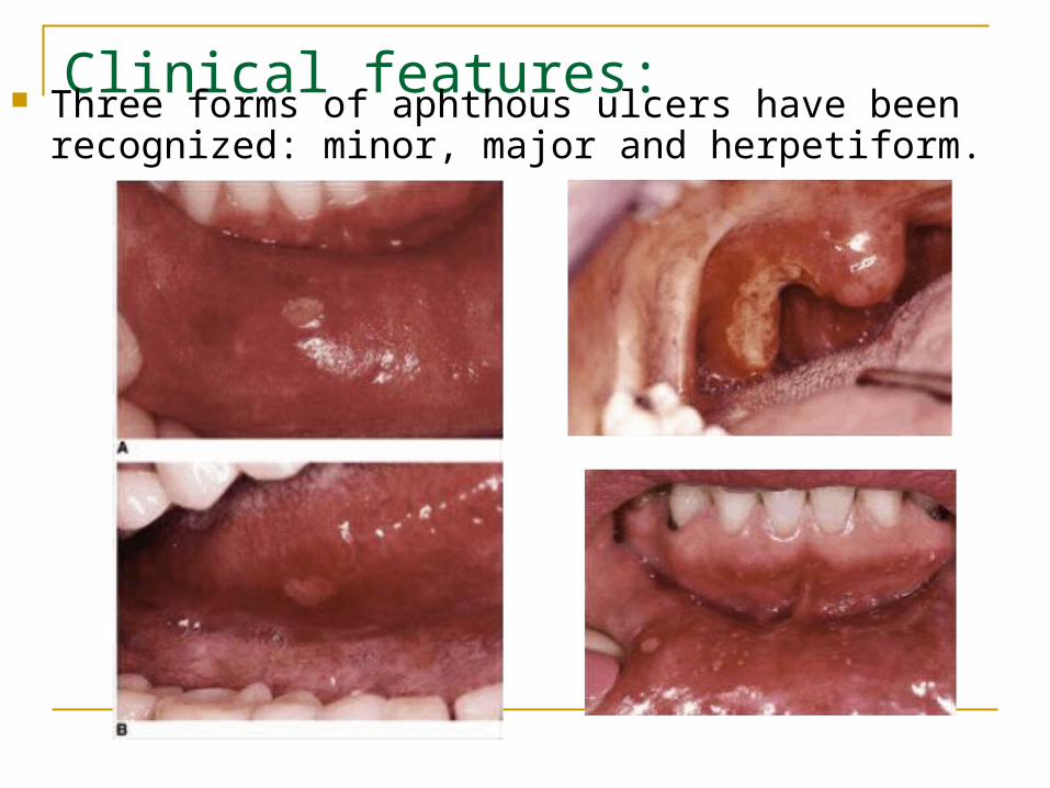
Clinical features: Three forms of aphthous ulcers have been recognized:
minor, major and herpetiform.

§ Major Ulcerations (> 0,5cm)—10% of lesions o extremely painful (odynophagia)... typically heal with scar formation o Appearance- deeply cratered ulcers o Duration- > 1 month for healing. o Location- posterior oral cavity. 0 Number – 1- 10 o NOTE- THINK OF HIV DISEASE PROGRESSION OR IMMUNOSUPPRESION § Minor Ulceration (1-5 mm)—85% of lesions o painful… heal without scar formation o Duration- 7-10 days o Location- anterior oral cavity 0 Number – 1-5 § Herpetiform Ulceration (1-3mm lesions that form clusters)—5% of lesions. o Number – 10-100 o Locational – any intraoral site

Minor ulcers:
Are the most commonly encountered form
Single, painful oval ulcer less than 0,6 mm, covered by a yellow fibrinous membrane
Last 7 to 10 days Periods of freedom
from disease – 2-3 weeks to year
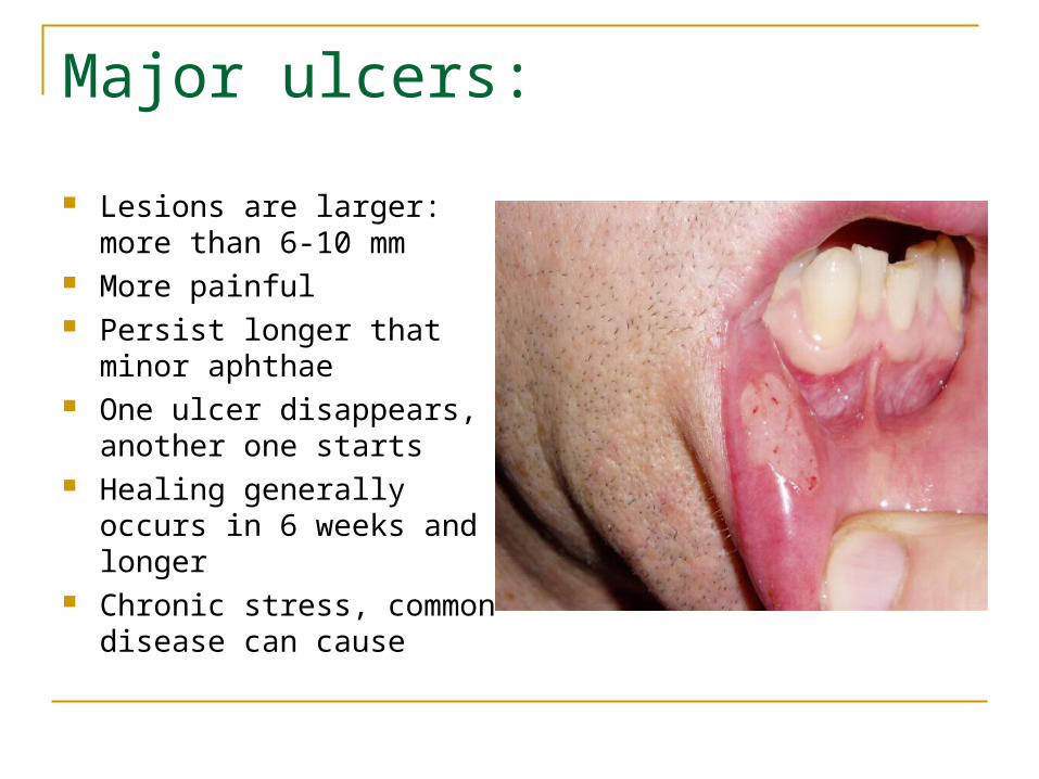
Major ulcers:
Lesions are larger: more than 6-10 mm
More painful Persist longer that minor
aphthae One ulcer disappears,
another one starts Healing generally occurs in
6 weeks and longer Chronic stress, common
disease can cause

Herpetiform ulcers
Recurrent crops of small ulcers
Healing generally occurs in 1 to 2 weeks
Not preceded by vesicle and exhibit no virus-infected cells

Treatment:
Oral hygiene Avoiding spicy food Oral rising with sodium bicarbonate Analgesics Antiseptics Anti – inflammatory agents Antimicrobial and corticosteroids