transposition of the great arteries - smgebooks.com · transposition of the great arteries....
TRANSCRIPT
1Echocardiography | www.smgebooks.comCopyright Agha HM.This book chapter is open access distributed under the Creative Commons Attribution 4.0 International License, which allows users to download, copy and build upon published articles even for commercial purposes, as long as the author and publisher are properly credited.
Gr upSMTransposition of the Great Arteries
INTRODUCTIONThe term transposition of great arteries (TGA) is used to describe connection of the great
vessels from the inappropriate ventricles i.e. the aorta arises from the morphologic right ventricle and the pulmonary artery arises from the morphologic left ventricle (ventriculo-arterial discordance). It accounts for 5– 7% of all CHD.
There are two forms of TGA; D and L-TGA. In D-TGA, there is atrio-ventricular concordance and the morphologic right ventricle lies to the right of the morphologic left ventricle. In L-TGA, there is ventricular inversion and atrio-ventricular discordance. Thus, the morphologic RV is to the left of the morphologic LV In both cases, the great arteries arise from the “incorrect” ventricle. Transposition can exist with either situs solitus or situs inversus. For simplicity, this section is a discussion of transposition in the presence of situs solitus only.
In the current era, echocardiography has become the diagnostic method of choice in the patient with TGA. It provides all the anatomic and functional information needed for the management of infants with D-TGA. The important echocardiographic features include:
Hala Mounir Agha1*, Mohamed Moustafa2 and Ibrahim Abou Farag2
1Pediatric Cardiology Division, Cairo University Specialized Pediatric Hospital, Egypt2Pediatric Cardiology Unit, El Azhar University, Egypt
*Corresponding author: Hala Mounir Agha, Pediatric Cardiology Division, Cairo University Specialized Pediatric Hospital, Egypt. Email: [email protected]
Published Date: October 13, 2016
2Echocardiography | www.smgebooks.comCopyright Agha HM.This book chapter is open access distributed under the Creative Commons Attribution 4.0 International License, which allows users to download, copy and build upon published articles even for commercial purposes, as long as the author and publisher are properly credited.
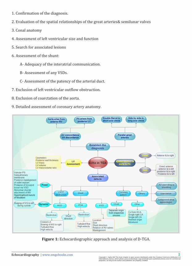
1. Confirmation of the diagnosis.
2. Evaluation of the spatial relationships of the great arteries& semilunar valves
3. Conal anatomy
4. Assessment of left ventricular size and function
5. Search for associated lesions
6. Assessment of the shunt:
A- Adequacy of the interatrial communication.
B- Assessment of any VSDs.
C- Assessment of the patency of the arterial duct.
7. Exclusion of left ventricular outflow obstruction.
8. Exclusion of coarctation of the aorta.
9. Detailed assessment of coronary artery anatomy.
Figure 1: Echocardiographic approach and analysis of D-TGA.
3Echocardiography | www.smgebooks.comCopyright Agha HM.This book chapter is open access distributed under the Creative Commons Attribution 4.0 International License, which allows users to download, copy and build upon published articles even for commercial purposes, as long as the author and publisher are properly credited.
CONFIRMATION OF THE DIAGNOSIS1. Concordant atrio-ventricular and discordant ventriculo-arterial connections.
The segmental anatomy can be abbreviated {S,D,D}:
1. Normal atrial situs.
2. Normal ventricular looping.
3. D-TGA; Aorta arises from the RV, PA arises from the LV.
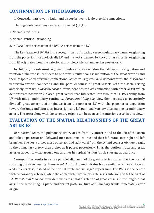
The key feature of D-TGA is the recognition a bifurcating vessel (pulmonary trunk) originating from the posterior morphologically LV and the aorta (defined by the coronary arteries originating from it) originates from the anterior morphologically RV and arches posteriorly.
In children, the subcostal imaging provides a flexible window that allows wide angulation and rotation of the transducer beam to optimize simultaneous visualization of the great arteries and their respective ventricular connections. Subcostal sagittal view demonstrates the discordant ventriculo-arterial connection and the parallel course of great vessels with the aorta arising anteriorly from RV. Subcostal coronal view identifies the AV connection with anterior tilt which demonstrate posteriorly placed great vessel that bifurcates into two, that is, PA arising from LV with mitral–pulmonary continuity. Parasternal long-axis view demonstrates a “posteriorly divided” great artery that originates from the posterior LV with sharp posterior angulation toward the lungs and bifurcates into a right and left pulmonary artery thus making it a pulmonary artery. The aorta along with the coronary origins can be seen as the anterior vessel in this view.
EVALUATION OF THE SPATIAL RELATIONSHIPS OF THE GREAT ARTERIES
In a normal heart, the pulmonary artery arises from RV anterior and to the left of the aorta and takes a posterior and leftward turn into initial course and then bifurcates into right and left branches. The aorta arises more posterior and rightward from the LV and courses obliquely right to the pulmonary artery then arches as it passes posteriorly. Thus, the outflow tracts and great arteries appear to wrap around one another in a spiral fashion (circle sausage appearance).
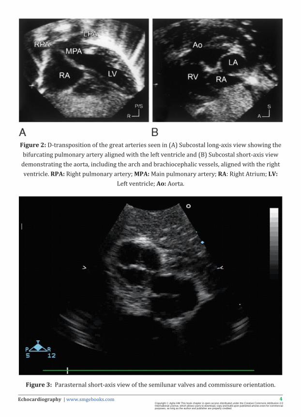
Transposition results in a more parallel alignment of the great arteries rather than the normal spiraling or criss-crossing. Parasternal short axis demonstrates both semilunar valves en face as a “double-circles”, instead of the normal circle and sausage” appearance. The PA is in the center with no coronary arteries, while the aorta with its coronary arteries is anterior and to the right of PA. Parasternal long-axis view demonstrates parallel relation of great vessels in the longitudinal axis in the same imaging plane and abrupt posterior turn of pulmonary trunk immediately after origin.
4Echocardiography | www.smgebooks.comCopyright Agha HM.This book chapter is open access distributed under the Creative Commons Attribution 4.0 International License, which allows users to download, copy and build upon published articles even for commercial purposes, as long as the author and publisher are properly credited.
Figure 2: D-transposition of the great arteries seen in (A) Subcostal long-axis view showing the bifurcating pulmonary artery aligned with the left ventricle and (B) Subcostal short-axis view
demonstrating the aorta, including the arch and brachiocephalic vessels, aligned with the right ventricle. RPA: Right pulmonary artery; MPA: Main pulmonary artery; RA: Right Atrium; LV:
Left ventricle; Ao: Aorta.
Figure 3: Parasternal short-axis view of the semilunar valves and commissure orientation.
5Echocardiography | www.smgebooks.comCopyright Agha HM.This book chapter is open access distributed under the Creative Commons Attribution 4.0 International License, which allows users to download, copy and build upon published articles even for commercial purposes, as long as the author and publisher are properly credited.
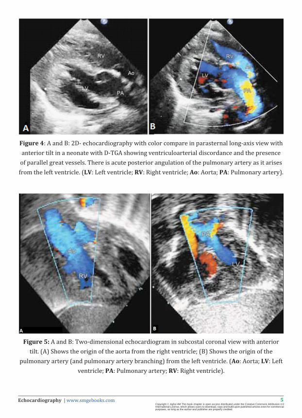
Figure 4: A and B: 2D- echocardiography with color compare in parasternal long-axis view with anterior tilt in a neonate with D-TGA showing ventriculoarterial discordance and the presence
of parallel great vessels. There is acute posterior angulation of the pulmonary artery as it arises from the left ventricle. (LV: Left ventricle; RV: Right ventricle; Ao: Aorta; PA: Pulmonary artery).
Figure 5: A and B: Two-dimensional echocardiogram in subcostal coronal view with anterior tilt. (A) Shows the origin of the aorta from the right ventricle; (B) Shows the origin of the
pulmonary artery (and pulmonary artery branching) from the left ventricle. (Ao: Aorta; LV: Left ventricle; PA: Pulmonary artery; RV: Right ventricle).
6Echocardiography | www.smgebooks.comCopyright Agha HM.This book chapter is open access distributed under the Creative Commons Attribution 4.0 International License, which allows users to download, copy and build upon published articles even for commercial purposes, as long as the author and publisher are properly credited.
Spatial relationship of the semilunar valves
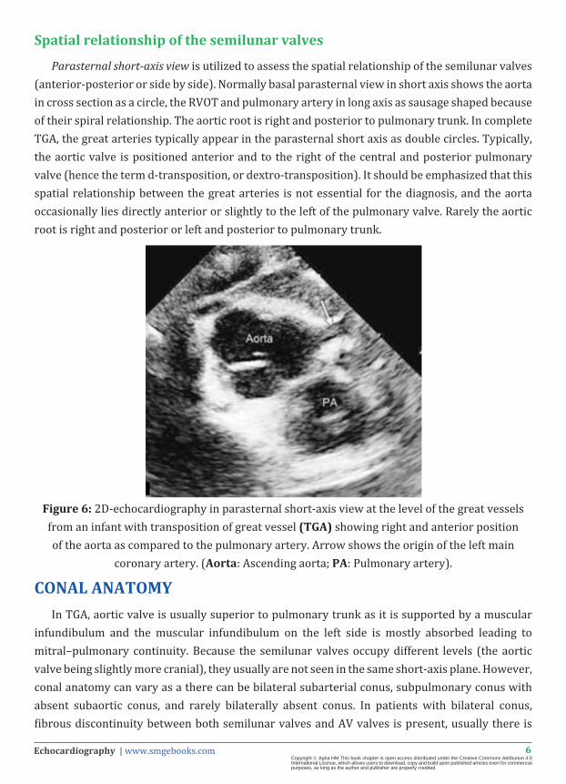
Parasternal short-axis view is utilized to assess the spatial relationship of the semilunar valves (anterior-posterior or side by side). Normally basal parasternal view in short axis shows the aorta in cross section as a circle, the RVOT and pulmonary artery in long axis as sausage shaped because of their spiral relationship. The aortic root is right and posterior to pulmonary trunk. In complete TGA, the great arteries typically appear in the parasternal short axis as double circles. Typically, the aortic valve is positioned anterior and to the right of the central and posterior pulmonary valve (hence the term d-transposition, or dextro-transposition). It should be emphasized that this spatial relationship between the great arteries is not essential for the diagnosis, and the aorta occasionally lies directly anterior or slightly to the left of the pulmonary valve. Rarely the aortic root is right and posterior or left and posterior to pulmonary trunk.
Figure 6: 2D-echocardiography in parasternal short-axis view at the level of the great vessels from an infant with transposition of great vessel (TGA) showing right and anterior position of the aorta as compared to the pulmonary artery. Arrow shows the origin of the left main
coronary artery. (Aorta: Ascending aorta; PA: Pulmonary artery).
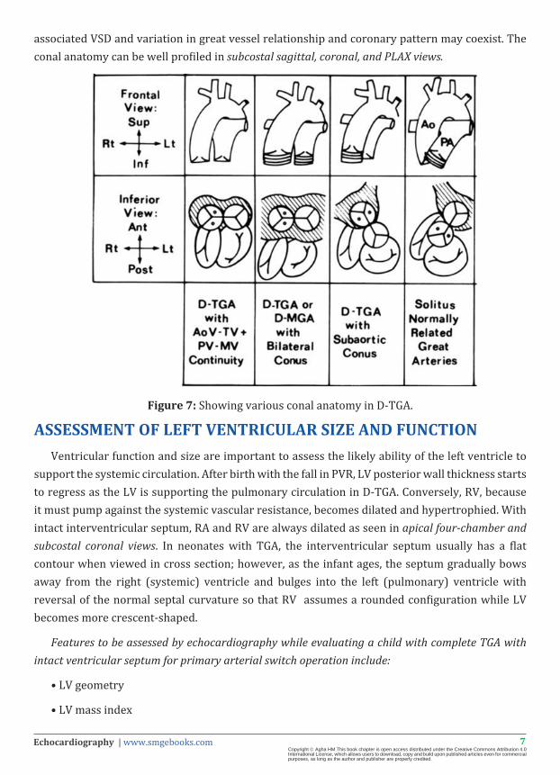
CONAL ANATOMYIn TGA, aortic valve is usually superior to pulmonary trunk as it is supported by a muscular
infundibulum and the muscular infundibulum on the left side is mostly absorbed leading to mitral–pulmonary continuity. Because the semilunar valves occupy different levels (the aortic valve being slightly more cranial), they usually are not seen in the same short-axis plane. However, conal anatomy can vary as a there can be bilateral subarterial conus, subpulmonary conus with absent subaortic conus, and rarely bilaterally absent conus. In patients with bilateral conus, fibrous discontinuity between both semilunar valves and AV valves is present, usually there is
7Echocardiography | www.smgebooks.comCopyright Agha HM.This book chapter is open access distributed under the Creative Commons Attribution 4.0 International License, which allows users to download, copy and build upon published articles even for commercial purposes, as long as the author and publisher are properly credited.
associated VSD and variation in great vessel relationship and coronary pattern may coexist. The conal anatomy can be well profiled in subcostal sagittal, coronal, and PLAX views.
Figure 7: Showing various conal anatomy in D-TGA.
ASSESSMENT OF LEFT VENTRICULAR SIZE AND FUNCTIONVentricular function and size are important to assess the likely ability of the left ventricle to
support the systemic circulation. After birth with the fall in PVR, LV posterior wall thickness starts to regress as the LV is supporting the pulmonary circulation in D-TGA. Conversely, RV, because it must pump against the systemic vascular resistance, becomes dilated and hypertrophied. With intact interventricular septum, RA and RV are always dilated as seen in apical four-chamber and subcostal coronal views. In neonates with TGA, the interventricular septum usually has a flat contour when viewed in cross section; however, as the infant ages, the septum gradually bows away from the right (systemic) ventricle and bulges into the left (pulmonary) ventricle with reversal of the normal septal curvature so that RV assumes a rounded configuration while LV becomes more crescent-shaped.
Features to be assessed by echocardiography while evaluating a child with complete TGA with intact ventricular septum for primary arterial switch operation include:
• LV geometry
• LV mass index
8Echocardiography | www.smgebooks.comCopyright Agha HM.This book chapter is open access distributed under the Creative Commons Attribution 4.0 International License, which allows users to download, copy and build upon published articles even for commercial purposes, as long as the author and publisher are properly credited.
• LV posterior wall thickness index (<0.3 mm taken as regressed)
• LV volume index
• LV mass/volume ratio.
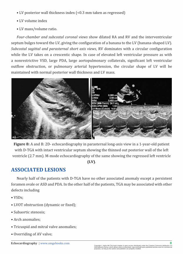
Four-chamber and subcostal coronal views show dilated RA and RV and the interventricular septum bulges toward the LV, giving the configuration of a banana to the LV (banana-shaped LV). Subcostal sagittal and parasternal short axis views, RV dominates with a circular configuration while the LV takes on a crescentic shape. In case of elevated left ventricular pressure as with a nonrestrictive VSD, large PDA, large aortopulmonary collaterals, significant left ventricular outflow obstruction, or pulmonary arterial hypertension, the circular shape of LV will be maintained with normal posterior wall thickness and LV mass.
Figure 8: A and B: 2D- echocardiography in parasternal long-axis view in a 1-year-old patient with D-TGA with intact ventricular septum showing the thinned out posterior wall of the left
ventricle (2.7 mm). M-mode echocardiography of the same showing the regressed left ventricle (LV).
ASSOCIATED LESIONS Nearly half of the patients with D-TGA have no other associated anomaly except a persistent
foramen ovale or ASD and PDA. In the other half of the patients, TGA may be associated with other defects including
• VSDs;
• LVOT obstruction (dynamic or fixed);
• Subaortic stenosis;
• Arch anomalies;
• Tricuspid and mitral valve anomalies;
• Overriding of AV valve;
9Echocardiography | www.smgebooks.comCopyright Agha HM.This book chapter is open access distributed under the Creative Commons Attribution 4.0 International License, which allows users to download, copy and build upon published articles even for commercial purposes, as long as the author and publisher are properly credited.
• Straddling of AV valves;
• Any evidence of a cleft in an AV valve;
• Attachment of tricuspid valve chordae to outlet septum or to the crest of ventricular septum;
• Outlet septum malalignment defects;
• Anomalies of systemic and pulmonary venous connections;
• Anomalies of the coronary arteries;
• Juxtaposed atrial appendages;
The presence of associated defects may influence the choice of palliation or repair.
PRESENCE OR ABSENCE AND SIZE OF SHUNTS The presence of ventriculo-arterial discordance alone will necessarily result in the creation
of two parallel circuits and is incompatible with life. Therefore, admixture of arterial and venous blood is a prerequisite for survival and can occur at any level.
A-At the Atrial Level (ASD)
The most common interatrial communication is a patent foramen ovale or ASD secundum. Good intracardiac mixing occurs if ASD is of adequate size. The atrial profile communication is best visualized from the subcostal four- chamber and sagittal views. The color flow mapping is used to assess the direction of shunt, which is usually bidirectional (left to right during systole, and right to left during diastole). It is very important to assess adequacy of the atrial communication in TGA with intact ventricular septum or small VSD for better mixing, and with large VSD or PDA to decrease left atrial pressure and provide good mixing at the atrial level.
When venous mixing is inadequate, an atrial septostomy is often performed if the arterial switch operation is not planned immediately.
In restrictive interatrial communication, echocardiography shows:
• Dilated LA,
• Bowing of the interatrial septum toward the RA,
• Turbulent flow across the interatrial communication with Doppler color flow mapping.
• Increased velocity across the interatrial communication by the PW Doppler interrogation >1.5cm/sec.
The adequacy of the septostomy under echocardiography is judged by the size of the resulting defect which is typically large and unrestrictive, with a mobile flap of the torn fossa ovalis waving to and fro across the defect.
10Echocardiography | www.smgebooks.comCopyright Agha HM.This book chapter is open access distributed under the Creative Commons Attribution 4.0 International License, which allows users to download, copy and build upon published articles even for commercial purposes, as long as the author and publisher are properly credited.
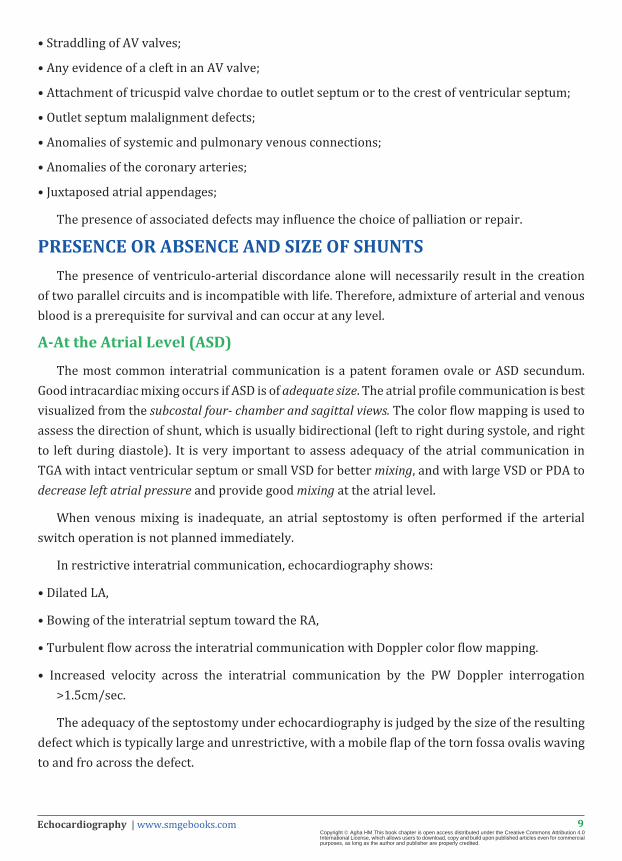
Figure 9: 2D- echocardiography in subcostal view in a case of D-TGA with color flow mapping showing a restricted patent foramen ovale (PFO; arrow). (LA: Left atrium; RA: Right atrium).
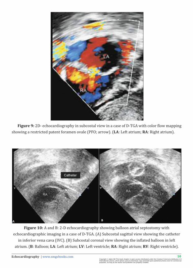
Figure 10: A and B: 2-D echocardiography showing balloon atrial septostomy with echocardiographic imaging in a case of D-TGA. (A) Subcostal sagittal view showing the catheter
in inferior vena cava (IVC). (B) Subcostal coronal view showing the inflated balloon in left atrium. (B: Balloon; LA: Left atrium; LV: Left ventricle; RA: Right atrium; RV: Right ventricle).
11Echocardiography | www.smgebooks.comCopyright Agha HM.This book chapter is open access distributed under the Creative Commons Attribution 4.0 International License, which allows users to download, copy and build upon published articles even for commercial purposes, as long as the author and publisher are properly credited.
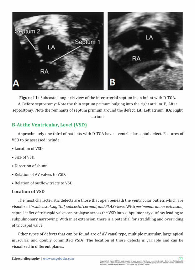
Figure 11: Subcostal long-axis view of the interarterial septum in an infant with D-TGA. A, Before septostomy: Note the thin septum primum bulging into the right atrium. B, After
septostomy: Note the remnants of septum primum around the defect. LA: Left atrium; RA: Right atrium
B-At the Ventricular, Level (VSD)
Approximately one third of patients with D-TGA have a ventricular septal defect. Features of VSD to be assessed include:
• Location of VSD.
• Size of VSD.
• Direction of shunt.
• Relation of AV valves to VSD.
• Relation of outflow tracts to VSD.
Location of VSD
The most characteristic defects are those that open beneath the ventricular outlets which are visualized in subcostal sagittal, subcostal coronal, and PLAX views. With perimembranous extension, septal leaflet of tricuspid valve can prolapse across the VSD into subpulmonary outflow leading to subpulmonary narrowing. With inlet extension, there is a potential for straddling and overriding of tricuspid valve.
Other types of defects that can be found are of AV canal type, multiple muscular, large apical muscular, and doubly committed VSDs. The location of these defects is variable and can be visualized in different planes.
12Echocardiography | www.smgebooks.comCopyright Agha HM.This book chapter is open access distributed under the Creative Commons Attribution 4.0 International License, which allows users to download, copy and build upon published articles even for commercial purposes, as long as the author and publisher are properly credited.
- Perimembranous is seen in the subcostal and parasternal short-axis views.
- Doubly committed ventricular septal defect is seen in subcostal coronal and sagittal views, and PLAX and PSAX view.
- AV canal defect is seen in the apical four-chamber, subcostal and parasternal short-axis views.
- Anterior muscular defect is seen in parasternal short-axis views.
- Posterior and apical muscular defect is seen in the apical four-chamber, short-axis subcostal and short-axis parasternal views.
Size of VSD
The VSD can be small or large. With restrictive VSD, a turbulent jet from right to LV will be found. CWD interrogation shows a pressure gradient across the VSD as well as the direction of shunt.
Number
The VSD can be single, or multiple
Direction of shunt
Color flow mapping helps in defining the direction of shunt. The shunt through VSD is bidirectional; left to right during diastole and right to left during systole.
Relation of AV valves to VSD
Four-chamber (subcostal coronal and apical) views are also useful to profile overriding and straddling of AV valves valve if present. In addition, a modified subcostal sagittal view at the level of AV valves (en face view) can also define the presence of overriding or straddling. Mitral valve straddling can also be profiled from PLAX view as the mitral valve chordae cross the VSD and insert on the opposite side of septum.
In the presence of straddling of AV valve, biventricular repair will not be possible and the patient would need to undergo univentricular repair.
Relation of outflow tracts to VSD
Anterior and posterior malalignment of the outlet septum with overriding of one of the great vessels, most commonly the PA (posterior great vessel) may occur and can be profiled from subcostal sagittal view, subcostal coronal and sagittal views, and PLAX view.
Posterior malalignment of the outlet septum can cause pulmonary stenosis. Because of the posterior deviation of outlet septum, there is a direct route from LV to aorta making these children good candidates for intraventricular re-routing from LV to aorta. If the VSD is smaller in relation to aortic valve annulus, then it needs enlargement during intraventricular repair.
13Echocardiography | www.smgebooks.comCopyright Agha HM.This book chapter is open access distributed under the Creative Commons Attribution 4.0 International License, which allows users to download, copy and build upon published articles even for commercial purposes, as long as the author and publisher are properly credited.
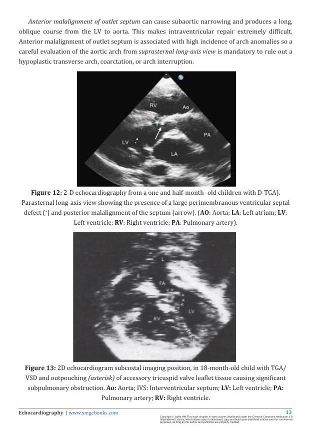
Anterior malalignment of outlet septum can cause subaortic narrowing and produces a long, oblique course from the LV to aorta. This makes intraventricular repair extremely difficult. Anterior malalignment of outlet septum is associated with high incidence of arch anomalies so a careful evaluation of the aortic arch from suprasternal long-axis view is mandatory to rule out a hypoplastic transverse arch, coarctation, or arch interruption.
Figure 12: 2-D echocardiography from a one and half-month -old children with D-TGA). Parasternal long-axis view showing the presence of a large perimembranous ventricular septal defect (∗) and posterior malalignment of the septum (arrow). (AO: Aorta; LA: Left atrium; LV:
Left ventricle; RV: Right ventricle; PA: Pulmonary artery).
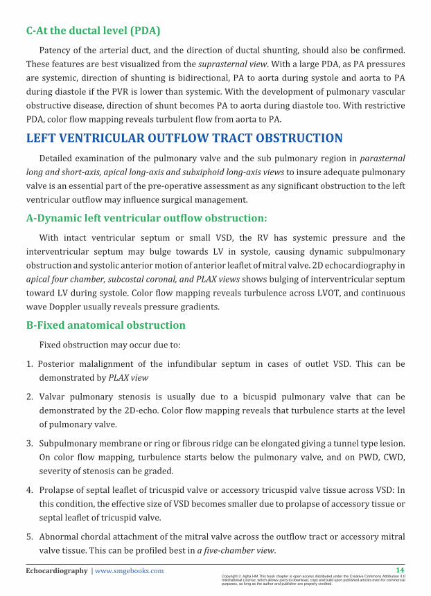
Figure 13: 2D echocardiogram subcostal imaging position, in 18-month-old child with TGA/VSD and outpouching (asterisk) of accessory tricuspid valve leaflet tissue causing significant subpulmonary obstruction. Ao: Aorta; IVS: Interventricular septum; LV: Left ventricle; PA:
Pulmonary artery; RV: Right ventricle.
14Echocardiography | www.smgebooks.comCopyright Agha HM.This book chapter is open access distributed under the Creative Commons Attribution 4.0 International License, which allows users to download, copy and build upon published articles even for commercial purposes, as long as the author and publisher are properly credited.
C-At the ductal level (PDA)
Patency of the arterial duct, and the direction of ductal shunting, should also be confirmed. These features are best visualized from the suprasternal view. With a large PDA, as PA pressures are systemic, direction of shunting is bidirectional, PA to aorta during systole and aorta to PA during diastole if the PVR is lower than systemic. With the development of pulmonary vascular obstructive disease, direction of shunt becomes PA to aorta during diastole too. With restrictive PDA, color flow mapping reveals turbulent flow from aorta to PA.
LEFT VENTRICULAR OUTFLOW TRACT OBSTRUCTIONDetailed examination of the pulmonary valve and the sub pulmonary region in parasternal
long and short-axis, apical long-axis and subxiphoid long-axis views to insure adequate pulmonary valve is an essential part of the pre-operative assessment as any significant obstruction to the left ventricular outflow may influence surgical management.
A-Dynamic left ventricular outflow obstruction:
With intact ventricular septum or small VSD, the RV has systemic pressure and the interventricular septum may bulge towards LV in systole, causing dynamic subpulmonary obstruction and systolic anterior motion of anterior leaflet of mitral valve. 2D echocardiography in apical four chamber, subcostal coronal, and PLAX views shows bulging of interventricular septum toward LV during systole. Color flow mapping reveals turbulence across LVOT, and continuous wave Doppler usually reveals pressure gradients.
B-Fixed anatomical obstruction
Fixed obstruction may occur due to:
1. Posterior malalignment of the infundibular septum in cases of outlet VSD. This can be demonstrated by PLAX view
2. Valvar pulmonary stenosis is usually due to a bicuspid pulmonary valve that can be demonstrated by the 2D-echo. Color flow mapping reveals that turbulence starts at the level of pulmonary valve.
3. Subpulmonary membrane or ring or fibrous ridge can be elongated giving a tunnel type lesion. On color flow mapping, turbulence starts below the pulmonary valve, and on PWD, CWD, severity of stenosis can be graded.
4. Prolapse of septal leaflet of tricuspid valve or accessory tricuspid valve tissue across VSD: In this condition, the effective size of VSD becomes smaller due to prolapse of accessory tissue or septal leaflet of tricuspid valve.
5. Abnormal chordal attachment of the mitral valve across the outflow tract or accessory mitral valve tissue. This can be profiled best in a five-chamber view.
15Echocardiography | www.smgebooks.comCopyright Agha HM.This book chapter is open access distributed under the Creative Commons Attribution 4.0 International License, which allows users to download, copy and build upon published articles even for commercial purposes, as long as the author and publisher are properly credited.
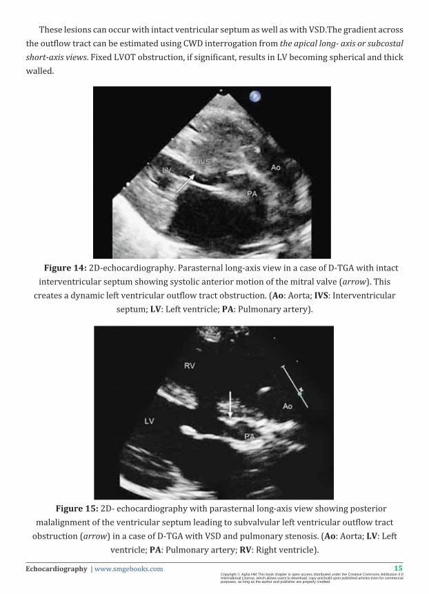
These lesions can occur with intact ventricular septum as well as with VSD.The gradient across the outflow tract can be estimated using CWD interrogation from the apical long- axis or subcostal short-axis views. Fixed LVOT obstruction, if significant, results in LV becoming spherical and thick walled.
Figure 14: 2D-echocardiography. Parasternal long-axis view in a case of D-TGA with intact interventricular septum showing systolic anterior motion of the mitral valve (arrow). This
creates a dynamic left ventricular outflow tract obstruction. (Ao: Aorta; IVS: Interventricular septum; LV: Left ventricle; PA: Pulmonary artery).
Figure 15: 2D- echocardiography with parasternal long-axis view showing posterior malalignment of the ventricular septum leading to subvalvular left ventricular outflow tract
obstruction (arrow) in a case of D-TGA with VSD and pulmonary stenosis. (Ao: Aorta; LV: Left ventricle; PA: Pulmonary artery; RV: Right ventricle).
16Echocardiography | www.smgebooks.comCopyright Agha HM.This book chapter is open access distributed under the Creative Commons Attribution 4.0 International License, which allows users to download, copy and build upon published articles even for commercial purposes, as long as the author and publisher are properly credited.
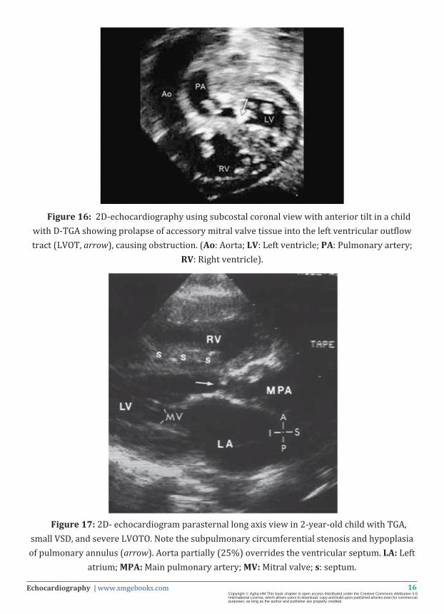
Figure 16: 2D-echocardiography using subcostal coronal view with anterior tilt in a child with D-TGA showing prolapse of accessory mitral valve tissue into the left ventricular outflow tract (LVOT, arrow), causing obstruction. (Ao: Aorta; LV: Left ventricle; PA: Pulmonary artery;
RV: Right ventricle).
Figure 17: 2D- echocardiogram parasternal long axis view in 2-year-old child with TGA, small VSD, and severe LVOTO. Note the subpulmonary circumferential stenosis and hypoplasia of pulmonary annulus (arrow). Aorta partially (25%) overrides the ventricular septum. LA: Left
atrium; MPA: Main pulmonary artery; MV: Mitral valve; s: septum.
17Echocardiography | www.smgebooks.comCopyright Agha HM.This book chapter is open access distributed under the Creative Commons Attribution 4.0 International License, which allows users to download, copy and build upon published articles even for commercial purposes, as long as the author and publisher are properly credited.
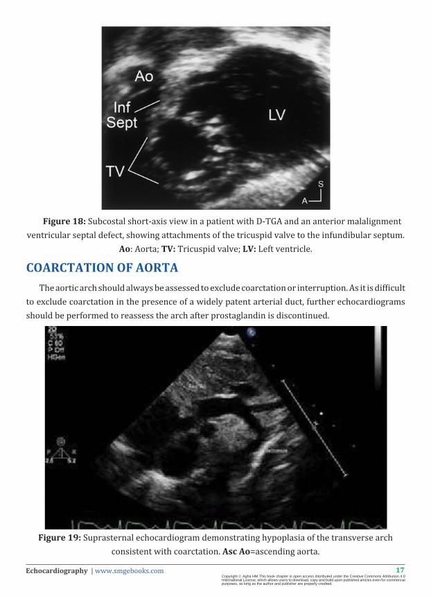
Figure 18: Subcostal short-axis view in a patient with D-TGA and an anterior malalignment ventricular septal defect, showing attachments of the tricuspid valve to the infundibular septum.
Ao: Aorta; TV: Tricuspid valve; LV: Left ventricle.
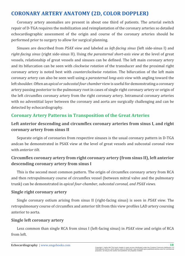
COARCTATION OF AORTAThe aortic arch should always be assessed to exclude coarctation or interruption. As it is difficult
to exclude coarctation in the presence of a widely patent arterial duct, further echocardiograms should be performed to reassess the arch after prostaglandin is discontinued.
Figure 19: Suprasternal echocardiogram demonstrating hypoplasia of the transverse arch consistent with coarctation. Asc Ao=ascending aorta.
18Echocardiography | www.smgebooks.comCopyright Agha HM.This book chapter is open access distributed under the Creative Commons Attribution 4.0 International License, which allows users to download, copy and build upon published articles even for commercial purposes, as long as the author and publisher are properly credited.
CORONARY ARTERY ANATOMY (2D, COLOR DOPPLER)Coronary artery anomalies are present in about one third of patients. The arterial switch
repair of D-TGA requires the mobilization and reimplantation of the coronary arteries so detailed echocardiographic assessment of the origin and course of the coronary arteries should be performed prior to surgery to allow for surgical planning.
Sinuses are described from PSAX view and labeled as left-facing sinus (left side-sinus I) and right-facing sinus (right side-sinus II). Using the parasternal short-axis view at the level of great vessels, relationship of great vessels and sinuses can be defined. The left main coronary artery and its bifurcation can be seen with clockwise rotation of the transducer and the proximal right coronary artery is noted best with counterclockwise rotation. The bifurcation of the left main coronary artery can also be seen well using a parasternal long-axis view with angling toward the left shoulder. Often an apical or subcostal four chamber view is useful for demonstrating a coronary artery passing posterior to the pulmonary root in cases of single right coronary artery or origin of the left circumflex coronary artery from the right coronary artery. Intramural coronary arteries with no adventitial layer between the coronary and aorta are surgically challenging and can be detected by echocardiography.
Coronary Artery Patterns in Transposition of the Great Arteries
Left anterior descending and circumflex coronary arteries from sinus I, and right coronary artery from sinus II
Separate origin of coronaries from respective sinuses is the usual coronary pattern in D-TGA andcan be demonstrated in PSAX view at the level of great vessels and subcostal coronal view with anterior tilt.
Circumflex coronary artery from right coronary artery (from sinus II), left anterior descending coronary artery from sinus I
This is the second most common pattern. The origin of circumflex coronary artery from RCA and then retropulmonary course of circumflex vessel (between mitral valve and the pulmonary trunk) can be demonstrated in apical four-chamber, subcostal coronal, and PSAX views.
Single right coronary artery
Single coronary ostium arising from sinus II (right-facing sinus) is seen in PSAX view. The retropulmonary course of circumflex and anterior tilt from this view profiles LAD artery coursing anterior to aorta.
Single left coronary artery
Less common than single RCA from sinus I (left-facing sinus) in PSAX view and origin of RCA from left.
19Echocardiography | www.smgebooks.comCopyright Agha HM.This book chapter is open access distributed under the Creative Commons Attribution 4.0 International License, which allows users to download, copy and build upon published articles even for commercial purposes, as long as the author and publisher are properly credited.
Inverted origin of coronaries
Demonstration of separate origin of coronaries in PSAX view. Imaging the retropulmonary course of LCA arising from right-facing sinus (sinus II) and its bifurcation into LAD and circumflex branches. This can be profiled in PSAX, apical four-chamber, and subcostal coronal views.
Intramural coronary artery
An intramural coronary is a potentially significant risk factor for transfer of the coronaries as part of the arterial switch operation for transposition of great arteries. Preoperative diagnosis is advantageous because it helps to prevent accidental injury to the intramural coronary artery during transection of the aortic root and excision of the coronary artery ostium from the aorta.
• Origin of coronary artery from contralateral sinus, as in case of left main or LAD artery origin from right-facing sinus or RCA from left-facing sinus profiled in PSAX view. Their orifices are placed adjacent to the commissure (usually intercoronary).
• Identification of major coronary artery coursing between the two arterial roots in PSAX, apical four-chamber, and subcostal coronal views with posterior to anterior tilt. After origin, the proximal portion of coronary artery runs parallel to the aortic wall and perpendicular to the intercoronary commissure, giving a “double border appearance” to the posterior aortic wall.
In approximately 10% of cases, the orifice of a coronary artery will arise close to one of the valve commissures that can be defined in PSAX view at the level of great vessels. With coronary ostia near the commissure, chances of neopulmonary regurgitation are higher after arterial switch operation. Less commonly, ostia can also arise above the commissures in the tubular portion of the aorta (higher origin of coronary).
Pattern Origin of right and left coronaries Origin of circumflex Retropulmonary course
Usual Separate origin from respective sinuses) LCA Absent
Circumflex Coronary from Right Coronary Separate origin from respective sinuses RCA Present (circumflex)
Single right coronary Single ostium from sinus II LCA Present (LCA or circumflex)
Single left coronary Single ostium from sinus I LCARCA (rare) Rare (circumflex)
Inverted Origin of Coronaries Separate origin from contralateral sinus LCASeparate from sinus II Present (LCA or circumflex)
20Echocardiography | www.smgebooks.comCopyright Agha HM.This book chapter is open access distributed under the Creative Commons Attribution 4.0 International License, which allows users to download, copy and build upon published articles even for commercial purposes, as long as the author and publisher are properly credited.
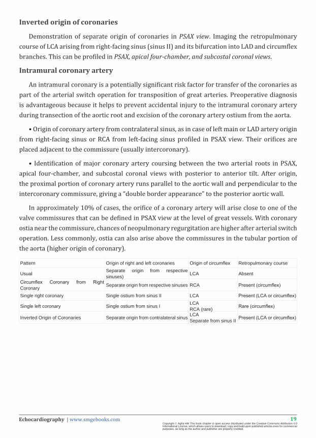
Figure 20: Diagram of the coronary artery anatomy in D-TGA. The orientation of the figures is that of a parasternal short-axis echocardiographic view. LAD: Left anterior descending artery;
LCCA: Left circumflex coronary artery; RCA: Right coronary artery.
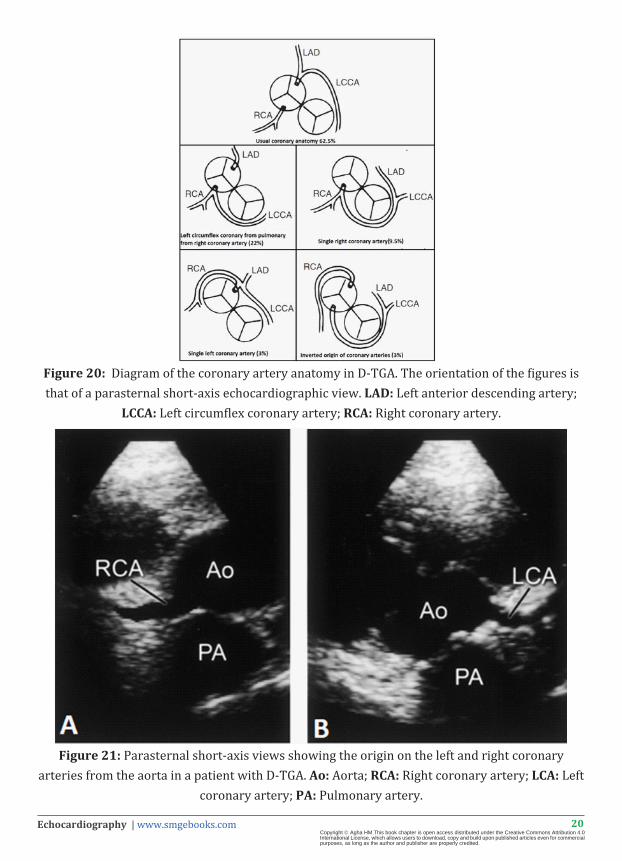
Figure 21: Parasternal short-axis views showing the origin on the left and right coronary arteries from the aorta in a patient with D-TGA. Ao: Aorta; RCA: Right coronary artery; LCA: Left
coronary artery; PA: Pulmonary artery.
21Echocardiography | www.smgebooks.comCopyright Agha HM.This book chapter is open access distributed under the Creative Commons Attribution 4.0 International License, which allows users to download, copy and build upon published articles even for commercial purposes, as long as the author and publisher are properly credited.
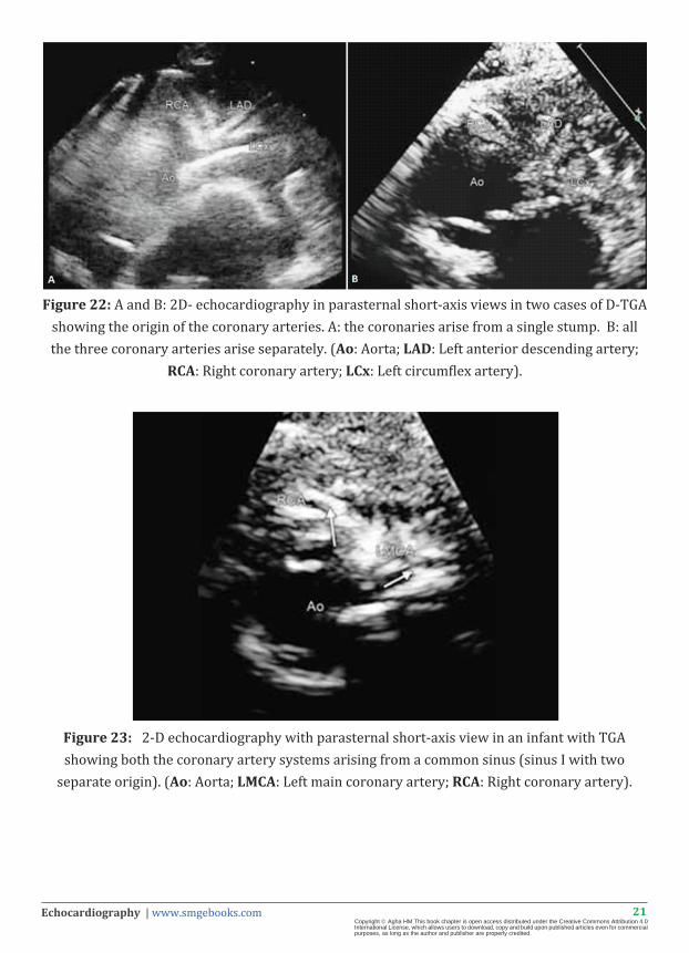
Figure 22: A and B: 2D- echocardiography in parasternal short-axis views in two cases of D-TGA showing the origin of the coronary arteries. A: the coronaries arise from a single stump. B: all the three coronary arteries arise separately. (Ao: Aorta; LAD: Left anterior descending artery;
RCA: Right coronary artery; LCx: Left circumflex artery).
Figure 23: 2-D echocardiography with parasternal short-axis view in an infant with TGA showing both the coronary artery systems arising from a common sinus (sinus I with two
separate origin). (Ao: Aorta; LMCA: Left main coronary artery; RCA: Right coronary artery).
22Echocardiography | www.smgebooks.comCopyright Agha HM.This book chapter is open access distributed under the Creative Commons Attribution 4.0 International License, which allows users to download, copy and build upon published articles even for commercial purposes, as long as the author and publisher are properly credited.
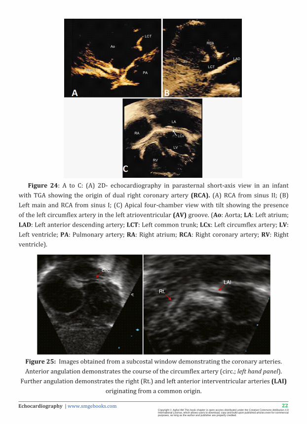
Figure 24: A to C: (A) 2D- echocardiography in parasternal short-axis view in an infant with TGA showing the origin of dual right coronary artery (RCA). (A) RCA from sinus II; (B) Left main and RCA from sinus I; (C) Apical four-chamber view with tilt showing the presence of the left circumflex artery in the left atrioventricular (AV) groove. (Ao: Aorta; LA: Left atrium; LAD: Left anterior descending artery; LCT: Left common trunk; LCx: Left circumflex artery; LV: Left ventricle; PA: Pulmonary artery; RA: Right atrium; RCA: Right coronary artery; RV: Right ventricle).
Figure 25: Images obtained from a subcostal window demonstrating the coronary arteries. Anterior angulation demonstrates the course of the circumflex artery (circ.; left hand panel).
Further angulation demonstrates the right (Rt.) and left anterior interventricular arteries (LAI) originating from a common origin.
23Echocardiography | www.smgebooks.comCopyright Agha HM.This book chapter is open access distributed under the Creative Commons Attribution 4.0 International License, which allows users to download, copy and build upon published articles even for commercial purposes, as long as the author and publisher are properly credited.
References1. Awasthy N, Shrivastava S. M-Mode and Two-Dimensional Echocardiography in Congenital Heart Disease. In: Navin Nanda.
Comprehensive Textbook of Echocardiography New Delhi: Jaypee Brothers Medical Publishers. 2014; 1526-1790.
2. Otto C. The Adult with Congenital Heart Disease. In: Otto Textbook of clinical Echocardiography, 5th ed. Philadelphia: Saunders Elsevier. 2013; 443-474.
3. Armstrong W and Ryan T. Congenital Heart Diseases. In: Feigenbaum’s Echocardiography, 7th Edition, Philadelphia: Lippincott Williams & Wilkins. 2010; 560 -634.
4. Wernovsky G. Transposition of the Great Arteries. In: Moss & Adams heart disease in infants, children, and adolescents: including the fetus and young adult, 8th ed. Philadelphia: Lippincott Williams & Wilkins. 2013; 1097-1147.
5. Schultz A. Transposition of the Great Arteries. In: Echocardiography in Congenital Heart Disease. Philadelphia: Saunders Elsevier. 2012; 104-123.
6. Mertens L, Vogt M, Marek J and Cohen M. Transposition of the Great Arteries. In: Echocardiography in Pediatric and Congenital Heart Disease: From Fetus to Adult. Oxford: Blackwell Publishing Ltd. 2009; 398-438.
7. Rao S, Garekar S: D-transposition of Great Arteries. In: A Comprehensive Approach to Congenital Heart Diseases. New Delhi: Jaypee Brothers Medical Publishers (P) Ltd. 2013; 619-623.
8. Komarlu R, Morell V, Kreutzer J, and Dextro R. Transposition of the Great Arteries Critical Care of Children with Heart Disease: Basic Medical and Surgical Concepts. New York: Springer-Verlag London Ltd. 2010; 359-375.
9. Karl T and Kirshbom P. Transposition of the Great Arteries and the Arterial Switch Operation. In : Critical Heart Disease in Infants and Children, 2nd ed. Philadelphia, Mosby Elsevier. 2006.
10. Salih C, Brizard C, Penny D, Anderson R. Transposition In: Paediatric cardiology 3rd edition. 2010; 795-819.
11. Schultz A and Soriano B. D-Transposition of the great arteries In: Echocardiography in Pediatric And Congenital Heart Disease. 2013.
12. Cohen MS, Eidem BW, Cetta F, Fogel MA, Frommelt PC. Multimodality Imaging Guidelines of Patients with Transposition of the Great Arteries: A Report from the American Society of Echocardiography Developed in Collaboration with the Society for Cardiovascular Magnetic Resonance and the Society of Cardiovascular Computed Tomography. J Am Soc Echocardiogr. 2016; 29: 571-621.
13. Brown LM, Duffy CE, Mitchell C, Young L. A practical guide to pediatric coronary artery imaging with echocardiography. J Am Soc Echocardiogr. 2015; 28: 379-391.
14. Villafañe J, Lantin-Hermos M. D -Transposition of the Great Arteries. Journal of the American College of Cardiology. 2014; 64: 499-511.
15. Lowry AW, Olabiyi OO, Adachi I, Moodie DS, Knudson JD. Coronary artery anatomy in congenital heart disease. Congenit Heart Dis. 2013; 8: 187-202.
16. Thadani SR, Foster E. Echocardiographic evaluation in transposition of the great arteries in the adult. Echocardiography. 2015; 32: S157-165.
17. Mahle WT, Gonzalez JH, Kreeger J, Marx G, Duldani G. Echocardiography of transposition of the great arteries. Cardiol Young. 2012; 22: 664-670.
18. Sithamparanathan S, Padley SP, Rubens MB, Gatzoulis MA, Ho SY. Great vessel and coronary artery anatomy in transposition and other coronary anomalies: a universal descriptive and alphanumerical sequential classification. JACC Cardiovasc Imaging. 2013; 6: 624-630.
19. Files MD, Arya B. Preoperative Physiology, Imaging, and Management of Transposition of the Great Arteries. Semin Cardiothorac Vasc Anesth. 2015; 19: 210-222.