transmitter-phenotypes of commissural interneurons …ffffffff-b516-1b08-ffff-ffffa3dcd198/... ·...
TRANSCRIPT

Transmitter-Phenotypes of Commissural Interneurons inthe Lumbar Spinal Cord of Newborn Mice
CARLOS ERNESTO RESTREPO,1 LINE LUNDFALD,1 GABOR SZABO,2 FERENC ERDELYI,2HANNS ULRICH ZEILHOFER,3 JOEL C. GLOVER,4 AND OLE KIEHN1*1Mammalian Locomotor Laboratory, Department of Neuroscience, Karolinska Institutet, 17177 Stockholm, Sweden2Gene Technology and Developmental Neuroscience, Institute of Experimental Medicine, H-1450, Budapest, Hungary3Institute of Pharmacology and Toxicology, University of Zurich, Zurich, Switzerland4Department of Physiology, University of Oslo, 0317 Oslo, Norway
ABSTRACTCommissural interneurons (CINs) are a necessary compo-nent of central pattern generators (CPGs) for locomotionbecause they mediate the coordination of left and rightmuscle activity. The projection patterns and relative loca-tions of different classes of CINs in the ventromedial part ofthe rodent lumbar cord have been described (Eide et al.[1999] J Comp Neurol 403:332–345; Stokke et al. [2002]J Comp Neurol 446:349–359; Nissen et al. [2005] J CompNeurol 483:30–47). However, the distribution and relativeprevalence of different CIN neurotransmitter phenotypes inthe ventral region of the mammalian spinal cord where thelocomotor CPG is localized is unknown. In this study wedescribe the relative proportions and anatomical locationsof putative inhibitory and excitatory CINs in the lumbar spi-nal cord of newborn mice. To directly visualize potentialneurotransmitter phenotypes we combined retrograde la-
beling of CINs with in situ hybridization against the glycinetransporter, GlyT2, or the vesicular glutamate transporter,vGluT2, in wildtype mice and in transgenic mice expressingeGFP driven by the promoters of glutamic acid decarboxyl-ase (GAD) 65, GAD67, or GlyT2. Our study shows that puta-tive glycinergic, GABAergic, and glutamatergic CINs areexpressed in almost equal numbers, with a small proportionof CINs coexpressing GlyT2 and GAD67::eGFP, indicating aputative combined glycinergic/GABAergic phenotype.These different CIN phenotypes were intermingled in lami-nas VII and VIII. Our results suggest that glycinergic,GABAergic, and glutamatergic CINs are the principal CINphenotypes in the CPG region of the lumbar spinal cord inthe newborn mouse. We compare these results to descrip-tions of CIN neurotransmitter phenotypes in other verte-brate species. J. Comp. Neurol. 517:177–192, 2009.© 2009 Wiley-Liss, Inc.
Indexing terms: locomotion; CPG; commissural interneurons; spinal cord; motor neurons;glutamate; GABA; glycine
The timing and coordination of rhythmic movements suchas swimming and walking in vertebrates are in large partproduced by neural networks localized in the spinal cord.These networks are called central pattern generators (CPGs)because they can generate functional rhythmic motor activityindependently of sensory input. A critical function of CPGs isto coordinate muscular activity on the left and right sides ofthe body. This CPG function is mediated by activity in com-missural interneurons (CINs), whose axons cross the midlineto synapse on neurons on the opposite side. Initial studies ofthe functional organization of CINs in vertebrate CPGs werecarried out in aquatic vertebrates (Buchanan, 1982, 1999;Buchanan and Cohen, 1982; Clarke and Roberts, 1984; Dale,1985; Buchanan and Grillner, 1987; Fetcho, 1990; Soffe et al.,2001; Grillner, 2003). These studies unveiled inhibitory glycin-ergic descending CINs (dCINs) as the CPG neurons mediatingleft–right alternation during swimming. The dCINs serve thisrole by projecting to and inhibiting contralateral motor neu-rons and contralateral CPG interneurons. Glutamatergic CINs
have also been identified in the lamprey and zebrafish (Hi-gashijima et al., 2004a,b; Biro et al., 2008). In the zebrafishthese crossed excitatory CINs are also thought to play a role
Additional Supporting Information may be found in the online version ofthis article.
Grant sponsor: National Institutes of Health (NIH); Grant number:5R01NS40795-09 (to O.K.); Grant sponsor: Swedish Research Council (toO.K.); Grant sponsor: Wings for Life (to O.K.); Grant sponsor: HedlundFoundation (to O.K.); Grant sponsor: Soderberg Foundation (to O.K.); Grantsponsor: the HFSP (to J.C.G., O.K.); Grant sponsor: Norwegian ResearchCouncil (to J.C.G.).
*Correspondence to: Professor Ole Kiehn, Department of Neuroscience,Karolinska Instiutet, Retzius vag 8, 17177 Stockholm, Sweden.E-mail: [email protected]
Received 16 January 2009; Revised 20 March 2009; Accepted 4 July2009
DOI 10.1002/cne.22144Published online July 16, 2009 in Wiley InterScience (www.interscience.wiley.
com).
The Journal of Comparative Neurology 517:177–192 (2009)
Research in Systems Neuroscience
© 2009 Wiley-Liss, Inc.

in mediating slow swimming (McLean et al., 2007). Studies oflocomotor-related CIN projections in mammals have de-scribed the presence of both direct excitatory and directinhibitory CIN connections to contralateral motor neurons(Butt and Kiehn, 2003; Jankowska et al., 2003, 2005a,b, 2006;Quinlan and Kiehn, 2007) as well as polysynaptic inhibitoryand excitatory CIN pathways onto motor neurons (Butt andKiehn, 2003; Jankowska et al., 2005b, 2006; Nishimaru et al.,2006; Quinlan and Kiehn, 2007). The synaptic nature of theseCIN connections has been shown electrophysiologically inrats and mice to be either glutamatergic, glycinergic, or com-bined glycinergic/GABAergic, whereas the polysynaptic inhib-itory connections have been shown to be mediated by gluta-matergic CINs synapsing onto combined glycinergic/GABAergic interneurons (Butt and Kiehn, 2003; Quinlan andKiehn, 2007). Likewise, anatomical studies in the cat and rathave shown that CIN axon terminals in close proximity tomotor neurons as well as in the intermediate area of theventral spinal cord are putatively glutamatergic, glycinergic, orGABAergic (Bannatyne et al., 2003; Weber et al., 2007). Thiscomplex system of CINs appears to be involved in segmentalleft–right alternation and in segmental and intersegmentalcrossed motor synergies during locomotion in tetrapods(Kiehn, 2006). No description has yet been made, however, ofthe distribution and relative prevalence of different CIN neu-rotransmitter phenotypes of the entire population of CINs inthe ventral spinal cord (the intermediate area and lamina VIII)where the locomotor CPG is localized (Kiehn and Kjaerulff,1998).
Here we visualize putative CIN neurotransmitter phenotypesby combining retrograde labeling of the entire CIN populationin lumbar segments of the ventral spinal cord of newborn micewith in situ hybridization and neurotransmitter-specific GFPexpression in transgenic mice. Our results show that putativeglycinergic, GABAergic, and glutamatergic CINs are ex-pressed in almost equal numbers, with a small proportion ofCINs potentially having a combined glycinergic/GABAergicphenotype. These different CIN phenotypes were found inoverlapping regions within the ventral spinal cord, with puta-tive GABAergic CINs occupying a somewhat more medialposition. We thus provide anatomical information that is es-sential to understand the complex functions of CINs in themouse spinal cord and which allows a comparison with theorganization of CINs in other vertebrate CPGs.
MATERIALS AND METHODSAnimals
All experimental procedures were approved by the SwedishAnimal Welfare Agency. Wildtype mice (C57BL/6) were usedto assess potential glutamatergic and glycinergic phenotypesof CINs using immunohistochemistry or in situ hybridizationagainst mRNA. Putative glycinergic CINs were also visualizedin GlyT2::eGFP mice that express eGFP under the control ofthe promoter for the neuronal glycine transporter 2 gene(GlyT2) (Zeilhofer et al., 2005). eGFP was placed in frame atthe beginning of exon 2 in the Glyt2 gene in a 179,508 bp-longBAC-clone that contained the entire GlyT2 gene and addi-tional 105 kb and 21 kb of DNA flanking the gene upstreamand downstream, respectively (Zeilhofer et al., 2005). PutativeGABAergic CINs were visualized in glutamic acid decarbox-
ylase (GAD) 67::eGFP knockin mice (Tamamaki et al., 2003)and/or in transgenic mice expressing eGFP under the controlof the GAD65 promoter. In the GAD67::eGFP knockin mouse,cDNA encoding eGFP was targeted to exon 1 encodingGAD67. The transgene in the GAD65 mouse includes a 6.3-kbsegment of the GAD65 gene covering the first two exons, theintron in between, and a 5.5-kb upstream region of the gene.eGFP without its own translation site was fused in-frame withexon 2 (Lopez-Bendito et al., 2004; Hughes et al., 2005).
The present study shows that eGFP reliably labels putativeglycinergic neurons in the spinal cord of the Glyt2::eGFP mouse(see Results). The eGFP expression in the GAD67::eGFP mousehas previously been thoroughly described and verified in brain(Tamamaki et al., 2003) and spinal cord tissue (Huang et al.,2008a,b). These studies showed using double immunohisto-chemistry or combined methods of immunohistochemistry andin situ hybridization that the vast majority (80–96%) of GFP-positive neurons in the GAD67::eGFP mouse are either GAD67mRNA-positive or GABA-immunoreactive. None of these studiesreported ectopic expression. Furthermore, in a previous studyknown groups of excitatory interneurons in the spinal cord werenot GFP-positive in the GAD67::eGFP mouse (Lundfald et al.,2007). The GAD65::eGFP transgenic mouse line has been shownto label many GAD65-positive terminals in the gray matter of thespinal cord, although some synaptic terminals express GAD65immunoreactivity without GFP expression (Hughes et al., 2005).Moreover, the distribution of the GAD65-GFP in the spinal cordwas observed to be identical to that seen in nontransgenicanimals with some apparent ectopic expression in fibers in thedorsal column white matter.
DissectionSpinal cords were prepared from wildtype and heterozy-
gous eGFP-expressing mice aged 0–2 days (P0–P2, where P0is day of birth). The animals were decapitated under deepisoflurane anesthesia after which they were eviscerated asdescribed previously (Kiehn and Kjaerulff, 1996). The thoraco-lumbar spinal cord (Th11-L6) was then surgically isolated andtransferred to a Petri dish filled with low Ca2! Ringer’s solu-tion (in mM: 111.14 NaCl, 3.09 KCl, 25 NaHCO3, 1.10 KH2PO4,3.73 MgSO4, 0.25 CaCl2, and 11.10 glucose aerated with 5%CO2 in O2 to maintain pH at 7.4).
Dextran labeling and fixationAfter dissection, CINs were retrogradely labeled by applying
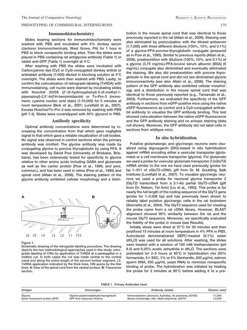
crystals of a fluorescent dextran amine tracer, Texas Reddextran amine (TxRDA, 3,000 kDa) in the ventral commissureof the L2 segment as previously described (Glover, 1995;Stokke et al., 2002; Nissen et al., 2005) (Fig. 1). Spinal cordsthat were accidentally labeled dorsal to the central canal wereexcluded from further analysis.
Preparations were then incubated for 5–7 hours in the darkin oxygenated normal Ringer’s solution (with normal calciumand magnesium concentrations: 2.5 mM CaCl2 and 1.43MgSO4) at room temperature.
After incubation the spinal cords were immersed in 4%paraformaldehyde (PFA) in phosphate-buffered saline (PBS)overnight at 4°C. The fixed tissue was then cryoprotected byincubation in 20% sucrose in PBS overnight at 4°C. Twelve"m cryosections were mounted on Menzer-glaser SuperFrostslides. The slides were kept at #80°C until used for immuno-histochemistry or in situ hybridization.
Research in Systems Neuroscience The Journal of Comparative Neurology
178 C.E. RESTREPO ET AL.
ImmunohistochemistrySlides bearing sections for immunohistochemistry were
washed with PBS and incubated with 5% donkey serum(Jackson Immunochemicals, West Grove, PA) for 1 hour inPBS to block nonspecific binding sites. Then the slides wereplaced in PBS containing rat antiglycine antibody (Table 1) orrabbit anti-GFP (Table 1) overnight at 4°C.
After washing with PBS the slides were incubated withCarbocyanine dye (Cy2 or Cy3)-conjugated donkey antirat orantirabbit antibody (1:500) diluted in blocking solution at 4°Covernight. The slides were then washed with PBS. Lastly, toconfirm the colocalization of retrograde labeling (TxRDA) withimmunostaining, cell nuclei were stained by incubating slideswith Hoechst 33258 (2!-(4-hydroxyphenyl)-5-(4-methyl-1-piperazinyl)-2,5!-bi(1H-benzimidazole) or To-Pro-3 (mono-meric cyanine nucleic acid stain) (1:10,000) for 5 minutes atroom temperature (Bink et al., 2001; Lundfald et al., 2007).Excess Hoechst/To-Pro-3 was removed by washing with PBS(pH 7.4). Slides were coverslipped with 50% glycerol in PBS.
Antibody specificityOptimal antibody concentrations were determined by in-
creasing the concentration from that which gave negligiblesignal to that which gave a reliable visualization of cell bodies.No signal was observed in control sections when the primaryantibody was omitted. The glycine antibody was made byconjugating glycine to porcine thyroglobulin by using PFA. Itwas developed by David Pow (University of Brisbane, Bris-bane), has been extensively tested for specificity to glycinerelative to other amino acids including GABA and glutamateas well as the carrier protein (Pow et al., 1995; and pers.commun.), and has been used in retina (Pow et al., 1995) andspinal cord (Allain et al., 2006). The staining pattern of theglycine antibody exhibited cellular morphology and a distri-
bution in the mouse spinal cord that was identical to thosepreviously reported in the rat (Allain et al., 2006). Staining wasalso eliminated by preincubation with the diluted antiserum(1:7,000) with three different dilutions (100%, 10%, and 0.1%)of a glycine-PFA-porcine-thyroglobolin conjugate (preparedas in Pow et al., 1995). Similar to previous reports (Allain et al.,2006), preabsorbtion with dilutions (100%, 10%, and 0.1%) ofa glycine (3.75 mg/mL)-PFA-bovine serum albumin (BSA) (5mg/mL) conjugate also diminished and eventually eliminatedthe staining. We also did preabsorbtion with porcine thyro-globulin in the spinal cord and did not see diminished glycineimmunoreactivity (see also Allain et al., 2006). The stainingpattern of the GFP antibody also exhibited cellular morphol-ogy and a distribution in the mouse spinal cord that wasidentical to those previously reported (e.g., Tamamaki et al.,2003). Furthermore, we evaluated the specificity of the GFPantibody in sections from eGFP-positive mice using the nativeeGFP fluorescence as control and a Cy3-conjugated antirab-bit antibody to visualize the GFP antibody binding. This testshowed colocalization between the native eGFP fluorescenceand the GFP antibody staining and no ectopic staining (datanot shown). Moreover, the GFP antibody did not label cells insections from wildtype mice.
In situ hybridizationPutative glutamatergic and glycinergic neurons were visu-
alized using digoxygenin (DIG)-based in situ hybridizationagainst mRNA encoding either a vesicular transporter (gluta-mate) or a cell membrane transporter (glycine). For glutamatewe used a probe for vesicular glutamate transporter 2 (vGluT2)mRNA similar to the one we have used previously (probe forbp 1–811 of vGluT2-cDNA, gift from Dr. M. Goulding, SalkInstitute) (Lundfald et al., 2007). To visualize glycinergic neu-rons we used a probe for neuronal glycine transporter 2(GlyT2) transcribed from a 3.1-kb partial GlyT2-cDNA (giftfrom Dr. Nelson, Tel Aviv) (Liu et al., 1993). This probe is fornearly the full length of the coding sequence of the GlyT2 gene(probe for 1–3.508 bp) and has previously been shown toreliably label putative glycinergic cells in the rat brainstem(Stornetta et al., 2004). The GlyT2 sequence used for creatingthe probe came from a rat cDNA library. However, BLASTalignment showed 95% similarity between the rat and themouse GlyT2 sequence. Moreover, we specifically evaluatedthe fidelity of the probe in mouse (see Results).
Initially slices were dried at 37°C for 20 minutes and thenpostfixed 15 minutes at room temperature in 4% PFA in PBS.Autoclaved demineralizered DEPC-treated (0.1%) water(dH2O) was used for all solutions. After washing, the slideswere treated with a solution of 100 mM triethanolamine (pH8.0) and 0.25% acetic anhydride in dH2O. The sections werepretreated for 2–3 hours at 60°C in hybridization mix (50%formamide, 5$ SSC, 1% or 5% Denhardts, 500 "g/mL salmonsperm DNA, 250 "g/mL yeast RNA) to minimize nonspecificbinding of probe. The hybridization was initiated by heatingthe probe for 2 minutes at 90°C before adding it to a pre-
Figure 1.Schematic drawing of the retrograde labeling procedure. The drawingdepicts the two methodological approaches used in this study: retro-grade labeling of CINs by application of TxRDA at a parasagittal or amidline cut. In both cases the cut was made ventral to the centralcanal and along the entire length of the second lumbar segment, L2.TxRDA application indicated by the thick lines, CIN axons by the thinlines. A: View of the spinal cord from the ventral surface. B: Transversesection.
TABLE 1. Primary Antibodies Used
Antigen Immunogen Antibody details Dilution used
Glycine Glycine-paraformaldehyde-thyroglobulin Immunosolution (Jesmond, Australia), rat polyclonal, IG1002 1:7,000Green fluorescent protein (GFP) GFP from Aequorea Victoria. Abcam (Cambridge, UK), rabbit polyclonal, ab3751 1:2,000
Research in Systems NeuroscienceThe Journal of Comparative Neurology
179PHENOTYPING OF COMMISSURAL INTERNEURONS

warmed (60°C) hybridization mix. The concentration of DIG-labeled probe was 500 ng/mL. After adding the hybridizationmix to the slides and covering them with Parafilm, the slideswere placed overnight in a humidified chamber at 60°C. At theend of incubation the slides were rinsed twice with 1$ SSC at60°C, 1$ SSC at 60°C (15 minutes), twice with 2$ SSC atroom temperature, 30 minutes with RNase (20 "g/mL) in 2$SSC at 37°C, 2$ SSC at room temperature, 1$ SSC at 60°C (1hour), 0.2$ SSC at room temperature. Nonspecific immuno-staining was then blocked with 5% heat-inactivated normalgoat serum for 30 minutes, after which the slides were incu-bated at 4°C in alkaline phosphatase-coupled anti-DIG anti-body (Roche, Nutley, NJ; 1:2,000). The slides were thenwashed with a large volume of buffer (3 $ 5 minutes). Prior tocolor development the slides were incubated in 5 mM levami-sole. Development was carried out using 5 mM levamisole andBCIP/NBT (Roche) in buffer. The color reaction was allowed todevelop in the dark for up to 12 hours at room temperature.The reaction was stopped by removing the developing mix byrinsing with PBS. The slices were then incubated for 5 minuteswith Hoechst (1:2,000) or To-Pro-3 (1:10,000) in order to visu-alize nuclear DNA. Sense probes for GlyT2 and vGlut2 wereused as controls and gave no signal (see also Lundfald et al.,2007).
To investigate the potential colocalization of GABA andglycine, GlyT2 in situ hybridization was performed in trans-genic GAD67::eGFP mice. The sections were processedpost-in situ with antibodies against GFP to reveal the GFPsignal as described above (immunohistochemistry).
MicroscopyImages of TxRDA and immunohistochemically labeled sec-
tions were obtained using a Zeiss LSM 510 laser scanningmicroscope equipped with Argon 488 nm for excitation of Cy2and GFP, a helium-neon 543 nm laser for excitation of Cy3 andTexas Red, and a helium-neon 633 laser for excitation ofTo-Pro-3. The respective filters used for detection were: 500–550 nm for Cy2 and GFP, 560–615 nm for Texas Red or Cy3,and 659–719 for To-Pro-3 and Cy5. The sections werescanned using 10$ or 20$ objectives and optical slices of 3"m were obtained for separate channels obtained with aresolution of 1024 $ 1024 (pixels per line $ lines per frame). Ineach experiment a control section (without the secondaryantibody) was used to measure the autofluorescence inten-sity. The photomultiplier gain was adjusted to a value belowthe autofluorescence emission level in order to subtract thepart of the signal originating from the autofluorescence. Pic-tures were saved as individual TIFF files for later analysis inAdobe Photoshop (San Jose, CA).
Slides for in situ hybridization were viewed in a NikonMicroPhoto-TX light microscope equipped with a fluorescentlamp and filter sets that allowed To-Pro-3 FITC and Texas Redexcitation and detection. Using an AxoCam (Zeiss, Thorn-wood, NY) camera, photographs were first taken in normallight to detect the DIG-positive cells and then under fluores-cent light to detect TxRDA or GFP. Images were obtainedusing the highest available resolution of the camera (3900 $3090) and then saved as TIFF files for later analysis. When insitu hybridization was carried out in the GAD67::eGFP micethe confocal microscope’s transmitted light channel,equipped with a photomultiplier, was used to obtain bright-field images of the situ hybridization. Subsequently, fluores-
cent images of TxRDA (CINs) and (GAD67-eGFP signal) wereobtained using the settings described above.
For reproduction, image quality was improved in AdobePhotoshop using only brightness and contrast scaling.
AnalysisCounting of neuronal profiles was performed in Adobe Pho-
toshop on high-resolution images of individual sections. Onlyneurons located in the ventral part of the spinal cord, whereevidence for the location of the CPG has been clearly estab-lished (Kiehn and Kjaerulff, 1998), were included in the study.The ventral spinal cord was in this case defined as the part ofthe cord ventral to a horizontal line extending laterally from thedorsal border of the central canal (Fig. 1). For each neuro-transmitter marker, counts were made in three to four differentanimals, in two to seven sections from each animal. Sectionswere taken more than 50 "m apart and spaced from therostral to caudal end of the L2 segment. Neuronal profileslabeled with each of the neurotransmitter markers, TxRDA-labeled CIN profiles, and double-labeled profiles were identi-fied using the zoom and multiple layer functions (see Fig. 3E).The Hoechst (light-microscopy images) or the To-Pro-3 (con-focal images) staining was used to identify nuclei within eachneuronal profile so that colocalization could be judged as realand not due to superimposition of neuronal profiles (see Fig.3E). Any case in which more than one nucleus was evidentwithin what appeared to be a single neuronal profile wasdiscarded. Neuronal profiles were not counted within thesmear of red fluorescence from TxRDA that was often seennear the midline, and which is probably due to contaminationfrom TxRDA that may spread along the midline and centralcanal after application.
In each section we counted in the ventral spinal cord thetotal number of putative glycinergic, GABAergic or glutama-tergic neuronal profiles, the total number of TxRDA-labeledCIN profiles, and the number of putative glycinergic, GABAer-gic, or glutamatergic CIN profiles. With these counts of neuronprofiles we calculated the proportion of putative glycinergic,GABAergic, and glutamatergic CIN profiles relative to the en-tire ventral population of CIN-profiles and to the entire ventralpopulations of putative glycinergic, GABAergic, and glutama-tergic neuronal profiles. Averages from all the sections fromeach animal were then averaged across animals to providegrand means % standard deviations (SD). Hence, the presentstudy estimates colocalization events that are normalized tocounts of neuron profiles in the same sections (e.g., all the CINprofiles or all the glycinergic, GABAergic, or glutamatergicneuronal profiles), with the sections spaced so far apart thatdouble counting neuronal profiles is impossible. The countingwe provide is therefore aimed at giving relative proportions ofidentified neuronal profiles rather than absolute numbers ofneurons in a given volume of the spinal cord. For convenience,we typically refer to neuronal profiles in the description belowas neurons except when the quantitative data are presented.
Schematic section maps were drawn of each image andsuperimposed on a standard section outline to illustrate therelative positions of the different CIN phenotypes. We usedthe midline and the most ventral part of the section outline asreference landmarks for the alignment of sections. Althoughall sections came from the L2 segment in age-matched ani-mals there was expected to be some variation in section size.We did not attempt to correct for this variation when super-
Research in Systems Neuroscience The Journal of Comparative Neurology
180 C.E. RESTREPO ET AL.

imposing the schematic maps (for example, by stretching orshrinking them). This means that the maps do not provide aprecise quantitative but rather a qualitative assessment ofrelative neuron positions as obtained by the profile locations.
RESULTSTo include CINs that are putatively involved in coordinating
locomotor movements, we focus our study on the upper lum-bar (L) spinal cord, which in rodents has been shown tocontain the most rhythmogenic part of the CPG (Kudo andYamada, 1987; Cazalets et al., 1996; Kjaerulff and Kiehn, 1996;Cowley and Schmidt, 1997; Kremer and Lev-Tov, 1997; Bon-not et al., 1998, 2002; Kiehn and Kjaerulff, 1998; Gabbay et al.,2002; Christie and Whelan, 2005). Previous lesions studieshave shown that all CINs that are involved in coordinatinglocomotor activity in the rodent are located in the ventralspinal cord (lamina VII, VIII, and X) and have their axonscrossing in the ventral commissure (Kjaerulff and Kiehn, 1996;Nakayama et al., 2002). Consequently, to label such CINs weapplied TxRDA to a longitudinal cut in the spinal cord ventralto the central canal along the entire length of the L2 segment(as defined by the ventral L2 rootlets leaving the spinal cord)(Fig. 1) and focused the analysis on CINs located in the ventralarea. This CIN population includes both intrasegmentally andintersegmentally projecting CINs whose axons cross in thesame segment as where their somata are located.
In initial experiments we applied TxRDA lateral to the mid-line (Fig. 1) in an attempt to avoid labeling of ipsilateral neu-rons that might have dendrites crossing or in close proximityto the midline. We then found that more CINs were labeledfrom midline than from parasagittal applications, and that thepattern and distribution of retrogradely labeled CINs weresimilar using the two protocols and similar to what has beenreported in previous studies (Stokke et al., 2002; Nissen et al.,2005). For most of the experiments we therefore used midlineapplications (see below).
Putative glycinergic CINsInitially we attempted to evaluate the glycinergic nature of
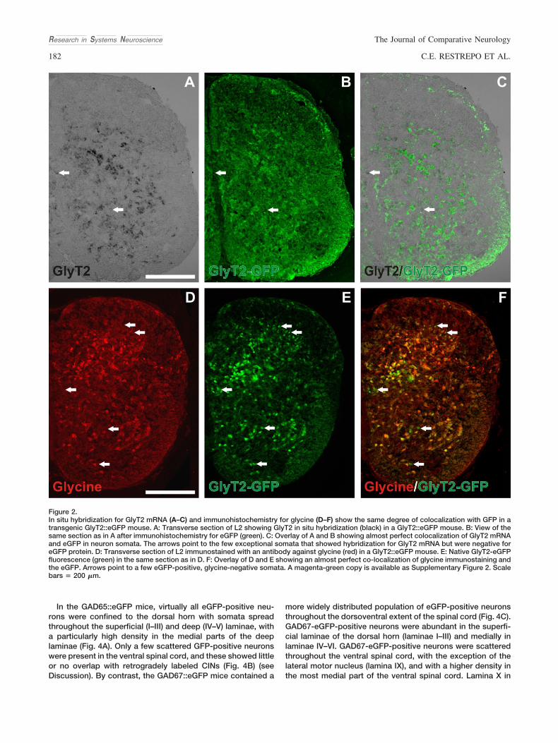
CINs using antibodies against glycine. Although neuronal so-mata were well labeled using immunohistochemistry, the gly-cine immunoreactivity disappeared from somata in the ventralspinal cord (see Suppl. Fig. S1) after cutting CIN axons in theventral commissure. Therefore, to visualize putative glyciner-gic CINs we used in situ hybridization against mRNA encodingthe membrane-bound glycine transporter GlyT2. Of the twoidentified glycine transporters, GlyT1 and GlyT2, GlyT1 ismainly expressed in glial cells, whereas GlyT2 has beenshown to be highly expressed in certain identified populationsof glycinergic neurons (Poyatos et al., 1997; Zafra et al., 1997).The GlyT2 riboprobe we used for these experiments origi-nated from a rat cDNA clone (Zafra et al., 1997) and it reliablylabels putative glycinergic neurons in the brainstem (Stornettaet al., 2004). The rat GlyT2 gene shows 95% sequence overlapwith the mouseGlyT2 gene (see Materials and Methods). Toevaluate the fidelity of the in situ hybridization obtained withthis probe in mouse we performed the following control ex-periments. First, glycine immunoreactivity (Allain et al., 2006)and GlyT2 in situ hybridization reactivity showed comparablepatterns: intensive neuronal labeling in the deep dorsal horn(corresponding to lamina V–VII), and substantial labeling in the
intermediate (laminae VII and X) and ventral (lamina VIII) spinalcord except for the motor nuclei in lamina IX (compare Fig.2A,D; see also Allain et al., 2006). Second, we attempted tocombine in situ hybridization for GlyT2 and immunohisto-chemistry for glycine to directly reveal colocalization. Unfor-tunately, the glycine antibody did not react in sections after insitu hybridization, making this approach impossible. Wetherefore performed in situ hybridization for GlyT2 in a trans-genic mouse expressing the eGFP protein under the control ofthe GlyT2 promoter (Zeilhofer et al., 2005). We found an al-most complete colocalization of GlyT2 in situ hybridizationreactivity with eGFP immunoreactivity (Fig. 2A–C). Addition-ally, glycine immunohistochemistry colocalized perfectly withthe GlyT2-eGFP-positive cells (Fig. 2D–F). Very few eGFP-positive cells were not glycine-immunoreactive (arrows Fig.2D–F). We conclude from these experiments that the rat GlyT2riboprobe efficiently labels putative glycinergic neurons in theventral spinal cord of the mouse.
We then used this GlyT2 riboprobe to identify putative gly-cinergic CINs. Figure 3 shows transverse sections of one sideof the L2 ventral spinal cord with retrogradely labeled CINs(Fig. 3A), GlyT2 mRNA-positive neurons (Fig. 3B), andHoechst-stained nuclei (Fig. 3C). The photos are superim-posed in Figure 3D with arrows pointing to CINs that wereGlyT2 mRNA-positive. Figure 3E shows a GlyT2 mRNA-positive and a GlyT2 mRNA-negative CIN in enlargements.
When the relative locations of the putative glycinergic CINsis plotted separately for midline versus parasagittal labelingprotocols, the picture appears quite similar, with neuronsdistributed throughout the ventral spinal cord with no obviousmediolateral or dorsoventral clustering (Fig. 3F: midline; Fig.3G: parasagittal). The proportion of CIN profiles that wereGlyT2 mRNA-positive was about one-third in both of the ret-rograde labeling protocols (midline labeling: 30% % 5% [n &3], Fig. 3H, first bar; parasagittal labeling: 27% % 3.7% [n & 3],Fig. 3G, third bar). The proportion of GlyT2 mRNA-positiveneuron profiles that were CINs was also about one-third (27%% 5.3% [n & 3] using midline labeling, Fig. 3H, second bar;32% % 2.4% [n & 3] using parasagittal labeling, Fig. 3H, fourthbar). When we compared the number of double-labeled neu-ron profiles in the two labeling protocols they were very similar(midline labeling: 40 % 8.0; parasagittal: 33 % 4.9). In all of thefollowing experiments we only used the midline applicationprotocol for retrograde labeling.
Putative GABAergic CINsGABA is synthesized by the enzyme GAD, which is found in
two isoforms, GAD65 and GAD67. The presence of one or theother or both of these enzymes, particularly in synaptic termi-nals, is considered a good indicator of a GABAergic pheno-type. However, whereas antibodies against GAD65 andGAD67 label axon terminals strongly the same antibodies failto label neuronal somata in the rodent spinal cord (Mackie etal., 2003; Weber et al., 2007). To reveal putative GABAergicCINs we therefore used transgenic mice that express eGFPunder control of the GAD65 promotor (Hughes et al., 2005), orknockin mice with eGFP in the GAD67 gene (Tamamaki et al.,2003). Both of these mice reliably express eGFP in certainidentified populations of GABAergic neurons, and we there-fore used them here to provide an indication of which ventralCINs may be GABAergic.
Research in Systems NeuroscienceThe Journal of Comparative Neurology
181PHENOTYPING OF COMMISSURAL INTERNEURONS
In the GAD65::eGFP mice, virtually all eGFP-positive neu-rons were confined to the dorsal horn with somata spreadthroughout the superficial (I–III) and deep (IV–V) laminae, witha particularly high density in the medial parts of the deeplaminae (Fig. 4A). Only a few scattered GFP-positive neuronswere present in the ventral spinal cord, and these showed littleor no overlap with retrogradely labeled CINs (Fig. 4B) (seeDiscussion). By contrast, the GAD67::eGFP mice contained a
more widely distributed population of eGFP-positive neuronsthroughout the dorsoventral extent of the spinal cord (Fig. 4C).GAD67-eGFP-positive neurons were abundant in the superfi-cial laminae of the dorsal horn (laminae I–III) and medially inlaminae IV–VI. GAD67-eGFP-positive neurons were scatteredthroughout the ventral spinal cord, with the exception of thelateral motor nucleus (lamina IX), and with a higher density inthe most medial part of the ventral spinal cord. Lamina X in
Figure 2.In situ hybridization for GlyT2 mRNA (A–C) and immunohistochemistry for glycine (D–F) show the same degree of colocalization with GFP in atransgenic GlyT2::eGFP mouse. A: Transverse section of L2 showing GlyT2 in situ hybridization (black) in a GlyT2::eGFP mouse. B: View of thesame section as in A after immunohistochemistry for eGFP (green). C: Overlay of A and B showing almost perfect colocalization of GlyT2 mRNAand eGFP in neuron somata. The arrows point to the few exceptional somata that showed hybridization for GlyT2 mRNA but were negative foreGFP protein. D: Transverse section of L2 immunostained with an antibody against glycine (red) in a GlyT2::eGFP mouse. E: Native GlyT2-eGFPfluorescence (green) in the same section as in D. F: Overlay of D and E showing an almost perfect co-localization of glycine immunostaining andthe eGFP. Arrows point to a few eGFP-positive, glycine-negative somata. A magenta-green copy is available as Supplementary Figure 2. Scalebars & 200 "m.
Research in Systems Neuroscience The Journal of Comparative Neurology
182 C.E. RESTREPO ET AL.

Figure 3.Detection of putative glycinergic CINs in the ventral mouse spinal cord. A: CINs in the ventral spinal cord of a transverse section from L2retrogradely labeled with TxRDA (red) applied to the midline. B: Putative glycinergic neurons identified by in situ hybridization against GlyT2mRNA (purple) in the same section. C: Hoechst labeling of nuclei (blue) in the same section. D: Merged view of A–C showing GlyT2mRNA-positive CINs. E: Enlargements from A–D (box areas) showing identification of Glyt2 mRNA-positive and Glyt2 mRNA-negative neurons.F: Cumulative plot of GlyT2 mRNA-positive CINs from three sections in each of three animals where CINs were retrogradely labeled by applyingTxRDA to a midline cut. G: Cumulative plot of GlyT2 mRNA-positive CINs from three sections in each of three animals where CINs wereretrogradely labeled by TxRDA applied to a parasagittal cut. H: The percentage of CINs that are GlyT2 mRNA-positive (first and third bar) andthe percentage of GlyT2 mRNA-positive neurons that are CINs (second and fourth bar) are shown for the midline (white) and parasagittal (gray)labeling protocols. Scale bar & 200 "m.

particular was always strongly populated by GAD67-eGFP-positive neurons. Particularly bright GAD67-eGFP-positiveneurons were seen medial to the lateral motor neurons. Theselatter neurons have been shown to be Renshaw cells (Nishi-maru et al., 2005; Alvarez et al., 2005; Nishimaru et al., 2006).
The ventrally located GAD67-eGFP-positive cells intermingledwith CINs (Fig. 4D) and a fraction was CINs (Fig. 4D–F).GAD67-eGFP-positive CINs were located mainly in laminae VIIand VIII, but a very few were found in the most lateral part oflamina VII (Fig. 4E). The proportion of CIN profiles that were
Figure 4.Detection of putative GABAergic CINs in the ventral mouse spinal cord. Putative GABAergic neurons were visualized using transgenic mice thatexpress eGFP under the promoters for one of the two GABA synthesizing enzymes GAD-65 (A,B) and GAD-67 (C,D). A: Transverse section from L2showing eGFP-positive neurons in the GAD65::eGFP mouse. Note that there are hardly any eGFP-positive neurons in the ventral cord. B: TxRDAretrogradely labeled CINs (red) in a transverse section of L2 from a GAD65::eGFP mouse (green, eGFP). Native eGFP fluorescence. C: Transversesection from L2 showing eGFP-positive neurons in the GAD67::eGFP mouse. Note the widespread distribution of eGFP-positive neurons both dorsallyand ventrally. D: TxRDA retrogradely labeled CINs (red) in a transverse section of L2 from a GAD67::eGFP mouse (green, eGFP). Native eGFPfluorescence. E: Cumulative plot of GAD67-eGFP-positive CINs from three sections in each of three animals in which CINs were retrogradely labeledfrom the midline. F: The percentage of retrogradely labeled CINs that are GAD67-eGFP-positive (first bar) and the percentage of GAD67-eGFP-positiveneurons that are CINs (second bar). A magenta-green copy is available as Supplementary Figure 3. Scale bars & 200 "m.
Research in Systems Neuroscience The Journal of Comparative Neurology
184 C.E. RESTREPO ET AL.

GAD67-eGFP-positive was 20 % 3.38% (n & 3, Fig. 4F, firstbar), and the proportion of GAD67-eGFP-positive neuronalprofiles that were CINs was 18 % 7%, (n & 3, Fig. 4F, secondbar).
Putative combined GABAergic-glycinergic CINsCotransmission of GABA and glycine has been documented
in the spinal cord (Jonas et al., 1998), and colocalization of
GABA and glycine is particularly prominent in the spinal cordembryonically and early postnatally (Jonas et al., 1998; Allainet al., 2006). We therefore assessed the colocalization of thesetransmitters in CINs by using in situ hybridization for GlyT2mRNA in the GAD67::eGFP mice.
Figure 5A–D shows transverse sections of one side of the L2ventral spinal cord with retrogradely labeled CINs (Fig. 5A), insitu labeling of GlyT2 mRNA-positive neuron (Fig. 5B), and
Figure 5.Detection of putative combined glycinergic/GABAergic CINs in the ventral mouse spinal cord. A–C: Transverse section from L2 showingretrogradely labeled CINs. TxRDA applied to a midline cut (A), in situ hybridization against GlyT2 mRNA (black) (B), and GAD67-eGFP expression(green) (C). D: Overlay of A–C. The arrows point to triple-labeled cells. E: CINs positive for both GAD67-eGFP and GlyT2 mRNA plotted from threesections in each of three animals where CINs were retrogradely labeled from the midline. F: The percentage of all labeled CINs that areGAD67-eGFP-positive and GlyT2-eGFP-positive. A magenta-green copy is available as Supplementary Figure 4. Scale bar & 200 "m.
Research in Systems NeuroscienceThe Journal of Comparative Neurology
185PHENOTYPING OF COMMISSURAL INTERNEURONS

GAD67-eGFP-positive neuron (Fig. 5C). The photos are super-imposed in Figure 5D with arrows pointing to CINs that areboth GlyT2 mRNA-positive and GAD67-eGFP-positive. Theseare restricted to the most medial part of layers VII and VIII andare absent from lamina X (Fig. 5E). The percentage of CINprofiles in the ventral spinal cord that were both GAD67-eGFP- and GlyT2 mRNA-positive was 6 % 0.28% (n & 3, Fig.5F). In general, these putative GABAergic/glycinergic CIN pro-files were distributed in a similar pattern to that of the putativeGABAergic CINs, with a somewhat higher density mediallythan laterally.
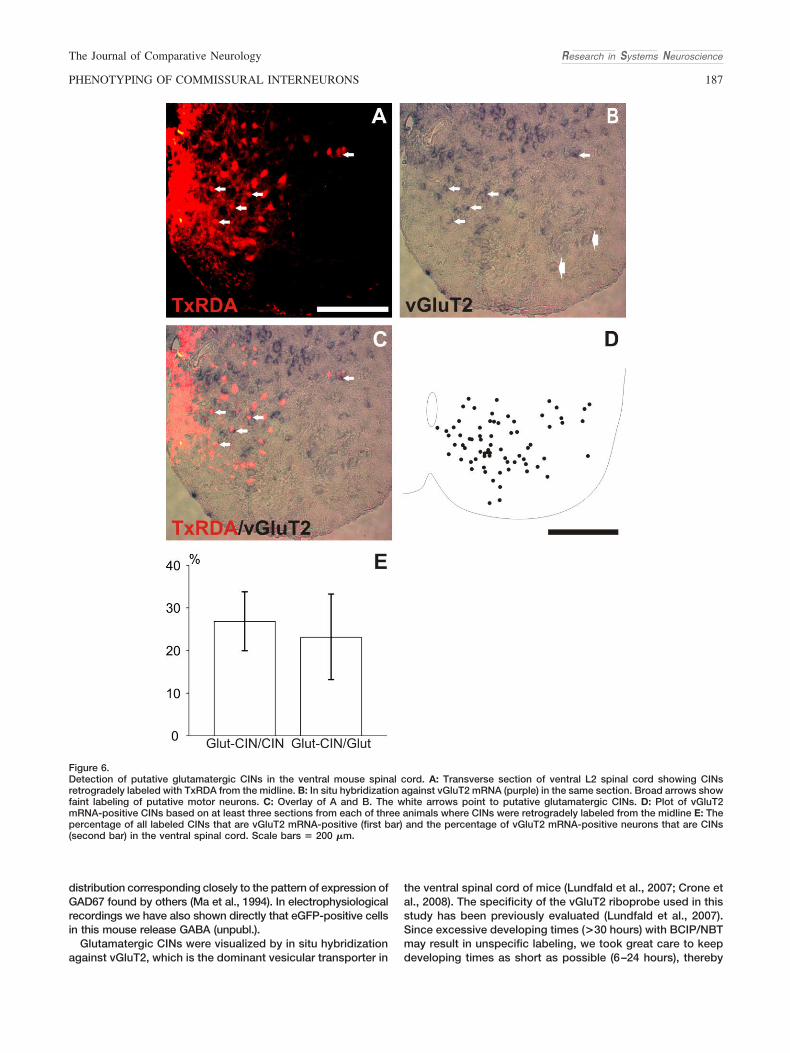
Putative glutamatergic CINsWe used in situ hybridization against vGluT2 mRNA in combi-
nation with retrograde labeling to visualize putative glutamater-gic CINs. Previous experiments from our group and others haveshown that among the three known vesicular glutamate trans-porters 1–3 (vGluT1–3) selectively expressed in glutamatergicneurons, vGluT2 is the dominant transporter expressed in neu-rons in the ventral part of the mouse and rat spinal cord (Kul-lander et al., 2003; Oliveira et al., 2003; Herzog et al., 2004; Wilsonet al., 2005; Lundfald et al., 2007). VGluT1 transcripts are mainlyexpressed in somata in the medial part of dorsal spinal cord(Oliveira et al., 2003; Kullander et al., 2003; see, however, Herzoget al., 2004) and the dorsal root ganglion cells, and vGluT3transcripts are not detected in the ventral spinal cord at all or invery few neurons (Oliveira et al., 2003).
In accordance with previous observations (Lundfald et al.,2007; Crone et al., 2008), we found intensely labeled vGluT2-positive neurons in laminae VII, VIII, and X, although the densityappeared to be lower in the lateral part of layer VII (Fig. 6B,D).Faintly labeled neurons were also seen in lamina X (Herzog et al.,2004; Nishimaru et al., 2005). vGluT2-positive CINs (Fig. 6C,D)were present throughout the ventral spinal cord (except for themotor nucleus), with a somewhat lower density in the lateral partof the cord (Fig. 6D). The distribution of the putative glutamater-gic CINs was similar to that of the putative glycinergic CINs (Fig.6D, compare to Fig. 3E,F). The percentage of retrogradely la-beled CIN profiles that expressed vGluT2 mRNA was 26 %0.67% (n & 3, Fig. 6E, first bar) and the percentage of vGluT2mRNA-positive neuronal profiles that were CINs was 22 % 2.3%(n & 3, Fig. 6E, second bar).
DISCUSSIONSpinal CINs play an essential role in coordinating rhythmic
motor activity between the left and right sides of the bodyduring locomotion. From physiological studies in mammals itis known that this coordination involves glycinergic, GABAer-gic, and glutamatergic crossed connections. In the presentstudy we describe anatomically the relative proportions anddistributions of putative glycinergic, GABAergic, combinedglycinergic/GABAergic, and glutamatergic CINs in the loco-motor region (laminae VII, VIII, and X) of the lumbar spinal cordin the newborn mouse.
Our study shows that the putative glycinergic, GABAergic,and glutamatergic phenotypes are found in comparablenumbers whereas a smaller proportion of CINs may have acombined glycinergic/GABAergic phenotype. There was noparticular differential distribution of these different CIN phe-notypes in the ventral spinal cord, aside from a somewhatmore medial distribution of putative GABAergic CINs relative
to the others. Our data suggest that in addition to the glycin-ergic CINs that constitute the core of the left–right coordinat-ing circuits in locomotor models of aquatic vertebrates, therodent CPG may encompass equally strong GABAergic andglutamatergic left–right coordinating circuits.
Technical considerationsUnlike glutamatergic neurons, where the presence of func-
tional vesicular transporter proteins (e.g., vGluT2), is consid-ered an indicator of a glutamatergic phenotype, GABAergicand glycinergic neurons share the same vesicular inhibitoryamino acid transporter, VIAAT (Wojcik et al., 2006). Therefore,establishing the presence of either of these transmitter phe-notypes anatomically requires a differential evaluation of spe-cific characters, such as synthesizing enzymes (GAD65/67) orreuptake transporters (GlyT2). There are also excellent anti-bodies against GABA and glycine that label neuronal somata(Storm-Mathisen et al., 1983; Allain et al., 2006). Unfortunately,glycine immunoreactivity was drastically weakened in theneonatal spinal cord by the axotomy required to retrogradelylabel the CINs. This change in immunoreactivity occurredalready after 2–3 hours and was substantial after 5–7 hours.Similar findings have been described for choline acetyltrans-ferase and vesicular acetylcholine transporter immunoreactiv-ity in chronically axotomixed cholinergic neurons (Rende etal., 1995; Matsuura et al., 1997). The depletion of glycineimmunoreactivity was not a consequence of the in vitro incu-bation because this did not occur in nonaxotomized interneu-rons in the same sections.
We were therefore forced to use methods other than glycineand GABA immunohistochemistry to identify putativeGABAergic and glycinergic CINs in the present study. Anti-bodies against the GABA-synthesizing enzymes GAD65 andGAD67 or against the neuronal glycine transporter GlyT2 alsotypically gave poor labeling of neuronal somata in the neona-tal mouse and rat spinal cord (unpubl. obs.), presumably be-cause the proteins are localized principally in synaptic termi-nals. To visualize putative inhibitory CIN somata directly wetherefore used transgenic animals that express eGFP under thecontrol of GAD65 or GAD67 promoters (GABA) and in situ hy-bridization against GlyT2 mRNA (glycine). In the case of theGAD65::GFP mice there was a complete absence of eGFP-positive neurons in the ventral horn, precluding the identificationof any GAD65-positive CINs. This absence may reflect a relativedifference in the expression levels of the two GAD enzymes.Although GAD65 and GAD67 have been reported to be coex-pressed in most GABAergic neurons in the adult, GAD67 mRNAlevels are generally higher than GAD65 mRNA levels in the spinalcord, especially in the ventral horn (Ma et al., 1994; Feldblum etal., 1995). When evaluated at the protein level, GAD67 is alwaysexpressed at higher levels than GAD65 throughout the spinalcord (Mackie. et al., 2003). Since the relative difference is seen atthe level of transcripts as well as protein, the expression of eGFPdriven by the GAD65 promoter may be expected to be similarlylow in the ventral spinal cord compared to that of eGFP driven bythe GAD67 promoter.
The best indication of potential GABA synthesis in ventralspinal CINs was therefore obtained in the GAD67::eGFP mice.The specificity of eGFP expression in the GAD67::eGFP mousehas been previously evaluated in the brain and spinal cord usingin situ hybridization for GAD67 showing a perfect match betweeneGFP and GAD67 expression (Tamamaki et al., 2003) and a
Research in Systems Neuroscience The Journal of Comparative Neurology
186 C.E. RESTREPO ET AL.
distribution corresponding closely to the pattern of expression ofGAD67 found by others (Ma et al., 1994). In electrophysiologicalrecordings we have also shown directly that eGFP-positive cellsin this mouse release GABA (unpubl.).
Glutamatergic CINs were visualized by in situ hybridizationagainst vGluT2, which is the dominant vesicular transporter in
the ventral spinal cord of mice (Lundfald et al., 2007; Crone etal., 2008). The specificity of the vGluT2 riboprobe used in thisstudy has been previously evaluated (Lundfald et al., 2007).Since excessive developing times (>30 hours) with BCIP/NBTmay result in unspecific labeling, we took great care to keepdeveloping times as short as possible (6–24 hours), thereby
Figure 6.Detection of putative glutamatergic CINs in the ventral mouse spinal cord. A: Transverse section of ventral L2 spinal cord showing CINsretrogradely labeled with TxRDA from the midline. B: In situ hybridization against vGluT2 mRNA (purple) in the same section. Broad arrows showfaint labeling of putative motor neurons. C: Overlay of A and B. The white arrows point to putative glutamatergic CINs. D: Plot of vGluT2mRNA-positive CINs based on at least three sections from each of three animals where CINs were retrogradely labeled from the midline E: Thepercentage of all labeled CINs that are vGluT2 mRNA-positive (first bar) and the percentage of vGluT2 mRNA-positive neurons that are CINs(second bar) in the ventral spinal cord. Scale bars & 200 "m.
Research in Systems NeuroscienceThe Journal of Comparative Neurology
187PHENOTYPING OF COMMISSURAL INTERNEURONS

avoiding any background signal in our preparations. The weakvGluT2 mRNA expression in motor neurons has been ob-served previously and may be explained by the corelease ofglutamate from motor neurons in early postnatal animals(Mentis et al., 2005; Nishimaru et al., 2006).
In the case of the GlyT2 riboprobe, specificity is supported bycolocalization with glycine immunoreactivity and with eGFP im-munoreactivity in the transgenic GlyT2::eGFP mice.
Retrograde labeling did not diminish the vGluT2 and GlyT2mRNA signals or the eGFP immunoreactivity, as it did glycineimmunoreactivity, suggesting that axotomy has less effect onthe stability of these transcripts than on the neurotransmittersthemselves.
We conclude that the combination of phenotypic markersused here provides a means to specifically distinguish puta-tive glutamatergic, GABAergic, and glycinergic phenotypes inretrogradely labeled spinal CINs. However, although severalof the markers involved have been correlated with the physi-ological utilization of the respective transmitter in certain neu-ronal systems, they cannot be considered definitive markersof neurotransmitter phenotype in the absence of physiologicalconfirmation. First of all, neither the use of reporter genesdriven by regulatory elements for enzymes or transporters northe use of in situ hybridization to detect mRNA for transport-ers provides any direct information about the degree of pro-tein expression because posttranscriptional regulatory eventsmay affect the level of expression of protein. Moreover, evenif the relevant proteins were detected immunohistochemicallyin neuronal somata, this would not be proof that they werefunctional or properly localized to generate neurotransmitterrelease within the synapse. For these reasons we have de-cided to exercise caution by referring to the phenotypes asputative neurotransmitter phenotypes, with the anticipationthat the advantages of the neonatal mouse spinal cord prep-aration will permit a physiological confirmation of many ofthese phenotypes in the future.
Although the phenotyping appears to be specific, it clearlydoes not account for all of the retrogradely labeled CINs.
Thus, the sum of putative inhibitory and excitatory CIN phe-notypes is not 100% but only 70%. This is similar to what hasbeen found for CIN synaptic terminal phenotyping in the rat(85%; Weber et al., 2007) and the cat (67%; Bannatyne et al.,2003; Jankowska et al., 2009) (see below). One possible ex-planation for the discrepancy is that the remaining CINs mayexpress other neurotransmitters. In adult cat there is indeed asmall number of cholinergic neurons with commissural pro-jections (Huang et al., 2000) and it is possible that this groupof neurons also exist in the neonatal mouse and may accountfor a proportion of unidentified CINs. Another possibility isthat CINs utilizing peptide neurotransmitters account forsome of the shortfall (Ryge et al., 2008), which we did not testfor here. We cannot exclude such alternative transmitters, butwe feel that a more likely explanation for the deficit in ourstudy is that neither the RNA (detected by the in situ hybrid-ization) nor the eGFP protein in the transgenic mice is ex-pressed in sufficient levels in all the neurons to be detected bythe techniques used. Presumably, neurons missing for thisreason would be equivalently distributed among the differentphenotypes and therefore would not change the relative spa-tial distributions or proportions. Therefore, to facilitate thecomparison of our data with other anatomical and electro-physiological studies of CIN distributions in mammals we willhereafter express CIN transmitter phenotypes as proportionsrelative to the population of transmitter-identified CINs ratherthan as proportions relative to the total population of retro-gradely labeled CINs (Table 2).
Organization of ventrally located CINs in therodent
In the present study we find that putative glycinergic andGABAergic CINs comprise about 60% of the transmitter-identified CINs in the ventral spinal cord, while the remaindercomprises putative glutamatergic CINs (Table 2, column 2).CINs expressing only glycinergic markers (30%) are slightlymore abundant than those expressing only GABAergic mark-ers (21%). The putative glycinergic and GABAergic pheno-
TABLE 2. Summary of CIN Phenotypes in Rodents
Anatomy
Mouse1: Percentage inrelationship to the totalpopulation of labeled
CINs
Mouse1: Percentage inrelationship to the
population oftransmitter-identified
CINs
Rat2: Percentage inrelationship to the totalpopulation of labeled
CIN terminals
Rat2: Percentage inrelationship to the
population oftransmitter-identified
CIN terminals
Glycine 20% 30% 27% 32%GABA 14% 21% 14% 17%Glycine/GABA 6% 9% 16% 19%Total inhibitory 40% 60% 58% 69%Glutamate 26% 39% 27% 31%Unknown 35% 0% 15% 0%Total 100% 100% 100% 100%
Electrophysiology
Mouse3: Percentage inrelationship to the totalpopulation of labeled
CINs
Mouse3: Percentage inrelationship to the
population oftransmitter-identified
CINs
Rat4: Percentage inrelationship to the totalpopulation of labeled
CINs
Rat4: Percentage inrelationship to the
population oftransmitter-identified
CINs
Inhibitory 11% 30% 17% 24%Excitatory 33% 70% 55% 77%Unknown 47% 0% 28% 0%Total 100% 100% 100% 100%
1Present study.2Weber et al., 2007.3Quinlan and Kiehn, 2007.4Butt and Kiehn, 2003.
Research in Systems Neuroscience The Journal of Comparative Neurology
188 C.E. RESTREPO ET AL.
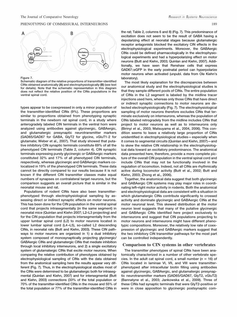
types appear to be coexpressed in only a minor population ofthe transmitter-identified CINs (9%). These proportions aresimilar to proportions obtained from phenotyping synapticterminals in the newborn rat spinal cord, in a study whereanterogradely labeled CIN terminals in the ventral horn wereanalyzed using antibodies against glycinergic, GABAergic,and glutamatergic presynaptic neurotransmitter markers(GAD65/GAD67 for GABA, GlyT2 for glycine, vGluT1-2 forglutamate; Weber et al., 2007). That study showed that puta-tive inhibitory CIN synaptic terminals constitute 69% of all thephenotyped CIN terminals (Table 2, column 4). CIN synapticterminals expressing purely glycinergic or GABAergic markersconstituted 32% and 17% of all phenotyped CIN terminals,respectively, whereas glycinergic and GABAergic markers co-localized in 19% of the phenotyped CIN terminals. These datacannot be directly compared to our results because it is notknown if the different CIN transmitter classes make equalnumbers of synapses on contralateral target neurons, but thecomparison suggests an overall picture that is similar in theneonatal mouse and rat.
Populations of rodent CINs have also been transmitter-phenotyped through electrophysiological sampling by as-sessing direct or indirect synaptic effects on motor neurons.This has been done for the CIN population in the ventral spinalcord that projects intrasegmentally (in the same segment) inneonatal mice (Quinlan and Kiehn 2007; L2–L3 projecting) andfor the CIN population that projects intersegmentally from theupper lumbar spinal cord (L2) to motor neurons located inlower lumbar spinal cord (L4–L5), so-called L2 descendingCINs, in neonatal rats (Butt and Kiehn, 2003). These CIN path-ways to motor neurons are organized in 1) a dual inhibitorysystem composed of monosynaptically projecting glycinergic/GABAergic CINs and glutamatergic CINs that mediate inhibitionthrough local inhibitory interneurons, and 2) a single excitatorysystem of glutamatergic CINs that excite motor neurons. Whencomparing the relative contribution of phenotypes obtained byelectrophysiological sampling of CINs with the data obtainedfrom the anatomical sampling here the results appear very dif-ferent (Fig. 7). Thus, in the electrophysiological studies most ofthe CINs were determined to be glutamatergic both for intraseg-mental (Quinlan and Kiehn, 2007) and for intersegmental (Buttand Kiehn, 2003) connections (33% of the total population or70% of the transmitter-identified CINs in the mouse and 55% ofthe total population or 77% of the transmitter-identified CINs in
the rat; Table 2, columns 6 and 8) (Fig. 7). This predominance ofexcitation does not seem to be the result of GABA having adepolarizing effect at neonatal stages because glutamatergicreceptor antagonists blocked the excitatory CIN effects in theelectrophysiological experiments. Moreover, the GABAergicCINs could be defined pharmacologically in the electrophysio-logical experiments and had a hyperpolarizing effect on motorneurons (Butt and Kiehn, 2003; Quinlan and Kiehn, 2007). Addi-tionally, we have seen that Renshaw cells that expressGAD67::eGFP in the early postnatal period can hyperpolarizemotor neurons when activated (unpubl. data from Ole Kiehn’slaboratory).
The most likely explanation for the discrepancies betweenour anatomical study and the electrophysiological studies isthat they sample different pools of CINs. The entire populationof CINs in the L2 segment is labeled by the midline tracerinjections used here, whereas only those CINs that have director indirect synaptic connections to motor neurons are de-tected electrophysiologically (Fig. 7). The electrophysiologicalsampling of motor neurons therefore excludes CINs that ter-minate exclusively on interneurons, whereas the population ofCINs labeled retrogradely from the midline includes CINs thatproject to motor neurons as well as to interneurons only(Birinyi et al., 2003; Matsuyama et al., 2004, 2006). This con-dition seems to leave a relatively large proportion of CINsunidentified in electrophysiological studies—especially whenit comes to segmental circuits (Quinlan and Kiehn, 2007)—andto skew the relative CIN relationship in the electrophysiolog-ical data toward an excitatory predominance. The anatomicaldata presented here, therefore, provide a more complete pic-ture of the overall CIN population in the ventral spinal cord andinclude CINs that may not be functionally involved in theregulation of locomotion. Indeed, not all CINs are rhythmicallyactive during locomotor activity (Butt et al., 2002; Butt andKiehn, 2003; Zhong et al., 2006).
Together, the anatomical data suggest that both glycinergicand GABAergic CIN systems may play major roles in coordi-nating left–right motor activity in rodents. Both the anatomicaland electrophysiological data are consistent with a situation inwhich glutamatergic CINs contribute significantly to crossedactivity and dominate glycinergic and GABAergic CINs at themotor neuronal level. This skewed distribution at the motorneuron level suggests that many of the putative glycinergicand GABAergic CINs identified here project exclusively tointerneurons and suggest that CIN populations projecting tomotor neurons and interneurons might have different pheno-typic compositions. Moreover, the relatively low rate of coex-pression of glycinergic and GABAergic markers suggest thatthe two inhibitory CIN transmitter pathways for the most partcan be controlled independently.
Comparison to CIN systems in other vertebratesThe transmitter phenotypes of spinal CINs have been ana-
tomically characterized in a number of other vertebrate spe-cies. In the adult cat spinal cord, a small number (n & 16) ofCINs located in laminae VI, VII, and VIII were transmitter-phenotyped after intracellular biotin filling using antibodiesagainst glycinergic, GABAergic, and glutamatergic presynap-tic neurotransmitter markers (GAD65/GAD67, GlyT2, vGluT2)(Bannatyne et al., 2003; Jankowska et al., 2009). Three ofthese CINs had synaptic terminals that were GlyT2-positive orwere in close apposition to glycinergic postsynaptic com-
Figure 7.Schematic diagram of the relative proportions of transmitter-identifiedCINs obtained anatomically (A) and electrophysiologically (B) (see textfor details). Note that the schematic representation in this diagramdoes not reflect the relative position of the CINs populations in theventral spinal cord.
Research in Systems NeuroscienceThe Journal of Comparative Neurology
189PHENOTYPING OF COMMISSURAL INTERNEURONS

plexes, whereas 13 CINs had vGluT2-positive terminals. Thus,in this small sample, of the phenotyped CINs, those that wereputatively inhibitory were glycinergic and made up a smallpercentage (19%) of the CINs, whereas those that were puta-tively excitatory made up a large majority (81%) of the CINs.CINs located in laminae VI–VII were all putative excitatory,whereas CINs located in lamina VIII represented a mixture ofputative glycininergic and glutamatergic CINs (40% and 60%,respectively). We found no sign of such a differential laminardistribution of CIN neurotransmitter phenotypes in the neona-tal mouse in the present study. Using retrograde tracing incombination with immunohistochemistry in the adult lamprey!65% of the total population of phenotyped CINs was foundto be putatively glycinergic, whereas the remaining CINs wereputatively glutamatergic (Mahmood et al., Eur J Neurosci,submitted). In the embryonic zebrafish the putative transmit-ter phenotypes of anatomically defined spinal CINs have beencharacterized using transgenic fish that express eGFP underthe control of genes for glycinergic, GABAergic, and glutama-tergic marker proteins (Higashijima et al., 2004a,b). By com-paring the results of this transmitter phenotyping with thedifferent classes of CINs in the zebrafish spinal cord it hasbeen calculated that !35% of spinal CINs (including ventraland dorsal CINs) are glycinergic, whereas the remaining 65%are glutamatergic (Fetcho, 1990; Higashijima et al., 2004a,b;Nissen et al., 2008). In the tadpole embryo there are no reportsof excitatory CINs and all CINs appear to be glycinergic (Daleet al., 1986; Roberts, 2000).
From this comparison it is clear that, with the notable ex-ception of the Xenopus tadpole, all investigated species haveboth inhibitory (glycinergic and/or GABAergic) and excitatory(glutamatergic) CIN populations in the cord. However, therelative sizes of putative inhibitory and excitatory CIN popu-lations vary. The strong GABAergic component of the CINsystem found in rodents clearly deviates from what is found inall other vertebrates investigated. The GABAergic componentmight be an early developmental phenotype in mammals thatis replaced by a purely glycinergic component later in adultlife. The difference between neonatal rodents and the aquaticvertebrates, however, seems not to reflect a developmentaldifference but rather a fundamental difference in spinal cordorganization, since putative GABAergic CINs were absentboth in zebrafish embryos, Xenopus tadpoles, and lampreyadults. Together, the comparison of the reported data sug-gests that most vertebrate phyla independent of their devel-opmental stage use a combined inhibitory and excitatory CINsystem to coordinate left–right spinal cord activity. It shouldbe noted that we are aware that comparing different speciesand developmental stages is not straightforward. For exam-ple, the fully independent autonomy of movements observedat early developmental stages in Xenopus larvae and ze-brafish do not have a counterpart in mammals. Thus, althoughthe some of the CIN populations so far identified in fish or frogmay be evolutionarily conserved, others might belong to aspecies-specific repertoire.
Functional considerations of CIN organization invertebrate CPGs
Physiologically, most species so far investigated have bothinhibitory (glycinergic) and excitatory (glutamatergic) crossedprojections within the locomotor CPG, the former mediatingleft–right alternation and the latter mediating left–right syn-
chrony (Buchanan and Cohen, 1982; Dale, 1985; Bracci et al.,1996; Cazalets et al., 1996; Cowley and Schmidt, 1997; Rob-erts, 2000; Grillner, 2003; Hinckley et al., 2005). However, inrodents it has been shown that segmental crossed inhibitionis not only mediated by glycinergic CINs but also indirectly byexcitatory CINs connecting through inhibitory interneuronslocated on the contralateral side (Quinlan and Kiehn, 2007).This segmental dual inhibitory system may also be present inthe cat (Jankowska, 2008; Kiehn et al., 2008). Recent experi-ments from the lamprey have also shown that the CPG con-trolling fin movements contains a dual inhibitory pathway(Roberts, 2000; Mentel et al., 2006), suggesting that such adual inhibitory CIN system might have evolved to control limbor limb-like (fin) movements. In addition, electrophysiologicalstudies (Quinlan and Kiehn, 2007) and this study show thatputative GABAergic CINs also constitute part of the inhibitorycrossed projections and might be involved in left–right alter-nation in the spinal cord of neonatal rodents (Hinckley et al.,2005).
Excitatory CINs are also active in binding left–right motorsynergies along the length of the lumbar cord during locomo-tion in rodents (Butt and Kiehn, 2003). Whether glutamatergicCINs play a similar role in the lamprey (Biro et al., 2008) orother vertebrate species is unknown (Roberts, 2000; Grillner,2003; Kiehn, 2006; Jankowska, 2008). However, populationsof glutamatergic CINs found in both fish and mammals seemto be directly involved in coordinating the left–right synchro-nous activity observed after blocking all glycinergic and/orGABAergic synaptic transmission in the cord (Roberts, 2000;Grillner, 2003; Kiehn, 2006; Jankowska, 2008).
In addition to coordinating left–right activities in the CPG,CINs seem to play a role in setting the speed of locomotion.Thus, selective activation of the locomotor network on oneside of the cord (Kjaerullf and Kiehn, 1997) or in the surgicallyisolated hemicord (Bonnot et al., 2002) in rodents alwaysleads to a slower rhythm than observed in the intact cord. Achange in the speed of fictive locomotion has also been re-ported after hemisection in the lamprey (Cangiano and Grill-ner, 2003). Here, the changes in swimming frequencies de-pend on the initial pharmacology or stimulation used to evokeswimming before the hemisection. In the zebrafish, ablation ofa type of glutamatergic CINs—the ventrally located multipolarcommissural descending interneurons—has been shown toaffect slow frequency movements but not fast movements(McLean et al., 2007). Genetic ablation experiments in themouse spinal cord have also ascribed to ventrally locatedputative excitatory CINs a role in stabilizing the rhythmic mo-tor output (Zhang et al., 2008). Together, these experimentssuggest a direct role of CINs in rhythm generation in additionto a role in left–right coordination.
CONCLUSIONThe present study shows that CIN projections in the area of
the upper lumbar spinal cord of the neonatal rodent thatcontains locomotor CPG circuits are heterogeneous, with atleast four different putative neurotransmitter phenotypes. Therelative proportions of putative excitatory and inhibitory pro-jections suggest that both glycinergic and GABAergic CINsystems may play major roles in coordinating left–right motoractivity in neonatal rodents. The low incidence of coexpres-sion of putative glycinergic and GABAergic phenotypes sug-
Research in Systems Neuroscience The Journal of Comparative Neurology
190 C.E. RESTREPO ET AL.
gests that these inhibitory CIN transmitter pathways may becontrolled independently. Finally, we did not observe any to-pographic segregation of the four CIN phenotypes. Under-standing the anatomical organization of the CIN pathways willfacilitate the comparison of different CIN populations amongvertebrate species and among transgenic mouse models inwhich different patterns of locomotion may result from spe-cific changes in the balance of crossed excitation and inhibi-tion.
ACKNOWLEDGMENTSWe thank Mette Kirkegaard for comments on a previous
version of the article, and Lovisa Kallman and Anna Erikssonfor caretaking and breeding of the different transgenic linesused in this study.
LITERATURE CITEDAllain AE, Bairi A, Meyrand P, Branchereau P. 2006. Expression of the
glycinergic system during the course of embryonic development in themouse spinal cord and its co-localization with GABA immunoreactivity.J Comp Neurol 496:832– 846.
Alvarez FJ, Jonas PC, Sapir T, Hartley R, Berrocal MC, Geiman EJ, ToddAJ, Goulding M. 2005. Postnatal phenotype and localization of spinalcord V1 derived interneurons. J Comp Neurol 493:177–192.
Bannatyne BA, Edgley SA, Hammar I, Jankowska E, Maxwell DJ. 2003.Networks of inhibitory and excitatory commissural interneurons medi-ating crossed reticulospinal actions. Eur J Neurosci 18:2273–2284.
Bink K, Walch A, Feuchtinger A, Eisenmann H, Hutzler P, Hofler H, WernerM. 2001. TO-PRO-3 is an optimal fluorescent dye for nuclear counter-staining in dual-colour FISH on paraffin sections. Histochem Cell Biol115:293–299.
Birinyi A, Viszokay K, Weber I, Kiehn O, Antal M. 2003. Synaptic targets ofcommissural interneurons in the lumbar spinal cord of neonatal rats.J Comp Neurol 461:429 – 440.
Biro Z, Hill RH, Grillner S. 2008. The activity of spinal commissural inter-neurons during fictive locomotion in the lamprey. J Neurophysiol 100:716 –722.
Bonnot A, Morin D, Viala D. 1998. Organization of rhythmic motor patternsin the lumbosacral spinal cord of neonate mouse. Ann N Y Acad Sci860:432– 435.
Bonnot A, Whelan PJ, Mentis GZ, O’Donovan MJ. 2002. Locomotor-likeactivity generated by the neonatal mouse spinal cord. Brain Res BrainRes Rev 40:141–151.
Bracci E, Ballerini L, Nistri A. 1996. Localization of rhythmogenic networksresponsible for spontaneous bursts induced by strychnine and bicu-culline in the rat isolated spinal cord. J Neurosci 16:7063–7076.
Buchanan JT. 1982. Identification of interneurons with contralateral, cau-dal axons in the lamprey spinal cord: synaptic interactions and mor-phology. J Neurophysiol 47:961–975.
Buchanan JT. 1999. The roles of spinal interneurons and motoneurons inthe lamprey locomotor network. Prog Brain Res 123:311–321.
Buchanan JT, Cohen AH. 1982. Activities of identified interneurons, mo-toneurons, and muscle fibers during fictive swimming in the lampreyand effects of reticulospinal and dorsal cell stimulation. J Neurophysiol47:948 –960.
Buchanan JT, Grillner S. 1987. Newly identified ‘glutamate interneurons’and their role in locomotion in the lamprey spinal cord. Science 236:312–314.
Butt SJ, Kiehn O. 2003. Functional identification of interneurons respon-sible for left-right coordination of hindlimbs in mammals. Neuron 38:953–963.
Cangiano L, Grillner S. 2003. Fast and slow locomotor burst generation inthe hemispinal cord of the lamprey. J Neurophysiol 89:2931–2942.
Cazalets JR, Borde M, Clarac F. 1996. The synaptic drive from the spinallocomotor network to motoneurons in the newborn rat. J Neurosci16:298 –306.
Christie KJ, Whelan PJ. 2005. Monoaminergic establishment of rostrocau-dal gradients of rhythmicity in the neonatal mouse spinal cord. J Neu-rophysiol 94:1554 –1564.
Clarke JD, Roberts A. 1984. Interneurones in the Xenopus embryo spinalcord: sensory excitation and activity during swimming. J Physiol 354:345–362.
Cowley KC, Schmidt BJ. 1997. Regional distribution of the locomotorpattern-generating network in the neonatal rat spinal cord. J Neuro-physiol 77:247–259.
Crone SA, Quinlan KA, Zagoraiou L, Droho S, Restrepo CE, Lundfald L,Endo T, Setlak J, Jessell TM, Kiehn O, Sharma K. 2008. Geneticablation of V2a ipsilateral interneurons disrupts left-right locomotorcoordination in mammalian spinal cord. Neuron 60:70 – 83.
Dale N. 1985. Reciprocal inhibitory interneurones in the Xenopus embryospinal cord. J Physiol 363:61–70.
Dale N, Ottersen OP, Roberts A, Storm-Mathisen J. 1986. Inhibitory neu-rones of a motor pattern generator in Xenopus revealed by antibodiesto glycine. Nature 324:255–257.
Eide AL, Glover J, Kjaerulff O, Kiehn O. 1999. Characterization of com-missural interneurons in the lumbar region of the neonatal rat spinalcord. J Comp Neurol 403:332–345.
Feldblum S, Dumoulin A, Anoal M, Sandillon F, Privat A. 1995. Compara-tive distribution of GAD65 and GAD67 mRNAs and proteins in the ratspinal cord supports a differential regulation of these two glutamatedecarboxylases in vivo. J Neurosci Res 42:742–757.
Fetcho JR. 1990. Morphological variability, segmental relationships, andfunctional role of a class of commissural interneurons in the spinal cordof goldfish. J Comp Neurol 299:283–298.
Gabbay H, Delvolve I, Lev-Tov A. 2002. Pattern generation in caudal-lumbar and sacrococcygeal segments of the neonatal rat spinal cord.J Neurophysiol 88:732–739.
Glover J. 1995. Retrograde and anterograde axonal tracing with fluores-cent dextrans in the embryonic nervous system. Neurosci Protoc 30:1–13.
Grillner S. 2003. The motor infrastructure: from ion channels to neuronalnetworks. Nat Rev Neurosci 4:573–586.
Herzog E, Landry M, Buhler E, Bouali-Benazzouz R, Legay C, HendersonCE, Nagy F, Dreyfus P, Giros B, El Mestikawy S. 2004. Expression ofvesicular glutamate transporters, VGLUT1 and VGLUT2, in cholinergicspinal motoneurons. Eur J Neurosci 20:1752–1760.
Higashijima S, Mandel G, Fetcho JR. 2004a. Distribution of prospectiveglutamatergic, glycinergic, and GABAergic neurons in embryonic andlarval zebrafish. J Comp Neurol 480:1–18.
Higashijima S, Schaefer M, Fetcho JR. 2004b. Neurotransmitter propertiesof spinal interneurons in embryonic and larval zebrafish. J CompNeurol 480:19 –37.
Hinckley C, Seebach B, Ziskind-Conhaim L. 2005. Distinct roles of glycin-ergic and GABAergic inhibition in coordinating locomotor-like rhythmsin the neonatal mouse spinal cord. Neuroscience 131:745–758.
Huang J, Wang, Y, Wang W, Li Y, Wu S. 2008a. Proenkephalin mRNA isexpressed in a subpopulation of GABAergic neurons in the spinaldorsal horn of the GAD67-GFP knock in mouse. Anat Record 291:1334 –1341.
Huang J, Wang Y, Wang W, Li Y, Tamamaki N, Wu S. 2008b. 5-HT3areceptor subunit is expressed in a subpopulation of GABAergic andenkephalinergic neurons in the mouse spinal cord. Neurosci Lett 441:1– 6.
Hughes DI, Mackie M, Nagy GG, Riddell JS, Maxwell DJ, Szabo G, ErdelyiF, Veress G, Szucs P, Antal M, Todd AJ. 2005. P boutons in lamina IXof the rodent spinal cord express high levels of glutamic aciddecarboxylase-65 and originate from cells in deep medial dorsal horn.Proc Natl Acad Sci U S A 102:9038 –9043.
Jankowska E. 2008. Spinal interneuronal networks in the cat: elementarycomponents. Brain Res Rev 57:46 –55.
Jankowska E, Hammar I, Slawinska U, Maleszak K, Edgley SA. 2003.Neuronal basis of crossed actions from the reticular formation on felinehindlimb motoneurons. J Neurosci 23:1867–1878.
Jankowska E, Edgley SA, Krutki P, Hammar I. 2005a. Functional differen-tiation and organization of feline midlumbar commissural interneu-rones. J Physiol 565(Pt 2):645– 658.
Jankowska E, Krutki P, Matsuyama K. 2005b. Relative contribution of Iainhibitory interneurones to inhibition of feline contralateral motoneu-rones evoked via commissural interneurones. J Physiol 568(Pt 2):617–628.
Jankowska E, Stecina K, Cabaj A, Pettersson LG, Edgley SA. 2006.Neuronal relays in double crossed pathways between feline motorcortex and ipsilateral hindlimb motoneurones. J Physiol 575(Pt 2):527–541.
Research in Systems NeuroscienceThe Journal of Comparative Neurology
191PHENOTYPING OF COMMISSURAL INTERNEURONS

Jankowska E, Bannatyne BA, Stecina K, Hammar I, Cabaj A, Maxwell DJ.2009. Commissural interneurons with input from group I and II muscleafferents in feline lumbar segments: neurotransmitters, projections andtarget cells. J Physiol 587:401– 418.
Jonas P, Bischofberger J, Sandkuhler J. 1998. Corelease of two fastneurotransmitters at a central synapse. Science 281:419 – 424.
Kiehn O. 2006. Locomotor circuits in the mammalian spinal cord. Annu RevNeurosci 29:279 –306.
Kiehn O, Kjaerulff O. 1998. Distribution of central pattern generators forrhythmic motor outputs in the spinal cord of limbed vertebrates. Ann NY Acad Sci 860:110 –129.
Kiehn O, Quinlan KA, Restrepo CE, Lundfald L, Borgius L, Talpalar AE,Endo T. 2008. Excitatory components of the mammalian locomotorCPG. Brain Res Rev 57:56 – 63.
Kjaerulff O, Kiehn O. 1996. Distribution of networks generating and coor-dinating locomotor activity in the neonatal rat spinal cord in vitro: alesion study. J Neurosci 16:5777–5794.
Kremer E, Lev-Tov A. 1997. Localization of the spinal network associatedwith generation of hindlimb locomotion in the neonatal rat and orga-nization of its transverse coupling system. J Neurophysiol 77:1155–1170.
Kudo N, Yamada T. 1987. N-methyl-D,L-aspartate-induced locomotoractivity in a spinal cord-hindlimb muscles preparation of the newbornrat studied in vitro. Neurosci Lett 75:43– 48.
Kullander K, Butt SJ, Lebret JM, Lundfald L, Restrepo CE, Rydstrom A,Klein R, Kiehn O. 2003. Role of EphA4 and EphrinB3 in local neuronalcircuits that control walking. Science 299:1889 –1892.
Lopez-Bendito G, Sturgess K, Erdelyi F, Szabo G, Molnar Z, Paulsen O.2004. Preferential origin and layer destination of GAD65-GFP corticalinterneurons. Cereb Cortex 14:1122–1133.
Lundfald L, Restrepo CE, Butt SJ, Peng CY, Droho S, Endo T, ZeilhoferHU, Sharma K, Kiehn O. 2007. Phenotype of V2-derived interneuronsand their relationship to the axon guidance molecule EphA4 in thedeveloping mouse spinal cord. Eur J Neurosci 26:2989 –3002.
Ma W, Behar T, Chang L, Barker JL. 1994. Transient increase in expressionof GAD65 and GAD67 mRNAs during postnatal development of ratspinal cord. J Comp Neurol 346:151–160.
Mackie M, Hughes DI, Maxwell DJ, Tillakaratne NJ, Todd AJ. 2003. Dis-tribution and colocalisation of glutamate decarboxylase isoforms in therat spinal cord. Neuroscience 119:461– 472.
Matsuura J, Ajiki K, Ichikawa T, Misawa H. 1997. Changes of expressionlevels of choline acetyltransferase and vesicular acetylcholine trans-porter mRNAs after transection of the hypoglossal nerve in adult rats.Neurosci Lett 236:95–98.
Matsuyama K, Nakajima K, Mori F, Aoki M, Mori S. 2004. Lumbar com-missural interneurons with reticulospinal inputs in the cat: morphologyand discharge patterns during fictive locomotion. J Comp Neurol 474:546 –561.
Matsuyama K, Kobayashi S, Aoki M. 2006. Projection patterns of laminaVIII commissural neurons in the lumbar spinal cord of the adult cat: ananterograde neural tracing study. Neuroscience 140:203–218.
McLean DL, Fan J, Higashijima S, Hale ME, Fetcho JR. 2007. A topo-graphic map of recruitment in spinal cord. Nature 446:71–75.
Mentel T, Krause A, Pabst M, El Manira A, Buschges A. 2006. Activity of finmuscles and fin motoneurons during swimming motor pattern in thelamprey. Eur J Neurosci 23:2012–2026.
Mentis GZ, Alvarez FJ, Bonnot A, Richards DS, Gonzalez-Forero D, ZerdaR, O’Donovan MJ. 2005. Noncholinergic excitatory actions of mo-toneurons in the neonatal mammalian spinal cord. Proc Natl Acad SciU S A 102:7344 –7349.
Nakayama K, Nishimaru H, Kudo N. 2002. Basis of changes in left-rightcoordination of rhythmic motor activity during development in the ratspinal cord. J Neurosci 22:10388 –10398.
Nishimaru H, Restrepo CE, Ryge J, Yanagawa Y, Kiehn O. 2005. Mamma-lian motor neurons corelease glutamate and acetylcholine at centralsynapses. Proc Natl Acad Sci U S A 102:5245–5249.
Nishimaru H, Restrepo CE, Kiehn O. 2006. Activity of Renshaw cells duringlocomotor-like rhythmic activity in the isolated spinal cord of neonatalmice. J Neurosci 26:5320 –5328.
Nissen UV, Mochida H, Glover JC. 2005. Development of projection-specific interneurons and projection neurons in the embryonic mouseand rat spinal cord. J Comp Neurol 483:30 – 47.
Oliveira AL, Hydling F, Olsson E, Shi T, Edwards RH, Fujiyama F, KanekoT, Hokfelt T, Cullheim S, Meister B. 2003. Cellular localization of threevesicular glutamate transporter mRNAs and proteins in rat spinal cordand dorsal root ganglia. Synapse 50:117–129.
Pow DV, Wright LL, Vaney DI. The immunocytochemical detection ofamino-acid neurotransmitters in paraformaldehyde-fixed tissues.1995. J Neurosci Methods 56:115–123.
Poyatos I, Ponce J, Aragon C, Gimenez C, Zafra F. 1997. The glycinetransporter GLYT2 is a reliable marker for glycine-immunoreactiveneurons. Brain Res Mol Brain Res 49:63–70.
Quinlan KA, Kiehn O. 2007. Segmental, synaptic actions of commissuralinterneurons in the mouse spinal cord. J Neurosci 27:6521– 6530.
Rende M, Giambanco I, Buratta M, Tonali P. 1995. Axotomy induces adifferent modulation of both low-affinity nerve growth factor receptorand choline acetyltransferase between adult rat spinal and brainstemmotoneurons. J Comp Neurol 363:249 –263.
Roberts A. 2000. Early functional organization of spinal neurons in devel-oping lower vertebrates. Brain Res Bull 53:585–593.
Ryge J, Westerdahl AC, Alstrøm P, Kiehn O. 2008. Gene expressionprofiling of two distinct neuronal populations in the rodent spinal cord.PLoS ONE. 3:e3415.
Soffe SR, Zhao FY, Roberts A. 2001. Functional projection distances ofspinal interneurons mediating reciprocal inhibition during swimming inXenopus tadpoles. Eur J Neurosci 13:617– 627.
Stokke MF, Nissen UV, Glover JC, Kiehn O. 2002. Projection patterns ofcommissural interneurons in the lumbar spinal cord of the neonatal rat.J Comp Neurol 446:349 –359.
Storm-Mathisen J, Leknes AK, Bore AT, Vaaland JL, Edminson P, HaugFM, Ottersen OP. 1983. First visualization of glutamate and GABA inneurones by immunocytochemistry. Nature 301:517–520.
Stornetta RL, McQuiston TJ, Guyenet PG. 2004. GABAergic and glyciner-gic presympathetic neurons of rat medulla oblongata identified byretrograde transport of pseudorabies virus and in situ hybridization.J Comp Neurol 479:257–270.
Tamamaki N, Yanagawa Y, Tomioka R, Miyazaki J, Obata K, Kaneko T.2003. Green fluorescent protein expression and colocalization withcalretinin, parvalbumin, and somatostatin in the GAD67-GFP knock-inmouse. J Comp Neurol 467:60 –79.
Weber I, Veress G, Szucs P, Antal M, Birinyi A. 2007. Neurotransmittersystems of commissural interneurons in the lumbar spinal cord ofneonatal rats. Brain Res 1178:65–72.
Wilson NR, Kang J, Hueske EV, Leung T, Varoqui H, Murnick JG, EricksonJD, Liu G. 2005. Presynaptic regulation of quantal size by the vesicularglutamate transporter VGLUT1. J Neurosci 25:6221– 6234.
Wojcik SM, Katsurabayashi S, Guillemin I, Friauf E, Rosenmund C, BroseN, Rhee JS. 2006. A shared vesicular carrier allows synaptic coreleaseof GABA and glycine. Neuron 50:575–587.
Zafra F, Aragon C, Gimenez C. 1997. Molecular biology of glycinergicneurotransmission. Mol Neurobiol 14:117–142.
Zeilhofer HU, Studler B, Arabadzisz D, Schweizer C, Ahmadi S, Layh B,Bosl MR, Fritschy JM. 2005. Glycinergic neurons expressing enhancedgreen fluorescent protein in bacterial artificial chromosome transgenicmice. J Comp Neurol 482:123–141.
Zhang Y, Narayan S, Geiman E, Lanuza GM, Velasquez T, Shanks B, AkayT, Dyck J, Pearson K, Gosgnach S, Fan CM, Goulding M. 2008. V3spinal neurons establish a robust and balanced locomotor rhythmduring walking. Neuron 60:84 –96.
Zhong G, Dıaz-Rıos M, Harris-Warrick RM. 2006. Intrinsic and functionaldifferences among commissural interneurons during fictive locomotionand serotonergic modulation in the neonatal mouse. J Neurosci 26:6509 – 6517.
Research in Systems Neuroscience The Journal of Comparative Neurology
192 C.E. RESTREPO ET AL.