transitional vertebrae radiology
TRANSCRIPT
TRANSITIONAL VERTEBRAE
AJNR 2010 31: 1778-1786
Dr Mohit Goel
30/6/12
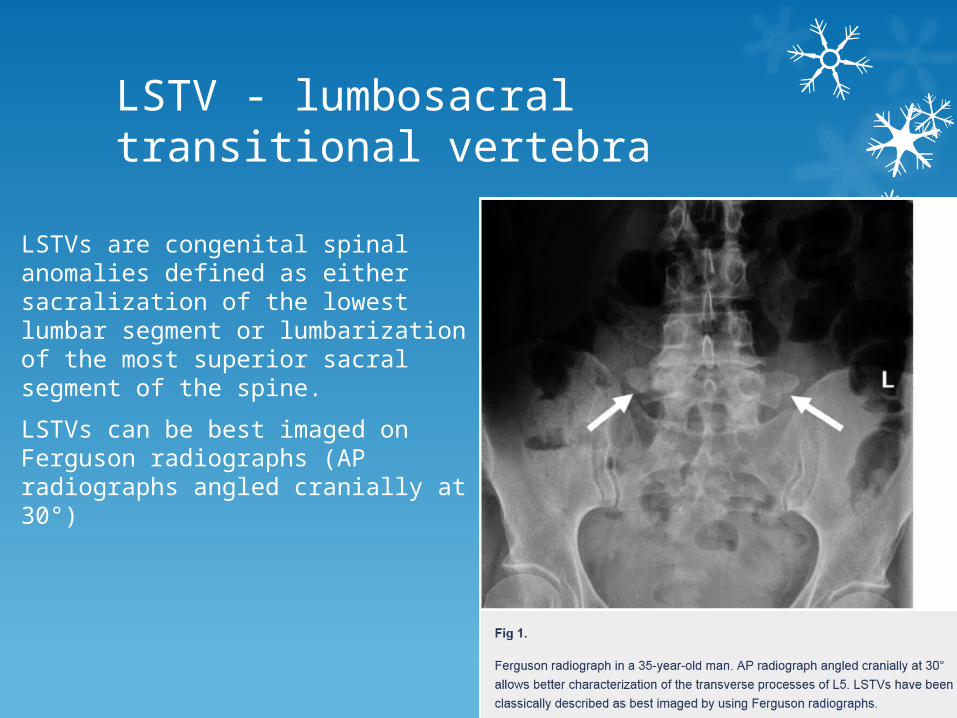
LSTV - lumbosacral transitional vertebra
LSTVs are congenital spinal anomalies defined as either sacralization of the lowest lumbar segment or lumbarization of the most superior sacral segment of the spine.
LSTVs can be best imaged on Ferguson radiographs (AP radiographs angled cranially at 30°)
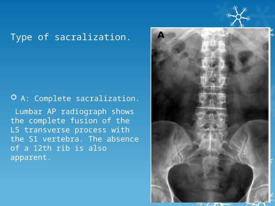
Type of sacralization.
A: Complete sacralization.
Lumbar AP radiograph shows the complete fusion of the L5 transverse process with the S1 vertebra. The absence of a 12th rib is also apparent.
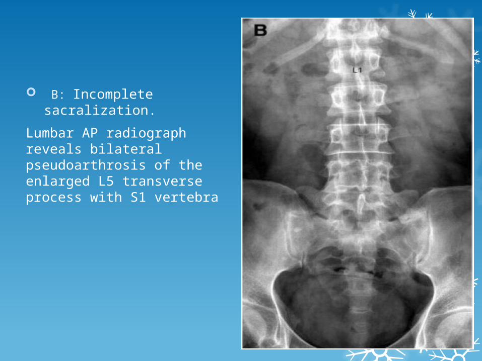
B: Incomplete sacralization.
Lumbar AP radiograph reveals bilateral pseudoarthrosis of the enlarged L5 transverse process with S1 vertebra
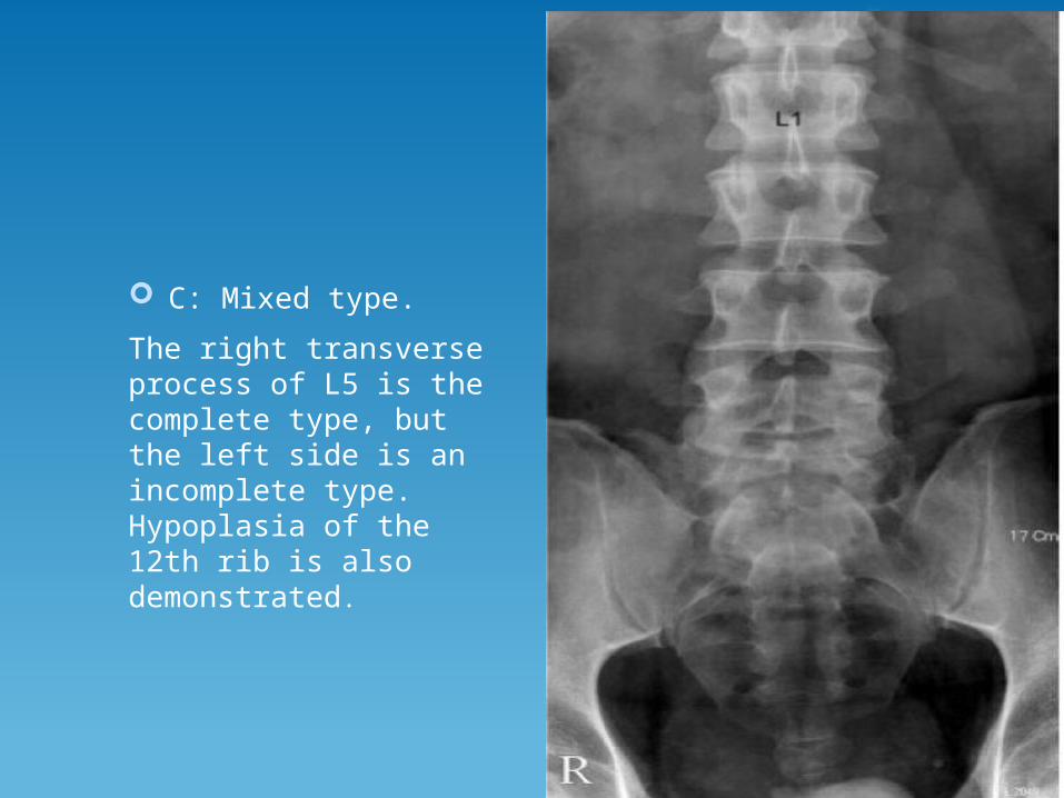
C: Mixed type.
The right transverse process of L5 is the complete type, but the left side is an incomplete type. Hypoplasia of the 12th rib is also demonstrated.
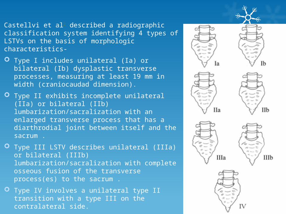
Castellvi et al2 described a radiographic classification system identifying 4 types of LSTVs on the basis of morphologic characteristics-
Type I includes unilateral (Ia) or bilateral (Ib) dysplastic transverse processes, measuring at least 19 mm in width (craniocaudad dimension).
Type II exhibits incomplete unilateral (IIa) or bilateral (IIb) lumbarization/sacralization with an enlarged transverse process that has a diarthrodial joint between itself and the sacrum .
Type III LSTV describes unilateral (IIIa) or bilateral (IIIb) lumbarization/sacralization with complete osseous fusion of the transverse process(es) to the sacrum .
Type IV involves a unilateral type II transition with a type III on the contralateral side.
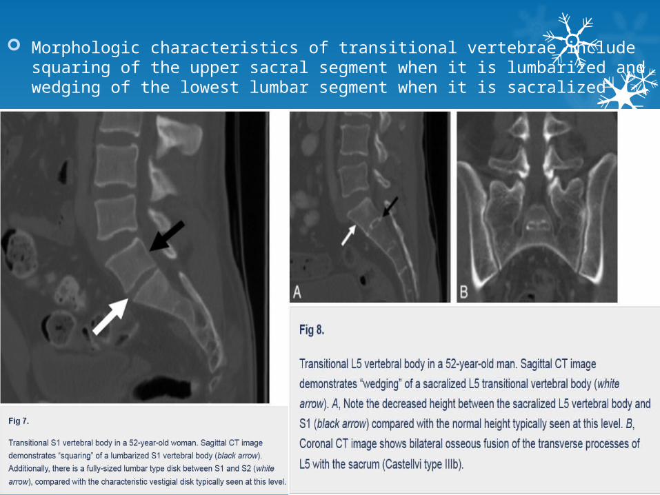
Morphologic characteristics of transitional vertebrae include squaring of the upper sacral segment when it is lumbarized and wedging of the lowest lumbar segment when it is sacralized
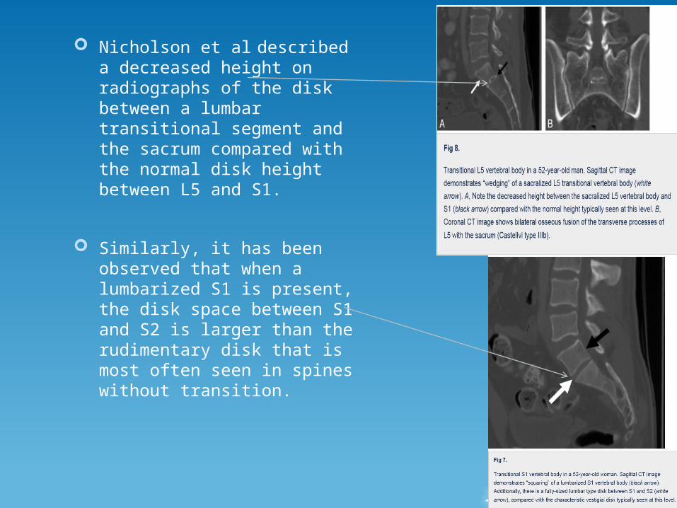
Nicholson et al described a decreased height on radiographs of the disk between a lumbar transitional segment and the sacrum compared with the normal disk height between L5 and S1.
Similarly, it has been observed that when a lumbarized S1 is present, the disk space between S1 and S2 is larger than the rudimentary disk that is most often seen in spines without transition.
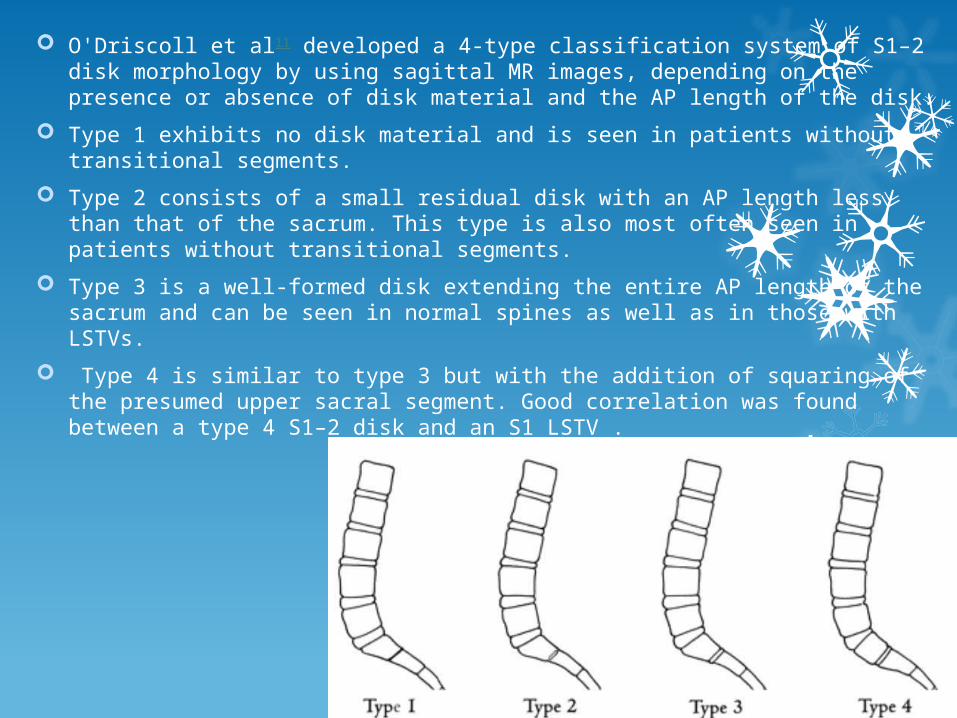
O'Driscoll et al11 developed a 4-type classification system of S1–2 disk morphology by using sagittal MR images, depending on the presence or absence of disk material and the AP length of the disk.
Type 1 exhibits no disk material and is seen in patients without transitional segments.
Type 2 consists of a small residual disk with an AP length less than that of the sacrum. This type is also most often seen in patients without transitional segments.
Type 3 is a well-formed disk extending the entire AP length of the sacrum and can be seen in normal spines as well as in those with LSTVs.
Type 4 is similar to type 3 but with the addition of squaring of the presumed upper sacral segment. Good correlation was found between a type 4 S1–2 disk and an S1 LSTV .
Numbering Technique
With a sagittal MR localizer, the vertebrae may be counted in a caudad direction from C2 rather than cephalad from L5.
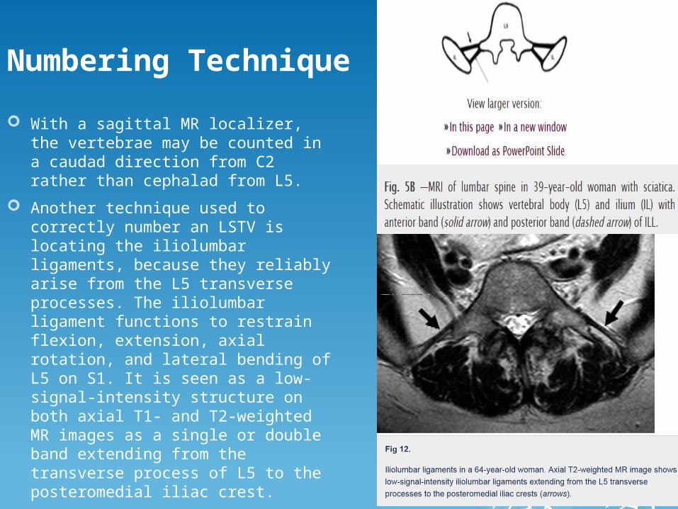
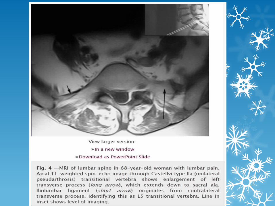
Another technique used to correctly number an LSTV is locating the iliolumbar ligaments, because they reliably arise from the L5 transverse processes. The iliolumbar ligament functions to restrain flexion, extension, axial rotation, and lateral bending of L5 on S1. It is seen as a low-signal-intensity structure on both axial T1- and T2-weighted MR images as a single or double band extending from the transverse process of L5 to the posteromedial iliac crest.
When an iliolumbar ligament was seen to arise above the LSTV, then the vertebral body with the iliolumbar ligament was labeled L5 and the LSTV, as S1.
In case of L5 transition(sacralisation), the ligament was absent or smaller and no ligament was seen at the level.
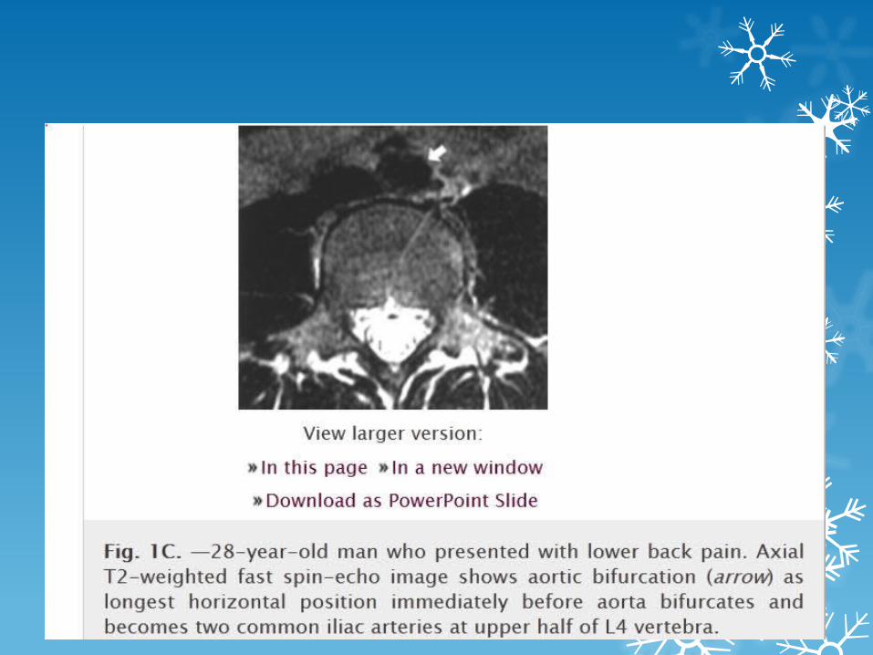
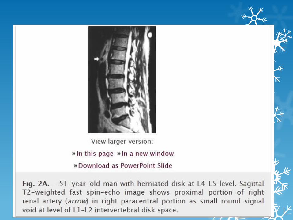
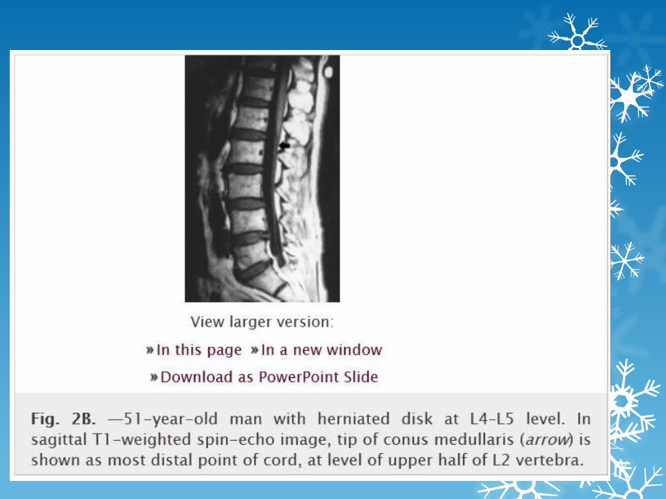
The use of anatomic markers, including the aortic bifurcation, right renal artery, and conus medullaris has been reported to be least reliable. Although the right renal artery is usually located at the L1–2 disk space, 25% of the time it is either not imaged or is present at another location. Variability may be seen in the position of the aortic bifurcation as it has been found at L4 in 83% of patients.
Methods for numbering the lumbar vertebrae on lumbosacral radiographs –
1. the transverse diameter of the pedicle and the interpedicular distance are frequently largest at the L5 vertebra.
2. the L3 vertebra usually exhibits the longest transverse process of all lumbar segments.
3. the size of the neural foramen gradually increases to the last lumbar vertebra.
4. the iliac wings are also generally found at the level between the L4 and L5 vertebrae.
Essentially without high-quality imaging of the entire spine, there is no foolproof method for accurately numbering a transitional segment.
Bertolotti Syndrome
The low back pain of this syndrome is currently thought to be of varying
etiologies, subsequently arising from different locations:
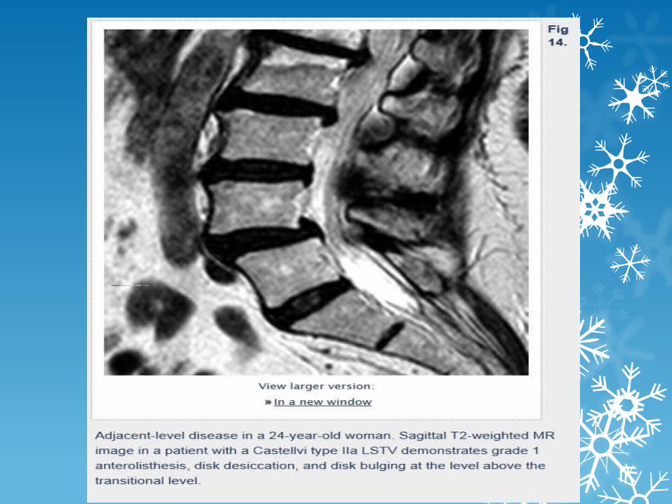
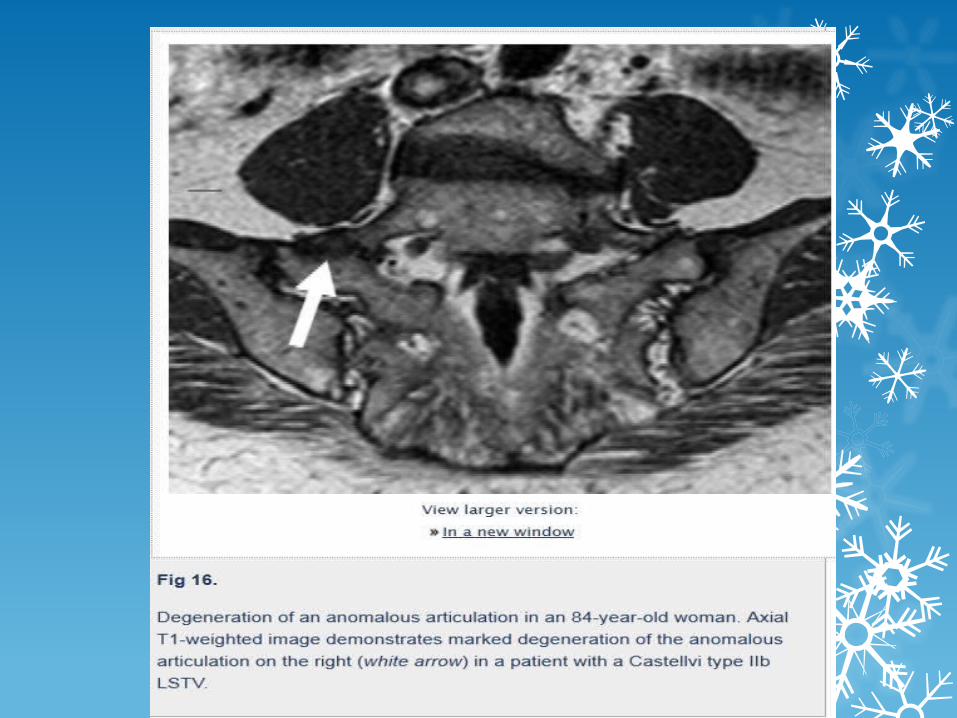
1) disk, spinal canal, and posterior element pathology at the level
above a transition.
2) degeneration of the anomalous articulation between an LSTV and the
sacrum.
3) facet joint arthrosis contralateral to a unilateral fused or articulating
LSTV.
4) extraforaminal stenosis secondary to the presence of a broadened
transverse process
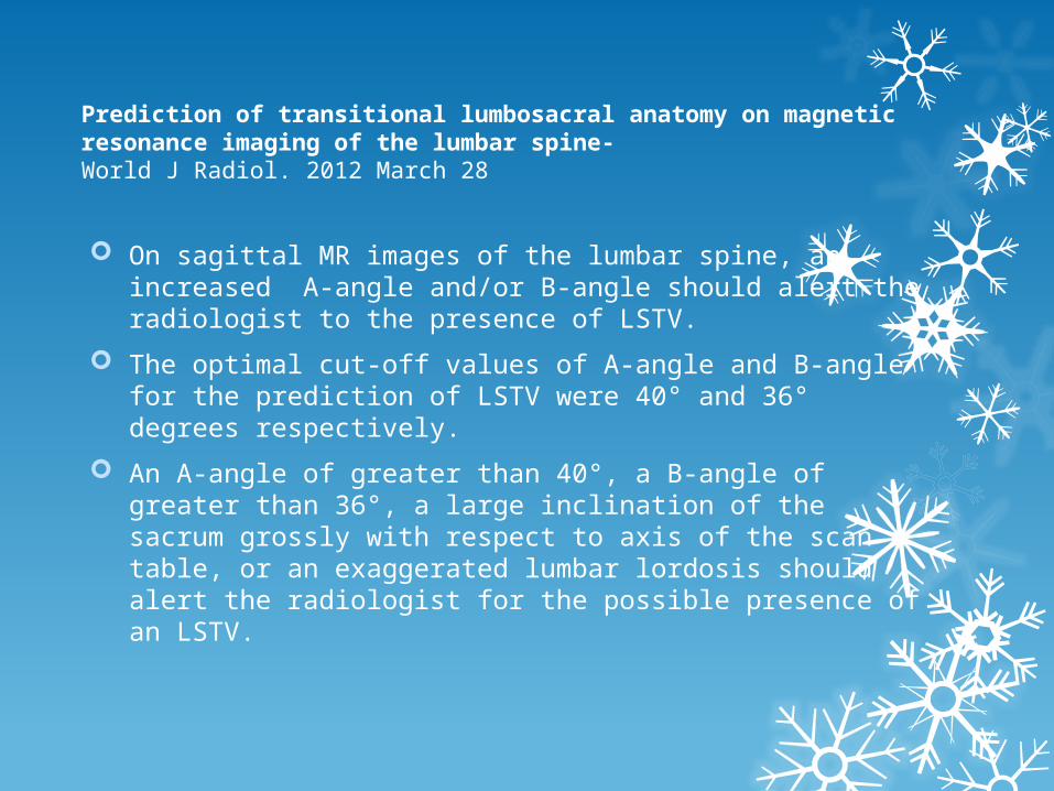
Prediction of transitional lumbosacral anatomy on magnetic resonance imaging of the lumbar spine-World J Radiol. 2012 March 28
On sagittal MR images of the lumbar spine, an increased A-angle and/or B-angle should alert the radiologist to the presence of LSTV.
The optimal cut-off values of A-angle and B-angle for the prediction of LSTV were 40° and 36° degrees respectively.
An A-angle of greater than 40°, a B-angle of greater than 36°, a large inclination of the sacrum grossly with respect to axis of the scan table, or an exaggerated lumbar lordosis should alert the radiologist for the possible presence of an LSTV.
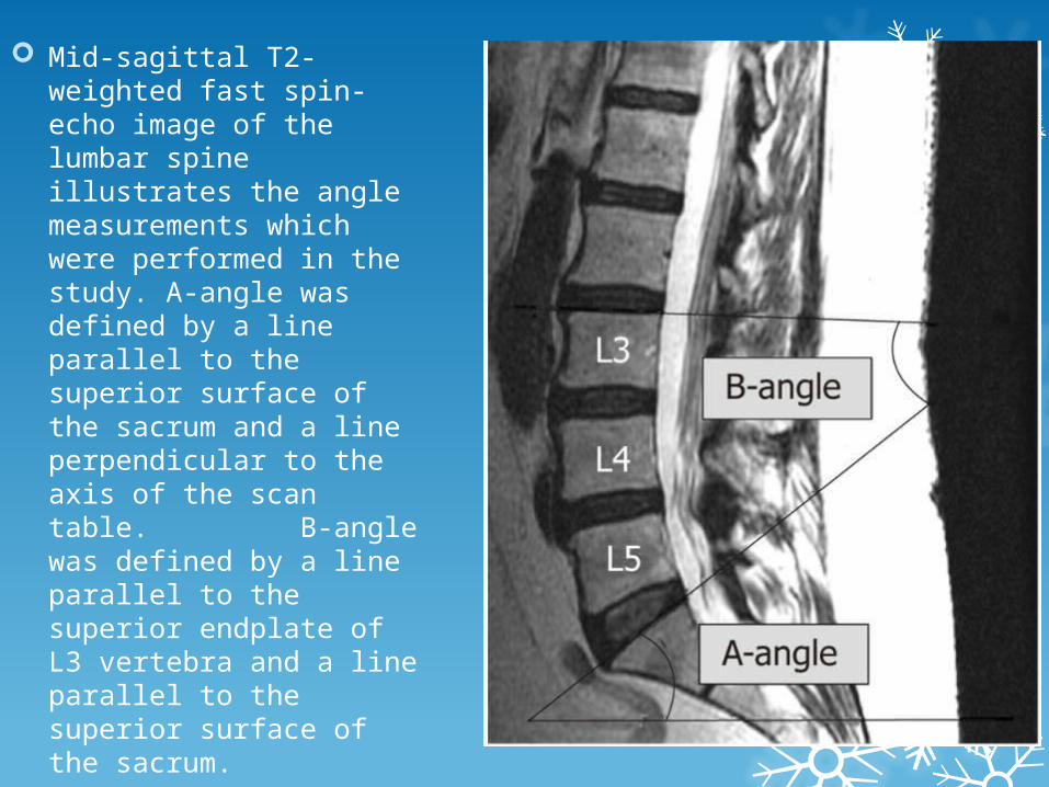
Mid-sagittal T2-weighted fast spin-echo image of the lumbar spine illustrates the angle measurements which were performed in the study. A-angle was defined by a line parallel to the superior surface of the sacrum and a line perpendicular to the axis of the scan table. B-angle was defined by a line parallel to the superior endplate of L3 vertebra and a line parallel to the superior surface of the sacrum.
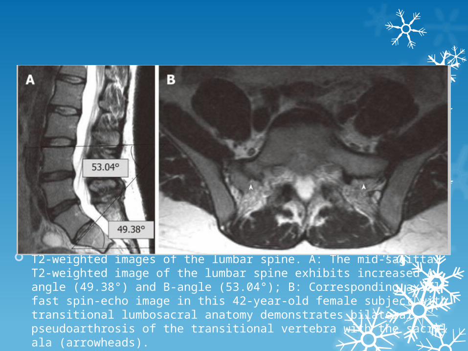
T2-weighted images of the lumbar spine. A: The mid-sagittal T2-weighted image of the lumbar spine exhibits increased A-angle (49.38°) and B-angle (53.04°); B: Corresponding axial fast spin-echo image in this 42-year-old female subject with transitional lumbosacral anatomy demonstrates bilateral pseudoarthrosis of the transitional vertebra with the sacral ala (arrowheads).

THANK U