transdermal drug delivery: innovative pharmaceutical ...€¦ · transdermal drug delivery (tdd)....
TRANSCRIPT
Pharmaceutics 2015, 7, 438-470; doi:10.3390/pharmaceutics7040438
pharmaceutics ISSN 1999-4923
www.mdpi.com/journal/pharmaceutics
Review
Transdermal Drug Delivery: Innovative Pharmaceutical Developments Based on Disruption of the Barrier Properties of the stratum corneum
Ahlam Zaid Alkilani 1,2,*, Maelíosa T.C. McCrudden 1 and Ryan F. Donnelly 1
1 School of Pharmacy, 97 Lisburn Road, Queens University Belfast, Belfast BT9 7BL, Northern
Ireland, UK; E-Mails: [email protected] (M.T.C.M.); [email protected] (R.F.D.) 2 Faculty of Pharmacy, Zarqa University, Zarqa 132222, Jordan
* Author to whom correspondence should be addressed; E-Mail: [email protected];
Tel.: +962-0-795-294-329; Fax: +962-0-5832-1120.
Academic Editor: Diganta B. Das
Received: 19 August 2015 / Accepted: 13 October 2015 / Published: 22 October 2015
Abstract: The skin offers an accessible and convenient site for the administration of
medications. To this end, the field of transdermal drug delivery, aimed at developing safe
and efficacious means of delivering medications across the skin, has in the past and continues
to garner much time and investment with the continuous advancement of new and innovative
approaches. This review details the progress and current status of the transdermal drug
delivery field and describes numerous pharmaceutical developments which have been
employed to overcome limitations associated with skin delivery systems. Advantages and
disadvantages of the various approaches are detailed, commercially marketed products are
highlighted and particular attention is paid to the emerging field of microneedle technologies.
Keywords: transdermal; drug delivery; velocity based device; ultrasound; thermal ablation;
mechanical and electrical approaches; microneedle
OPEN ACCESS

Pharmaceutics 2015, 7 439
1. Introduction
The most common routes of drug delivery are the oral and parenteral routes with the majority of small
molecule drugs conventionally delivered orally [1,2]. The oral route has the advantage of pre-determined
doses, portability and patient self-administration. For these reasons, the oral route remains the most
convenient means of delivering medications [3,4]. However, most therapeutic peptides or proteins are
not delivered by the oral route, due to rapid degradation in the stomach and size-limited transport across
the epithelium [5]. The primary mode of administering macromolecules is therefore via injection [1,5,6]
which is not without limitations, such as the invasive nature of injections eliciting pain and lower
acceptance/compliance by patients, in addition to the requirement for administration by a trained
administrator [5–7]. Rationally, the conventional routes of medication delivery have many inherent
limitations which could potentially be overcome by advanced drug delivery methodologies such as
transdermal drug delivery (TDD).
2. Transdermal Drug Delivery (TDD)
TDD is a painless method of delivering drugs systemically by applying a drug formulation onto intact
and healthy skin [2,5]. The drug initially penetrates through the stratum corneum and then passes through
the deeper epidermis and dermis without drug accumulation in the dermal layer. When
drug reaches the dermal layer, it becomes available for systemic absorption via the dermal
microcirculation [8,9]. TDD has many advantages over other conventional routes of drug
delivery [10–12]. It can provide a non-invasive alternative to parenteral routes, thus circumventing issues
such as needle phobia [2]. A large surface area of skin and ease of access allows many placement options
on the skin for transdermal absorption [5]. Furthermore, the pharmacokinetic profiles of drugs are more
uniform with fewer peaks, thus minimizing the risk of toxic side effects [2]. It can improve patient
compliance due to the reduction of dosing frequencies and is also suitable for patients who are
unconscious or vomiting, or those who rely on self-administration [13]. TDD avoids pre-systemic
metabolism, thus improving bioavailability [2,4]. With reference to the use of the skin as a novel site for
vaccination strategies, this organ is known to be replete with dendritic cells in both the epidermal and
dermal layers which play a central role in immune responses making TDD an attractive vaccination route
for therapeutic proteins and peptides [3,14]. The requirement for an inexpensive and non-invasive means
of vaccination, especially in the developing world [3,14,15], has given rise to substantial research
focused on the development of simple, needle-free systems such as TDD for vaccination purposes. This
theme will be explored further in Section 4.5.2 of this review.
3. A Brief Review of Skin Structure
Skin is the most accessible and largest organ of the body with a surface area of 1.7 m2, compromising
16% of the total body mass of an average person [16–18]. The main function of the
skin is to provide a protective barrier between the body and the external environment against
microorganisms, the permeation of ultraviolet (UV) radiation, chemicals, allergens and the loss of water
[19]. Skin can be divided into three main regions: (1) the outermost layer, the epidermis, which contains

Pharmaceutics 2015, 7 440
the stratum corneum; (2) the middle layer, the dermis and (3) the inner most layer, the hypodermis (Figure
1) [5,20,21].
Figure 1. Anatomy of the skin. (Reprinted from [22] with permission. Copyright 2012 Elsevier).
3.1. Epidermis
The epidermis is the outermost layer of the skin and varies in thickness with approximately 0.8 mm
on the palms of the hands and soles of the feet [19]. It consists of multi-layered regions of epithelial cells
and the viable epidermis is often referred to as the epidermal layers below the stratum
corneum [8,19]. The cellular content of the epidermis consists predominantly of keratinocytes
(approximately 95% of cells), with other cells of the epidermal layers including melanocytes,
Langerhans cells and merkel cells [14].The stratum corneum is the most superficial layer of the
epidermis [19,23,24]. It is in direct contact with the external environment and its barrier properties may
be partly related to its very high density (1.4 g/cm3 in the dry state) and its low hydration of
15%–20% [25]. The cells of the stratum corneum are composed mainly of insoluble keratins (70%) and
lipid (20%) [25]. Water in the stratum corneum is associated with keratin in the corneocytes [19,26].
3.2. Dermis
The dermis is approximately 2–3 mm thick and consists of collagenous (70%) and elastin fibres which
give strength and elasticity to the skin [17]. Blood vessels found in the dermis provide nutrients for both
the dermis and epidermis. Nerves, macrophages and lymphatic vessels are also present in the dermis
layer, as depicted in Figure 1 [23].

Pharmaceutics 2015, 7 441
3.3. Hypodermis
The hypodermis or subcutaneous layer is the deepest layer of the skin and consists of a network of
fat cells [17]. It is the contact layer between the skin and the underlying tissues of the body, such as
muscles and bone. Therefore, the major functions of the hypodermis are protection against physical
shock, heat insulation and support and conductance of the vascular and neural signals of the skin [27].
Hypodermis-resident fat cells account for approximately 50% of the body’s fat with the other
predominant cells of the hypodermis consisting of fibroblasts and macrophages [28].
3.4. Drug Penetration Routes
There are two possible routes of drug penetration across the intact skin, namely the transepidermal
and transappendegeal pathways, which have been diagrammatically presented in Figure 2. The
transepidermal pathway involves the passage of molecules through the stratum corneum, an
architecturally diverse, multi-layered and multi-cellular barrier. Transepidermal penetration can be
termed intra- or inter-cellular [29]. The intra-cellular route through corneocytes, terminally differentiated
keratinocytes, allows the transport of hydrophilic or polar solutes. Transport via inter-cellular spaces
allows diffusion of lipophilic or non-polar solutes through the continuous lipid matrix. The
transappendegeal route involves the passage of molecules through sweat glands and across the hair
follicles [5,30].
Figure 2. Possible drug penetration routes across human skin. (Reprinted from [30] with
permission. Copyright 2012 Elsevier).

Pharmaceutics 2015, 7 442
3.5. Kinetics of TDD
An understanding of the kinetics of skin permeation is necessary for development of successful TDD
systems. In order to evaluate any TDD, the assessment of percutaneous absorption of molecules is a very
important step. Percutaneous absorption is the penetration of substances into various layers of skin and
permeation across the skin into the systemic circulation [8,31–33]. Percutaneous absorption of molecules
is a step wise process involving:
i. Penetration: The entry of a substance into a particular layer of the skin;
ii. Partitioning from the stratum corneum into the aqueous viable epidermis;
iii. Diffusion through the viable epidermis and into the upper dermis;
iv. Permeation: The penetration of molecules from one layer into another, which is different both
functionally and structurally from the first layer;
v. Absorption: The uptake of a substance into the systemic circulation.
In delivery systems involving transdermal patches, the drug is stored in a reservoir (reservoir type) or
drug dissolved in a liquid or gel-based reservoir (matrix type).The starting point for the evaluation of the
kinetics of drug release from a transdermal patch is an estimation of the drug compound’s maximum
flux across the skin (flux (J)) which is typically expressed in units of μg/cm2/h) (Equation 1) (Figure 3).
Based on Fick’s law of diffusion, the transport of therapeutic molecules across skin will be maintained
until the concentration gradient ceases to exist [33–35].
Figure 3. Description of flux across the skin from a transdermal patch where J is the
molecular flux, C2 is the concentration of the active molecule in the patch, C1 is the
concentration of the active molecule in the body, D is the diffusion coefficient; L is the cross
sectional thickness of diffusion, and t is the diffusion time. The equation indicates Fick’s law
of diffusion. (Reprinted from [33] with permission. Copyright 2013 Elsevier).
= − (1)
where D is the diffusion coefficient and dc / dt is the concentration gradient.
Conventional TDD is possible only if the drug possesses certain physiochemical properties. The rate
of permeation across the skin (dQ / dt) is given by [35]: / = ( − ) (2)

Pharmaceutics 2015, 7 443
where Cd and Cr are the concentration of the skin penetrant in the donor compartment (i.e., on the surface
of stratum corneum) and in the receptor compartment (i.e., body), respectively. P is the permeability
coefficient of the skin tissue to the penetrant. = ∗ / (3)
where D is he diffusion coefficient obtained from the permeability coefficient, P, the solute partition
coefficient, K, and L is the overall thickness of skin tissues.
From equation (2) it is clear that a constant rate of drug permeation can be obtained only when
Cd >> Cr i.e., the drug concentration at the surface of the stratum corneum. Cd is consistently and
substantially greater than the drug concentration in the body Cr. The equation becomes: / = ∗ (4)
The cumulative amount permeating (Q) the barrier with the effective surface area of permeation (A)
at a given time (t) is calculated by using Equation (5) [35]: = (5)
The permeability coefficient (P) can be obtained from the slope of a plot of cumulative permeation
of diffusant vs. time obtained from an experimental permeation study. A typical plot of permeation study
is shown in Figure 4.
Figure 4. A typical plot of permeation study.
L is the swollen membrane thickness. As shown in Figure 4, the cumulative permeation curve has
two portions. The initial portion of the curve represents non-steady state diffusion and the linear
portion corresponds to steady state diffusion. The non-steady portion of the curve can be described
mathematically by Fick’s second law, while the linear portion can be expressed by Fick’s first
law [35]. The time required to reach steady state is called the lag time (tLag). The lag time can be
determined by extrapolating the linear portion of permeation vs. the time curve to the time axis. With lag
time, Equation (6) is rewritten as (7) [35].

Pharmaceutics 2015, 7 444
= [ ( − )]/ (6)
= ( − ) (7)
The lag time can be calculated by Equation (8) [35]: = / (8)
Transdermal systems should be formulated to provide the maximum thermodynamic driving force
for passive diffusion across the skin which is saturated with a sufficient payload of the drug to ensure
delivery of drugs across the skin. The ability of approved transdermal drugs to penetrate the skin
varies widely from the extremely permeable nicotine to compounds, such as buprenorphine and the
progestins, which have very low predicted fluxes.
The first transdermal patch approved for systemic delivery in 1979 was a patch for the sustained,
three days delivery of scopolamine in the treatment of motion sickness [1,34]. Transdermal delivery is
currently restricted to approximately 17 drug molecules that are approved by the US Food and Drug
Administration (FDA) (Table 1) [2,4]. The limited number of drug molecules seen in s Table 1 reflects
the difficulty of meeting the dual challenge of potent pharmacological activity and the correct
physicochemical properties to enable skin penetration [34,36].These approved molecules are all of low
molecular weight (MW < 500 Da), a balanced lipophilicity (log P = 1–3), and a measurable solubility
both in oil and in water because TDD systems require both breaching the lipophilic stratum corneum
and resorption into the aqueous central compartment of the systemic circulation [8,34]. Moreover, high
pharmacological potency of drug molecules is required to become a feasible candidate for TDD [8,36,37].
The limited permeability of molecules is due to the outermost layer of the skin, the stratum corneum [38,39].
This “dead” layer of tissue has the ability to prevent the permeation of foreign compounds including
drug molecules and therefore acts as a very effective barrier [39,40]. In order to enhance drug permeation
across the skin, a number of chemical and physical methods have been devised [3,5,34].

Pharmaceutics 2015, 7 445
Table 1. Daily dose ranges and selected physicochemical and pharmacokinetic properties of currently approved transdermally delivered drugs.
(Reprinted from [34] with permission. Copyright 2014 Elsevier).
Drug (Year of Approval) Dose/Day (mg) MW (Da) Log P a Cl (L/h) T1/2 (h)b F (%) c Cp,eff (ng/mL) e
Scopolamine (1979) 0.3 303 0.98 672 2.9 27 0.04
Glyceryl trinitrate (1981) 2.4–15 227 01.62 966 0.04 <1 0.1–5
Clonidine (1984) 0.1–0.3 230 2.42 ± 0.52 13 6–20 95 0.2–2.0
Estradiol (1986) 0.025–0.1 272 4.01 615–790 0.05 3-5 0.04–0.06
Fentanyl (1990) 0.288–2.4 337 4.05 27–75 3–12 32 1.0
Nicotine (1991) 7–21 162 1.17 78 2 30 10–30
Testosterone (1993) 0.3–5 288 3.32 0.17–1.7 <1 10–100
Estradiol & Norethisterone
Acetate (1998) 0.025–0.050 0.125–0.250 272 340 4.01 3.99 2–3 6–8 d 3–5 64 0.04–0.07 0.8–1.1
Norelgestromin &
EthinylEstradiol (2001) 0.025–0.050 0.125–0.250 327 296 3.90 ± 0.47 3.67 28 17 d 40 0.8 0.05
Estradiol & Levonorgestrel
(2003) 0.025–0.050 0.125–0.250 272 312 4.01 3.72 ± 0.49 3 28 d 3-5 0.03–0.05 0.1–0.2
Oxybutynin (2003) 3.9 357 4.02 ± 0.52 2 6 1.0–5.0
Selegeline (2006) 6–12 187 2.90 84 10 10 2.0–3.0
Methylphenidate (2006) 26–80 233 2.15 ± 0.42 20 2–3 5–20 5.0–25
Rotigotine (2007) 1–3 315 4.58 ± 0.72 600 5–7 d n/a ~1.0
Rivastigmine (2007) 4.6–9.5 250 2.34 ± 0.16 108 1.5 40 ~10
Granisetron (2008) 3.1 312 2.55 ± 0.28 33–76 healthy
15–34 patients
4–6 healthy
9–12 patients 60 0.7–9.5
Buprenorphine (2010) 0.12–1.68 468 4.98 55 22–36 d n/a 0.1–0.4
a Log{octanol-water partition coefficient (P)}: either experimental or calculated (mean ± SD) values; b Terminal half-life post-oral or IV dosing; c Oral bioavailability; d
Terminal half-life following transdermal delivery; e Pharmacologically effective plasma concentration.

Pharmaceutics 2015, 7 446
Due to the aforementioned challenges associated with successful drug permeation across the skin, a
number of different, innovative approaches have been explored and developed to overcome these
challenges. These will be discussed in the subsequent sections of this review.
4. Techniques for Enhancement of Skin Permeabilisation
Technologies used to modify the barrier properties of the stratum corneum can be divided into
passive/chemical or active/physical methodologies (Figure 5). Passive methods include the influencing
of drug and vehicle interactions and optimization of formulation, in order to modify the stratum corneum
structure [29,41,42]. Passive methods are relatively easy to incorporate into transdermal patches such as
chemical enhancers and emulsions [43]. However, the main drawback of passive methods may be a lag
time in drug release incurred with obvious negative influence on rapid onset drugs, such as insulin.
Figure 5. Approaches for enhancing drug transport across the skin.
One of the most widely used passive approaches is the use of chemical penetration enhancers which
facilitate drug permeation across the skin by increasing drug partitioning into the barrier domain of the
stratum corneum, without long-term damage to the skin [11,44]. Penetration enhancers have several
mechanisms of action such as: increasing the fluidity of the stratum corneum lipid bilayers, interaction
with intercellular proteins, disruption or extraction of intercellular lipids, increase of the drug’s
thermodynamic activity and increase in stratum corneum hydration [11,44,45]. Several types of
penetration enhancers are known and they can be divided into several groups based on their chemical
structure, rather than their mechanism of action [32,44]. Most of these have mixed modes of action so
it is difficult to classify them according to this characteristic. Examples of commonly investigated
penetration enhancers are alcohols, sulphoxides, azone, pyrrolidones, essential oil, terpenes and terpenoids,
fatty acids, water and urea [44,45]. However, the major limitation for penetration enhancers is that their
efficacy is often closely correlated with the occurrence of skin irritation [32,45]. Gels have been used in
TDD and recent developments in the technology have introduced new variations of semisolid vehicles
such as proniosomes and microemulsion gels into the field of penetration enhancers [43]. Proniosomes
are non-ionic based surfactant vesicles, they are known as ‘‘dry niosomes’’ because they may require
Pharmaceutics 2015, 7 447
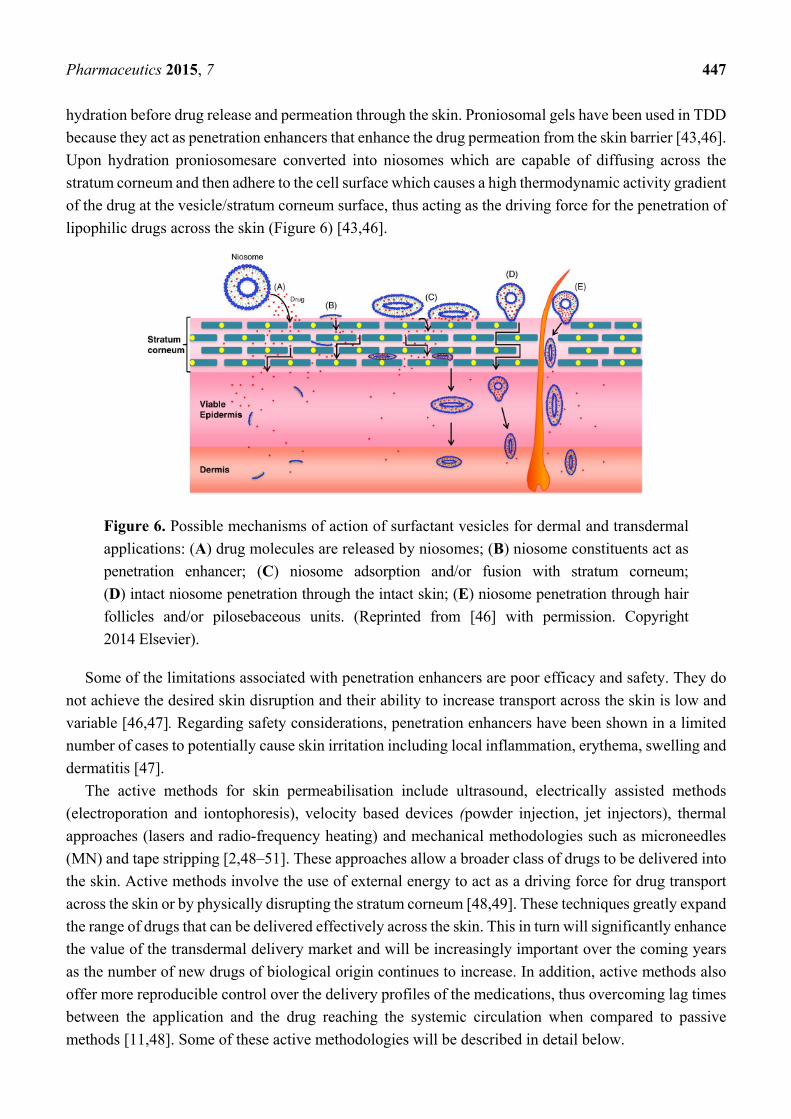
hydration before drug release and permeation through the skin. Proniosomal gels have been used in TDD
because they act as penetration enhancers that enhance the drug permeation from the skin barrier [43,46].
Upon hydration proniosomesare converted into niosomes which are capable of diffusing across the
stratum corneum and then adhere to the cell surface which causes a high thermodynamic activity gradient
of the drug at the vesicle/stratum corneum surface, thus acting as the driving force for the penetration of
lipophilic drugs across the skin (Figure 6) [43,46].
Figure 6. Possible mechanisms of action of surfactant vesicles for dermal and transdermal
applications: (A) drug molecules are released by niosomes; (B) niosome constituents act as
penetration enhancer; (C) niosome adsorption and/or fusion with stratum corneum;
(D) intact niosome penetration through the intact skin; (E) niosome penetration through hair
follicles and/or pilosebaceous units. (Reprinted from [46] with permission. Copyright
2014 Elsevier).
Some of the limitations associated with penetration enhancers are poor efficacy and safety. They do
not achieve the desired skin disruption and their ability to increase transport across the skin is low and
variable [46,47]. Regarding safety considerations, penetration enhancers have been shown in a limited
number of cases to potentially cause skin irritation including local inflammation, erythema, swelling and
dermatitis [47].
The active methods for skin permeabilisation include ultrasound, electrically assisted methods
(electroporation and iontophoresis), velocity based devices (powder injection, jet injectors), thermal
approaches (lasers and radio-frequency heating) and mechanical methodologies such as microneedles
(MN) and tape stripping [2,48–51]. These approaches allow a broader class of drugs to be delivered into
the skin. Active methods involve the use of external energy to act as a driving force for drug transport
across the skin or by physically disrupting the stratum corneum [48,49]. These techniques greatly expand
the range of drugs that can be delivered effectively across the skin. This in turn will significantly enhance
the value of the transdermal delivery market and will be increasingly important over the coming years
as the number of new drugs of biological origin continues to increase. In addition, active methods also
offer more reproducible control over the delivery profiles of the medications, thus overcoming lag times
between the application and the drug reaching the systemic circulation when compared to passive
methods [11,48]. Some of these active methodologies will be described in detail below.

Pharmaceutics 2015, 7 448
4.1. Ultrasound Devices
Ultrasound is an oscillating sound pressure wave that has long been used for many research areas
including physics, chemistry, biology, engineering and others in a wide range of frequencies [2,50].
Ultrasound, sonophoresis, or phonophoresis can be defined as the transport of drugs across the skin by
application of ultrasound perturbation at frequencies of 20 kHz–16 MHz which has a sufficient intensity
to reduce the resistance of skin [2,5]. The use of ultrasound has resulted in the effective delivery of
various different categories and classes of drugs, regardless of their electrical characteristics, by increasing
skin permeability. These drugs have included hydrophilic and large molecular weight drugs [39].
However, the mechanism of action is still not clearly understood or characterized [50]. The proposed
mechanisms by which ultrasound effects tissues and cells include thermal effects and cavitation effects
caused by collapse and acoustic streaming which can be explained as oscillation of cavitation bubbles
in the ultrasound field [5]. Ultrasound can increase the temperature of the insonated medium (the skin)
by the absorption of the sound waves with a frequency greater than the upper limit of the human hearing
range. Obviously, the higher the medium’s absorption coefficient, the higher the increase in temperature
and thus the greater the thermal effect [50]. All recent studies point out that cavitation is believed to be
the predominant mechanism in the enhancement of TDD via ultrasound treatment [50].
The concept of ultrasound for use in TDD was initially reported by Fellinger and Schmidt in
1950 for the successful treatment of polyarthritis using hydrocortisone ointment combined with sonophoresis
[52–54]. However, the first ultrasound device for transdermal application was approved in 2004 by the
FDA for the delivery of local dermal anesthesia by the Sontra Medical, SonoPrep® (Figure 7). Since that
time, ultrasound has been widely used as a TDD system in the treatment of many other diseases including
bone joint diseases and bursitis [2]. Many challenges must be overcome before such devices gain
commercial acceptance however. Some of these challenges include: availability of easy-to-use devices;
the determination of the duration of treatment required; gaining a full understanding of how the
technology functions; broadening of the range of drugs that can be delivered and evaluation of the safety
profiles of the devices [5,39,55,56]. Examples of undesirable side effects of ultrasound approaches were
observed by Singer et al. (1998) when it was shown that low-intensity ultrasound caused minor skin
reactions in dogs while high-intensity ultrasound was capable of inducing second-degree burns [56].
Limitations such as this must be overcome before these innovations can garner full acceptance.

Pharmaceutics 2015, 7 449
Figure 7. The SonoPrep® ultrasound device (Reprinted from [50] with permission. Copyright
2014 Elsevier).
4.2. Electrical Techniques
4.2.1. Electroporation
The two major means of electrically-facilitated TDD are iontophoresis and electroporation [2,4].
In electroporation, cells are temporarily exposed to high intensities of electric pulses that lead to the
formation of aqueous pores in the lipid bilayers of the stratum corneum, thus allowing the diffusion of
drugs across skin [5,57–59]. The technique was first described by Neumann et al. in 1982 [59]. Usage
of high voltage pulses (50–500 V) for short times of only one second have been shown to increase
transport across the skin for different molecular weight drugs ranging from small e.g., fentanyl,
timolol [60,61], orcalcein [62], to high molecular weight drugs such as LHRH, calcitonin, heparin or
FITC–dextran with molecular weights up to 40 kDa [58,63–66]. However, the main drawbacks are the
lack of quantitative delivery, cell death with high fields and potential damage to labile drugs, e.g., those
of protein origin [57,67].
4.2.2. Iontophoresis
Iontophoresis involves the application of physiologically acceptable electrical currents (0.1–1.0 mA/cm2) to
drive charged permeants into the skin through electrostatic effects and make ionic drugs pass through
the skin into the body by its potential gradient [5,20,58,68–71]. Unlike other transdermal enhancement
methodologies, it acts mainly by involving a second driving force, the electrical potential gradient as
companion to the concentration gradient across the skin since uncharged species can also be delivered
through electroosmosis (Figure 8) [5,70].

Pharmaceutics 2015, 7 450
Figure 8. Schematic representation of an iontophoresis patch (Reprinted from [40] with
permission. Copyright 2000 Elsevier).
Phoresor®, Lidosite®, and E-trans® are examples of three commercially developed iontophoretic
delivery systems (Figure 9). The first approved commercial iontophoretic patch system was LidoSite®,
which was developed to deliver lidocaine for fast dermal anaesthesia. The system was composed of a
disposable pre-filled patch, re-usable battery-powered controller and a flexible interconnect module [20].
Iontophoresis has a minor effect on skin structure over short treatment periods due to the low-voltage
nature of the applied electric current, when compared to electroporation [5].
(a) (b)
Figure 9. Commercially developed iontophoretic delivery systems: (a) Phoresor® and
(b) Lidosite®.
Several factors affect iontophoretic TDD, including pH of the donor solution, electrode type, buffer
concentration, current strength and the type of current employed [20,69,72,73]. The molecular size of
the solute/drug is an important factor in determining its feasibility for successful iontophoretic delivery.
The flux of smaller and more hydrophilic ions is faster than larger ions [72–74]. A plethora of studies
correlating flux as a function of molecular weight have been conducted and it was found that the transport
of compounds decreased with increase in molecular weight (chloride > amino acid > nucleotide >
tripeptide > insulin) [22,72,75–78]. There is a linear relationship between the current and drug flux
across the skin but the current is limited to 1 mA in order to facilitate patient comfort and consider safety

Pharmaceutics 2015, 7 451
concerns as with increasing current, the risk of nonspecific vascular reactions (vasodilatation) also
increases [72]. Furthermore, the maximum time that the devices can be applied is 3 min, in order to
prevent local skin irritation or burns. The maximum physiologically acceptable iontophoretic current is
0.5 mA/cm2 [79]. The current should be adequately high to provide a desired flux rate but it should not
irritate the skin [80]. The use of continuous direct current (DC) can decrease the drugs flux due to its
polarization effect on the skin [69]. In order to overcome this problem, pulsed current has been used [81].
Overall, only a limited number of studies have been carried out comparing pulsed direct current
iontophoresis vs. continuous direct current iontophoresis. Recently, Kotzki et al. 2015 showed that
pulsed iontophoresis of treprostinil significantly enhanced cutaneous blood flow compared with
continuous iontophoresis [69]. The most common electrodes that are used in iontophoresis are aluminum
foil, platinum and silver/silver chloride electrodes [73]. However, the preferred one is Ag/AgCl since it
resists the changes in pH. In addition, the electrode materials used for iontophoretic delivery should be
harmless to the body and flexible so as to be applied closely to the body surface [73].
The maximum molecular weight for iontophoretic delivery has not been extensively studied, although
it is estimated that molecules with a molecular weight less than 12,000 Da may be successfully delivered
across skin via iontophoresis [79]. In order to deliver molecules greater than 12,000 Da, an alternate
means of overcoming the barrier properties of the stratum corneum must be sought. However, it was
found that a small protein, cytochrome c (12.4 kDa) was delivered non-invasively across intact skin
[82,83]. Afterwards, ribonuclease A, with isoelectric point of 8.64 (13.6 kDa), was successfully delivered
across porcine and human skin [84]. More recently, it was shown that transdermal iontophoresis was
also able to deliver biologically active human basic fibroblast growth factor (hbFGF; 17.4 kDa) in
therapeutically relevant amounts corresponding to those used in clinical trials and animal studies [85,86].
The applications of iontophoresis can be classified into therapeutic and diagnostic applications.
Iontophoresis has been used in various diagnostic applications e.g. diagnosing cystic fibrosis [87] and
recently for monitoring blood glucose levels [88].The major advantage of iontophoresis in diagnostic
applications is that there is no mechanical penetration or disruption of the skin involved in this
approach [89,90].
4.3. Velocity Based Devices
Velocity based devices, either powder or liquid jet injections, employ a high-velocity jet with
velocities ranging from 100 to 200 m/s to puncture the skin and deliver drugs using a power source
(compressed gas or a spring) [91]. The concept of jet injectors for use in drug delivery was first explored
in the early of 1930s by Arnold Sutermesiter [11]. Since then, interest in this method of drug delivery
has expanded significantly and two types of liquid jet injectors have been developed; single-dose jet
injectors (disposable cartridge jet injectors) and multi-use-nozzle jet injectors (MUNJIs) [91]. Jet
injections have been used for more than 50 years for parenteral delivery of vaccines, as well as small
molecules, such as anesthetics and antibiotics [11]. A jet injector is a needle free device capable of
delivering electronically controlled doses of medication which result in improved consistency of delivery
and reduced pain for the patient (Figure 10) [48,92].

Pharmaceutics 2015, 7 452
Figure 10. Methods for intradermal injection. (Reprinted from [93] with permission.
Copyright 2005 Elsevier).
Liquid-jet injectors propel liquid from a nozzle with an orifice diameter ranging from 50 to 360 μm,
which is much smaller than the outer diameter of a standard hypodermic needle (810 μm for a 21G
needle) [20,93,94]. The jet can deliver drug into different layers of skin e.g., intradermal (i.d.),
subcutaneous (s.c.) or intramuscular (i.m.), by changing the jet velocity and orifice diameter [20]. The
major advantage of using needle free devices relates to concerns regarding safe needle disposal and
avoidance of accidental needle stick injuries [20]. However, the risk of cross contamination is not
excluded, since splash back of interstitial liquid from the skin may contaminate the nozzle [95].
Therefore the use of multi-use nozzle jet injectors has been terminated and such devices are now only
used for multi-dose drug delivery to the same individual, e.g., the Tjet®device which delivers somatropin
(human growth hormone (hGH)) (Figure 11).
Figure 11. Commercially available jet injector Tjet®device.
Powder jet injectors have an advantage over liquid jet injectors of delivering solid drugs or vaccines
to the skin, so the stability of the formulation will be increased and the necessity for cold storage will be
avoided, which simplifies transportation and reduces associated costs. Powder jet injectors may be
formulated from nano-or micro-particles containing the active or lyophilised drugs and antigens [20,96].
Excellent bioavailability for a number of drugs has been reported but the intermittent pain and bruising
caused to patients has restricted wide acceptance of jet injectors [91]. Regarding the levels of pain

Pharmaceutics 2015, 7 453
experienced by volunteers, some reports state no difference in the pain recorded when comparing jet
injectors to conventional needle injections [97] but others have reported higher pain scores [98].
The basic design of solid jet injectors consists of compressed gas as the power source, drug loaded
compartment containing solid drug formulation, and a nozzle to direct the flow of particles towards the
skin [99]. By triggering the actuation mechanism, compressed gas expands and forces drug powder
through a nozzle into the skin. Upon impacting on the skin, particles create micronsized holes and deposit
in the stratum corneum or viable epidermis. The most important parameters that govern particle delivery
across the stratum corneum are particle properties (size, density) and impact velocity e.g., for DNA
vaccination, the particle size range should be between 0.5 and 3 µm [11].
4.4. Thermal Approaches (Lasers and Radio-Frequency Heating)
Thermal ablation is a method used to deliver drugs systemically through the skin by heating the
surface of the skin, which depletes the stratum corneum selectively at that site of heating only, without
damaging deeper tissues [49,100]. Many methods could be used to cause thermal ablation such as
laser [101], radiofrequency [49,102], in addition to electrical heating elements [49]. In order to generate
the high temperatures needed to ablate the stratum corneum without damaging the underlined epidermis,
the thermal exposure should be short, so the temperature gradient across the stratum corneum can be
high enough to keep the skin surface extremely hot but the temperature of the viable epidermis does not
experience a significant temperature rise [100].
4.4.1. Laser Thermal Ablation
Laser methodologies have been used in clinical therapies for the treatment of dermatological
conditions such as pigmented lesions [101,103,104]. The main mechanism of laser thermal ablation of
the skin is the selective removal of the stratum corneum without damaging deeper tissues, thus enhancing
the delivery of lipophilic and hydrophilic drugs into skin layers [26,45,104,105]. Lasers ablate the
stratum corneum by deposition of optical energy, which causes evaporation of water and formation of
microchanels in the skin [106]. In addition, such approaches have been used to extract interstitial fluid
for subsequent measurement of glucose levels in diabetic patients [49,101,103]. However, the degree of
barrier disruption achieved is controlled by wavelength, pulse length, tissue thickness, pulse energy, tissue
absorption coefficient, pulse number, duration of laser exposure and pulse repetition rate [48,58,107].
Baron et al., 2003 demonstrated that pre-treatment with the laser followed by lidocaine cream was found
to reduce the onset of lidocaine action to 3–5 min in human volunteers [106]. However, the structural
changes in the skin must be assessed, especially at the higher intensities of laser employed that may be
needed to enhance the transport of large molecular weight therapeutics [108,109].
4.4.2. Radiofrequency (RF) Thermal Ablation
Radiofrequency (RF) thermal ablation involves the placement of a thin, needle-like electrode directly
into the skin and application of high frequency alternating current (~100 kHz) which produces
microscopic pathways in the stratum corneum, through which drugs can permeate (Figure 12) [49,100].
Exposure of skin cells to a high frequency (100–500 kHz) causes ionic vibrations within the tissue which

Pharmaceutics 2015, 7 454
attempts to localize the heating to a specific area of the skin and thus ablate the cells in that region,
resulting in drug transport across the skin [110]. This technology may enable transdermal delivery of a
wide variety of hydrophilic drugs and macromolecules using a low-cost, fully disposable device [49].
Figure 12. Schematic diagram of drug delivery using thermal ablation: (a) micro-electrodes
are pressed against the skin, (b) skin is ablated via heating due to RF energy or resistive
heating in the electrodes, (c) after removing the ablation device, (d) micropores formed.
(Reprinted from [11] with permission. Copyright 2008 Elsevier).
4.5. Mechanical Approaches to Mediate Skin Permeation
The use of hypodermic needles, often associated with phobia, pain and the risk of needle-stick injuries
have been used to overcome some of the delivery limitations often experienced when delivering
macromolecular compounds [111,112]. Some innovative methodologies have been explored to
overcome these issues and include the use of MN and tape stripping. These concepts will be described
further below.
4.5.1. Tape Stripping
Tape stripping is a simple method for removing the stratum corneum layer by repeated application of
adhesive tapes [113]. The amount of stratum corenum removed by a single adhesive tape depends on
many factors such as the thickness of the stratum corenum, the age of the patient, the composition and
amount of lipid which varies depending on the anatomical site and finally, skin parameters such as
transepidermal water loss (TEWL) and pH. In addition, other factors also affect the amount of stratum
corneum removed by tape stripping, such as the force of removal of the tape from the skin and the
duration of pressure on the skin [113,114]. Tape stripping is a robust and simple method. However, many
parameters should be taken into consideration before and during the application of this procedure, such as
the duration of pressure on the skin, in order to remove the stratum corneum homogeneously.
4.5.2. Microneedle (MN) Arrays
MN arrays, minimally invasive drug delivery systems, were developed to overcome some of the
disadvantages commonly associated with hypodermic needle usage and in order to address and improve
patient compliance. MN arrays have the potential to be used as an alternative to hypodermic and
subcutaneous needle technologies (Figure 13) [12,34,111,112]. MN technologies have been subject to
intensive research and development efforts by both academic and industrial researchers with some
devices currently in clinical development and others awaiting FDA approval [1,34]. Also the number of

Pharmaceutics 2015, 7 455
publications describing MN as novel minimally invasive devices for drug delivery purposes has grown
exponentially in recent years [1,34,112,115]. As MN combine the ease of use of a transdermal patch
with the effectiveness of delivery achieved using conventional hypodermic needle and syringes, they
continue to elicit interest and investment [34,116].
Figure 13. Schematic representation of the mechanism of action of a microneedle array
device. The device perforates the stratum corneum (SC) providing direct access of drugs to
the underlying viable epidermis, without reaching blood vessels and nerve fibres located in
the dermis (Reprinted from [12] with permission. Copyright 2013 Elsevier).
MN are multiple microscopic projections typically assembled on one side of a supporting base or
patch, generally ranging from 25 to 2000 μm in height [5,12], 50 to 250 μm in base width and 1 to
25 μm in tip diameter [20,112,117,118]. The needles should be of suitable length, width and shape to
avoid nerve contact when inserted into skin layers [117–119]. They are usually designed in arrays in
order to improve the surface contact with the skin and facilitate penetration of therapeutic molecules into
the skin [112,120]. MN are designed to create transient aqueous conduits across the skin, thereby
enhancing flux of the molecules ranging from small hydrophilic molecules such as alendronate [52] to
macromolecules, including low molecular weight heparins [4,121], insulin [122] and vaccines [123], in
a pain-free manner [112,124]. Besides the aspect of pain-free delivery, there are many other advantages
of MN technologies, such as: the fact that they do not cause bleeding [125]; eliminate transdermal dosing
variability of small molecules [45,126]; only minimal introduction of pathogens through MN-induced
holes [124,127]; potential for self-administration [1,128]; the potential to overcome and reduce instances
of accidental needle-sticks injuries and the risk of transmitting infections [12,112], in addition to the
ease of MN waste disposal [11,112].
As conceded previously in this review, one of the most attractive applications of MN arrays is to use
them in vaccination and indeed, self-vaccination strategies. The skin contains high concentrations of
adaptive and innate immune cells including macrophages, Langerhans cells, and dermal dendritic cells.
To date, only oral typhoid vaccine is approved for self-administration in patients’ homes [129]. Injecting
vaccines into the epidermis or dermis is immunologically superior to injecting into the muscle where
much lower populations of immune cells reside and this MN approach therefore offers excellent
amplification potential for the desired immune response [21,130]. As a result, the dose required to

Pharmaceutics 2015, 7 456
vaccinate through the skin via MN will be much lower than that require dosing of a conventional needle
and syringe injection into the muscle. Vaccine delivery via the skin offers easier and painless
administration. Moreover, these MN vaccination devices can be manufactured inexpensively [5,34,112].
The first two commercially marketed MN-based products are Intanzia® and Micronjet® which are
based on metal and silicon MN, respectively (Figure 14) [131]. Intanza® is the first influenza vaccine
that targets the dermis, a highly immunogenic area. It was developed and licensed by Sanofi Pasteur
MSD Limited and is being marketed in two strengths; Intanza® 9 µg for adults aged between 18 and
59 years and Intanza® 15 µg for adults of 60 years and above. The Intanza® influenza vaccine system
has a needle length of 1.5 mm [132]. MicronJet is a single use, MN-based device for intradermal delivery
of vaccines and drugs. It was developed and licensed by NanoPass.
(a) (b)
Figure 14. Current commercial MNs-based products (a) Intanza® and (b) MicronJet®.
Several companies have been working towards the development of MN-based drug or vaccine
products, including 3M, Clearside Biomedical, NanoPass Technologies, Corium International,
TheraJect, Circassia, Radius Health, Lohmann Therapeutic Systems (LTS) and Zosano Pharma. Zosano
has developed a transdermal patch consisting of an array of titanium MN coated with parathyroid
hormone (PTH) (20 to 40 μg) attached to an adhesive patch and applied via a reusable applicator across
the skin [1,133]. A second study involving the Zosano titanium MN patch system was carried out by
Ameri et al. 2014 to evaluate the feasibility of titanium MN usage to deliver recombinant human growth
hormone (rhGH) [126]. In this study, it was found that the bioavailability of the rhGH MNpatch and the
current subcutaneous injection products (Norditropin®) were similar which indicates that this MN
product can be used as a patient-friendly alternative to subcutaneous injection of Norditropin® [126,133].
The 3M Microneedle Technologies (MTS) has developed coated MN to deliver water-soluble, polar and
ionic molecules, such as lidocaine, through the skin. This system has successfully delivered drugs to the
skin within seconds and provide rapid onset of local analgesia (~1 min) which facilitates routine or
emergency procedures [51,134].
The shape and geometry of MN is critical during design and fabrication [22,135–137]. The needles
must be capable of inserting into the skin without breaking and the needles should be of suitable length,
width and shape to avoid nerve contact and create efficient pathways for the delivery of small drugs,
macromolecules and nanoparticles, as well as for fluid extraction, depending on the objectives of each
device [115,117,119,138]. The elastic properties of human skin may prevent effective MN penetration

Pharmaceutics 2015, 7 457
by twisting of the skin fibers around the needles during application, particularly in the case of blunt and
short MN [117]. To date, many papers have described the fabrication of various MN from different
materials using various micro-moulding processes or other methods, such as lasers [112,139,140].
Generally, there are four strategies of TDD using MN (Figure 15) [22,123]. These are solid, coated,
dissolvable and hollow MN. A novel fifth MN-type, namely hydrogel MN have garnered much interest
in the recent past and are presented in Figure 16.
Figure 15. A schematic representation of four different MN application methods used to
facilitate drug delivery transdermally. (a) Solid MNs for increasing the permeability of a
drug formulation by creating micro-holes across the skin; (b) Coated MNs for rapid
dissolution of the coated drug into the skin; (c) Dissolvable MNs for rapid or controlled
release of the drug incorporated within the microneedles; (d) Hollow MNs used to puncture
the skin and enable release of a liquid drug following active infusion or diffusion of the
formulation through the needle bores. (Reprinted from [11] with permission. Copyright
2008 Elsevier).

Pharmaceutics 2015, 7 458
(1) Hollow MN are used to deliver drug solutions via the “poke and flow” method; which involves
insertion of the MN into tissue and then a drug solution can be transported through the bore of the MN
in similar fashion to a hypodermic needles [141,142] but hollow MN usually require very precise and
high cost manufacturing technology [111]. Passive diffusion of the drug solution may occur through the
MN, but active delivery allows for more rapid rates of delivery. Active delivery requires a driving force,
a syringe can be used to drive the solution through the MN into the tissue but some studies have
combined the MN systems with a pump or pressurised gas [143,144].
(2) “Poke and patch” mainly for solid MN by piercing the upper layers of the skin with solid MN and
creating microchannels followed by application of a drug formulation (e.g., patch, gel) at that site
piercing [5,112]. The skin pretreatment creates micro-conduits in the skin, thereby enhancing flux of the
molecules through the skin.
(3) “Coat and poke” by piercing the skin with drug coated solid MN, which solve the problem of two-
step application and provide extremely quick drug delivery [111,145]. Delivery from coated MN was
found to be attractive especially for high molecular weight molecules [146]. However, drug delivery is
limited due to the small dimensions of the MN shaft and tip [146–148].
(4) “Poke and release” for dissolving/porous/hydrogel forming MN through which drug will diffuse
into systemic circulation (Figure 16). The materials from which the MN are produced act as drug depots
holding the drugs until the trigger for release occurs, i.e., dissolution in the case of dissolvable MN or
swelling in the case of hydrogel MN [22,131,149]. This strategy eliminates the need for sharps disposal,
and the possibility of accidental reuse of MN. Moreover, dissolvable MN patches have been reported to
successfully deliver both small (MW 500 Da) and macro molecules (MW 500 Da) in “poke and release”
approaches [25,26].
A wide variety of MN types and designs have been shown to be effective for the transdermal delivery
of a diverse range of molecules, both in vitro and in vivo [10,12]. The potential now exists to greatly
expand the range and types of drugs that can be delivered effectively across the skin. This will
significantly enhance the value of the transdermal delivery market and will be increasingly important
over the coming years as the number of new drugs of biological origin continues to increase. Future
studies will be needed to address potential regulatory concerns over the use of MN devices, as well as
focusing on the design and development of processes to enable a low cost, efficient means for MN mass
production. A number of other physical approaches such as sonophoresis, electroporation, ultrasound
and iontophoresis have been combined with MN in order to enhance permeation of drugs. Kolli et al.,
2012 determined that the transdermal delivery of Prochlorperazine Edisylate was significantly enhanced
when MN were used in conjunction with iontophoresis [150]. Moreover, the delivery of ropinirole
hydrochloride by MN and iontophoresis was significantly higher compared to modulated iontophoresis
alone [151].

Pharmaceutics 2015, 7 459
Figure 16. Novel hydrogel-forming MN facilitate controlled transdermal drug delivery.
(a) An expanded view of the backing layer, drug-loaded adhesive patch and solid crosslinked
hydrogel MN array which constitutes an integrated hydrogel MN patch; (b) Application of
the integrated hydrogel MN patch to the skin surface; (c) Diffusion of water into the MN
array leading to controlled swelling of the arrays and diffusion of drug molecules from the
adhesive patch through the hydrogel conduit; (d) Intact hydrogel MN arrays following
removal from the skin. (Reprinted from [12] with permission. Copyright 2013 Elsevier).
5. MN Overcome Many of the Limitations Associated with Other TDD Methodologies
Various limitations associated with each of the outlined TDD approaches have been documented
throughout this review. To this end, MN methodologies may prove an efficacious, cost-effective and
patient friendly alternative in choosing a TDD system for delivery of a host of drug molecules. With this
in mind, some of the advantages of MN approaches over other TDD systems are outlined below.
As a novel and minimally invasive approach, MN are capable of creating superficial pathways across
the skin for small drugs, macromolecules, nanoparticles, or fluid extractions to achieve enhanced
transdermal drug delivery [152].Their sharp tips are short enough to limit contact with skin nerves, thus
preventing pain sensation [125] and they are narrow enough to induce minimal trauma and reduce the
opportunities for infections to develop following insertion [127]. This method combines the efficacy of
conventional injection needles with the convenience of transdermal patches, while minimizing the
disadvantages of these administration methods [152,153]. Moreover, MN can be manufactured using
various types of material e.g., polymers, metal or silicon. Biocompatible and biodegradable polymers
can be safely applied to the skin and are generally cost-effective. Various polymeric materials such as
poly-L-lactic acid, poly-glycolic acid, poly-carbonate, poly-lactic-co-glycolic acid (PLGA), poly-
dimethylsiloxane, a copolymer of methyl vinyl ether and maleic anhydride, carboxymethyl cellulose,
maltose, dextrin and galactose have all been used to fabricate MN [139]. MN can also deliver a wide

Pharmaceutics 2015, 7 460
range of drugs ranging from small molecular weight e.g., ibuprofen [124] to high molecular weight e.g.,
ovalbumin compounds [131]. Immunization programs in developing countries via MN could be applied
with minimal medical training and with lower associated costs. In addition, MN arrays have recently
been used as an alternative approach in the minimally-invasive sampling of fluids from patients, without
causing pain or bleeding in the advancement of novel therapeutic drug monitoring systems [12].
Although MN technologies show tremendous promise in the field of TDD, there are still relatively few
FDA approved MN devices. A number of challenges which must be addressed before MN become
widely available include scale up manufacture to industrial levels which will require considerable
planning and standardization. In addition, MN device regulatory considerations must be established and
addressed. These issues may include but are not limited to, issues surrounding product sterility; the
potential for accidental reuse of certain MN modalities (e.g., solid MN), appropriate packaging and
manufacturing aspects and the potential for undesired immunological effects. These must all be
addressed before MN devices receive widespread approval. Moreover, the choice of appropriate
biomaterials for preparation of MN is limited due to lack of mechanical strength, poor control of drug
delivery, and limitation of drug loading dose [154,155].
6. Conclusions
In conclusion, the TDD sector continues to grow and develop with rapid expansion in fundamental
knowledge feeding industrial development. In time, it is hoped that technological advancements in TDD
will lead to enhanced disease prevention, diagnosis and control, with concomitant improvement in
health-related quality of life for patients worldwide. To this end, this review has charted the development
of numerous novel TDD methodologies, highlighting the advantages and disadvantages of each
approach. Due to the exponential growth in investment and interest in MN technologies and the
numerous associated advantages of this approach, particular attention was paid to this TDD system.
Author Contributions
Ahlam Zaid Alkilani and Maelíosa T.C. McCrudden conceived, researched and wrote the paper. Ryan
F. Donnelly critiqued the paper.
Conflicts of Interest
The authors declare no conflict of interest.
References
1. Anselmo, A.C.; Mitragotri, S. An Overview of Clinical and Commercial Impact of Drug Delivery
Systems. J. Control. Release 2014, 190, 15–28.
2. Han, T.; Das, D.B. Potential of Combined Ultrasound and Microneedles for Enhanced Transdermal
Drug Permeation: A Review. Eur. J. Pharm. Biopharm. 2015, 89, 312–328.
3. Brambilla, D.; Luciani, P.; Leroux, J. Breakthrough Discoveries in Drug Delivery Technologies:
The Next 30 years. J. Control. Release 2014, 190, 9–14.

Pharmaceutics 2015, 7 461
4. Ita, K. Transdermal Drug Delivery: Progress and Challenges. J. Drug Deliv. Sci. Technol. 2014,
24, 245–250.
5. Schoellhammer, C.M.; Blankschtein, D.; Langer, R. Skin Permeabilization for Transdermal Drug
Delivery: Recent Advances and Future Prospects. Expert Opin. Drug Deliv. 2014, 11, 393–407.
6. McCrudden, M.T.; Singh, T.R.R.; Migalska, K.; Donnelly, R.F. Strategies for Enhanced Peptide
and Protein Delivery. Ther. Deliv. 2013, 4, 593–614.
7. Kermode, M. Unsafe Injections in Low-Income Country Health Settings: Need for Injection Safety
Promotion to Prevent the Spread of Blood-Borne Viruses. Health. Promot. Int. 2004, 19, 95–103.
8. Donnelly, R.F.; Singh, T.R.R.; Morrow, D.I.; Woolfson, A.D. Microneedle-Mediated Transdermal
and Intradermal Drug Delivery; Wiley: Hoboken, NJ, USA, 2012.
9. Kretsos, K.; Kasting, G.B. A Geometrical Model of Dermal Capillary Clearance. Math. Biosci.
2007, 208, 430–453.
10. Donnelly, R.F.; Singh, T.R.R.; Garland, M.J.; Migalska, K.; Majithiya, R.; McCrudden, C.M.;
Kole, P.L.; Mahmood, T.M.T.; McCarthy, H.O.; Woolfson, A.D. Hydrogel‐Forming Microneedle
Arrays for Enhanced Transdermal Drug Delivery. Adv. Funct. Mater. 2012, 22, 4879–4890.
11. Arora, A.; Prausnitz, M.R.; Mitragotri, S. Micro-Scale Devices for Transdermal Drug Delivery.
Int. J. Pharm. 2008, 364, 227–236.
12. Tuan-Mahmood, T.; McCrudden, M.T.; Torrisi, B.M.; McAlister, E.; Garland, M.J.; Singh, T.R.R.;
Donnelly, R.F. Microneedles for Intradermal and Transdermal Drug Delivery. Eur. J. Pharm. Sci.
2013, 50, 623–637.
13. Prausnitz, M.R.; Langer, R. Transdermal Drug Delivery. Nat. Biotechnol. 2008, 26, 1261–1268.
14. Suh, H.; Shin, J.; Kim, Y. Microneedle Patches for Vaccine Delivery. Clin. Exp. Vaccine Res.
2014, 3, 42–49.
15. Peasah, S.K.; Azziz-Baumgartner, E.; Breese, J.; Meltzer, M.I.; Widdowson, M. Influenza Cost
and Cost-Effectiveness Studies globally—A Review. Vaccine 2013, 31, 5339–5348.
16. Menon, G.K. New Insights into Skin Structure: Scratching the Surface. Adv. Drug Deliv. Rev.
2002, 54, S3–S17.
17. Liu, X.; Kruger, P.; Maibach, H.; Colditz, P.B.; Roberts, M.S. Using Skin for Drug Delivery and
Diagnosis in the Critically Ill. Adv. Drug Deliv. Rev. 2014, 77, 40–49.
18. Williams, A.C.; Barry, B.W. Penetration Enhancers. Adv. Drug Deliv. Rev. 2012, 64, 128–137.
19. Benson, H.A.; Watkinson, A.C. Topical and Transdermal Drug Delivery: Principles and Practice;
Wiley: Hoboken, NJ, USA, 2012.
20. Gratieri, T.; Alberti, I.; Lapteva, M.; Kalia, Y.N. Next Generation Intra-and Transdermal
Therapeutic Systems: Using Non-and Minimally-Invasive Technologies to Increase Drug Delivery
into and Across the Skin. Eur. J. Pharm. Sci. 2013, 50, 609–622.
21. Lambert, P.H.; Laurent, P.E. Intradermal Vaccine Delivery: Will New Delivery Systems
Transform Vaccine Administration? Vaccine 2008, 26, 3197–3208.
22. van der Maaden, K.; Jiskoot, W.; Bouwstra, J. Microneedle Technologies for (Trans) Dermal Drug
and Vaccine Delivery. J. Control. Release 2012, 161, 645–655.
23. Domínguez-Delgado, C.L.; Rodríguez-Cruz, I.M.; López-Cervantes, M.; Escobar-Chávez, J.;
Merino, V. The Skin a Valuable Route for Administration of Drugs. Current Technologies to
Increase the Transdermal Delivery of Drugs; Bentham Science: Sharjah, UAE, 2010; pp. 1–22.

Pharmaceutics 2015, 7 462
24. El Maghraby, G.; Barry, B.; Williams, A. Liposomes and Skin: From Drug Delivery to Model
Membranes. Eur. J. Pharm. Sci. 2008, 34, 203–222.
25. Walters, K.A. Dermatological and Transdermal Formulations; CRC Press: Boca Raton, FL,
USA, 2002.
26. Alexander, A.; Dwivedi, S.; Giri, T.K.; Saraf, S.; Saraf, S.; Tripathi, D.K. Approaches for Breaking
the Barriers of Drug Permeation through Transdermal Drug Delivery. J. Control. Release 2012,
164, 26–40.
27. Sherwood, A.; Bower, J.K.; McFetridge-Durdle, J.; Blumenthal, J.A.; Newby, L.K.; Hinderliter, A.L.
Age Moderates the Short-Term Effects of Transdermal 17β-Estradiol on Endothelium-Dependent
Vascular Function in Postmenopausal Women. Arterioscler. Thromb. Vasc. Biol. 2007, 27,
1782–1787.
28. McLennan, D.N.; Porter, C.J.; Charman, S.A. Subcutaneous Drug Delivery and the Role of the
Lymphatics. Drug Discov. Today Technol. 2005, 2, 89–96.
29. Schuetz, Y.B.; Naik, A.; Guy, R.H.; Kalia, Y.N. Emerging Strategies for the Transdermal Delivery
of Peptide and Protein Drugs. Expert Opin. Drug Deliv. 2005, 2, 533–548.
30. Shahzad, Y.; Louw, R.; Gerber, M.; du Plessis, J. Breaching the Skin Barrier through Temperature
Modulations. J. Control. Release 2015, 202, 1–13.
31. Dhote, V.; Bhatnagar, P.; Mishra, P.K.; Mahajan, S.C.; Mishra, D.K. Iontophoresis: A Potential
Emergence of a Transdermal Drug Delivery System. Sci. Pharm. 2012, 80, 1–28.
32. Dragicevic, N.; Maibach, H.I. Percutaneous Penetration Enhancers Chemical Methods in
Penetration Enhancement: Drug Manipulation Strategies and Vehicle Effects; Springer: New
York, NY, USA, 2015.
33. Subramony, J.A. Needle Free Parenteral Drug Delivery: Leveraging active transdermal
technologies for pediatric use. Int. J. Pharm. 2013, 455, 14–18.
34. Wiedersberg, S.; Guy, R.H. Transdermal Drug Delivery: 30 Years of War and Still Fighting!
J. Control. Release 2014, 190, 150–156.
35. Ghosh, T.K.; Jasti, B.R. Theory and Practice of Contemporary Pharmaceutics; CRC press: Boca
Raton, FL, USA, 2004.
36. Choy, Y.B.; Prausnitz, M.R. The Rule of Five for Non-Oral Routes of Drug Delivery: Ophthalmic,
Inhalation and Transdermal. Pharm. Res. 2011, 28, 943–948.
37. Lipinski, C.A.; Lombardo, F.; Dominy, B.W.; Feeney, P.J. Experimental and Computational
Approaches to Estimate Solubility and Permeability in Drug Discovery and Development Settings.
Adv. Drug Deliv. Rev. 2012, 64, 4–17.
38. Barry, B. Novel Mechanisms and Devices to Enable Successful Transdermal Drug Delivery. Eur.
J. Pharm. Sci. 2001, 14, 101–114.
39. Park, D.; Park, H.; Seo, J.; Lee, S. Sonophoresis in Transdermal Drug Deliverys. Ultrasonics 2014,
54, 56–65.
40. Naik, A.; Kalia, Y.N.; Guy, R.H. Transdermal Drug Delivery: Overcoming the skin’s Barrier
Function. Pharm. Sci. Technol. Today 2000, 3, 318–326.
41. Chen, Y.; Shen, Y.; Guo, X.; Zhang, C.; Yang, W.; Ma, M.; Liu, S.; Zhang, M.; Wen, L.
Transdermal Protein Delivery by a Coadministered Peptide Identified Via Phage Display. Nat.
Biotechnol. 2006, 24, 455–460.

Pharmaceutics 2015, 7 463
42. El Maghraby, G.M.; Williams, A.C.; Barry, B.W. Can drug‐bearing Liposomes Penetrate Intact
Skin? J. Pharm. Pharmacol. 2006, 58, 415–429.
43. Rehman, K.; Zulfakar, M.H. Recent Advances in Gel Technologies for Topical and Transdermal
Drug Delivery. Drug Dev. Ind. Pharm. 2013, 40, 433–440.
44. Zorec, B.; Préat, V.; Miklavčič, D.; Pavšelj, N. Active Enhancement Methods for Intra-and
Transdermal Drug Delivery: A Review. Zdravniški Vestnik 2013, 82, 339–356. (In Slovenian)
45. Paudel, K.S.; Milewski, M.; Swadley, C.L.; Brogden, N.K.; Ghosh, P.; Stinchcomb, A.L.
Challenges and Opportunities in dermal/transdermal Delivery. Ther. Deliv. 2010, 1, 109–131.
46. Marianecci, C.; Di Marzio, L.; Rinaldi, F.; Celia, C.; Paolino, D.; Alhaique, F.; Esposito, S.; Carafa,
M. Niosomes from 80s to Present: The State of the Art. Adv. Colloid Interface Sci. 2014, 205,
187–206.
47. Karande, P.; Mitragotri, S. Enhancement of Transdermal Drug Delivery via Synergistic Action of
Chemicals. Biochim. Biophys. Acta Biomembr. 2009, 1788, 2362–2373.
48. Mitragotri, S. Devices for Overcoming Biological Barriers: The use of physical forces to disrupt
the barriers. Adv. Drug Deliv. Rev. 2013, 65, 100–103.
49. Lee, J.W.; Gadiraju, P.; Park, J.; Allen, M.G.; Prausnitz, M.R. Microsecond Thermal Ablation of
Skin for Transdermal Drug Delivery. J. Control. Release 2011, 154, 58–68.
50. Azagury, A.; Khoury, L.; Enden, G.; Kost, J. Ultrasound Mediated Transdermal Drug Delivery.
Adv. Drug Deliv. Rev. 2014, 72, 127–143.
51. Zhang, D.; Rielly, C.D.; Das, D.B. Microneedle-Assisted Microparticle Delivery by Gene Guns:
Experiments and Modeling on the Effects of Particle Characteristics. Drug Deliv. 2014, 22, 1–16.
52. Katsumi, H.; Liu, S.; Tanaka, Y.; Hitomi, K.; Hayashi, R.; Hirai, Y.; Kusamori, K.; Quan, Y.;
Kamiyama, F.; Sakane, T. Development of a Novel self‐dissolving Microneedle Array of
Alendronate, a nitrogen‐containing Bisphosphonate: Evaluation of Transdermal Absorption,
Safety, and Pharmacological Effects After Application in Rats. J. Pharm. Sci. 2012, 101,
3230–3238.
53. Simonin, J. On the Mechanisms of in Vitro and in Vivo Phonophoresis. J. Control. Release 1995,
33, 125–141.
54. Skauen, D.M.; Zentner, G.M. Phonophoresis. Int. J. Pharm. 1984, 20, 235–245.
55. Polat, B.E.; Hart, D.; Langer, R.; Blankschtein, D. Ultrasound-Mediated Transdermal Drug
Delivery: Mechanisms, Scope, and Emerging Trends. J. Control. Release 2011, 152, 330–348.
56. Singer, A.J.; Homan, C.S.; Church, A.L.; McClain, S.A. Low‐frequency Sonophoresis: Pathologic
and Thermal Effects in Dogs. Acad. Emerg. Med. 1998, 5, 35–40.
57. Adamo, A.; Roushdy, O.; Dokov, R.; Sharei, A.; Jensen, K. Microfluidic Jet Injection for
Delivering Macromolecules into Cells. J. Micromech. Microeng. 2013, 23, 35026–35033.
58. Lakshmanan, S.; Gupta, G.K.; Avci, P.; Chandran, R.; Sadasivam, M.; Jorge, A.E.S.; Hamblin,
M.R. Physical Energy for Drug Delivery; Poration, Concentration and Activation. Adv. Drug
Deliv. Rev. 2014, 71, 98–114.
59. Neumann, E.; Schaefer-Ridder, M.; Wang, Y.; Hofschneider, P.H. Gene Transfer into Mouse
Lyoma Cells by Electroporation in High Electric Fields. EMBO J. 1982, 1, 841–845.
60. Preat, V.; Vanbever, R. Skin Electroporation for Transdermal and Topical Drug Delivery.
Transdermal Drug Deliv. 2002, 123, 227–254.

Pharmaceutics 2015, 7 464
61. Denet, A.; Preat, V. Transdermal Delivery of Timolol by Electroporation through Human Skin.
J. Control. Release 2003, 88, 253–262.
62. Prausnitz, M.R.; Bose, V.G.; Langer, R.; Weaver, J.C. Electroporation of Mammalian Skin: A
Mechanism to Enhance Transdermal Drug Delivery. Proc. Natl. Acad. Sci. USA 1993, 90,
10504–10508.
63. Prausnitz, M.R.; Edelman, E.; Gimm, J.; Langer, R.; Weaver, J. Transdermal Delivery of Heparin
by Skin Electroporation. Biotechnology 1995, 13, 1205–1209.
64. Bommannan, D.B.; Tamada, J.; Leung, L.; Potts, R.O. Effect of Electroporation on Transdermal
Lontophoretic Delivery of Luteinizing Hormone Releasing Hormone (LHRH) in Vitro. Pharm.
Res. 1994, 11, 1809–1814.
65. Chang, S.; Hofmann, G.A.; Zhang, L.; Deftos, L.J.; Banga, A.K. The Effect of Electroporation on
Iontophoretic Transdermal Delivery of Calcium Regulating Hormones. J. Control. Release 2000,
66, 127–133.
66. Lombry, C.; Dujardin, N.; Préat, V. Transdermal Delivery of Macromolecules using Skin
Electroporation. Pharm. Res. 2000, 17, 32–37.
67. Yi, J.; Barrow, A.J.; Yu, N.; O'Neill, B.E. Efficient Electroporation of Liposomes Doped with Pore
Stabilizing Nisin. J. Liposome Res. 2013, 1–6.
68. Badkar, A.V.; Banga, A.K. Electrically Enhanced Transdermal Delivery of a Macromolecule.
J. Pharm. Pharmacol. 2002, 54, 907–912.
69. Kotzki, S.; Roustit, M.; Arnaud, C.; Godin-Ribuot, D.; Cracowski, J. Effect of Continuous Vs
Pulsed Iontophoresis of Treprostinil on Skin Blood Flow. Eur. J. Pharm. Sci. 2015, 72, 21–26.
70. Gratieri, T.; Kalia, Y.N. Mathematical Models to Describe Iontophoretic Transport in Vitro and in
Vivo and the Effect of Current Application on the Skin Barrier. Adv. Drug Deliv. Rev. 2013, 65,
315–329.
71. Toyoda, M.; Hama, S.; Ikeda, Y.; Nagasaki, Y.; Kogure, K. Anti-Cancer Vaccination by
Transdermal Delivery of Antigen Peptide-Loaded Nanogels via Iontophoresis. Int. J. Pharm. 2015,
483, 110–114.
72. Dixit, N.; Bali, V.; Baboota, S.; Ahuja, A.; Ali, J. Iontophoresis—An Approach for Controlled
Drug Delivery: A Review. Curr. Drug Deliv. 2007, 4, 1–10.
73. Khan, A.; Yasir, M.; Asif, M.; Chauhan, I.; Singh, A.P.; Sharma, R.; Singh, P.; Rai, S.
Iontophoretic Drug Delivery: History and Applications. J. Appl. Pharm. Sci. 2011, 1, 11–24.
74. Miller, L.L.; Blankespoor, R.L.; Zinger, B. Electrochemical controlled release drug delivery
system. U.S. Patent 4585652 A, 29 April 1986.
75. Green, P.G.; Hinz, R.S.; Cullander, C.; Yamane, G.; Guy, R.H. Lontophoretic Delivery of Amino
Acids and Amino Acid Derivatives Across the Skin in Vitro. Pharm. Res. 1991, 8, 1113–1120.
76. Green, P.G.; Hinz, R.S.; Kim, A.; Cullander, C.; Yamane, G.; Szoka Jr, F.C.; Guy, R.H.
Transdermal Iontophoresis of Amino Acids and Peptides in Vitro. J. Control. Release 1992, 21,
187–190.
77. Burnette, R.R.; Ongpipattanakul, B. Characterization of the Permselective Properties of Excised
Human Skin during Iontophoresis. J. Pharm. Sci. 1987, 76, 765–773.
78. van der Geest, R.; Hueber, F.; Szoka Jr, F.C.; Guy, R.H. Iontophoresis of Bases, Nucleosides, and
Nucleotides. Pharm. Res. 1996, 13, 553–558.

Pharmaceutics 2015, 7 465
79. Banga, A.K. Electrically Assisted Transdermal and Topical Drug Delivery; Taylor & Francis:
Oxford, UK, 1998.
80. Roustit, M.; Gaillard-Bigot, F.; Blaise, S.; Stanke-Labesque, F.; Cracowski, C.; Seinturier, C.;
Jourdil, J.; Imbert, B.; Carpentier, P.H.; Cracowski, J. Cutaneous Iontophoresis of Treprostinil in
Systemic Sclerosis: A Proof-of-Concept Study. Clin. Pharmacol. Ther. 2014, 95, 439–445.
81. Pillai, O.; Nair, V.; Panchagnula, R. Transdermal Iontophoresis of Insulin: IV. Influence of
Chemical Enhancers. Int. J. Pharm. 2004, 269, 109–120.
82. Cázares-Delgadillo, J.; Naik, A.; Ganem-Rondero, A.; Quintanar-Guerrero, D.; Kalia, Y.
Transdermal Delivery of Cytochrome C—A 12.4 kDa protein—across Intact Skin by constant–
current Iontophoresis. Pharm. Res. 2007, 24, 1360–1368.
83. Gratieri, T.; Kalia, Y.N. Targeted Local Simultaneous Iontophoresis of Chemotherapeutics for
Topical Therapy of Head and Neck Cancers. Int. J. Pharm. 2014, 460, 24–27.
84. Dubey, S.; Kalia, Y. Non-Invasive Iontophoretic Delivery of Enzymatically Active Ribonuclease
A (13.6 kDa) Across Intact Porcine and Human Skins. J. Control. Release 2010, 145, 203–209.
85. Dubey, S.; Perozzo, R.; Scapozza, L.; Kalia, Y. Non-Invasive Electrically-Assisted Transdermal
Delivery of Human Basic Fibroblast Growth Factor. Mol. Pharm. 2011, 8, 1322–1331.
86. Dubey, S.; Kalia, Y. Understanding the Poor Iontophoretic Transport of Lysozyme across the Skin:
When High Charge and High Electrophoretic Mobility are not enough. J. Control. Release 2014,
183, 35–42.
87. LeGrys, V.A.; Yankaskas, J.R.; Quittell, L.M.; Marshall, B.C.; Mogayzel Jr, P.J. Diagnostic Sweat
Testing: The Cystic Fibrosis Foundation Guidelines. J. Pediatr. 2007, 151, 85–89.
88. Sun, T.; Shieh, H.; Ching, C.T. Carbon Nanotube Composites for Glucose Biosensor Incorporated
with Reverse Iontophoresis Function for Noninvasive Glucose Monitoring. Inter. J. Nanomed.
2014, 9, 3069–3076.
89. Krueger, E.; Claudino Junior, J.L.; Scheeren, E.M.; Neves, E.B.; Mulinari, E.; Nohama, P.
Iontophoresis: Principles and Applications. Fisioterapia Movimento 2014, 27, 469–481.
90. Kalia, Y.; Naik, A.; Garrison, J.; Guy, R.; Naik, A.; Garrison, J.; Guy, R. Iontophoretic Drug
Delivery. Adv. Drug Deliv. Rev. 2004, 56, 619–658.
91. Mitragotri, S. Current Status and Future Prospects of Needle-Free Liquid Jet Injectors. Nat. Rev.
Drug Discov. 2006, 5, 543–548.
92. Stachowiak, J.C.; Li, T.H.; Arora, A.; Mitragotri, S.; Fletcher, D.A. Dynamic Control of Needle-
Free Jet Injection. J. Control. Release 2009, 135, 104–112.
93. Mitragotri, S. Immunization without Needles. Nat. Rev. Immunol. 2005, 5, 905–916.
94. Arora, A.; Hakim, I.; Baxter, J.; Rathnasingham, R.; Srinivasan, R.; Fletcher, D.A.; Mitragotri, S.
Needle-Free Delivery of Macromolecules Across the Skin by Nanoliter-Volume Pulsed Microjets.
Proc. Natl. Acad. Sci. USA 2007, 104, 4255–4260.
95. Kelly, K.; Loskutov, A.; Zehrung, D.; Puaa, K.; LaBarre, P.; Muller, N.; Guiqiang, W.; Ding, H.;
Hu, D.; Blackwelder, W.C. Preventing Contamination between Injections with Multiple-use
Nozzle Needle-Free Injectors: A Safety Trial. Vaccine 2008, 26, 1344–1352.

Pharmaceutics 2015, 7 466
96. Cassaday, R.D.; Sondel, P.M.; King, D.M.; Macklin, M.D.; Gan, J.; Warner, T.F.; Zuleger, C.L.;
Bridges, A.J.; Schalch, H.G.; Kim, K.M. A Phase I Study of Immunization using Particle-Mediated
Epidermal Delivery of Genes for gp100 and GM-CSF into Uninvolved Skin of Melanoma Patients.
Clin. Cancer Res. 2007, 13, 540–549.
97. Sarno, M.J.; Blase, E.; Galindo, N.; Ramirez, R.; Schirmer, C.L.; Trujillo-Juarez, D.F. Clinical
Immunogenicity of Measles, Mumps and Rubella Vaccine Delivered by the Injex Jet Injector:
Comparison with Standard Syringe Injection. Pediatr. Infect. Dis. J. 2000, 19, 839–842.
98. Jackson, L.A.; Austin, G.; Chen, R.T.; Stout, R.; DeStefano, F.; Gorse, G.J.; Newman, F.K.;
Yu, O.; Weniger, B.G.; Vaccine Safety Datalink Study Group. Safety and Immunogenicity of
Varying Dosages of Trivalent Inactivated Influenza Vaccine Administered by Needle-Free Jet
Injectors. Vaccine 2001, 19, 4703–4709.
99. Kendall, M.; Mitchell, T.; Wrighton-Smith, P. Intradermal Ballistic Delivery of Micro-Particles
into Excised Human Skin for Pharmaceutical Applications. J. Biomech. 2004, 37, 1733–1741.
100. Hussain, A.; Wahab, G.M.K.A.; ur Rahman, M.A.S.; Altaf, H.; Akhtar, N.; Qayyum, M.I. Potential
Enhancers for Transdermal Drug Delivery: A Review. Inter. J. Basic Med. Sci. Pharm. 2014, 4,
19–22.
101. Sklar, L.R.; Burnett, C.T.; Waibel, J.S.; Moy, R.L.; Ozog, D.M. Laser Assisted Drug Delivery: A
Review of an Evolving Technology. Lasers Surg. Med. 2014, 46, 249–262.
102. Giannos, S. Skin Microporation: Strategies to Enhance and Expand Transdermal Drug Delivery.
J. Drug Deliv. Sci. Technol. 2014, 24, 293–299.
103. Brown, M.B.; Traynor, M.J.; Martin, G.P.; Akomeah, F.K. Transdermal drug delivery systems:
Skin perturbation devices. In Drug Delivery Systems; Springer: New York, NY, USA, 2008;
pp. 119–139.
104. Gomez, C.; Costela, A.; García‐Moreno, I.; Llanes, F.; Teijon, J.M.; Blanco, D. Laser Treatments
on Skin Enhancing and Controlling Transdermal Delivery of 5‐fluorouracil. Lasers Surg. Med.
2008, 40, 6–12.
105. Nelson, J.S.; McCullough, J.L.; Glenn, T.C.; Wright, W.H.; Liaw, L.L.; Jacques, S.L. Mid-Infrared
Laser Ablation of Stratum Corneum Enhances in Vitro Percutaneous Transport of Drugs. J.
Investig. Dermatol. 1991, 97, 874–879.
106. Baron, E.D.; Harris, L.; Redpath, W.S.; Shapiro, H.; Hetzel, F.; Morley, G.; Bar-Or, D.; Stevens, S.R.
Laser-Assisted Penetration of Topical Anesthetic in Adults. Arch. Dermatol. 2003, 139, 1288.
107. Dhamecha, D.L.; Rajendra, V.B.; Rathi, A.A.; Ghadlinge, S.V.; Saifee, M.; Dehghan, M.H.G.
Physical Approaches to Penetration Enhancement. Inter. J. Health Res. 2010, 3, 57–70.
108. Kumar, R.; Philip, A. Modified Transdermal Technologies: Breaking the Barriers of Drug
Permeation via the Skin. Trop. J. Pharm. Res. 2007, 6, 633–644.
109. Wang, K.; Fang, J.; Hu, C.; Lee, W. Erbium: YAG Laser Pretreatment Accelerates the Response
of Bowen’s Disease Treated by Topical 5‐Fluorouracil. Dermatol. Surg. 2004, 30, 441–445.
110. Lin, C.H.; Aljuffali, I.A.; Fang, J.Y. Lasers as an Approach for Promoting Drug Delivery via Skin.
Expert Opin. Drug Deliv. 2014, 11, 599–614.
111. Hong, X.; Wu, Z.; Chen, L.; Wu, F.; Wei, L.; Yuan, W. Hydrogel Microneedle Arrays for
Transdermal Drug Delivery. Nano-Micro Lett. 2014, 6, 191–199.

Pharmaceutics 2015, 7 467
112. Indermun, S.; Luttge, R.; Choonara, Y.E.; Kumar, P.; du Toit, L.C.; Modi, G.; Pillay, V. Current
Advances in the Fabrication of Microneedles for Transdermal Delivery. J. Control. Release 2014,
185, 130–138.
113. Lademann, J.; Jacobi, U.; Surber, C.; Weigmann, H.; Fluhr, J. The Tape Stripping procedure—
Evaluation of some Critical Parameters. Eur. J. Pharm. Biopharm. 2009, 72, 317–323.
114. Escobar-Chavez, J.J.; Merino-Sanjuán, V.; López-Cervantes, M.; Urban-Morlan, Z.; Pinon-
Segundo, E.; Quintanar-Guerrero, D.; Ganem-Quintanar, A. The Tape-Stripping Technique as a
Method for Drug Quantification in Skin. J. Pharm. Pharm. Sci. 2008, 11, 104–130.
115. Liu, S.; Jin, M.; Quan, Y.; Kamiyama, F.; Kusamori, K.; Katsumi, H.; Sakane, T.; Yamamoto, A.
Transdermal Delivery of Relatively High Molecular Weight Drugs using Novel Self-Dissolving
Microneedle Arrays Fabricated from Hyaluronic Acid and their Characteristics and Safety after
Application to the Skin. Eur. J. Pharm. Biopharm. 2014, 86, 267–276.
116. Nayak, A.; Babla, H.; Han, T.; Das, D.B. Lidocaine Carboxymethylcellulose with Gelatine Co-
Polymer Hydrogel Delivery by Combined Microneedle and Ultrasound. Drug Deliv. 2014,
doi:10.3109/10717544.2014.935985.
117. McAllister, D.V.; Wang, P.M.; Davis, S.P.; Park, J.H.; Canatella, P.J.; Allen, M.G.; Prausnitz,
M.R. Microfabricated Needles for Transdermal Delivery of Macromolecules and Nanoparticles:
Fabrication Methods and Transport Studies. Proc. Natl. Acad. Sci. USA 2003, 100, 13755.
118. Yung, K.; Xu, Y.; Kang, C.; Liu, H.; Tam, K.; Ko, S.; Kwan, F.; Lee, T.M.H. Sharp Tipped Plastic
Hollow Microneedle Array by Microinjection Moulding. J. Micromech. Microeng. 2012,
22, 015016.
119. Park, J.H.; Allen, M.G.; Prausnitz, M.R. Biodegradable Polymer Microneedles: Fabrication,
Mechanics and Transdermal Drug Delivery. J. Control. Release 2005, 104, 51–66.
120. Pattani, A.; McKay, P.F.; Garland, M.J.; Curran, R.M.; Migalska, K.; Cassidy, C.M.; Malcolm, R.K.;
Shattock, R.J.; McCarthy, H.O.; Donnelly, R.F. Microneedle Mediated Intradermal Delivery of
Adjuvanted Recombinant HIV-1 CN54gp140 Effectively Primes Mucosal Boost Inoculations.
J. Control. Release 2012, 162, 529–537.
121. Gomaa, Y.A.; Garland, M.J.; McInnes, F.; El-Khordagui, L.K.; Wilson, C.; Donnelly, R.F. Laser-
Engineered Dissolving Microneedles for Active Transdermal Delivery of Nadroparin Calcium.
Eur. J. Pharm. Biopharm. 2012, 82, 299–307.
122. Ito, Y.; Hirono, M.; Fukushima, K.; Sugioka, N.; Takada, K. Two-Layered Dissolving
Microneedles Formulated with Intermediate-Acting Insulin. Int. J. Pharm. 2012, 436, 387–393.
123. Kim, Y.; Park, J.; Prausnitz, M.R. Microneedles for Drug and Vaccine Delivery. Adv. Drug Deliv.
Rev. 2012, 64, 1547–1568.
124. McCrudden, M.T.; Alkilani, A.Z.; McCrudden, C.M.; McAlister, E.; McCarthy, H.O.;
Woolfson, A.D.; Donnelly, R.F. Design and Physicochemical Characterisation of Novel
Dissolving Polymeric Microneedle Arrays for Transdermal Delivery of High Dose, Low Molecular
Weight Drugs. J. Control. Release 2014, 180, 71–80.
125. Gill, H.S.; Denson, D.D.; Burris, B.A.; Prausnitz, M.R. Effect of Microneedle Design on Pain in
Human Subjects. Clin. J. Pain 2008, 24, 585.
126. Ameri, M.; Kadkhodayan, M.; Nguyen, J.; Bravo, J.A.; Su, R.; Chan, K.; Samiee, A.; Daddona, P.E.
Human Growth Hormone Delivery with a Microneedle Transdermal System: Preclinical

Pharmaceutics 2015, 7 468
Formulation, Stability, Delivery and PK of Therapeutically Relevant Doses. Pharmaceutics 2014,
6, 220–234.
127. Donnelly, R.F.; Singh, T.R.R.; Alkilani, A.Z.; McCrudden, M.T.; O’Neill, S.; O’Mahony, C.;
Armstrong, K.; McLoone, N.; Kole, P.; Woolfson, A.D. Hydrogel-Forming Microneedle Arrays
Exhibit Antimicrobial Properties: Potential for Enhanced Patient Safety. Int. J. Pharm. 2013, 451,
76–91.
128. Sullivan, S.P.; Koutsonanos, D.G.; del Pilar Martin, M.; Lee, J.W.; Zarnitsyn, V.; Choi, S.; Murthy,
N.; Compans, R.W.; Skountzou, I.; Prausnitz, M.R. Dissolving Polymer Microneedle Patches for
Influenza Vaccination. Nat. Med. 2010, 16, 915–920.
129. Norman, J.J.; Arya, J.M.; McClain, M.A.; Frew, P.M.; Meltzer, M.I.; Prausnitz, M.R. Microneedle
Patches: Usability and Acceptability for Self-Vaccination Against Influenza. Vaccine 2014, 32,
1856–1862.
130. Nicolas, J.; Guy, B. Intradermal, Epidermal and Transcutaneous Vaccination: From Immunology
to Clinical Practice. Expert Rev. Vaccine 2008, 7, 1201–1214.
131. Donnelly, R.F.; Moffatt, K.; Alkilani, A.Z.; Vicente-Pérez, E.M.; Barry, J.; McCrudden, M.T.;
Woolfson, A.D. Hydrogel-Forming Microneedle Arrays can be Effectively Inserted in Skin by
Self-Application: A Pilot Study Centred on Pharmacist Intervention and a Patient Information
Leaflet. Pharm. Res. 2014, 31, 1989–1999.
132. Laurent, P.E.; Bonnet, S.; Alchas, P.; Regolini, P.; Mikszta, J.A.; Pettis, R.; Harvey, N.G.
Evaluation of the Clinical Performance of a New Intradermal Vaccine Administration Technique
and Associated Delivery System. Vaccine 2007, 25, 8833–8842.
133. Daddona, P.E.; Matriano, J.A.; Mandema, J.; Maa, Y. Parathyroid Hormone (1–34)-Coated
Microneedle Patch System: Clinical Pharmacokinetics and Pharmacodynamics for Treatment of
Osteoporosis. Pharm. Res. 2011, 28, 159–165.
134. Zhang, Y.; Brown, K.; Siebenaler, K.; Determan, A.; Dohmeier, D.; Hansen, K. Development of
Lidocaine-Coated Microneedle Product for Rapid, Safe, and Prolonged Local Analgesic Action.
Pharm. Res. 2012, 29, 170–177.
135. Donnelly, R.F.; Singh, T.R.R.; Woolfson, A.D. Microneedle-Based Drug Delivery Systems:
Microfabrication, Drug Delivery, and Safety. Drug Deliv. 2010, 17, 187–207.
136. Watanabe, T.; Hagino, K.; Sato, T. Evaluation of the Effect of Polymeric Microneedle Arrays of
Varying Geometries in Combination with a High-Velocity Applicator on Skin Permeability and
Irritation. Biomed. Microdevices 2014, 16, 591–597.
137. Wang, Q.; Yao, G.; Dong, P.; Gong, Z.; Li, G.; Zhang, K.; Wu, C. Investigation on Fabrication
Process of Dissolving Microneedle Arrays to Improve Effective Needle Drug Distribution. Eur. J.
Pharm. Sci. 2015, 66, 148–156.
138. Yang, S.; Feng, Y.; Zhang, L.; Chen, N.; Yuan, W.; Jin, T. A Scalable Fabrication Process of
Polymer Microneedles. Inter. J. Nanomed. 2012, 7, 1415.

Pharmaceutics 2015, 7 469
139. Donnelly, R.; Majithiya, R.; Thakur, R.; Morrow, D.; Garland, M.; Demir, Y.; Migalska, K.; Ryan,
E.; Gillen, D.; Scott, C. Design and Physicochemical Characterisation of Optimised Polymeric
Microneedle Arrays Prepared by a Novel Laser-Based Micromoulding Technique. Pharm. Res.
2011, 28, 41–57.
140. Ashraf, M.W.; Tayyaba, S.; Afzulpurkar, N.; Nisar, A.; Punyasai, C.; Saejok, K.; Supadech, J.;
Atthi, N.; Hruanun, C. Optimization of Fabrication Process for MEMS Based Microneedles using
ICP Etching Technology. Adv. Mater. Res. 2012, 403, 4611–4616.
141. Gardeniers, H.J.; Luttge, R.; Berenschot, E.J.; De Boer, M.J.; Yeshurun, S.Y.; Hefetz, M.; van’t
Oever, R.; van den Berg, A. Silicon Micromachined Hollow Microneedles for Transdermal Liquid
Transport. Microelectromech. Syst. 2003, 12, 855–862.
142. Martanto, W.; Moore, J.S.; Kashlan, O.; Kamath, R.; Wang, P.M.; O’Neal, J.M.; Prausnitz, M.R.
Microinfusion using Hollow Microneedles. Pharm. Res. 2006, 23, 104–113.
143. Gupta, J.; Felner, E.I.; Prausnitz, M.R. Minimally Invasive Insulin Delivery in Subjects with Type
1 Diabetes using Hollow Microneedles. Diabetes Technol. Ther. 2009, 11, 329–337.
144. Amirouche, F.; Zhou, Y.; Johnson, T. Current Micropump Technologies and their Biomedical
Applications. Microsyst. Technol. 2009, 15, 647–666.
145. Bariya, S.H.; Gohel, M.C.; Mehta, T.A.; Sharma, O.P. Microneedles: An Emerging Transdermal
Drug Delivery System. J. Pharm. Pharmacol. 2012, 64, 11–29.
146. Gill, H.S.; Prausnitz, M.R. Coated Microneedles for Transdermal Delivery. J. Control. Release
2007, 117, 227–237.
147. Choi, H.J.; Yoo, D.G.; Bondy, B.J.; Quan, F.S.; Compans, R.W.; Kang, S.M.; Prausnitz, M.R.
Stability of Influenza Vaccine Coated onto Microneedles. Biomaterials 2012, 33, 3756–3769.
148. Vrdoljak, A.; McGrath, M.G.; Carey, J.B.; Draper, S.J.; Hill, A.V.; O’Mahony, C.; Crean, A.M.;
Moore, A.C. Coated Microneedle Arrays for Transcutaneous Delivery of Live Virus Vaccines.
J. Control. Release 2012, 159, 34–42.
149. Demir, Y.K.; Akan, Z.; Kerimoglu, O. Sodium Alginate Microneedle Arrays Mediate the
Transdermal Delivery of Bovine Serum Albumin. PLoS ONE 2013, doi:10.1371/
journal.pone.0063819.
150. Kolli, C.S.; Xiao, J.; Parsons, D.L.; Babu, R.J. Microneedle Assisted Iontophoretic Transdermal
Delivery of Prochlorperazine Edisylate. Drug Dev. Ind. Pharm. 2012, 38, 571–576.
151. Singh, N.D.; Banga, A.K. Controlled Delivery of Ropinirole Hydrochloride through Skin using
Modulated Iontophoresis and Microneedles. J. Drug Target. 2013, 21, 354–366.
152. Liu, S.; Jin, M.; Quan, Y.; Kamiyama, F.; Katsumi, H.; Sakane, T.; Yamamoto, A. The
Development and Characteristics of Novel Microneedle Arrays Fabricated from Hyaluronic Acid,
and their Application in the Transdermal Delivery of Insulin. J. Control. Release 2012, 161,
933–941.
153. Prausnitz, M.R. Microneedles for Transdermal Drug Delivery. Adv. Drug Deliv. Rev. 2004, 56,
581–587.
154. Ita, K. Transdermal Delivery of Drugs with Microneedles—Potential and Challenges.
Pharmaceutics 2015, 7, 90–105.

Pharmaceutics 2015, 7 470
155. Donnelly, R.F.; Singh, T.R.R. Novel Delivery Systems for Transdermal and Intradermal Drug
Delivery; Wiley: Hoboken, NJ, USA, 2015.
© 2015 by the authors; licensee MDPI, Basel, Switzerland. This article is an open access article
distributed under the terms and conditions of the Creative Commons Attribution license
(http://creativecommons.org/licenses/by/4.0/).