transcription regulation in prokaryotes. background: major and minor grooves of dna
TRANSCRIPT
Transcription regulation in prokaryotes

Background: major and minor grooves of DNA
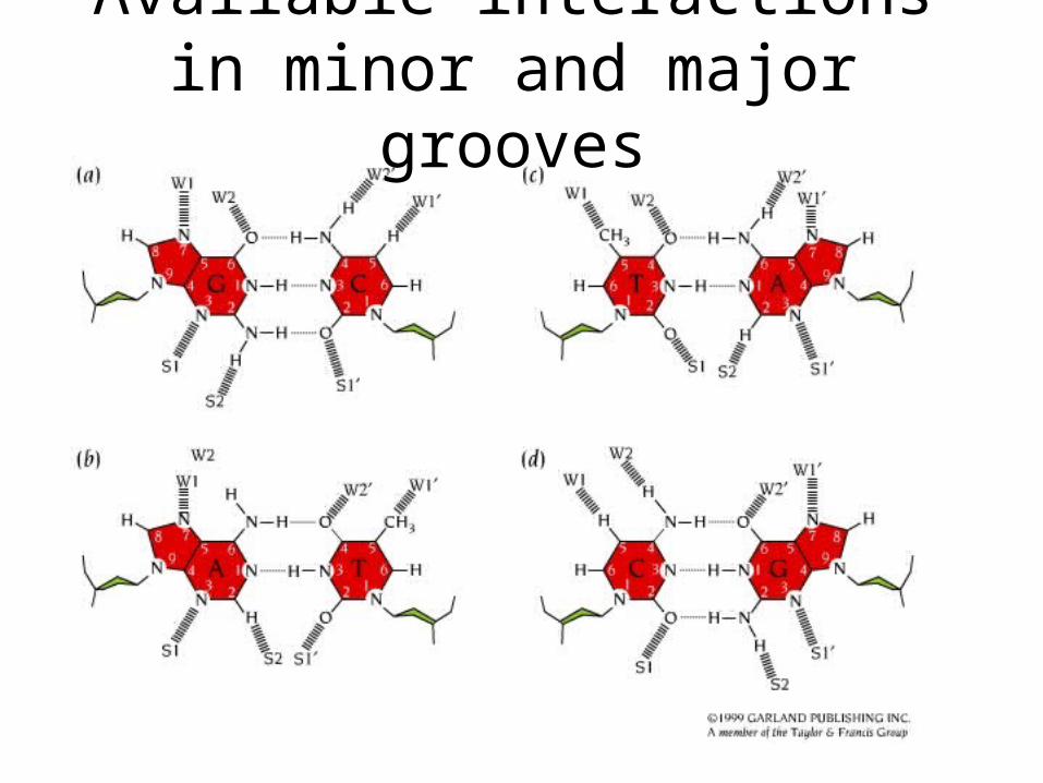
Available interactions in minor and major grooves
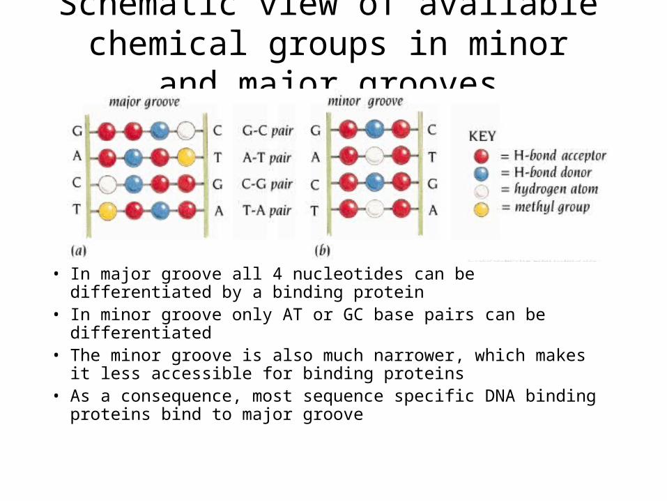
Schematic view of available chemical groups in minor and major grooves
• In major groove all 4 nucleotides can be differentiated by a binding protein
• In minor groove only AT or GC base pairs can be differentiated• The minor groove is also much narrower, which makes it less
accessible for binding proteins • As a consequence, most sequence specific DNA binding proteins
bind to major groove
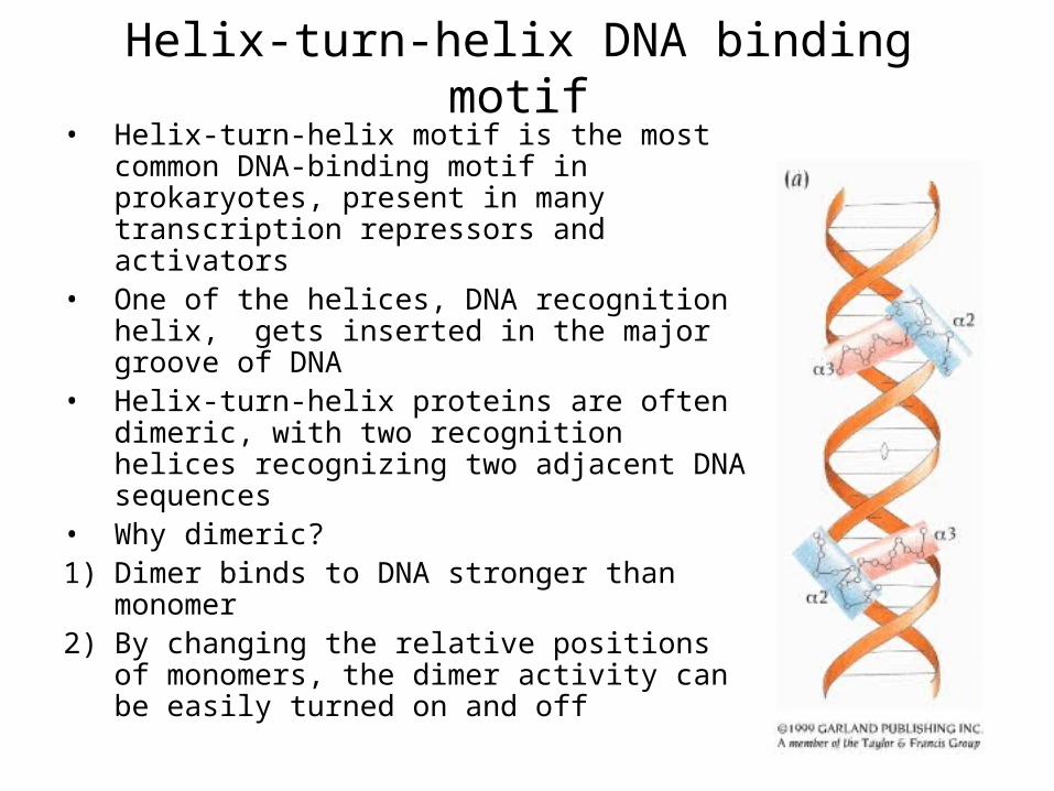
Helix-turn-helix DNA binding motif• Helix-turn-helix motif is the most common
DNA-binding motif in prokaryotes, present in many transcription repressors and activators
• One of the helices, DNA recognition helix, gets inserted in the major groove of DNA
• Helix-turn-helix proteins are often dimeric, with two recognition helices recognizing two adjacent DNA sequences
• Why dimeric?1) Dimer binds to DNA stronger than
monomer2) By changing the relative positions of
monomers, the dimer activity can be easily turned on and off
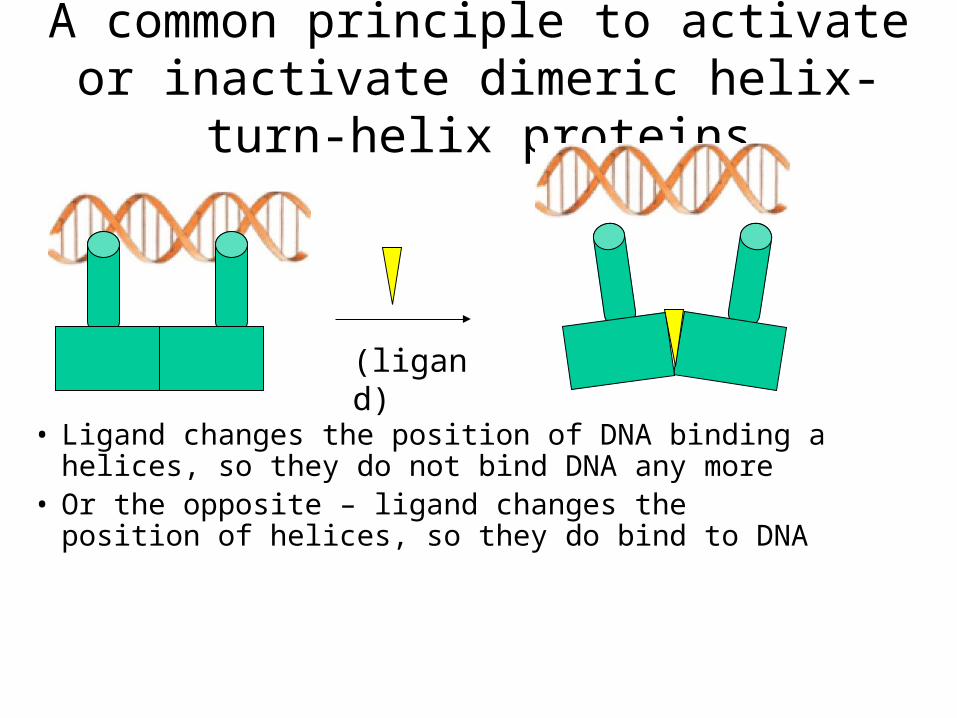
A common principle to activate or inactivate dimeric helix-turn-helix proteins
• Ligand changes the position of DNA binding a helices, so they do not bind DNA any more
• Or the opposite – ligand changes the position of helices, so they do bind to DNA
(ligand)

The main features of interactions between DNA and the helix-turn-helix motif in DNA-binding proteins
• 1. Sequence unspecific interactions contribute to overall stability of complex, but do not differentiate the ound DNA sequence
• 2. Sequence specific interactions determine, with which regions of DNA the interaction will occur
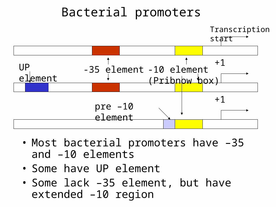
Bacterial promoters
• Most bacterial promoters have –35 and –10 elements
• Some have UP element• Some lack –35 element, but have extended –10
region
-35 element -10 element (Pribnow box)UP element
pre –10 element
+1
Transcription start
+1
The factors• factors are required for promoter recognition and
transcription initiation in prokaryotes• factors have analogous function as general transcription
factors in eukaryotes• A variety of factors exist in E.coli • For expression from most promoters 70 is required• For expression from some bacterial promoters one of other
subunits is needed instead• 70 is essential for cell growth in all conditions, while other
sigmas are required for special events, like nitrogen regulation (54), response to heat shock (32), sporulation, etc
•
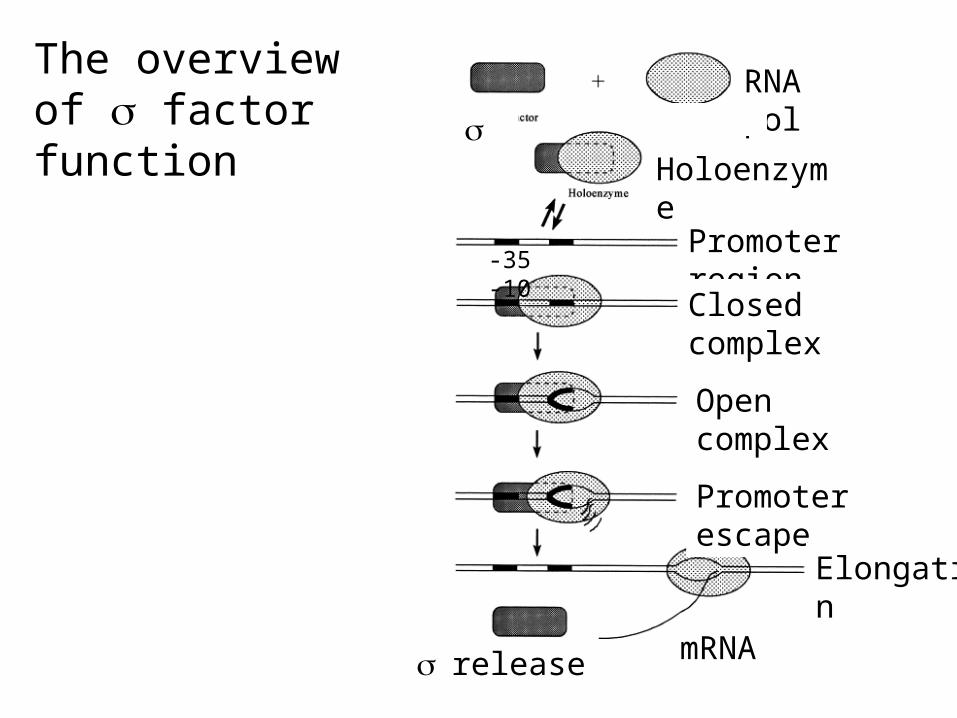
RNA pol
Holoenzyme
Promoter region
Closed complex
Open complex
Promoter escape
Elongation
mRNArelease
-35 -10
The overview of factor function

The promoter specificity of some factors in E.coli
70 TTGACA – 17 bp – TATAATN3-6-A-35 -10 +1
32 CTTGAAA – 16 bp – CCCCATNTN3-10-T/A -35 -10 +1
54 GG – N12 – GC/T – 12bp – A -24 -12 +1
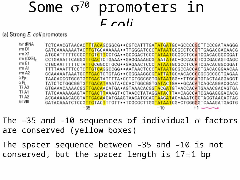
Some70 promoters in E.coli
The –35 and –10 sequences of individual factors are conserved (yellow boxes)
The spacer sequence between –35 and –10 is not conserved, but the spacer length is 171 bp

The 3D structure of bacterial RNA polymerase holoenzyme
N-term 1
Inhibition
2
-10 binding
3
-10 binding
4
-35 binding
factor domains :
3
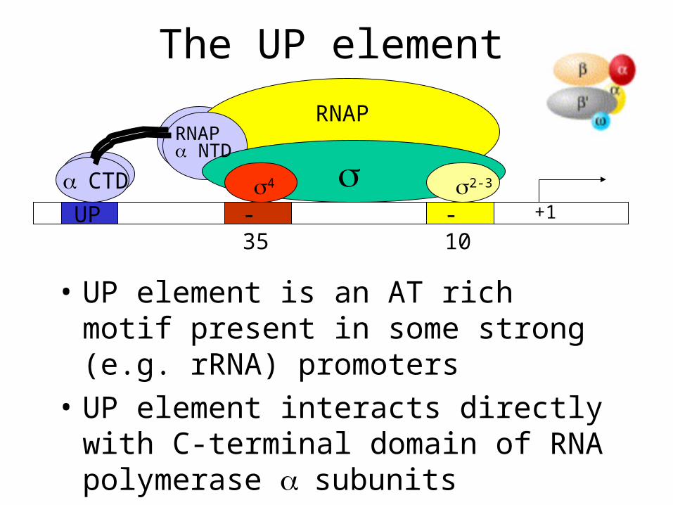
The UP element
• UP element is an AT rich motif present in some strong (e.g. rRNA) promoters
• UP element interacts directly with C-terminal domain of RNA polymerase subunits
-35 -10 UP +14 2-3
RNAP NTD
CTD
RNAP

Constitutive and inducible promoters
• Certain genes are transcribed at all times and circumstances
-Examples – tRNAs, rRNAs, ribosomal proteins, RNA polymerase
-Promoters of those genes are called constitutive• Most genes, however, need to be transcribed only
under certain circumstances or periods in cell life cycles
-The promoters of those genes are called inducible and they are subject to up- and down- regulation
Regulation at promoters
• Promoters can be regulated by repression and/or activation
• Many 70 promoters are controlled both by repression and activation, whereas, for example 54 promoters are controled solely by activation
Mechanisms of repression
• Repression by steric hindrance
• Inhibition of transition to open complex
• Inhibition of promoter clearance
• Anti-activation
• Anti-sigma factors
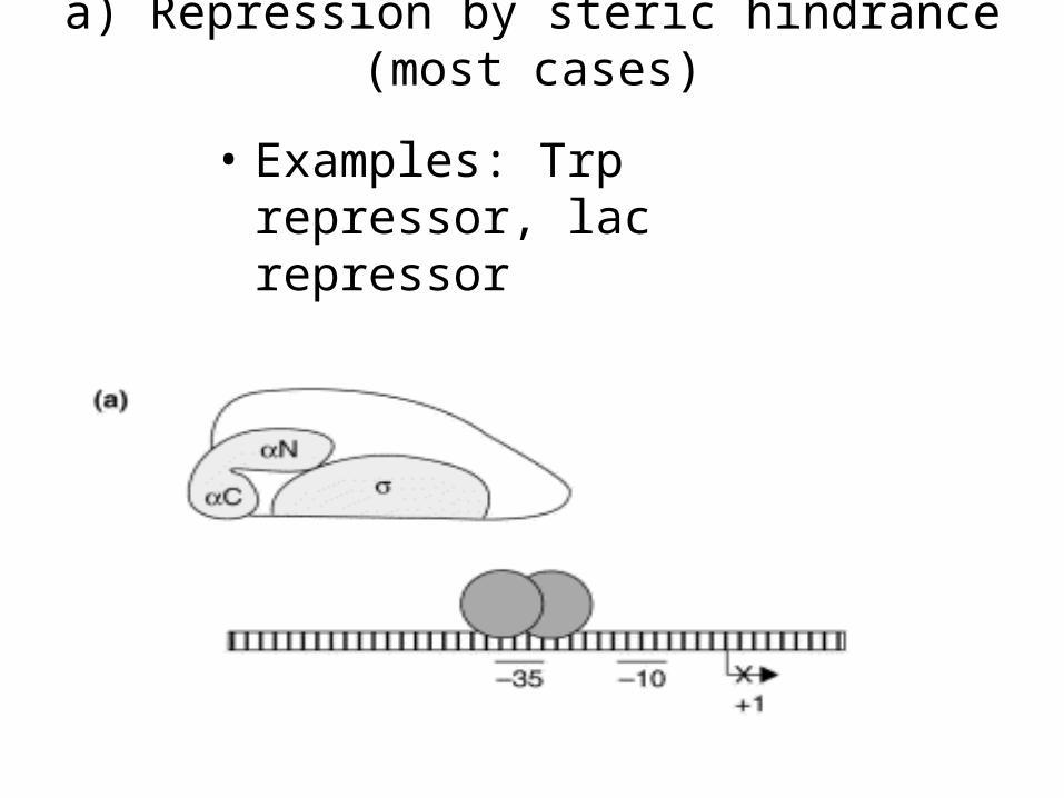
a) Repression by steric hindrance (most cases)
• Examples: Trp repressor, lac repressor
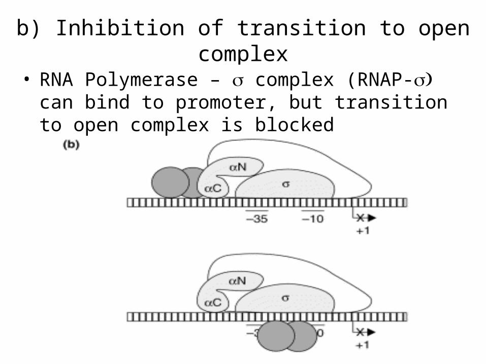
b) Inhibition of transition to open complex
• RNA Polymerase – complex (RNAP- can bind to promoter, but transition to open complex is blocked
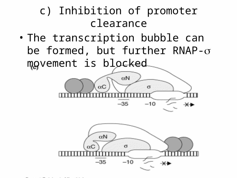
c) Inhibition of promoter clearance
• The transcription bubble can be formed, but further RNAP- movement is blocked

e) Anti-sigma factors• An anti- factor is defined by the ability to prevent its
cognate factor to compete for core RNA polymerase• Mostly used for factors, other than 70, for example in
life cycle regulation (sporulation, etc)• Some bacteriophages use their own anti- factors to
prevent expression of cellular proteins
-10 -35
RNAP
-10 -35
RNAP
anti-
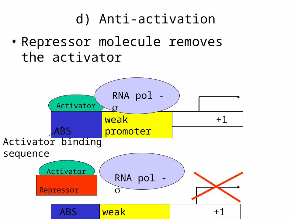
d) Anti-activation
• Repressor molecule removes the activator
weak promoter +1
weak promoter ABS +1
ABS
RNA pol - Activator
RNA pol - Activator
Repressor
Activator binding sequence

Two examples of steric hindrance
• Trp repressor
• Lac repressor
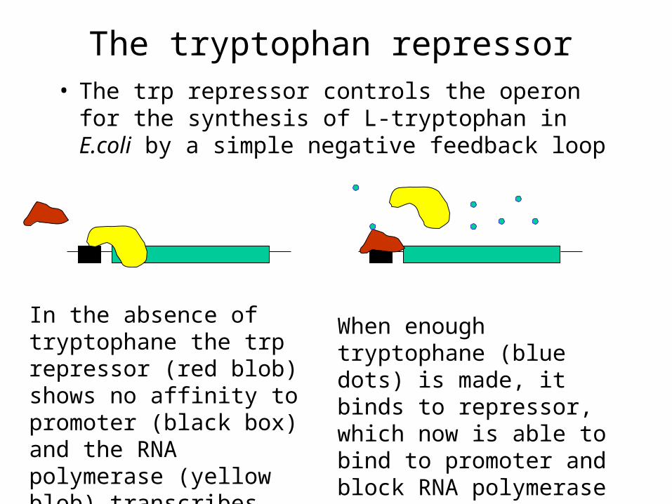
The tryptophan repressor• The trp repressor controls the operon for the
synthesis of L-tryptophan in E.coli by a simple negative feedback loop
When enough tryptophane (blue dots) is made, it binds to repressor, which now is able to bind to promoter and block RNA polymerase binding
In the absence of tryptophane the trp repressor (red blob) shows no affinity to promoter (black box) and the RNA polymerase (yellow blob) transcribes the operon

The 3-D structure of trp repressor
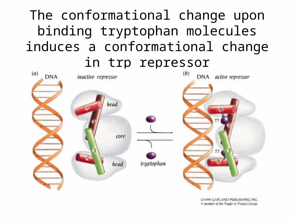
The conformational change upon binding tryptophan molecules induces a
conformational change in trp repressor

The lac promoter
Lac promoter is widely used in artifical plasmids, designed for protein production
For practical purposes it is easier to use non-hydrolyzable lactose analog – IPTG (isopropyl--thiogalactoside) instead of native lactose

The structure of lac repressor monomer
DNA binding domain
Tetramerization helix
Core N subdomain
Core C subdomain
Inducer binding pocket
Hinge helix

Functional lac repressor is a homotetramer
• Each dimer binds to a distinct DNA sequence at –82 and +11 respective to transcription start site
• This results in DNA looping, preventing the DNA polymerase from binding to –35 and –10 sequences
-82 +11
lac repressor
-35
-10
-82 +11

The lac repressor binds both to major and minor grooves of
DNA
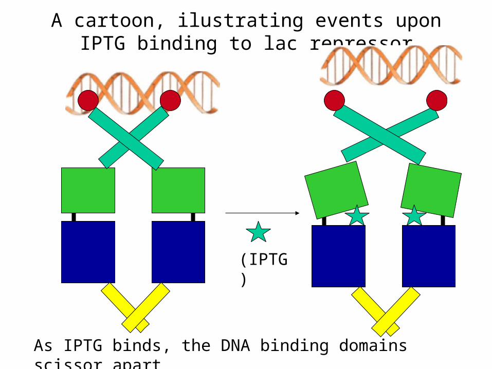
A cartoon, ilustrating events upon IPTG binding to lac repressor
As IPTG binds, the DNA binding domains scissor apart
(IPTG)
Mechanisms of activation
• a) Regulated recruitment
• b) Polymerase activation
• c) Promoter activation

a) Regulated recruitment
• Activator “extends” the binding site for RNA polymerase
weak promoter +1 ABS
RNA pol - Activator
strong affinity weak affinity
strong or weak affinity
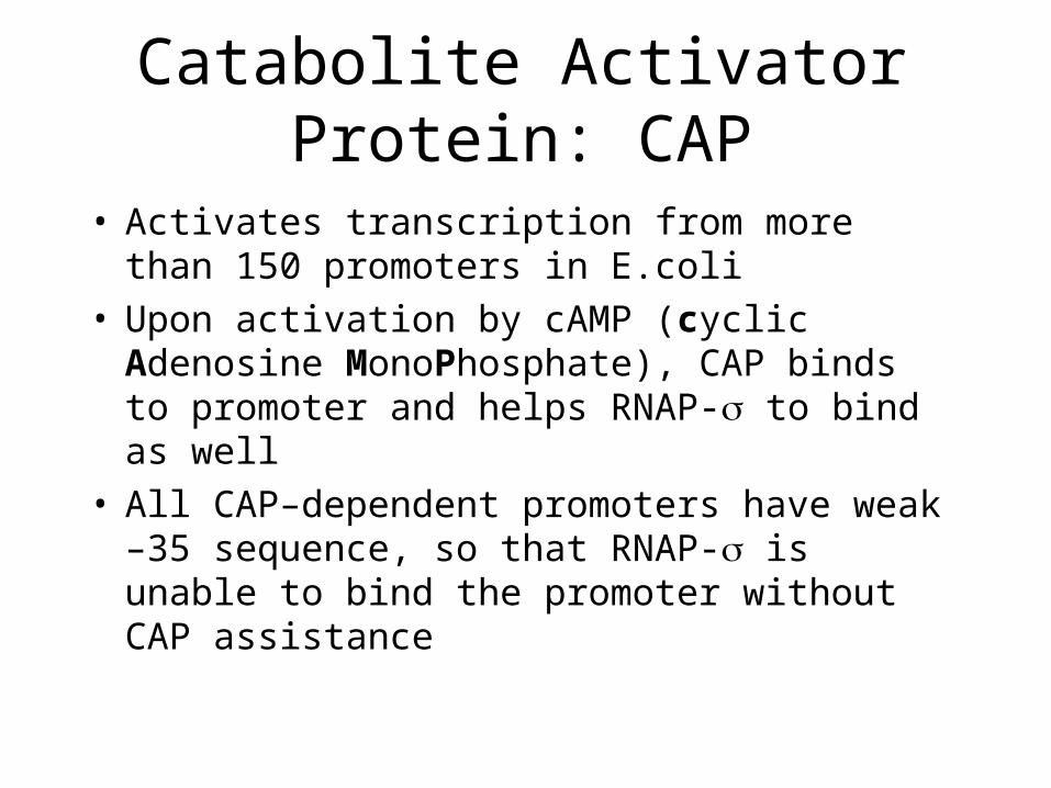
Catabolite Activator Protein: CAP
• Activates transcription from more than 150 promoters in E.coli
• Upon activation by cAMP (cyclic Adenosine MonoPhosphate), CAP binds to promoter and helps RNAP- to bind as well
• All CAP–dependent promoters have weak –35 sequence, so that RNAP- is unable to bind the promoter without CAP assistance

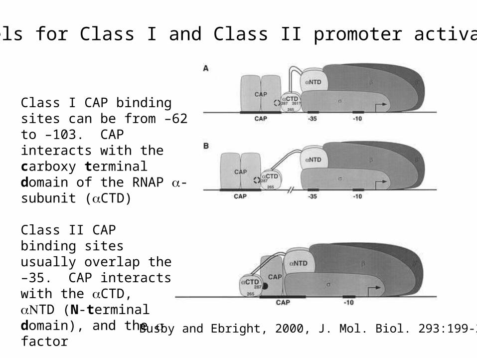
Models for Class I and Class II promoter activation
Busby and Ebright, 2000, J. Mol. Biol. 293:199-213
Class I CAP binding sites can be from –62 to –103. CAP interacts with the carboxy terminal domain of the RNAP -subunit (CTD)
Class II CAP binding sites usually overlap the –35. CAP interacts with the CTD, TD (N-terminal domain), and the factor
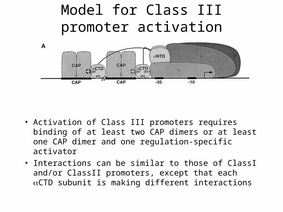
Model for Class III promoter activation
• Activation of Class III promoters requires binding of at least two CAP dimers or at least one CAP dimer and one regulation-specific activator
• Interactions can be similar to those of ClassI and/or ClassII promoters, except that each CTD subunit is making different interactions
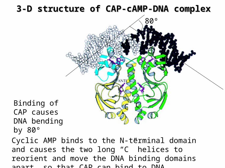
3-D structure of 3-D structure of CAPCAP-cAMP-DNA complex-cAMP-DNA complex
Cyclic AMP binds to the N-terminal domain and causes the two long “C” helices to reorient and move the DNA binding domains apart, so that CAP can bind to DNA
80º
Binding of CAP causes DNA bending by 80º
C C
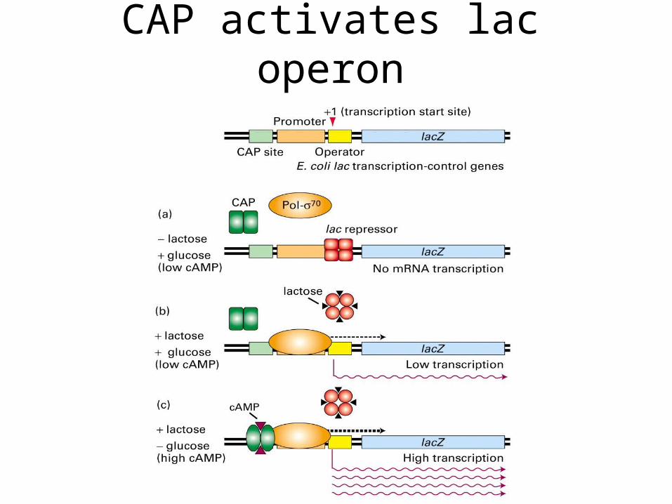
CAP activates lac operon

AraC – repressor and activator of arabinose promoter
RNAP-
RNAP-
Transcription
+ arabinose ( )
AraC
promoter
DNA binding domain of AraC
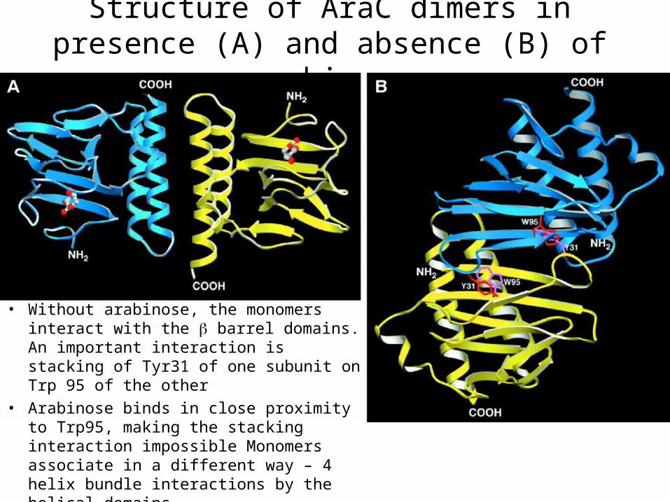
Structure of AraC dimers in presence (A) and absence (B) of arabinose
• Without arabinose, the monomers interact with the barrel domains. An important interaction is stacking of Tyr31 of one subunit on Trp 95 of the other
• Arabinose binds in close proximity to Trp95, making the stacking interaction impossible Monomers associate in a different way – 4 helix bundle interactions by the helical domains

b) Polymerase activation
• This works for 54 promoters
• RNAP-54 forms a stable complex with DNA, but needs to be activated to form an open complex

RNAP-54 activation
• RNAP-54 open complex formation requires ATP hydrolysis
• Activator protein with ATP-ase activity binds to “enhancer” site about 160 bp upstream from –24 sequence. DNA then gets looped and activator interacts with RNAP-54 resulting in the open bubble formation upon ATP hydrolysis
5454
ATP ATP+Pi
c) Example of promoter activation: MerR activator family
• MerR is an activator that controls genes involved in the response to mercury poisoning
• Other MerR family activators (CueR, BmrR, etc) respond to a variety of different toxic compounds such as other heavy metal atoms or drugs
• In MerR activated promoters, -10 and –35 regions are separated by 19bp instead of optimal 17bp
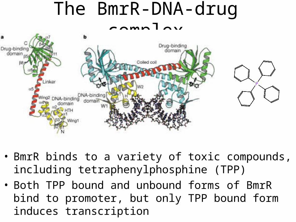
The BmrR-DNA-drug complex
• BmrR binds to a variety of toxic compounds, including tetraphenylphosphine (TPP)
• Both TPP bound and unbound forms of BmrR bind to promoter, but only TPP bound form induces transcription

• In 19-bp spacer variant, -35 and –10 binding regions not only are too far from each other, but also on the opposite sides of double helix
• In BmrR-TPP bound vairiant, DNA double helix is underwound, so that –10 and –35 regions are at the same distance as in regular 17-bp spacer
Transcription termination
• In prokaryotes two types of transcription termination occur – rho indepedent termination and rho dependent termination
• In rho independent case, the termination is achieved by a secondary structure of mRNA – RNA stem-loop, followed by an AU rich region
• A rho protein is required for rho-dependent termination
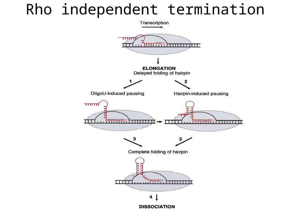
Rho independent termination
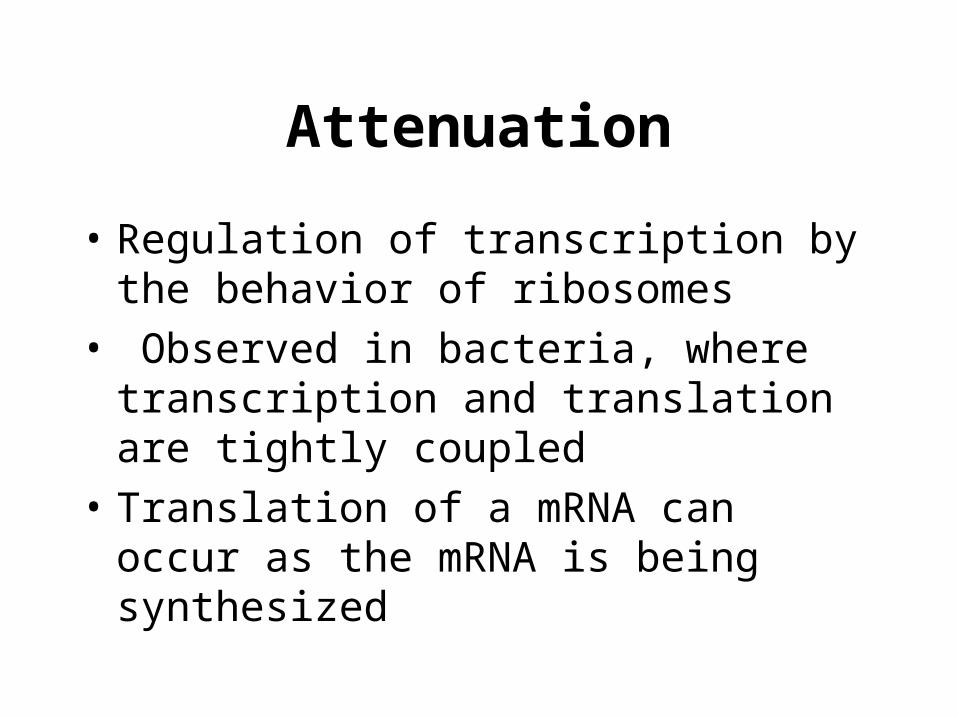
Attenuation
• Regulation of transcription by the behavior of ribosomes
• Observed in bacteria, where transcription and translation are tightly coupled
• Translation of a mRNA can occur as the mRNA is being synthesized

Attenuation in trp operon

Rho dependent terminationAs polymerase transcribes away from the promoter, rho factor binds to RNA and follows the polymerase
When polymerase reaches some sort of pause site, rho factor catches up with polymerase and unwinds the DNA-RNA hybrid, resulting in release of polymerase
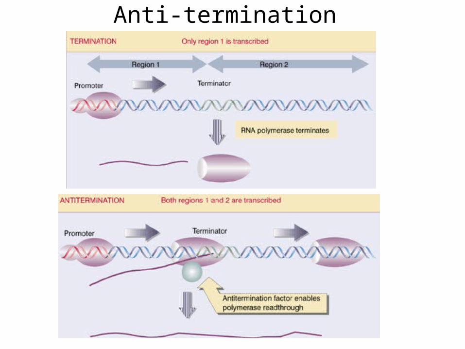
Anti-termination