toxoplasma gondii exploits host low-density lipoprotein ... filetoxoplasma gondii exploits host...
TRANSCRIPT
The Rockefeller University Press, 0021-9525/2000/04/167/14 $5.00The Journal of Cell Biology, Volume 149, Number 1, April 3, 2000 167–180http://www.jcb.org 167
Toxoplasma gondii
Exploits Host Low-Density LipoproteinReceptor-mediated Endocytosis for Cholesterol Acquisition
Isabelle Coppens, Anthony P. Sinai, and Keith A. Joiner
Infectious Diseases Section, Department of Internal Medicine, Yale University School of Medicine, New Haven, Connecticut 06520-8022
Abstract.
The obligate intracellular protozoan
Toxo-plasma gondii
resides within a specialized parasito-phorous vacuole (PV), isolated from host vesicular traf-fic. In this study, the origin of parasite cholesterol was investigated.
T
.
gondii
cannot synthesize sterols via the mevalonate pathway. Host cholesterol biosynthesis re-mains unchanged after infection and a blockade in host de novo sterol biosynthesis does not affect parasite growth. However, simultaneous limitation of exoge-nous and endogenous sources of cholesterol from the host cell strongly reduces parasite replication and para-site growth is stimulated by exogenously supplied cho-lesterol. Intracellular parasites acquire host cholesterol that is endocytosed by the low-density lipoprotein (LDL) pathway, a process that is specifically increased in infected cells. Interference with LDL endocytosis,
with lysosomal degradation of LDL, or with cholesterol translocation from lysosomes blocks cholesterol deliv-ery to the PV and significantly reduces parasite replica-tion. Similarly, incubation of
T
.
gondii
in mutant cells defective in mobilization of cholesterol from lysosomes leads to a decrease of parasite cholesterol content and proliferation. This cholesterol trafficking to the PV is independent of the pathways involving the host Golgi or endoplasmic reticulum. Despite being segregated from the endocytic machinery of the host cell, the
T
.
gondii
vacuole actively accumulates LDL-derived cho-lesterol that has transited through host lysosomes.
Key words:
Toxoplasma gondii
• parasitophorous vacuole • somatic cell mutant • LDL endocytic path-way • cholesterol transport
Introduction
Upon entering a host cell, many intracellular pathogensreside within membrane-bound vacuoles. Successful intra-cellular parasitism is dependent on the pathogen-drivencontrol of the biogenesis and maturation of the vacuole,allowing the establishment of a replication-permissiveniche. The obligate intracellular protozoan
Toxoplasmagondii
resides in a specialized parasitophorous vacuole(PV)
1
that neither acidifies nor fuses with organelles of theendocytic cascade and exocytic pathway and, as such, is to-
tally isolated from the host cell vesicular transport system(Jones et al., 1972; Sibley et al., 1985; Joiner et al., 1990;Mordue et al., 1999). This parasite is auxotrophic for sev-eral metabolites (see review by Sinai and Joiner, 1997) andmust exchange nutrients across the PV membrane (PVM),surrounding it to assure its survival and propagation. Thisraises the intriguing issue of how nutrients are obtainedfrom the host cell by
T
.
gondii
.Small soluble molecules of
,
1,400 D are able to crossthe PVM through functional pores (Schwab et al., 1994).Although devoid of transmembrane transporter/receptorsof host cell origin (Porchet-Hennere and Torpier, 1983),the PVM contains numerous secreted parasite proteinsthat might be implicated in metabolite transport (Sinaiand Joiner, 1997; Lingelbach and Joiner, 1998). Within thevacuolar space is a tubulo-reticular network connected tothe PVM, which likely increases the exchange surface be-tween the host cytoplasm and intravacuolar parasites (Sib-ley et al., 1995).
Of importance, the PVM of
T
.
gondii
is tightly en-shrouded by host mitochondria and endoplasmic reticu-lum (ER), the host cell lipid biosynthetic apparatus (Joneset al., 1972; Melo et al., 1992; Lindsay et al., 1993; Sinai et
Address correspondence to Dr. Keith A. Joiner, Infectious Diseases Sec-tion, Department of Internal Medicine, 808 LCI, 333 Cedar Street, New
Haven, CT 06520-8022. Tel.: (203) 785-4140. Fax: (203) 785-3864. E-mail:[email protected]
Dr. Sinai’s present address is Department of Microbiology and Immu-nology, University of Kentucky College of Medicine, Lexington, KY40536.
1
Abbreviations used in this paper:
CO, cholesteryl oleate; HFF, humanforeskin fibroblast; HMG-CoA, 3-hydroxy-3-methylglutaryl coenzyme A;LDL, low-density lipoprotein; LP, total lipoproteins; LPDS, LP-deficientserum; NBD-C, 22-(
N
-(7-nitrobenz-2-oxa-1,3-diazol-4-yl)amino)23,24-bisnor-5-cholen-3
b
)-ol; NPC, Niemann-Pick type C; PV, parasitophorousvacuole; PVM, PV membrane; SRD cells, mutant CHO cells with a sterolregulatory defective phenotype; SSD cells, mutant CHO cells with asqualene synthase deficiency; [
3
H-CO]-LDL, LDL that is labeled with[
3
H-CO]; [NBD-C]-LDL, LDL that is labeled with [NBD-C].
The Journal of Cell Biology, Volume 149, 2000 168
al., 1997). This organelle association has been postulatedto play a role in lipid and possibly membrane scavengingfrom these host organelles to the intravacuolar parasite atsites of PVM-organelle association (Sinai et al., 1997). In-deed,
T
.
gondii
seems to be deficient in its ability to syn-thesize selected phospholipids de novo (Sinai, A.P., K.A.Joiner, and D.R. Voelker, unpublished observations).
Toxoplasma
membranes contain cholesterol based onboth biochemical and morphological criteria (MonteiroCintra and de Souza, 1985; Gallois et al., 1988; Foussard etal., 1991a, 1991b). Cholesterol is concentrated in rhoptries,apical secretory organelles implicated in the extension ofthe PVM during invasion. Indeed, these organelles have avery high cholesterol/phospholipid molar ratio of 1.5(Foussard et al., 1991a). In higher eukaryotic cells, choles-terol homeostasis is finely regulated by transcriptional,translational, and posttranslational mechanisms (reviewedin Goldstein and Brown, 1990; Brown and Goldstein,1999). Cells have a number of options when it comes to theuse of cholesterol for membrane biogenesis or synthesis ofnew molecules derived from cholesterol. This latter is syn-thesized in the ER via the key enzyme of the mevalon-ate pathway, the 3-hydroxy-3-methylglutaryl coenzyme A(HMG-CoA) reductase. Newly synthesized cholesterolis transported rapidly to the caveolae domains of theplasma membrane from where it constitutively cycles withthe cell interior. Another important source of cholesterolis plasma low-density lipoprotein particles (LDL) that areinternalized by specific receptors and delivered to late en-dosomes/lysosomes for hydrolysis. When cholesterol iseffluxed from lysosomes, the bulk of cholesterol is trans-ported to the plasma membrane probably by a Golgi-dependent pathway involving caveolae, while a portion isdelivered to the ER by vesicular transport. Deposition ofexcess cellular cholesterol in the form of cholesteryl estersis catalyzed by the resident ER acyl-CoA:cholesterol acyl-transferase (ACAT), leading to the biogenesis of lipiddroplets (reviewed in Lange and Steck, 1996; Liscum andMunn, 1999).
Upon infection with
T
.
gondii
, the mammalian cell ac-quires a novel dynamic compartment, the PV, which con-tains live, dividing microorganisms. We have addressedthe following questions, relating to the origin of choles-terol for the parasite: Can
T
.
gondii
synthesize its own cho-lesterol via the classical mevalonate pathway? Is the PVaccessible to host cell cholesterol? If accessible, is it thecholesterol synthesized by the host cell or the exogenouscholesterol delivered by LDL endocytosis that can betransported into the parasite? If acquired exogenouslyfrom LDL, is cholesterol transported from lysosomes tothe PV by a direct transfer, a Golgi-, or an ER-dependentpathway? Is the host cell altered in its cholesterol biosyn-thesis or LDL uptake in response to parasitization? Is theparasite capable of replication in host cells unable eitherto synthesize cholesterol de novo, or to use LDL-deliveredcholesterol, or both?
Although the
Toxoplasma
PV remains segregated fromvesicular trafficking through the endo- and exocytoticpathways in the host cell, the results presented here dem-onstrate that this parasite can actively intercept host LDL-derived cholesterol, in transit from the lysosomes to othercellular compartments.
Materials and Methods
Chemicals and Antibody
All chemicals were obtained from either Sigma Chemical Co. or Boeh-ringer Mannheim Biochemicals, unless indicated otherwise. Analyticalgrade solvents were used for lipid analysis. The nitrobenzoxadiazole-cho-lesterol (NBD-C) was obtained from Molecular Probes, Inc. and stored at
2
20
8
C as a 10-mg/100-
m
l solution in dimethylformamide. U18666A wasfrom Biomol. Compactin was kindly provided by Dr. Akira Endo (NokoUniversity, Tokyo, Japan). The SDZ 215-918 cyclosporin A analogue wasprovided by Sandoz Pharma, Ltd. Radiolabeling reagents, purchased fromAmersham Corp., included [1
a
,2
a
(n)-
3
H]cholesteryl oleate (sp act, 43 Ci/mmol), 3-hydroxy-3-methyl[3-
14
C]glutaryl coenzyme A (sp act, 61 mCi/mmol), RS-[2-
14
C]mevalonic acid lactone (sp act, 58 mCi/mmol), [5,6-
3
H]uracil (sp act, 45 Ci/mmol), and [methyl-
3
H]thymidine (sp act, 6.7 Ci/mmol). Human [
125
I]-diferric transferrin was from DuPont NEN ResearchProducts. The monoclonal anti–low-density lipoprotein receptor antibodywas obtained from Amersham Corp.
Preparation of LDL, Lipoprotein-deficient Serum,LDL Labeled with NBD-C, or with[
3
H]cholesteryl oleate–LDL
Human LDL (density 1.019–1.063 g/ml) was isolated from fresh plasmaby zonal density gradient ultracentrifugation as described (Poumay andRonveaux-Dupal, 1984). Lipoprotein-deficient serum (LPDS) was pre-pared by ultracentrifugation of fetal bovine serum (Gemini Bio-Products,Inc.) after the density was increased to 1.215 g/ml with KBr (Havel et al.,1955). Total lipoproteins (LP) contained in the supernatant were used toreconstitute LPDS. The fluorescent lipid NBD-C was incorporated intoLDL by mixing 50
m
l of the lipid stock solution with 20 ml of filtered freshhuman plasma. After incubation for 16 h at 37
8
C, LDL was isolated as de-scribed above. The association of the NBD-C to the LDL fraction was es-timated by the specific uptake of the fluorescent LDL by microscopy. Thefluorescence observed after uptake of labeled LDL in the presence of a40-fold excess of nonlabeled LDL was negligible. Radiolabeling of LDLwith [
3
H]cholesteryl oleate ([
3
H-CO]) was previously described (Coppenset al., 1995).
Cell Lines and Culture Conditions
The cell lines used in this study included: Chinese hamster ovary cells, pri-mary human foreskin fibroblasts (HFF), and African green monkey(Vero) cells. Somatic mutants of CHO cells were generous gifts from Drs.M. Brown, J. Goldstein, and A. Nohturftt of University of Texas S.W.Medical Center (Dallas, TX): CHO-7 cells (Metherall et al., 1989), UT-1cells (Luskey et al., 1983), and a sterol regulatory-defective (SRD) mu-tant, SRD-1 cells (Metherall et al., 1989). Dr. L. Liscum (Tufts UniversitySchool of Medicine, Medford, MA) kindly provided the 2-2 mutant (Dahlet al., 1992, 1993) and Dr. R. Simoni (Stanford University, Stanford, CA)generously gave a squalene synthase-deficient (SSD) mutant (Bradfute etal., 1992). All cell lines were grown as monolayers at 37
8
C in an atmo-sphere of 5% CO
2
in
a
-minimum essential medium (prepared at the Me-dia Core Facility of the Department of Cell Biology, Yale University),supplemented with 2 mM
L
-glutamine and penicillin/streptomycin (100U/ml per 100
m
g/ml). This culture medium also contained variable % (vol/vol) of either FBS or LPDS that was reconstituted (or not) with LP, as in-dicated in each experiment. Specific requirements added in media for themutants of CHO cells were: 40
m
M compactin for the UT-1 cells, 1
m
g/mlof 25-hydroxycholesterol for the SRD-1 cells, 0.2 mg/ml of geneticin forthe SSD cells.
Parasite Culture and Purification
The RH strain tachyzoite of
Toxoplasma gondii
was used throughout thisstudy, and was maintained by passage in the peritoneum of Swiss-Webstermice or by in vitro culture in Vero cells or HFF, as previously described(Roos et al., 1994). A purification scheme of intracellular parasites basedon density gradient separation using Nycodenz and isopycnic centrifuga-tion was developed. Confluent HFF or CHO cell monolayers were in-fected with
T
.
gondii
, which were further harvested from the culture su-pernatants. After two passages through a 27-gauge needle to disrupt anycontaminating host cells, parasites were washed three times in PBS (137mM NaCl, 2.7 mM KCl, 10 mM NaH
2
PO
4
, 1.8 mM KH
2
PO
4
, adjusted to
Coppens et al.
Cholesterol Transport in Toxoplasma gondii–infected Cells
169
pH 7.4) by centrifugation at 1,000
g
in a GRP tabletop centrifuge (Beck-man Instruments, Inc.) for 10 min. Parasites were then resuspended in10% Nycodenz before application at the top of a linear Nycodenz gradientfrom 30 to 10%. After centrifugation in a swing-out rotor at 2,000
g
for 30min at room temperature, fractions were collected and analyzed for den-sity and protein content. Fractions containing parasites were equilibratedat a density of 1.09–1.11 g/ml of Nycodenz. They were then diluted 2.5-fold with PBS, centrifuged at 5,000
g
for 30 min, and washed three times inPBS before use for lipid extraction. The purity of the parasite preparationwas monitored by the comparison of the protein content in the 1.09–1.11-g/ml fractions after inoculation of the same number of parasites in eitherone or three dishes of cell monolayers. When parasites were purified fromthree dishes, no increase in protein concentration was observed in the par-asite fractions, as compared with parasites from one dish.
Determination of Parasite Concentration, Replication, and Viability
Parasite concentration was determined by numeration using a Haussercounting chamber at 400
3
magnification. The analysis of the number ofparasites per vacuole to estimate the parasite replication and the measure-ment of [
3
H]uracil incorporated into the parasites to evaluate their viabil-ity were determined as previously described (Nakaar et al., 1999). Parasitecultures were synchronized by removal of parasites that had not yet in-vaded 4 h after their inoculation into confluent cells.
Ligand Radiolabeling and Uptake Experiments
LDL was radiolabeled with
125
I by means of ICl (McFarlane, 1958) andspecific radioactivity of
125
I-LDL was 300–550 cpm/ng protein.
125
I-trans-ferrin was used at a specific radioactivity of 100–200 cpm/ng protein. Foruptake experiments, confluent cultures of CHO cells in a six-well platewere inoculated with 10
6
freshly lysed-out parasites and incubated for 4 hat 37
8
C before washing and resuspension in culture medium containing ei-ther 10% LPDS (for LDL uptake experiments) or 10% FBS (for transfer-rin and horseradish peroxidase uptake experiments). The presence or ab-sence of LP during the 24-h incubation is without influence on thesubsequent transferrin or HRP uptake. 24 h post-infection, cultures werewashed twice with PBS, incubated at 37
8
C for 2 h in culture medium (de-void of serum) containing 1% of serum albumin and different concentra-tions of
125
I-LDL,
125
I-transferrin, or HRP, and then chilled to 4
8
C andwashed twice in PBS plus 5% FBS. Cell monolayers were further incu-bated for 60 min at 4
8
C in the presence of 0.1% pronase (wt/vol) to re-move cell surface–bound ligand, washed twice in PBS and lysed in 0.01%Triton X-100. The pronase-resistant fraction was considered as internal-ized ligand. For comparison, noninfected CHO cells were used as controls,and processed identically. Cell-internalized radioiodinated ligand in thelysate was evaluated in a liquid scintillation system (LS 6000SC; BeckmanInstruments, Inc.) and cell-internalized HRP was determined by measure-ment of the peroxidase activity in the lysate by the stopped colorimetricassay using ortho-phenylenediamine as a substrate, according to Steinmanet al. (1976). Values of cell-internalized ligand in infected and uninfectedcells were normalized to total cell protein.
Incubation of Infected Cells with [
3
H-CO]-LDL and Lipid Analysis
Synchronized infected cells were incubated in culture medium containing10% FBS or 10% LPDS. After 24 h, cells were treated with the indicatedinhibitor, and then pulse-labeled with 5 mg of [
3
H-CO]-LDL. After wash-ing, parasites were isolated, their protein concentration determined, andlipids extracted to quantify the amount of [
3
H-C] associated with the ste-rol fraction, as described (Coppens et al., 1995). Results were calculated ascounts per minute per milligram cell protein, and then expressed in per-cent control values corresponding to the amount of [
3
H-C] associated withthe sterol fraction of infected cells that were incubated in the absence ofany inhibitor.
Incubation of Infected Cells with LDL Labeled with NBD-C, Filipin, Lipid Droplet Dyes, andFluorescence Microscopy
To visualize fluorescent cholesterol associated with
T
.
gondii
, synchro-nized infected cells were seeded on coverslips in a 24-well plate and incu-bated in culture medium containing 10% LPDS. After 24 h, cells were
treated with the indicated inhibitor, and then labeled with 0.1 mg of[NBC-C]-LDL for various pulse-chase times. Coverslips with live cellswere directly observed with an epifluorescence microscope (MicrophotFXA; Nikon Inc.). Images were captured with a CCD camera (Photomet-rics), processed with Image-Pro Plus (Media Cybernetics), and their con-trast was enhanced with Adobe Photoshop 5.0 (Adobe Systems Inc.).For cytochemical staining of
b
-hydroxysterols with filipin, infected cellsseeded on coverslips were incubated in culture medium containing 10%FBS, washed in PBS, and then fixed in 3% paraformaldehyde for 30 minat room temperature and washed again with PBS. Coverslips with fixedcells were incubated with 25
m
g/ml of filipin for 15 min, washed in PBS,and mounted with glycerol to be viewed by a fluorescence microscope us-ing an excitation filter of 350–410 nm. For detection of parasite lipid drop-lets, extracellular
T
.
gondii
from cell cultures incubated in culture mediumplus 10% FBS or 10% LPDS were fixed in paraformaldehyde and stainedwith either Oil Red O or Nile Red before observation by phase-contrastand fluorescence microscopy, as described (Greenspan et al., 1985; El-Jack et al., 1999).
Assay for HMG-CoA Reductase Activity
Confluent cultures of CHO cells in 10-cm plates were inoculated with 10
7
freshly lysed-out parasites and incubated for 4 h at 37
8
C before washingand resuspension in culture medium containing either 10% FBS or 10%LPDS. After 24 h of infection, the activity of HMG-CoA reductase wasmeasured in situ on digitonin-permeabilized cells using [
14
C]HMG-CoAas substrate, as described (Leonard and Chen, 1987; Geelen et al., 1991).In parallel, noninfected CHO cells were used as controls and similarlyprocessed. Reductase activity was expressed as picomoles of radioactiveHMG-CoA converted to radioactive mevalonate per minute per milli-gram detergent-solubilized protein.
Mevalonate and HMG-CoA Incorporation into Sterols
To study the biosynthesis of sterols in
T
.
gondii
, 500 nmol of [
14
C]meva-lonic acid or 300 nmol [
14
C]HMG-CoA was added for 3 h at 37
8
C to con-fluent cultures of CHO cells in 10-cm plates infected with 10
7
freshly ly-sed-out parasites for 24 h in medium containing 10% LPDS. For labelingwith HMG-CoA, infected cells were previously permeabilized with digito-nin (Leonard and Chen, 1987). After washing, parasites were isolated,their protein concentration determined, and lipids extracted to quantifythe amount of [
14
C]cholesterol associated with the sterol fraction, as de-scribed (Coppens et al., 1995). An assay of radioactive mevalonate orHMG-CoA incorporation was performed on extracellular
T
.
gondii
inconditions where parasites are metabolically active, as described previ-ously (Coppens et al., 1999).
Protein Determination
Protein content was determined by the bicinchoninic acid assay (Smith etal., 1985), using serum albumin as standard.
Statistical Analysis
For comparison of means,
P
was determined by analysis of varianceagainst control (ANOVA 2).
Results
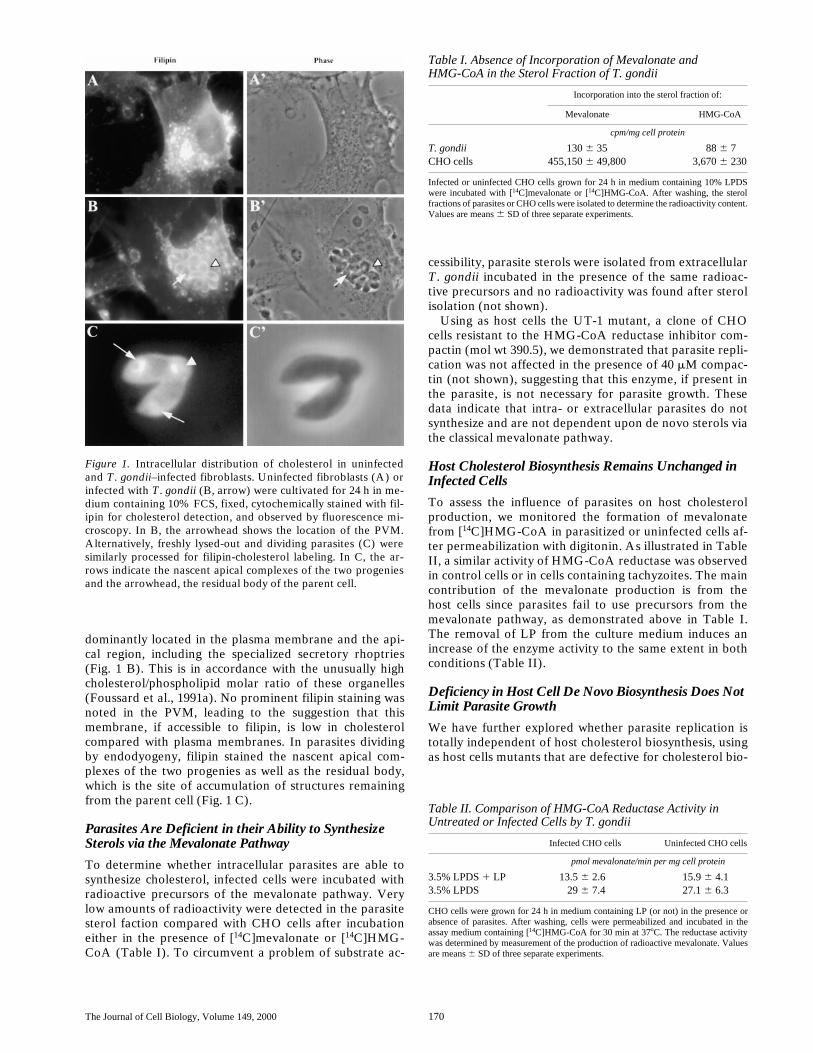
Cholesterol Is Not Uniformly Distributedwithin T. gondii
The polyene antibiotic filipin was used to visualize the dis-tribution of cholesterol or other
b
-hydroxy-sterols by lightfluorescence microscopy. In the presence of extracellularlipoproteins, uninfected fibroblasts showed a filipin-cho-lesterol staining in the plasma membrane and in intracellu-lar perinuclear organelles (Fig. 1 A) that have been identi-fied as lysosomes, endocytic recycling compartments, andGolgi structures (Butler et al., 1992; Neufeld et al., 1996;Mukherjee et al., 1998). Cholesterol distribution in para-sites inside fibroblasts was observed as intense and pre-
The Journal of Cell Biology, Volume 149, 2000 170
dominantly located in the plasma membrane and the api-cal region, including the specialized secretory rhoptries(Fig. 1 B). This is in accordance with the unusually highcholesterol/phospholipid molar ratio of these organelles(Foussard et al., 1991a). No prominent filipin staining wasnoted in the PVM, leading to the suggestion that thismembrane, if accessible to filipin, is low in cholesterolcompared with plasma membranes. In parasites dividingby endodyogeny, filipin stained the nascent apical com-plexes of the two progenies as well as the residual body,which is the site of accumulation of structures remainingfrom the parent cell (Fig. 1 C).
Parasites Are Deficient in their Ability to Synthesize Sterols via
the Mevalonate
Pathway
To determine whether intracellular parasites are able tosynthesize cholesterol, infected cells were incubated withradioactive precursors of the mevalonate pathway. Verylow amounts of radioactivity were detected in the parasitesterol faction compared with CHO cells after incubationeither in the presence of [
14
C]mevalonate or [
14
C]HMG-CoA (Table I). To circumvent a problem of substrate ac-
cessibility, parasite sterols were isolated from extracellular
T
.
gondii
incubated in the presence of the same radioac-tive precursors and no radioactivity was found after sterolisolation (not shown).
Using as host cells the UT-1 mutant, a clone of CHOcells resistant to the HMG-CoA reductase inhibitor com-pactin (mol wt 390.5), we demonstrated that parasite repli-cation was not affected in the presence of 40
m
M compac-tin (not shown), suggesting that this enzyme, if present inthe parasite, is not necessary for parasite growth. Thesedata indicate that intra- or extracellular parasites do notsynthesize and are not dependent upon de novo sterols viathe classical mevalonate pathway.
Host Cholesterol Biosynthesis Remains Unchanged in Infected Cells
To assess the influence of parasites on host cholesterolproduction, we monitored the formation of mevalonatefrom [
14
C]HMG-CoA in parasitized or uninfected cells af-ter permeabilization with digitonin. As illustrated in TableII, a similar activity of HMG-CoA reductase was observedin control cells or in cells containing tachyzoites. The maincontribution of the mevalonate production is from thehost cells since parasites fail to use precursors from themevalonate pathway, as demonstrated above in Table I.The removal of LP from the culture medium induces anincrease of the enzyme activity to the same extent in bothconditions (Table II).
Deficiency in Host Cell De Novo Biosynthesis Does Not Limit Parasite Growth
We have further explored whether parasite replication istotally independent of host cholesterol biosynthesis, usingas host cells mutants that are defective for cholesterol bio-
Figure 1. Intracellular distribution of cholesterol in uninfectedand T. gondii–infected fibroblasts. Uninfected fibroblasts (A) orinfected with T. gondii (B, arrow) were cultivated for 24 h in me-dium containing 10% FCS, fixed, cytochemically stained with fil-ipin for cholesterol detection, and observed by fluorescence mi-croscopy. In B, the arrowhead shows the location of the PVM.Alternatively, freshly lysed-out and dividing parasites (C) weresimilarly processed for filipin-cholesterol labeling. In C, the ar-rows indicate the nascent apical complexes of the two progeniesand the arrowhead, the residual body of the parent cell.
Table I. Absence of Incorporation of Mevalonate andHMG-CoA in the Sterol Fraction of T. gondii
Incorporation into the sterol fraction of:
Mevalonate HMG-CoA
cpm/mg cell protein
T. gondii
130
6
35 88
6
7CHO cells 455,150
6
49,800 3,670
6
230
Infected or uninfected CHO cells grown for 24 h in medium containing 10% LPDSwere incubated with [14C]mevalonate or [14C]HMG-CoA. After washing, the sterolfractions of parasites or CHO cells were isolated to determine the radioactivity content.Values are means 6 SD of three separate experiments.
Table II. Comparison of HMG-CoA Reductase Activity in Untreated or Infected Cells by T. gondii
Infected CHO cells Uninfected CHO cells
pmol mevalonate/min per mg cell protein
3.5% LPDS 1 LP 13.5 6 2.6 15.9 6 4.13.5% LPDS 29 6 7.4 27.1 6 6.3
CHO cells were grown for 24 h in medium containing LP (or not) in the presence orabsence of parasites. After washing, cells were permeabilized and incubated in theassay medium containing [14C]HMG-CoA for 30 min at 378C. The reductase activitywas determined by measurement of the production of radioactive mevalonate. Valuesare means 6 SD of three separate experiments.
Coppens et al. Cholesterol Transport in Toxoplasma gondii–infected Cells 171
synthesis and, therefore, auxotrophic for LDL-derivedcholesterol.
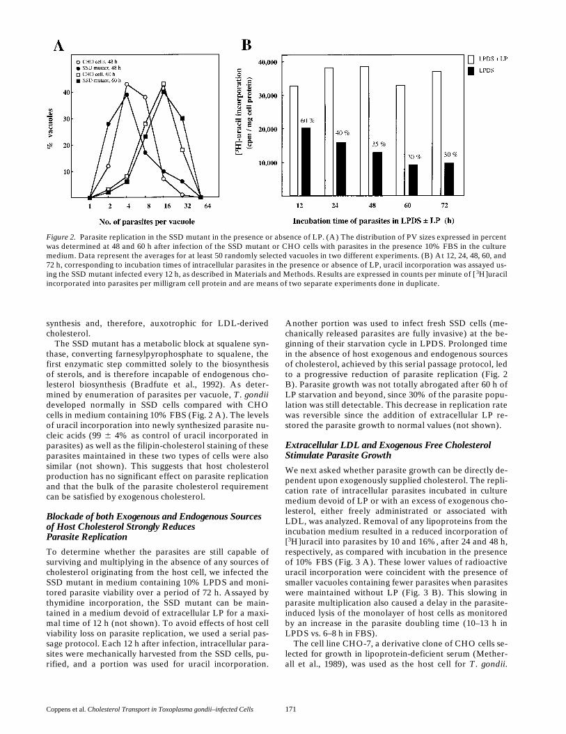
The SSD mutant has a metabolic block at squalene syn-thase, converting farnesylpyrophosphate to squalene, thefirst enzymatic step committed solely to the biosynthesisof sterols, and is therefore incapable of endogenous cho-lesterol biosynthesis (Bradfute et al., 1992). As deter-mined by enumeration of parasites per vacuole, T. gondiideveloped normally in SSD cells compared with CHOcells in medium containing 10% FBS (Fig. 2 A). The levelsof uracil incorporation into newly synthesized parasite nu-cleic acids (99 6 4% as control of uracil incorporated inparasites) as well as the filipin-cholesterol staining of theseparasites maintained in these two types of cells were alsosimilar (not shown). This suggests that host cholesterolproduction has no significant effect on parasite replicationand that the bulk of the parasite cholesterol requirementcan be satisfied by exogenous cholesterol.
Blockade of both Exogenous and Endogenous Sources of Host Cholesterol Strongly ReducesParasite Replication
To determine whether the parasites are still capable ofsurviving and multiplying in the absence of any sources ofcholesterol originating from the host cell, we infected theSSD mutant in medium containing 10% LPDS and moni-tored parasite viability over a period of 72 h. Assayed bythymidine incorporation, the SSD mutant can be main-tained in a medium devoid of extracellular LP for a maxi-mal time of 12 h (not shown). To avoid effects of host cellviability loss on parasite replication, we used a serial pas-sage protocol. Each 12 h after infection, intracellular para-sites were mechanically harvested from the SSD cells, pu-rified, and a portion was used for uracil incorporation.
Another portion was used to infect fresh SSD cells (me-chanically released parasites are fully invasive) at the be-ginning of their starvation cycle in LPDS. Prolonged timein the absence of host exogenous and endogenous sourcesof cholesterol, achieved by this serial passage protocol, ledto a progressive reduction of parasite replication (Fig. 2B). Parasite growth was not totally abrogated after 60 h ofLP starvation and beyond, since 30% of the parasite popu-lation was still detectable. This decrease in replication ratewas reversible since the addition of extracellular LP re-stored the parasite growth to normal values (not shown).
Extracellular LDL and Exogenous Free Cholesterol Stimulate Parasite Growth
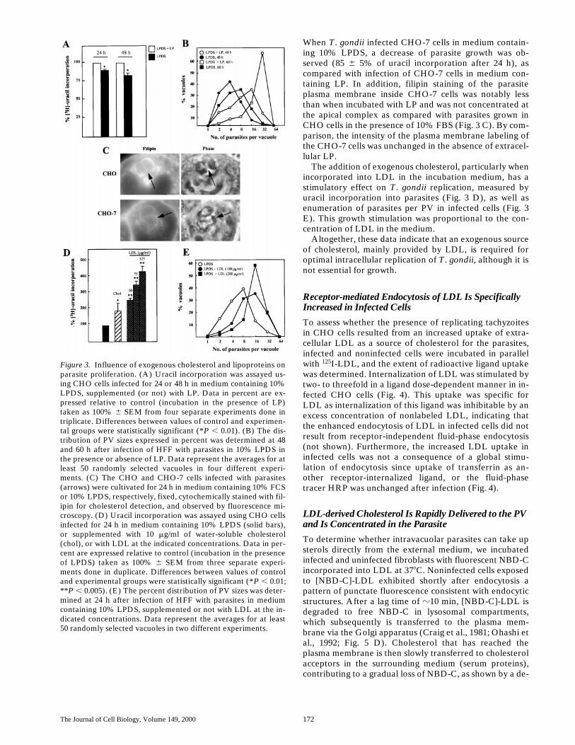
We next asked whether parasite growth can be directly de-pendent upon exogenously supplied cholesterol. The repli-cation rate of intracellular parasites incubated in culturemedium devoid of LP or with an excess of exogenous cho-lesterol, either freely administrated or associated withLDL, was analyzed. Removal of any lipoproteins from theincubation medium resulted in a reduced incorporation of[3H]uracil into parasites by 10 and 16%, after 24 and 48 h,respectively, as compared with incubation in the presenceof 10% FBS (Fig. 3 A). These lower values of radioactiveuracil incorporation were coincident with the presence ofsmaller vacuoles containing fewer parasites when parasiteswere maintained without LP (Fig. 3 B). This slowing inparasite multiplication also caused a delay in the parasite-induced lysis of the monolayer of host cells as monitoredby an increase in the parasite doubling time (10–13 h inLPDS vs. 6–8 h in FBS).
The cell line CHO-7, a derivative clone of CHO cells se-lected for growth in lipoprotein-deficient serum (Mether-all et al., 1989), was used as the host cell for T. gondii.
Figure 2. Parasite replication in the SSD mutant in the presence or absence of LP. (A) The distribution of PV sizes expressed in percentwas determined at 48 and 60 h after infection of the SSD mutant or CHO cells with parasites in the presence 10% FBS in the culturemedium. Data represent the averages for at least 50 randomly selected vacuoles in two different experiments. (B) At 12, 24, 48, 60, and72 h, corresponding to incubation times of intracellular parasites in the presence or absence of LP, uracil incorporation was assayed us-ing the SSD mutant infected every 12 h, as described in Materials and Methods. Results are expressed in counts per minute of [ 3H]uracilincorporated into parasites per milligram cell protein and are means of two separate experiments done in duplicate.
The Journal of Cell Biology, Volume 149, 2000 172
When T. gondii infected CHO-7 cells in medium contain-ing 10% LPDS, a decrease of parasite growth was ob-served (85 6 5% of uracil incorporation after 24 h), ascompared with infection of CHO-7 cells in medium con-taining LP. In addition, filipin staining of the parasiteplasma membrane inside CHO-7 cells was notably lessthan when incubated with LP and was not concentrated atthe apical complex as compared with parasites grown inCHO cells in the presence of 10% FBS (Fig. 3 C). By com-parison, the intensity of the plasma membrane labeling ofthe CHO-7 cells was unchanged in the absence of extracel-lular LP.
The addition of exogenous cholesterol, particularly whenincorporated into LDL in the incubation medium, has astimulatory effect on T. gondii replication, measured byuracil incorporation into parasites (Fig. 3 D), as well asenumeration of parasites per PV in infected cells (Fig. 3E). This growth stimulation was proportional to the con-centration of LDL in the medium.
Altogether, these data indicate that an exogenous sourceof cholesterol, mainly provided by LDL, is required foroptimal intracellular replication of T. gondii, although it isnot essential for growth.
Receptor-mediated Endocytosis of LDL Is Specifically Increased in Infected Cells
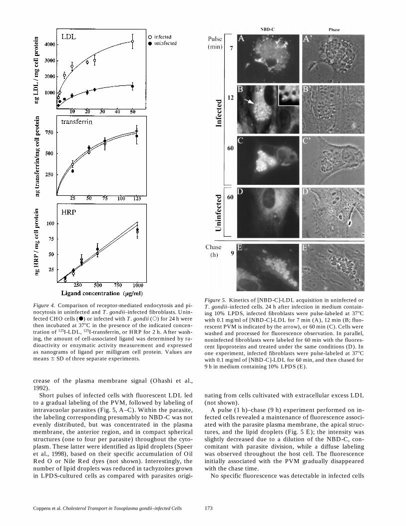
To assess whether the presence of replicating tachyzoitesin CHO cells resulted from an increased uptake of extra-cellular LDL as a source of cholesterol for the parasites,infected and noninfected cells were incubated in parallelwith 125I-LDL, and the extent of radioactive ligand uptakewas determined. Internalization of LDL was stimulated bytwo- to threefold in a ligand dose-dependent manner in in-fected CHO cells (Fig. 4). This uptake was specific forLDL as internalization of this ligand was inhibitable by anexcess concentration of nonlabeled LDL, indicating thatthe enhanced endocytosis of LDL in infected cells did notresult from receptor-independent fluid-phase endocytosis(not shown). Furthermore, the increased LDL uptake ininfected cells was not a consequence of a global stimu-lation of endocytosis since uptake of transferrin as an-other receptor-internalized ligand, or the fluid-phasetracer HRP was unchanged after infection (Fig. 4).
LDL-derived Cholesterol Is Rapidly Delivered to the PV and Is Concentrated in the Parasite
To determine whether intravacuolar parasites can take upsterols directly from the external medium, we incubatedinfected and uninfected fibroblasts with fluorescent NBD-Cincorporated into LDL at 378C. Noninfected cells exposedto [NBD-C]-LDL exhibited shortly after endocytosis apattern of punctate fluorescence consistent with endocyticstructures. After a lag time of z10 min, [NBD-C]-LDL isdegraded to free NBD-C in lysosomal compartments,which subsequently is transferred to the plasma mem-brane via the Golgi apparatus (Craig et al., 1981; Ohashi etal., 1992; Fig. 5 D). Cholesterol that has reached theplasma membrane is then slowly transferred to cholesterolacceptors in the surrounding medium (serum proteins),contributing to a gradual loss of NBD-C, as shown by a de-
Figure 3. Influence of exogenous cholesterol and lipoproteins onparasite proliferation. (A) Uracil incorporation was assayed us-ing CHO cells infected for 24 or 48 h in medium containing 10%LPDS, supplemented (or not) with LP. Data in percent are ex-pressed relative to control (incubation in the presence of LP)taken as 100% 6 SEM from four separate experiments done intriplicate. Differences between values of control and experimen-tal groups were statistically significant (*P , 0.01). (B) The dis-tribution of PV sizes expressed in percent was determined at 48and 60 h after infection of HFF with parasites in 10% LPDS inthe presence or absence of LP. Data represent the averages for atleast 50 randomly selected vacuoles in four different experi-ments. (C) The CHO and CHO-7 cells infected with parasites(arrows) were cultivated for 24 h in medium containing 10% FCSor 10% LPDS, respectively, fixed, cytochemically stained with fil-ipin for cholesterol detection, and observed by fluorescence mi-croscopy. (D) Uracil incorporation was assayed using CHO cellsinfected for 24 h in medium containing 10% LPDS (solid bars),or supplemented with 10 mg/ml of water-soluble cholesterol(chol), or with LDL at the indicated concentrations. Data in per-cent are expressed relative to control (incubation in the presenceof LPDS) taken as 100% 6 SEM from three separate experi-ments done in duplicate. Differences between values of controland experimental groups were statistically significant (*P , 0.01;**P , 0.005). (E) The percent distribution of PV sizes was deter-mined at 24 h after infection of HFF with parasites in mediumcontaining 10% LPDS, supplemented or not with LDL at the in-dicated concentrations. Data represent the averages for at least50 randomly selected vacuoles in two different experiments.
Coppens et al. Cholesterol Transport in Toxoplasma gondii–infected Cells 173
crease of the plasma membrane signal (Ohashi et al.,1992).
Short pulses of infected cells with fluorescent LDL ledto a gradual labeling of the PVM, followed by labeling ofintravacuolar parasites (Fig. 5, A–C). Within the parasite,the labeling corresponding presumably to NBD-C was notevenly distributed, but was concentrated in the plasmamembrane, the anterior region, and in compact sphericalstructures (one to four per parasite) throughout the cyto-plasm. These latter were identified as lipid droplets (Speeret al., 1998), based on their specific accumulation of OilRed O or Nile Red dyes (not shown). Interestingly, thenumber of lipid droplets was reduced in tachyzoites grownin LPDS-cultured cells as compared with parasites origi-
nating from cells cultivated with extracellular excess LDL(not shown).
A pulse (1 h)–chase (9 h) experiment performed on in-fected cells revealed a maintenance of fluorescence associ-ated with the parasite plasma membrane, the apical struc-tures, and the lipid droplets (Fig. 5 E); the intensity wasslightly decreased due to a dilution of the NBD-C, con-comitant with parasite division, while a diffuse labelingwas observed throughout the host cell. The fluorescenceinitially associated with the PVM gradually disappearedwith the chase time.
No specific fluorescence was detectable in infected cells
Figure 4. Comparison of receptor-mediated endocytosis and pi-nocytosis in uninfected and T. gondii–infected fibroblasts. Unin-fected CHO cells (d) or infected with T. gondii (s) for 24 h werethen incubated at 378C in the presence of the indicated concen-tration of 125I-LDL, 125I-transferrin, or HRP for 2 h. After wash-ing, the amount of cell-associated ligand was determined by ra-dioactivity or enzymatic activity measurement and expressedas nanograms of ligand per milligram cell protein. Values aremeans 6 SD of three separate experiments.
Figure 5. Kinetics of [NBD-C]-LDL acquisition in uninfected orT. gondii–infected cells. 24 h after infection in medium contain-ing 10% LPDS, infected fibroblasts were pulse-labeled at 378Cwith 0.1 mg/ml of [NBD-C]-LDL for 7 min (A), 12 min (B; fluo-rescent PVM is indicated by the arrow), or 60 min (C). Cells werewashed and processed for fluorescence observation. In parallel,noninfected fibroblasts were labeled for 60 min with the fluores-cent lipoproteins and treated under the same conditions (D). Inone experiment, infected fibroblasts were pulse-labeled at 378Cwith 0.1 mg/ml of [NBD-C]-LDL for 60 min, and then chased for9 h in medium containing 10% LPDS (E).
The Journal of Cell Biology, Volume 149, 2000 174
or in parasites when incubated in medium containing 10%LPDS and NBD-C administrated as lipid alone, indicatinga requirement for LDL in the acquisition of NBD-C by in-tracellular T. gondii (not shown).
LDL-derived Cholesterol Acquisition Requires Live Intracellular Parasites
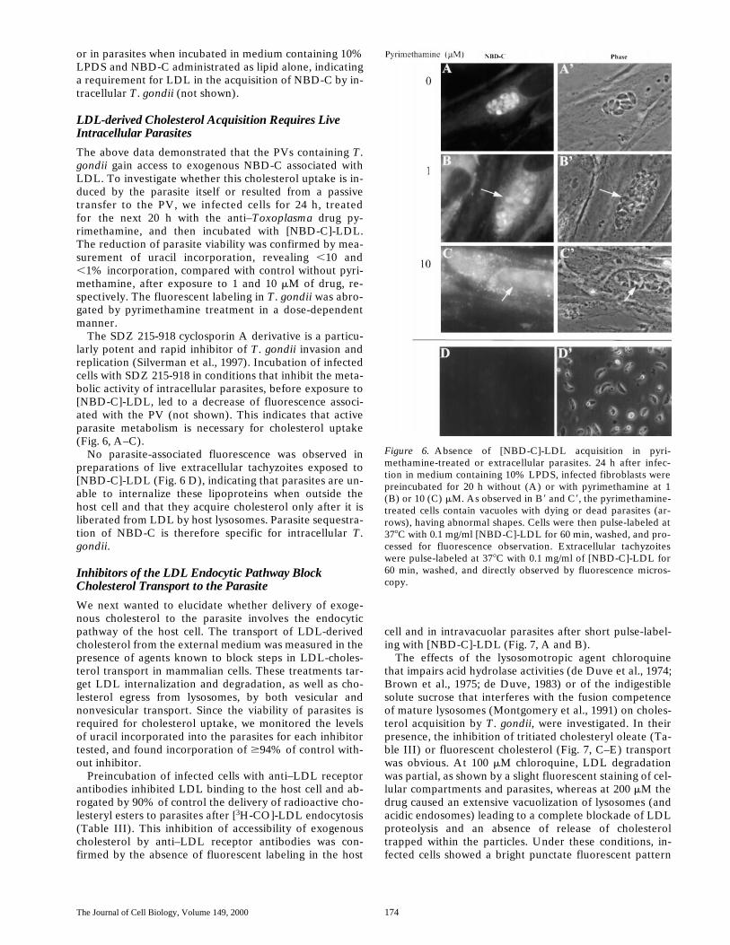
The above data demonstrated that the PVs containing T.gondii gain access to exogenous NBD-C associated withLDL. To investigate whether this cholesterol uptake is in-duced by the parasite itself or resulted from a passivetransfer to the PV, we infected cells for 24 h, treatedfor the next 20 h with the anti–Toxoplasma drug py-rimethamine, and then incubated with [NBD-C]-LDL.The reduction of parasite viability was confirmed by mea-surement of uracil incorporation, revealing ,10 and,1% incorporation, compared with control without pyri-methamine, after exposure to 1 and 10 mM of drug, re-spectively. The fluorescent labeling in T. gondii was abro-gated by pyrimethamine treatment in a dose-dependentmanner.
The SDZ 215-918 cyclosporin A derivative is a particu-larly potent and rapid inhibitor of T. gondii invasion andreplication (Silverman et al., 1997). Incubation of infectedcells with SDZ 215-918 in conditions that inhibit the meta-bolic activity of intracellular parasites, before exposure to[NBD-C]-LDL, led to a decrease of fluorescence associ-ated with the PV (not shown). This indicates that activeparasite metabolism is necessary for cholesterol uptake(Fig. 6, A–C).
No parasite-associated fluorescence was observed inpreparations of live extracellular tachyzoites exposed to[NBD-C]-LDL (Fig. 6 D), indicating that parasites are un-able to internalize these lipoproteins when outside thehost cell and that they acquire cholesterol only after it isliberated from LDL by host lysosomes. Parasite sequestra-tion of NBD-C is therefore specific for intracellular T.gondii.
Inhibitors of the LDL Endocytic Pathway Block Cholesterol Transport to the Parasite
We next wanted to elucidate whether delivery of exoge-nous cholesterol to the parasite involves the endocyticpathway of the host cell. The transport of LDL-derivedcholesterol from the external medium was measured in thepresence of agents known to block steps in LDL-choles-terol transport in mammalian cells. These treatments tar-get LDL internalization and degradation, as well as cho-lesterol egress from lysosomes, by both vesicular andnonvesicular transport. Since the viability of parasites isrequired for cholesterol uptake, we monitored the levelsof uracil incorporated into the parasites for each inhibitortested, and found incorporation of $94% of control with-out inhibitor.
Preincubation of infected cells with anti–LDL receptorantibodies inhibited LDL binding to the host cell and ab-rogated by 90% of control the delivery of radioactive cho-lesteryl esters to parasites after [3H-CO]-LDL endocytosis(Table III). This inhibition of accessibility of exogenouscholesterol by anti–LDL receptor antibodies was con-firmed by the absence of fluorescent labeling in the host
cell and in intravacuolar parasites after short pulse-label-ing with [NBD-C]-LDL (Fig. 7, A and B).
The effects of the lysosomotropic agent chloroquinethat impairs acid hydrolase activities (de Duve et al., 1974;Brown et al., 1975; de Duve, 1983) or of the indigestiblesolute sucrose that interferes with the fusion competenceof mature lysosomes (Montgomery et al., 1991) on choles-terol acquisition by T. gondii, were investigated. In theirpresence, the inhibition of tritiated cholesteryl oleate (Ta-ble III) or fluorescent cholesterol (Fig. 7, C–E) transportwas obvious. At 100 mM chloroquine, LDL degradationwas partial, as shown by a slight fluorescent staining of cel-lular compartments and parasites, whereas at 200 mM thedrug caused an extensive vacuolization of lysosomes (andacidic endosomes) leading to a complete blockade of LDLproteolysis and an absence of release of cholesteroltrapped within the particles. Under these conditions, in-fected cells showed a bright punctate fluorescent pattern
Figure 6. Absence of [NBD-C]-LDL acquisition in pyri-methamine-treated or extracellular parasites. 24 h after infec-tion in medium containing 10% LPDS, infected fibroblasts werepreincubated for 20 h without (A) or with pyrimethamine at 1(B) or 10 (C) mM. As observed in B9 and C9, the pyrimethamine-treated cells contain vacuoles with dying or dead parasites (ar-rows), having abnormal shapes. Cells were then pulse-labeled at378C with 0.1 mg/ml [NBD-C]-LDL for 60 min, washed, and pro-cessed for fluorescence observation. Extracellular tachyzoiteswere pulse-labeled at 378C with 0.1 mg/ml of [NBD-C]-LDL for60 min, washed, and directly observed by fluorescence micros-copy.
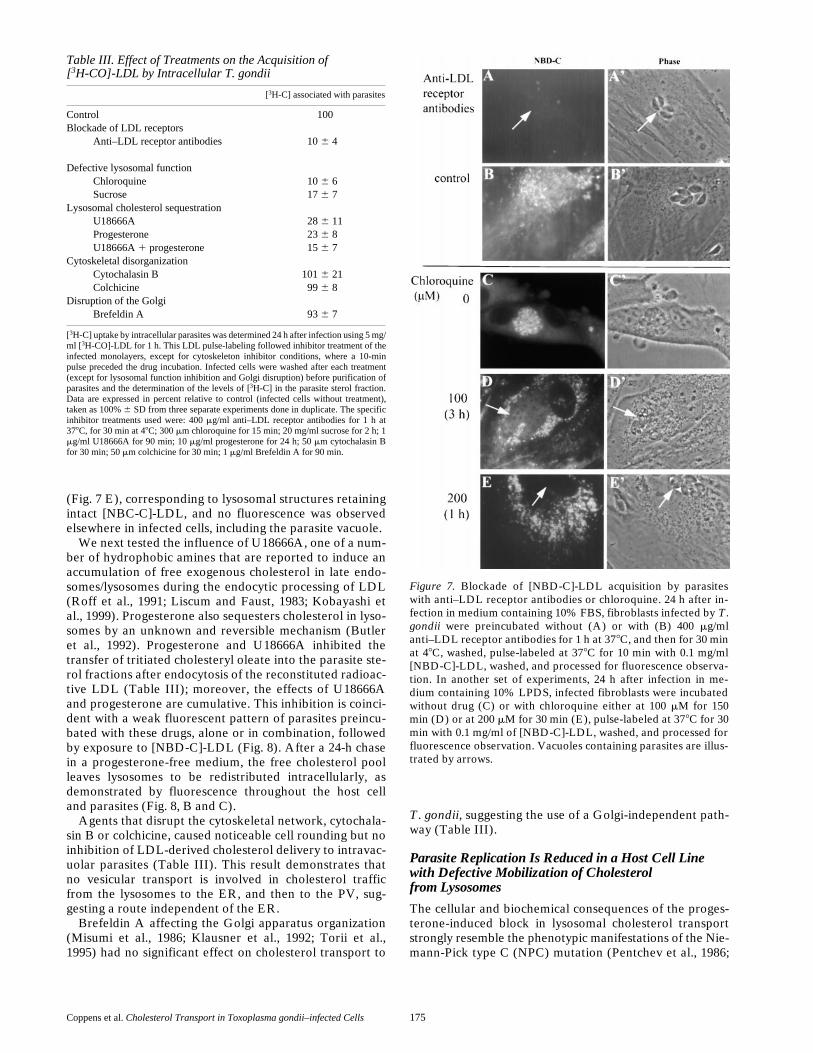
Coppens et al. Cholesterol Transport in Toxoplasma gondii–infected Cells 175
(Fig. 7 E), corresponding to lysosomal structures retainingintact [NBC-C]-LDL, and no fluorescence was observedelsewhere in infected cells, including the parasite vacuole.
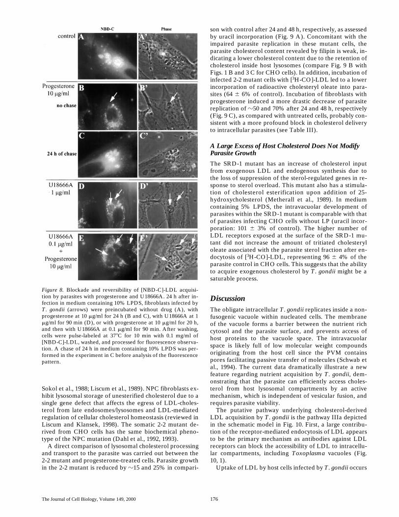
We next tested the influence of U18666A, one of a num-ber of hydrophobic amines that are reported to induce anaccumulation of free exogenous cholesterol in late endo-somes/lysosomes during the endocytic processing of LDL(Roff et al., 1991; Liscum and Faust, 1983; Kobayashi etal., 1999). Progesterone also sequesters cholesterol in lyso-somes by an unknown and reversible mechanism (Butleret al., 1992). Progesterone and U18666A inhibited thetransfer of tritiated cholesteryl oleate into the parasite ste-rol fractions after endocytosis of the reconstituted radioac-tive LDL (Table III); moreover, the effects of U18666Aand progesterone are cumulative. This inhibition is coinci-dent with a weak fluorescent pattern of parasites preincu-bated with these drugs, alone or in combination, followedby exposure to [NBD-C]-LDL (Fig. 8). After a 24-h chasein a progesterone-free medium, the free cholesterol poolleaves lysosomes to be redistributed intracellularly, asdemonstrated by fluorescence throughout the host celland parasites (Fig. 8, B and C).
Agents that disrupt the cytoskeletal network, cytochala-sin B or colchicine, caused noticeable cell rounding but noinhibition of LDL-derived cholesterol delivery to intravac-uolar parasites (Table III). This result demonstrates thatno vesicular transport is involved in cholesterol trafficfrom the lysosomes to the ER, and then to the PV, sug-gesting a route independent of the ER.
Brefeldin A affecting the Golgi apparatus organization(Misumi et al., 1986; Klausner et al., 1992; Torii et al.,1995) had no significant effect on cholesterol transport to
T. gondii, suggesting the use of a Golgi-independent path-way (Table III).
Parasite Replication Is Reduced in a Host Cell Line with Defective Mobilization of Cholesterolfrom Lysosomes
The cellular and biochemical consequences of the proges-terone-induced block in lysosomal cholesterol transportstrongly resemble the phenotypic manifestations of the Nie-mann-Pick type C (NPC) mutation (Pentchev et al., 1986;
Table III. Effect of Treatments on the Acquisition of[3H-CO]-LDL by Intracellular T. gondii
[3H-C] associated with parasites
Control 100Blockade of LDL receptors
Anti–LDL receptor antibodies 10 6 4
Defective lysosomal functionChloroquine 10 6 6Sucrose 17 6 7
Lysosomal cholesterol sequestrationU18666A 28 6 11Progesterone 23 6 8U18666A 1 progesterone 15 6 7
Cytoskeletal disorganizationCytochalasin B 101 6 21Colchicine 99 6 8
Disruption of the GolgiBrefeldin A 93 6 7
[3H-C] uptake by intracellular parasites was determined 24 h after infection using 5 mg/ml [3H-CO]-LDL for 1 h. This LDL pulse-labeling followed inhibitor treatment of theinfected monolayers, except for cytoskeleton inhibitor conditions, where a 10-minpulse preceded the drug incubation. Infected cells were washed after each treatment(except for lysosomal function inhibition and Golgi disruption) before purification ofparasites and the determination of the levels of [3H-C] in the parasite sterol fraction.Data are expressed in percent relative to control (infected cells without treatment),taken as 100% 6 SD from three separate experiments done in duplicate. The specificinhibitor treatments used were: 400 mg/ml anti–LDL receptor antibodies for 1 h at378C, for 30 min at 48C; 300 mm chloroquine for 15 min; 20 mg/ml sucrose for 2 h; 1mg/ml U18666A for 90 min; 10 mg/ml progesterone for 24 h; 50 mm cytochalasin Bfor 30 min; 50 mm colchicine for 30 min; 1 mg/ml Brefeldin A for 90 min.
Figure 7. Blockade of [NBD-C]-LDL acquisition by parasiteswith anti–LDL receptor antibodies or chloroquine. 24 h after in-fection in medium containing 10% FBS, fibroblasts infected by T.gondii were preincubated without (A) or with (B) 400 mg/mlanti–LDL receptor antibodies for 1 h at 378C, and then for 30 minat 48C, washed, pulse-labeled at 378C for 10 min with 0.1 mg/ml[NBD-C]-LDL, washed, and processed for fluorescence observa-tion. In another set of experiments, 24 h after infection in me-dium containing 10% LPDS, infected fibroblasts were incubatedwithout drug (C) or with chloroquine either at 100 mM for 150min (D) or at 200 mM for 30 min (E), pulse-labeled at 378C for 30min with 0.1 mg/ml of [NBD-C]-LDL, washed, and processed forfluorescence observation. Vacuoles containing parasites are illus-trated by arrows.
The Journal of Cell Biology, Volume 149, 2000 176
Sokol et al., 1988; Liscum et al., 1989). NPC fibroblasts ex-hibit lysosomal storage of unesterified cholesterol due to asingle gene defect that affects the egress of LDL-choles-terol from late endosomes/lysosomes and LDL-mediatedregulation of cellular cholesterol homeostasis (reviewed inLiscum and Klansek, 1998). The somatic 2-2 mutant de-rived from CHO cells has the same biochemical pheno-type of the NPC mutation (Dahl et al., 1992, 1993).
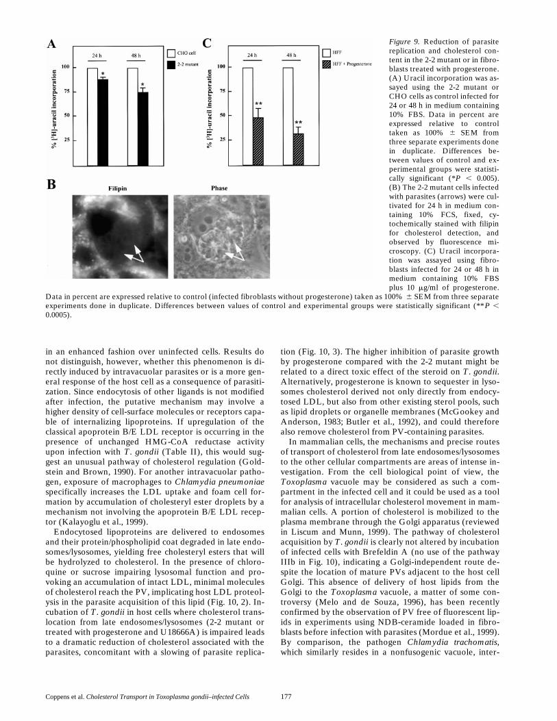
A direct comparison of lysosomal cholesterol processingand transport to the parasite was carried out between the2-2 mutant and progesterone-treated cells. Parasite growthin the 2-2 mutant is reduced by z15 and 25% in compari-
son with control after 24 and 48 h, respectively, as assessedby uracil incorporation (Fig. 9 A). Concomitant with theimpaired parasite replication in these mutant cells, theparasite cholesterol content revealed by filipin is weak, in-dicating a lower cholesterol content due to the retention ofcholesterol inside host lysosomes (compare Fig. 9 B withFigs. 1 B and 3 C for CHO cells). In addition, incubation ofinfected 2-2 mutant cells with [3H-CO]-LDL led to a lowerincorporation of radioactive cholesteryl oleate into para-sites (64 6 6% of control). Incubation of fibroblasts withprogesterone induced a more drastic decrease of parasitereplication of z50 and 70% after 24 and 48 h, respectively(Fig. 9 C), as compared with untreated cells, probably con-sistent with a more profound block in cholesterol deliveryto intracellular parasites (see Table III).
A Large Excess of Host Cholesterol Does Not Modify Parasite Growth
The SRD-1 mutant has an increase of cholesterol inputfrom exogenous LDL and endogenous synthesis due tothe loss of suppression of the sterol-regulated genes in re-sponse to sterol overload. This mutant also has a stimula-tion of cholesterol esterification upon addition of 25-hydroxycholesterol (Metherall et al., 1989). In mediumcontaining 5% LPDS, the intravacuolar development ofparasites within the SRD-1 mutant is comparable with thatof parasites infecting CHO cells without LP (uracil incor-poration: 101 6 3% of control). The higher number ofLDL receptors exposed at the surface of the SRD-1 mu-tant did not increase the amount of tritiated cholesteryloleate associated with the parasite sterol fraction after en-docytosis of [3H-CO]-LDL, representing 96 6 4% of theparasite control in CHO cells. This suggests that the abilityto acquire exogenous cholesterol by T. gondii might be asaturable process.
DiscussionThe obligate intracellular T. gondii replicates inside a non-fusogenic vacuole within nucleated cells. The membraneof the vacuole forms a barrier between the nutrient richcytosol and the parasite surface, and prevents access ofhost proteins to the vacuole space. The intravacuolarspace is likely full of low molecular weight compoundsoriginating from the host cell since the PVM containspores facilitating passive transfer of molecules (Schwab etal., 1994). The current data dramatically illustrate a newfeature regarding nutrient acquisition by T. gondii, dem-onstrating that the parasite can efficiently access choles-terol from host lysosomal compartments by an activemechanism, which is independent of vesicular fusion, andrequires parasite viability.
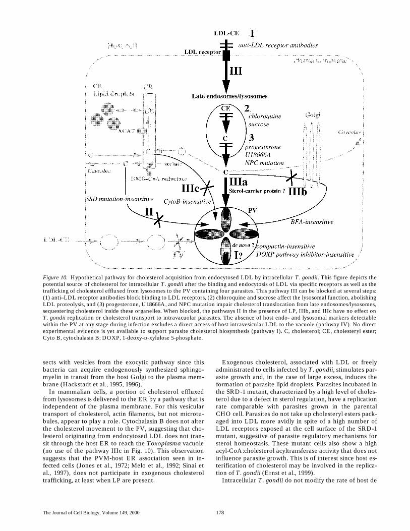
The putative pathway underlying cholesterol-derivedLDL acquisition by T. gondii is the pathway IIIa depictedin the schematic model in Fig. 10. First, a large contribu-tion of the receptor-mediated endocytosis of LDL appearsto be the primary mechanism as antibodies against LDLreceptors can block the accessibility of LDL to intracellu-lar compartments, including Toxoplasma vacuoles (Fig.10, 1).
Uptake of LDL by host cells infected by T. gondii occurs
Figure 8. Blockade and reversibility of [NBD-C]-LDL acquisi-tion by parasites with progesterone and U18666A. 24 h after in-fection in medium containing 10% LPDS, fibroblasts infected byT. gondii (arrows) were preincubated without drug (A), withprogesterone at 10 mg/ml for 24 h (B and C), with U18666A at 1mg/ml for 90 min (D), or with progesterone at 10 mg/ml for 20 h,and then with U18666A at 0.1 mg/ml for 90 min. After washing,cells were pulse-labeled at 378C for 10 min with 0.1 mg/ml of[NBD-C]-LDL, washed, and processed for fluorescence observa-tion. A chase of 24 h in medium containing 10% LPDS was per-formed in the experiment in C before analysis of the fluorescencepattern.
Coppens et al. Cholesterol Transport in Toxoplasma gondii–infected Cells 177
in an enhanced fashion over uninfected cells. Results donot distinguish, however, whether this phenomenon is di-rectly induced by intravacuolar parasites or is a more gen-eral response of the host cell as a consequence of parasiti-zation. Since endocytosis of other ligands is not modifiedafter infection, the putative mechanism may involve ahigher density of cell-surface molecules or receptors capa-ble of internalizing lipoproteins. If upregulation of theclassical apoprotein B/E LDL receptor is occurring in thepresence of unchanged HMG-CoA reductase activityupon infection with T. gondii (Table II), this would sug-gest an unusual pathway of cholesterol regulation (Gold-stein and Brown, 1990). For another intravacuolar patho-gen, exposure of macrophages to Chlamydia pneumoniaespecifically increases the LDL uptake and foam cell for-mation by accumulation of cholesteryl ester droplets by amechanism not involving the apoprotein B/E LDL recep-tor (Kalayoglu et al., 1999).
Endocytosed lipoproteins are delivered to endosomesand their protein/phospholipid coat degraded in late endo-somes/lysosomes, yielding free cholesteryl esters that willbe hydrolyzed to cholesterol. In the presence of chloro-quine or sucrose impairing lysosomal function and pro-voking an accumulation of intact LDL, minimal moleculesof cholesterol reach the PV, implicating host LDL proteol-ysis in the parasite acquisition of this lipid (Fig. 10, 2). In-cubation of T. gondii in host cells where cholesterol trans-location from late endosomes/lysosomes (2-2 mutant ortreated with progesterone and U18666A) is impaired leadsto a dramatic reduction of cholesterol associated with theparasites, concomitant with a slowing of parasite replica-
tion (Fig. 10, 3). The higher inhibition of parasite growthby progesterone compared with the 2-2 mutant might berelated to a direct toxic effect of the steroid on T. gondii.Alternatively, progesterone is known to sequester in lyso-somes cholesterol derived not only directly from endocy-tosed LDL, but also from other existing sterol pools, suchas lipid droplets or organelle membranes (McGookey andAnderson, 1983; Butler et al., 1992), and could thereforealso remove cholesterol from PV-containing parasites.
In mammalian cells, the mechanisms and precise routesof transport of cholesterol from late endosomes/lysosomesto the other cellular compartments are areas of intense in-vestigation. From the cell biological point of view, theToxoplasma vacuole may be considered as such a com-partment in the infected cell and it could be used as a toolfor analysis of intracellular cholesterol movement in mam-malian cells. A portion of cholesterol is mobilized to theplasma membrane through the Golgi apparatus (reviewedin Liscum and Munn, 1999). The pathway of cholesterolacquisition by T. gondii is clearly not altered by incubationof infected cells with Brefeldin A (no use of the pathwayIIIb in Fig. 10), indicating a Golgi-independent route de-spite the location of mature PVs adjacent to the host cellGolgi. This absence of delivery of host lipids from theGolgi to the Toxoplasma vacuole, a matter of some con-troversy (Melo and de Souza, 1996), has been recentlyconfirmed by the observation of PV free of fluorescent lip-ids in experiments using NDB-ceramide loaded in fibro-blasts before infection with parasites (Mordue et al., 1999).By comparison, the pathogen Chlamydia trachomatis,which similarly resides in a nonfusogenic vacuole, inter-
Figure 9. Reduction of parasitereplication and cholesterol con-tent in the 2-2 mutant or in fibro-blasts treated with progesterone.(A) Uracil incorporation was as-sayed using the 2-2 mutant orCHO cells as control infected for24 or 48 h in medium containing10% FBS. Data in percent areexpressed relative to controltaken as 100% 6 SEM fromthree separate experiments donein duplicate. Differences be-tween values of control and ex-perimental groups were statisti-cally significant (*P , 0.005).(B) The 2-2 mutant cells infectedwith parasites (arrows) were cul-tivated for 24 h in medium con-taining 10% FCS, fixed, cy-tochemically stained with filipinfor cholesterol detection, andobserved by fluorescence mi-croscopy. (C) Uracil incorpora-tion was assayed using fibro-blasts infected for 24 or 48 h inmedium containing 10% FBSplus 10 mg/ml of progesterone.
Data in percent are expressed relative to control (infected fibroblasts without progesterone) taken as 100% 6 SEM from three separateexperiments done in duplicate. Differences between values of control and experimental groups were statistically significant (**P ,0.0005).
The Journal of Cell Biology, Volume 149, 2000 178
sects with vesicles from the exocytic pathway since thisbacteria can acquire endogenously synthesized sphingo-myelin in transit from the host Golgi to the plasma mem-brane (Hackstadt et al., 1995, 1996).
In mammalian cells, a portion of cholesterol effluxedfrom lysosomes is delivered to the ER by a pathway that isindependent of the plasma membrane. For this vesiculartransport of cholesterol, actin filaments, but not microtu-bules, appear to play a role. Cytochalasin B does not alterthe cholesterol movement to the PV, suggesting that cho-lesterol originating from endocytosed LDL does not tran-sit through the host ER to reach the Toxoplasma vacuole(no use of the pathway IIIc in Fig. 10). This observationsuggests that the PVM-host ER association seen in in-fected cells (Jones et al., 1972; Melo et al., 1992; Sinai etal., 1997), does not participate in exogenous cholesteroltrafficking, at least when LP are present.
Exogenous cholesterol, associated with LDL or freelyadministrated to cells infected by T. gondii, stimulates par-asite growth and, in the case of large excess, induces theformation of parasite lipid droplets. Parasites incubated inthe SRD-1 mutant, characterized by a high level of choles-terol due to a defect in sterol regulation, have a replicationrate comparable with parasites grown in the parentalCHO cell. Parasites do not take up cholesteryl esters pack-aged into LDL more avidly in spite of a high number ofLDL receptors exposed at the cell surface of the SRD-1mutant, suggestive of parasite regulatory mechanisms forsterol homeostasis. These mutant cells also show a highacyl-CoA:cholesterol acyltransferase activity that does notinfluence parasite growth. This is of interest since host es-terification of cholesterol may be involved in the replica-tion of T. gondii (Ernst et al., 1999).
Intracellular T. gondii do not modify the rate of host de
Figure 10. Hypothetical pathway for cholesterol acquisition from endocytosed LDL by intracellular T. gondii. This figure depicts thepotential source of cholesterol for intracellular T. gondii after the binding and endocytosis of LDL via specific receptors as well as thetrafficking of cholesterol effluxed from lysosomes to the PV containing four parasites. This pathway III can be blocked at several steps:(1) anti–LDL receptor antibodies block binding to LDL receptors, (2) chloroquine and sucrose affect the lysosomal function, abolishingLDL proteolysis, and (3) progesterone, U18666A, and NPC mutation impair cholesterol translocation from late endosomes/lysosomes,sequestering cholesterol inside these organelles. When blocked, the pathways II in the presence of LP, IIIb, and IIIc have no effect onT. gondii replication or cholesterol transport to intravacuolar parasites. The absence of host endo- and lysosomal markers detectablewithin the PV at any stage during infection excludes a direct access of host intravesicular LDL to the vacuole (pathway IV). No directexperimental evidence is yet available to support parasite cholesterol biosynthesis (pathway I). C, cholesterol; CE, cholesteryl ester;Cyto B, cytochalasin B; DOXP, 1-deoxy-D-xylulose 5-phosphate.
Coppens et al. Cholesterol Transport in Toxoplasma gondii–infected Cells 179
novo cholesterol biosynthesis. Parasites are capable ofreplicating in cells altered in their sterol production suchas the SSD mutant. These data indicate that parasites arenot dependent upon host endogenous cholesterol as longas exogenous sterols are available. They do not interceptthe transport of this lipid from the ER in transit to theplasma membrane. These results corroborate the absenceof labeled cholesterol associated with the sterol fraction ofparasites after incubation of infected cells with radioactiveprecursors of the mevalonate pathway (no use of the path-way II in Fig. 10). The survival of 30% of the parasite pop-ulation in the SSD mutant in the absence of LP opens thequestion on the origin of other potential sources of sterolsfor T. gondii, such as cholesterol from cell debris, storedcholesteryl esters, probably present in their lipid droplets,or cholesterol biosynthesized by the parasite. However,the compactin insensitivity, as well as the absence of sterolsynthesis from precursors of the mevalonate pathway inthe parasites, suggests the absence of the classical meva-lonate pathway functional for isoprenoid biosynthesis in T.gondii, as observed for Plasmodium falciparum, an api-complexan parasite related to T. gondii. An alternativenonmevalonate pathway for the early steps in the biosyn-thesis of isoprenoids, the 1-deoxy-D-xylulose 5-phosphate(DOXP) pathway, has been proposed in the plasmodialapicoplast, a plastid acquired by members of the phylumApicomplexa by secondary endosymbiosis of an alga(Jomaa et al., 1999; McFadden and Roos, 1999). However,two lines of evidence suggest that sterol synthesis in the T.gondii apicoplast is not necessary for parasite replicationin the presence of lipoproteins. First, parasites that cannotpartition the apicoplast to daughter cells replicate nor-mally in the primary vacuole (D. Roos, personal commu-nication). Second, inhibitors of the DOXP pathway, whichblock plasmodial replication, do not inhibit T. gondiigrowth in presence of serum (D. Soldati, personal commu-nication).
The accumulation of cholesterol from host lysosomes in-side intravacuolar parasites suggests a largely unidirec-tional influx of this lipid and may be correlated to therapid division rates of T. gondii. Alternatively, the isola-tion of the PV from the external medium results in the ab-sence of transfer of cholesterol to extracellular cholesterolacceptors. The cholesterol transport pathway in cells in-fected with T. gondii clearly differs from those for theother cell organelles and is characterized by an absence ofinteraction with vesicles involved in export from the Golgiand ER, as well as those implicated in exchanges betweenthe plasma membrane and the cell interior. The route ofdelivery of exogenous cholesterol from lysosomes to thePV might involve a sterol-binding protein, mediating amolecular transport of cholesterol towards the PVM. T.gondii may actively either divert a host sterol-carrier pro-tein or synthesize such a protein since parasite viability isrequired for cholesterol acquisition. After crossing thePVM, cholesterol molecules must be translocated to theparasite. The elucidation of the mechanisms of these twosequential steps by generation of resistant mutants formsthe basis for our future experiments.The authors thank Norma Andrews, Laura Liscum, and Alex Nohturfttfor critical comments on the manuscript and the members of the K.A.Joiner lab for helpful discussions during the course of this work. We ac-
knowledge the individuals who generously provided mutant cell lines usedin this study (see Materials and Methods). We thank Achim Kaasch forthe conceptual idea of the parasite purification protocol using a Nycodenzgradient.
The work was supported by the National Institutes of Health grant AI-30060 and a Burroughs Wellcome Fund Molecular Parasitology ScholarAward to K.A. Joiner and an American Heart Association (CT affiliate)Postdoctoral Fellowship to A.P. Sinai.
Submitted: 20 December 1999Revised: 16 February 2000Accepted: 18 February 2000
References
Bradfute, D.L., C.J. Silva, and R.D. Simoni. 1992. Squalene synthase-deficientmutant of Chinese hamster ovary cells. J. Biol. Chem. 267:18308–18314.
Brown, M.S., S.E. Dana, and J.L. Goldstein. 1975. Receptor-dependent hydrol-ysis of cholesteryl esters contained in plasma low density lipoprotein. Proc.Natl. Acad. Sci. USA. 72:2925–2929.
Brown, M.S., and J.L. Goldstein. 1999. A proteolytic pathway that controls thecholesterol content of membranes, cell, and blood. Proc. Natl. Acad. Sci.USA. 96:11041–11048.
Butler, J.D., J. Blanchette-Mackie, E. Goldin, R.R. O’Neill, G. Carstea, C.F.Roff, M.C. Patterson, S. Patel, M.E. Comly, A. Cooney, M.T. Vanier, R.O.Brady, and P.G. Pentchev. 1992. Progesterone blocks cholesterol transloca-tion from lysosomes. J. Biol. Chem. 267:23797–23805.
Coppens, I., M. Andries, J.L. Liu, and M.F. Cesbron-Delauw. 1999. Intracellu-lar trafficking of dense granule proteins in Toxoplasma gondii and experi-mental evidences for a regulated exocytosis. Eur. J. Cell Biol. 78:463–472.
Coppens, I., T. Levade, and P.J. Courtoy. 1995. Host plasma low density lipo-protein particles as an essential source of lipids for the bloodstream forms ofTrypanosoma brucei. J. Biol. Chem. 270:5736–5741.
Craig, I.F., D.P. Via, W.W. Mantulin, H.J. Pownall, A.M. Gotto, Jr., and L.C.Smith. 1981. Low density lipoprotein reconstituted with steroids containingthe nitrobenzoxadiazole fluorophore. J. Lipid Res. 22:687–696.
Dahl, N.K., W.G. Gutheil, and L. Liscum. 1993. Abnormal regulation of lowdensity lipoprotein–sensitive events in a cholesterol transport mutant. J.Biol. Chem. 268:16979–16986.
Dahl, N.K., K.L. Reed, M.A. Daunais, J.R. Faust, and L. Liscum. 1992. Isola-tion and characterization of Chinese hamster ovary cells defective in the in-tracellular metabolism of LDL-derived cholesterol. J. Biol. Chem. 267:4889–4896.
de Duve, C. 1983. Lysosomes revisited. Eur. J. Biochem. 137:391–397.de Duve, C., T. de Barsy, B. Poole, A. Trouet, P. Tulkens, and F. Van Hoof.
1974. Lysosomotropic agents. Biochem. Pharmacol. 23:2495–2531.El-Jack, A.K., K.V. Kandror, and P.F. Pilch. 1999. The formation of an insulin-
responsive vesicular cargo compartment is an early event in 3T3-L1 adipo-cyte differentiation. Mol. Biol. Cell. 10:1581–1594.
Ernst, J.D., L.-M. Ting, S. Novak, and R.V. Farese. 1999. Fifth ToxoplasmosisConference. Marshall, CA.
Foussard, F., M.A. Leriche, and J.F. Dubremetz. 1991a. Characterization of thelipid content of Toxoplasma gondii rhoptries. Parasitology. 102:367–370.
Foussard, F., Y. Gallois, A. Girault, and J.F. Menez. 1991b. Lipids and fatty ac-ids of tachyzoites and purified pellicles of Toxoplasma gondii. Parasitol. Res.77:475– 477.
Gallois, Y., F. Foussard, A. Girault, J. Hodberg, A. Tricaud, G. Mauras, and C.Motta. 1988. Membrane fluidity of Toxoplasma gondii: a fluorescence polar-ization study. Biol. Cell. 62:11–15.
Geelen, M.J.H., J.S. Papiez, K. Girgis, and D.M. Gibson. 1991. In situ measure-ment of HMG-CoA reductase activity in digitonin-permeabilized hepato-cytes. Biochim. Biophys. Res. Commun. 180:525–530.
Goldstein, J.L., and M.S. Brown. 1990. Regulation of the mevalonate pathway.Nature. 343:425–430.
Greenspan, P., E.P. Mayer, and S.D. Fower. 1985. Nile red: a selective fluores-cent stain for intracellular lipid droplets. J. Cell Biol. 100:965–973.
Hackstadt, T., D.D. Rockey, R.A. Heinzen, and M.A. Scidmore. 1996. Chla-mydia trachomatis interrupts an exocytic pathway to acquire endogenouslysynthesized sphingomyelin in transit from the Golgi apparatus to the plasmamembrane. EMBO (Eur. Mol. Biol. Organ.) J. 15:964–977.
Hackstadt, T., M.A. Scidmore, and D.D. Rockey. 1995. Lipid metabolism inChlamydia trachomatis–infected cell: directed trafficking of Golgi-derivedsphingolipids to the chlamydial inclusion. Proc. Natl. Acad. Sci. USA. 92:4877–4881.
Havel, R.J., H.A. Eder, and J.H. Bragdon. 1955. The distribution and chemicalcomposition of ultracentrifugally separated lipoproteins in human serum. J.Clin. Invest. 34:1345–1351.
Joiner, K.A., S.A. Fuhrman, H. Mietinnen, L.L. Kasper, and I. Mellman. 1990.Toxoplasma gondii: fusion competence of parasitophorous vacuoles in Fc re-ceptor transfected fibroblasts. Science. 249:641–646.
Jomaa, H., J. Wiesner, S. Sanderbrand, B. Altincicek, C. Weidemeyer, M.Hintz, I. Turbachova, M. Eberl, J. Zeidler, H.K. Lichtenthaler, et al. 1999.
The Journal of Cell Biology, Volume 149, 2000 180
Inhibitors of the nonmevalonate pathway of isoprenoid biosynthesis as anti-malarial drugs. Science. 285:1573–1576.
Jones, T.C., S. Yeh, and J.G. Hirsch. 1972. The interaction between Toxo-plasma gondii and mammalian cells. I. Mechanism of entry and intracellularfate of the parasite. J. Exp. Med. 136:1157–1172.
Kalayoglu, M.V., G.S. Miranpuri, D.T. Golenbock, and G.I. Byrne. 1999. Char-acterization of low-density lipoprotein uptake by murine macrophages ex-posed to Chlamydia pneumoniae. Microb. Infect. 1:409–418.
Klausner, R.D., J.G. Donaldson, and J. Lippincott-Schwartz. 1992. Brefeldin A:insights into the control of membrane traffic and organelle structure. J. CellBiol. 116:1071–1080.
Kobayashi, T., M.H. Beuchat, M. Lindsay, S. Frias, R.D. Palmiter, H. Sakuraba,R.G. Parton, and J. Gruenberg. 1999. Late endosomes membranes richin lysobisphosphatidic acid regulate cholesterol transport. Nat. Cell Biol.1:113–118.
Lange, Y., and T.L. Steck. 1996. The role of intracellular cholesterol transportin cholesterol homeostasis. Trends Cell Biol. 6:205–208.
Leonard, D.A., and H.W. Chen. 1987. ATP-dependent degradation of 3-hy-droxy-3-methylglutaryl coenzyme A reductase in permeabilized cells. J.Biol. Chem. 262:7914–7919.
Lindsay, D.S., R.R. Mitscher, M.A. Toivio-Kinnucan, S.J. Upton, J.P. Dubey,and B.L. Blagburn. 1993. Association of host cell mitochondria with devel-oping Toxoplasma gondii tissue cysts. Am. J. Vet. Res. 54:1663–1667.
Lingelbach, K., and K.A. Joiner. 1998. The parasitophorous vacuole membranesurrounding Plasmodium and Toxoplasma: an unusual compartment in in-fected cells. J. Cell Sci. 111:1467–1475.
Liscum, L., and J.J. Klansek. 1998. Niemann-Pick disease type C. Curr. Opin.Lipidol. 9:131–135.
Liscum, L., and J.R. Faust. 1989. The intracellular transport of low density lipo-protein–derived cholesterol is inhibited in Chinese hamster ovary cells cul-tured with 3-b-[2-(diethylamino)ethoxy]androst-5-en-17-one. J. Biol. Chem.264:11796–11806.
Liscum, L., and N.J. Munn. 1999. Intracellular cholesterol transport. Biochim.Biophys. Acta. 1438:19–37.
Liscum, L., R.M. Ruggiero, and J.R. Faust. 1989. The intracellular transport oflow density lipoprotein–derived cholesterol is defective in Niemann-Picktype C fibroblasts. J. Cell Biol. 108:1625–1636.
Luskey, K.L., J.R. Faust, D.J. Chin, M.S. Brown, and J.L. Goldstein. 1983. Am-plification of the gene for 3-hydroxy-3-methylglutaryl coenzyme A reduc-tase, but not for the 53-kDa protein, in UT-1 cells. J. Biol. Chem. 258:8462–8469.
McFadden, G.I., and D.S. Roos. 1999. Apicomplexan plastids as drug targets.Trends Microbiol. 7:328–333.
McFarlane, A.S. 1958. Efficient trace-labelling of proteins with iodine. Nature.182:53.
McGookey, D.J., and G.W. Anderson. 1983. Morphological characterization ofthe cholesteryl ester cycle in cultured mouse macrophage foam cells. J. CellBiol. 97:1156–1165.
Melo, E.J.T., T.U. Carvalho, and W. de Souza. 1992. Penetration of Toxo-plasma gondii into host cells induces changes in the distribution of mito-chondria and the endoplasmic reticulum. Cell Struct. Funct. 17:311–317.
Melo, E.J.T, and W. de Souza. 1996. Pathway of C6-NBD-ceramide on the hostcell infected with Toxoplasma gondii. Cell Struct. Funct. 21:47–52.
Metherall, J.E., J.L. Goldstein, K.L. Luskey, and M.S. Brown. 1989. Loss oftranscriptional repression of three-sterol-regulated genes in mutant hamstercells. J. Biol. Chem. 264:15634–15641.
Misumi, Y., Y. Misumi, K. Miki, A. Takatsuki, G. Tamura, and Y. Ikehara.1986. Novel blockade by Brefeldin A of intracellular transport of secretoryproteins in cultured rat hepatocytes. J. Biol. Chem. 261:11398–11403.
Monteiro Cintra, W., and W. de Souza. 1985. Distribution of intramembranousparticles and filipin-sterol complexes in the cell membranes of Toxoplasmagondii. Eur. J. Cell Biol. 37:63–69.
Montgomery, R.R., P. Webster, and I. Mellman. 1991. Accumulation of indi-gestible substances reduces fusion competence of macrophage lysosomes. J.Immunol. 147:3087–3095.
Mordue, D.G., S. Hakansson, I. Niesman, and L.D. Sibley. 1999. Toxoplasma
gondii resides in a vacuole that avoids fusion with host cell endocytic andexocytic vesicular trafficking pathways. Exp. Parasitol. 92:87–99.
Mukherjee, S., X. Zha, I. Tabas, and F.R. Maxfield. 1998. Cholesterol distribu-tion in living cells: fluorescence imaging using dehydroergosterol as a fluo-rescent analog. Biophys. J. 75:1915–1925.
Nakaar, V., B.U. Samuel, E.O., and K.A. Joiner. 1999. Targeted reduction ofnucleoside triphosphate hydrolase by antisense RNA inhibits Toxoplasmagondii proliferation. J. Biol. Chem. 274:5083–5087.
Neufeld, E.B., A.M. Cooney, J. Pitha, E.A. Dawidowicz, N.K. Dwyer, P.G.Pentchev, and E.J. Blanchette-Mackie. 1996. Intracellular trafficking of cho-lesterol monitored with a cyclodextrin. J. Biol. Chem. 271:21604–21613.
Ohashi, M., M. Murata, and S. Ohnishi. 1992. A novel fluorescence method tomonitor the lysosomal disintegration of low density lipoprotein. Eur. J. CellBiol. 59:116–126.
Pentchev, P.G., H.S. Kruth, M.E. Comly, J.D. Butler, M.T. Vanier, D.A.Wenger, and S. Patel. 1986. Type C Niemann-Pick disease. A parallel loss ofregulatory responses in both the uptake and esterification of low density li-poprotein–derived cholesterol in cultured fibroblasts. J. Biol. Chem. 261:16775–16780.
Porchet-Hennere, E., and G. Torpier. 1983. Relations entre Toxoplasma et sacellule-hote. Protistologica. 19:357–370.
Poumay, Y., and M.F. Ronveaux-Dupal. 1984. Rapid preparative isolation ofconcentrated low-density lipoproteins and of lipoprotein-deficient serum us-ing vertical rotor gradient ultracentrifugation. J. Lipid Res. 26:1476–1480.
Roff, C.F., E. Goldin, M.E. Comly, A. Cooney, A. Brown, M.T. Vanier, S.P.F.Miller, R.O. Brady, and P.G. Pentchev. 1991. Type C Niemann-Pick disease:use of hydrophobic amines to study defective cholesterol transport. Dev.Neurosci. 13:315–320.
Roos, D.S., R.G.K. Donald, N.S. Morissette, and A.L.C. Moulton. 1994. Molec-ular tools for genetic dissection of the protozoan parasite Toxoplasma gon-dii. Methods Cell. Biol. 45:27–63.
Schwab, J.C., C.J.M. Beckers, and K.A. Joiner. 1994. The parasitophorous vac-uole membrane surrounding intracellular Toxoplasma gondii functions as amolecular sieve. Proc. Natl. Acad. Sci. USA. 91:509–513.
Sibley, L.D., I.R. Niesman, S.F. Parmley, and M.F. Cesbron-Delauw. 1995.Regulated secretion of multilamellar vesicles leads to formation of a tubulo-vesicular network in host-cell vacuoles occupied by Toxoplasma gondii. J.Cell Sci. 108:1669–1677.
Sibley, L.D., E. Weidner, and J.L. Krahenbulh. 1985. Phagosome acidificationblocked by intracellular Toxoplasma gondii. Nature. 315:416–419.
Silverman, J.A., M.L. Haynes, B.J. Luft, and K.A. Joiner. 1997. Characteriza-tion of anti-Toxoplasma activity of SDZ 215-918, a cyclosporin derivativelacking immunosuppressive and peptidyl-prolyl-isomerase-inhibiting activ-ity: possible role of a P glycoprotein in Toxoplasma physiology. Antimicrob.Agents Chemother. 41:1859–1866.
Sinai, A.P., and K.A. Joiner. 1997. Safe haven: the cell biology of nonfusogenicpathogen vacuoles. Annu. Rev. Microbiol. 51:415–462.
Sinai, A.P., P. Webster, and K.A. Joiner. 1997. Association of host cell endo-plasmic reticulum and mitochondria with the Toxoplasma gondii parasito-phorous vacuole membrane: a high affinity interaction. J. Cell Sci. 110:2117–2128.
Smith, P.K., R.I. Krohn, G.T. Hermanson, A.K. Mallia, F.H. Gartner, M.D.Provenzano, E.K. Fujimoto, N.M. Goeke, B.J. Olson, and D.C. Klenk. 1985.Measurement of protein using bicinchoninic acid. Anal. Biochem. 150:76–85.
Sokol, J., J. Blanchette-Mackie, H.S. Kruth, N.K. Dwyer, L.M. Amende, J.D.Butler, E. Robinson, S. Patel, R.O. Brady, M.E. Comly, et al. 1988. Type CNiemann-Pick disease. Lysosomal accumulation and defective intracellularmobilization of low density lipoprotein. J. Biol. Chem. 263:3411–3417.
Speer, C.A., S. Clark, and J.P. Dubey. 1998. Ultrastructure of the oocysts, spo-rocysts, and sporozoites of Toxoplasma gondii. J. Parasitol. 84:505–512.
Steinman, R.M., S.E. Brodie, and Z.A. Cohn. 1976. Membrane flow during pi-nocytosis. J. Cell Biol. 68:665–687.
Torii, S., B. Tomohiro, T. Watanabes, Y. Ikehara, K. Murakami, and K. Na-kayama. 1995. Cytotoxicity of Brefeldin A correlates with its inhibitory ef-fect on membrane binding of COP coat proteins. J. Biol. Chem. 270:11574–11580.