toxic epidermal necrolysis and stevens-johnson syndrome
TRANSCRIPT

Toxic epidermal necrolysis and Stevens-Johnson syndrome:A review*
Roland Gerull, MD; Mathias Nelle, MD; Thomas Schaible, MD
Several different manifestationsof drug-induced skin reactionshave been described. Mild con-ditions like maculopapular ex-
anthema, urticaria, photoallergic reactions,and fixed eruptions due to drugs occur fre-quently. More severe manifestations likeacute bullous exanthema are rare.
The aims of this review are to summa-rize definitions, causes, and clinicalcourses of and therapy for Stevens-Johnson syndrome (SJS) and toxic epi-dermal necrolysis (TEN). A systematic lit-erature search was performed over the
last 5 yrs using the following terms:“toxic epidermal necrolysis,” “Stevens-Johnson syndrome,” ‘child,” and “drugtoxicity.” This review was exempt fromapproval by the Research Ethics Commit-tee, University Hospital of Berne, Berne,Switzerland.
In 1956, Lyell (1) reported four casesof skin eruptions following either drugingestion or staphylococcal infection orof undetermined etiology. Over time, itbecame clear that he had described threedifferent diseases, including a case ofTEN. TEN is the most severe form ofdrug-induced skin reaction and is definedas epidermal detachment of �30% ofbody surface area. Similar diseases in-clude SJS, named after the 1922 descrip-tion by Stevens and Johnson (2) and er-ythema multiforme. SJS presents withepidermal detachment of �10% of bodysurface area, whereas involvement of10%–30% of body surface is defined asSJS/TEN overlap. Differences betweenTEN, SJS, and erythema multiforme aresummarized in Table 1. There is growingevidence that SJS and TEN are a single
disease with common causes and mech-anisms (3). Most authors give credit forthe first description of SJS to Hebra (4) in1860, but Rosenberg (5) mentions an1822 publication by Alibert and Bazinthat probably refers to the same disease.
The incidence of severe exfoliativeskin reactions are estimated at 1 to 7cases per million person-years for SJSand 0.4 to 1.5 cases per million person-years for TEN (6–10). In children, TENoccurs equally frequently in males andfemales, whereas in adults women aremore frequently affected by a ratio of 3:2to 2:1. Persons over 60 yrs seem to bemore likely to develop TEN (8, 11).
Causes of TEN and SJS
In 74%–94% of cases, TEN is triggeredeither by preceding medication or by aninfection of the upper respiratory tract(13–16).
The first large study to assess the riskof developing SJS or TEN included 245TEN-patients and 1,147 controls (17).This study distinguished between drugsusually used for short-term periods and
*See also p. 1600.From the Department of Neonatology (MN, RG),
Inselspital Bern, Bern, Switzerland; and Department ofNeonatology/Pediatric Intensive Care (TS), UniversityHospital Mannheim, Mannheim, Germany.
There are no sources of external funding.The authors have not disclosed any potential con-
flicts of interest.For information regarding this article, E-mail:
[email protected] © 2011 by the Society of Critical Care
Medicine and Lippincott Williams & Wilkins
DOI: 10.1097/CCM.0b013e31821201ed
Objectives: The aims of this review are to summarize thedefinitions, causes, and clinical course as well as the currentunderstanding of the genetic background, mechanism of disease,and therapy of toxic epidermal necrolysis and Stevens-Johnsonsyndrome.
Data Sources: PubMed was searched using the terms toxicepidermal necrolysis, Stevens-Johnson syndrome, drug toxicity,drug interaction, and skin diseases.
Data Synthesis: Toxic epidermal necrolysis and Stevens-John-son syndrome are acute inflammatory skin reactions. The onset isusually triggered by infections of the upper respiratory tract or bypreceding medication, among which nonsteroidal anti-inflamma-tory agents, antibiotics, and anticonvulsants are the most com-mon triggers. Initially the diseases present with unspecific symp-toms, followed by more or less extensive blistering and sheddingof the skin. Complete death of the epidermis leads to sloughingsimilar to that seen in large burns. Toxic epidermal necrolysis isthe most severe form of drug-induced skin reaction and includesdenudation of >30% of total body surface area. Stevens-Johnsonsyndrome affects <10%, whereas involvement of 10%–30% of body
surface area is called Stevens-Johnson syndrome/toxic epidermalnecrolysis overlap. Besides the skin, mucous membranes such asoral, genital, anal, nasal, and conjunctival mucosa are frequentlyinvolved in toxic epidermal necrolysis and Stevens-Johnson syn-drome. Toxic epidermal necrolysis is associated with a significantmortality of 30%–50% and long-term sequelae. Treatment includesearly admission to a burn unit, where treatment with precise fluid,electrolyte, protein, and energy supplementation, moderate mechan-ical ventilation, and expert wound care can be provided. Specifictreatment with immunosuppressive drugs or immunoglobulins didnot show an improved outcome in most studies and remains con-troversial. The mechanism of disease is not completely understood,but immunologic mechanisms, cytotoxic reactions, and delayed hy-persensitivity seem to be involved.
Conclusion: Profound knowledge of exfoliative skin diseases isneeded to improve therapy and outcome of these life-threateningillnesses. (Crit Care Med 2011; 39:1521–1532)
KEY WORDS: toxic epidermal necrolysis; Stevens-Johnson syn-drome; child; drug interactions; drug toxicity; skin diseases;hypersensitivity
1521Crit Care Med 2011 Vol. 39, No. 6

drugs used for months or years. Thehighest risk in the first group was docu-mented for trimethoprim-sulfomethoxa-zole and other sulfonamide antibiotics(crude relative risk [RR] 172), followed bychlormezanone (crude RR 62), cephalo-sporins (multivariate RR 14), quinolones(multivariate RR 10), and aminopenicil-lins (multivariate RR 6.7). For acetamin-ophen, the RR was calculated to be 0.6 inFrance but up to 9.3 in other countries.In the long-term-use group, the in-creased risk was confined largely to thefirst 2 months of treatment. Crude RRs ofother drugs were 90 for carbamazepinefollowed by oxicam-nonsteroidal anti-inflammatory drugs (RR 72), corticoste-roids (RR 54), phenytoin (RR 53), allo-purinol (RR 52), phenobarbital (RR 45),and valproic acid (RR 25).
The large EuroSCAR study (18) ana-lyzed RRs for several drugs. Among thenewer drugs, strong associations weredocumented for nevirapine (RR �22),tramadol (RR � 20), pantoprazole (RR �18), lamotrigine (RR �14), and sertraline(RR � 11). Analysis of older drugs largelyconfirmed the results from Roujeau et al(17) and showed the following univariateRRs: cotrimoxazole, 102; sulfonamides,53; carbamazepine, 33; phenytoin, 26;phenobarbital, 17; allopurinol, 11; andoxicam-nonsteroidal anti-inflammatorydrugs, 6.4.
Both of these studies included childrenand adults, with �10% of cases occurringin children. A pooled analysis was per-formed for children of �15 yrs (19). Theunivariate analysis showed that anti-infective sulfonamides, phenobarbital, lam-otrigine, and carbamazepine were stronglyassociated with SJS/TEN in children. Val-proic acid, acetaminophen, and nonsteroi-dal anti-inflammatory drugs as a group alsoincreased the risk of SJS/TEN.
Cross-reactivity of drugs might lead tofatal recurrent TEN (20). Potential cross-reactivity among �-lactam antibioticsand cephalosporins exists. Similarlycross-reactivity and recurrence of disease
might occur after administration of anti-epileptic drugs with the arene-oxide moi-ety (e.g., phenytoin, phenobarbital, car-bamazepine, etc.) and application ofother antiepileptics (e.g., valproic acid,levetiracetam, etc.) should be considered.
Other factors associated with SJS/TENare infectious diseases such as thosecaused by human immunodeficiency vi-rus (21), herpesvirus, Mycoplasma pneu-moniae (22–25), and hepatitis A virus(26) and noninfectious conditions includ-ing radiotherapy (27, 28), lupus erythem-atosus (29, 30), and collagen vascular dis-ease (31).
Genetics
Several genetic factors influence therisk of developing TEN/SJS. A statisti-cally significant increase in human leu-kocyte antigen (HLA)-B12 phenotypeamong 44 patients surviving TEN wasdocumented (32).
In Han Chinese, two other gene locihave been shown to increase risk forTEN: HLA-B*5801 was present in all pa-tients with severe cutaneous reactions toallopurinol and in only 15% of 135 toler-ant patients (33).
HLA-B*1502 was present in all Chi-nese patients experiencing carbamaz-epine-induced SJS, while HLA-B*1502was detected in only 3% of carbamaz-epine-tolerant patients (34). The risk of car-bamazepine-induced SJS/TEN was signifi-cantly higher in Thai patients with HLA-B*1502 (35) and in Indian patients (36).
Carbamazapine-induced SJS/TEN wasstrongly associated with HLA-B*1502 inChinese patients but patients with carba-mazepine-induced maculopapular erup-tions had an odds ratio similar to tolerantcontrols (37). In a European study of 12patients with carbamazepine-induced SJS/TEN, only four had the HLA-B*1502 allele(38). Remarkably, these four patients hadan Asian ancestry, whereas the others didnot. This shows that HLA-B*1502 seems tobe a useful predictive marker in the Asian
population but not in European patients.An HLA-B*1502 screening before treat-ment with carbamazepine in Asian patientswas proposed (39).
Immunopathogenesis
The etiology of TEN is not completelyunderstood. Several theories including im-munologic mechanisms, reactive drug me-tabolites, or interactions between these twohave been published in a review (40).
In summary, specific drug hypersensi-tivity leads to major histocompatibilityclass I-restricted drug presentation and isfollowed by an expansion of cytotoxic T-lymphocytes, leading to an infiltration ofskin lesions with cytotoxic T-lymphocytesand natural killer cells.
Evidence is supportive of a role for thedeath receptor Fas and its Fas ligand(CD95) in the pathogenesis of keratino-cyte apoptosis during TEN (41). Otherfindings suggest activation of the perfo-rin/granzyme pathway as a cytotoxicmechanism in SJS/TEN (42).
Recent publications show that granu-lysin probably is the key mediator fordisseminated keratinocyte death in SJS/TEN. Granulysin levels in the sera of pa-tients with SJS/TEN are much higherthan in patients with ordinary drug-induced skin reactions or healthy con-trols (43). Furthermore granulysin levelscorrelate with clinical severity (44).
Since the mechanism is not IgE me-diated, a desensitization of the triggeringdrug is not an option.
Clinical Course
Drug-induced TEN typically presentswith fever and influenza-like symptoms 1to 3 wks after the application of the sus-pected drug (45). One to 3 days later,signs begin in the mucous membranes,including eyes, mouth, nose, and genita-lia in up to 90% of cases. Skin lesionsmanifest as generalized macules withpurpuric centers. The macules progressto large confluating blisters with subse-quent epidermal detachment, yet nevershow involvement of the hair (Fig. 1). Inthe following 3 to 5 days, separation ofthe epidermis progresses and leads tolarge denuded areas (Fig. 2). The largewound area leads to extreme pain, mas-sive loss of fluid and protein, bleeding,evaporative heat loss with subsequent hy-pothermia, and infection (46).
Histopathology shows separation ofthe epidermis at the dermal-epidermal
Table 1. Classification of exfoliative skin reactions (12)
ReactionBullous Erythema
MultiformeStevens-Johnson
Syndrome
OverlapStevens-JohnsonSyndrome- TEN
TEN WithSpots
TEN WithoutSpots
Detachment �10% �10% 10%–30% �30% �10%Typical targets Yes No No No NoAtypical targets Raised Flat Flat FlatSpots No Yes Yes Yes No
TEN, toxic epidermal necrolysis.
1522 Crit Care Med 2011 Vol. 39, No. 6

junction of the skin, extracutaneous epi-thelium, and mucous membranes. Clini-cally, this can be detected by a positiveNikolsky sign, which describes detach-ment of the full-thickness epidermiswhen light lateral pressure is applied withthe examining finger.
Unlike the situation in full-thicknessburns, the epidermal appendages remainlargely intact, which allows re-epithelial-ization without scarring. Re-epithelial-ization of the epidermis begins about 1wk after onset of skin reactions and takesup to 3 wks (47).
Gastrointestinal involvement fre-quently occurs in the mouth and esoph-agus but also in the small bowl and colon(48–50). Patients with TEN usually donot develop a paralytic ileus, which al-lows early enteral nutrition (51). Involve-ment of the gastrointestinal tract maylead to stenosis or strictures and consec-utive long-term complications with dys-phagia and ileus-like symptoms. Vulvo-
vaginal involvement may also lead tovaginal stenosis or strictures (52, 53).
Hyper- and hypopigmentation occurin virtually all children and tend to fadewith time but usually do not resolve com-pletely (Fig. 3). Hypertrophic changesand scarring of the skin occur infre-quently (54). Long-term sequelae also in-volve fingernails and toenails, usually af-ter inflammation of the nail beds and lossof the nails during the acute phase ofTEN. Nails can develop deformities thatare usually not painful and not associatedwith significant functional disability.
In TEN, pulmonary edema and pro-gressive respiratory failure developwithin the first days and large ulcerationsand epithelial necrosis of the bronchialepithelium occur, which has to be sus-pected when dyspnea, bronchial hyperse-cretion, normal chest radiograph, and hy-poxemia are present during the earlystages of the disease (55).
Intubation and mechanical ventilationmay be required and is associated withhigher mortality (56). Permissive hyper-capnia with moderate respiratory acidosis(pH �7.20) can help to prevent baro-trauma and ventilator-induced lung in-jury (57). Treatment with inhaled nitricoxide might be helpful. PaO2/FIO2 ratioimproved an average of 162% after ad-ministration of low-dose inhaled nitricoxide (average 6.7 ppm), but nonsurvi-vors had a significantly less favorable ini-tial response (58).
SJS/TEN survivors may have persis-tent respiratory sequelae and a reductionin carbon monoxide diffusing capacity ofup to 35%–40% below normal even ifthey did not require mechanical ventila-tion (59).
Ophthalmic complications are seen inabout 30% of the surviving children, upto 74% in adults (60), and are severe in25% of cases (61).
The acute stage of ophthalmic involve-ment persists 2 to 6 wks and shows swol-len and erythematous eyelids, bacterialconjunctivitis, suppurative keratitis, orendophthalmitis. If there is significantinflammation, topical steroids can reducethe circle of cicatrization and eyelid de-formities (62). Extensive scarring due toovergrowth with conjunctival epithe-lium, membranous or pseudomembra-nous conjunctivitis, ankyloblepharon, orsymblepharon with additional complica-tions like entropion or lagophthalmosleads to a severe dry eye syndrome or lossof vision.
Persistent inflammation and ulcer-ation may lead to the destruction of lim-bal stem cells and stem cell deficiency(63, 64). Even after limbal stem celltransplantation, the prognosis is poor(65). Chronic ophthalmic complicationsoccur more frequently in patients withinitial ocular involvement (60), but lossof vision due to chronic corneal inflam-mation also occurs in patients withoutinitial affection of the eye and is consid-ered to be the most severe long-termcomplication in TEN/SJS survivors (61).
Long-term treatment with steroidsand vigorous lubrication helps to preventlate complications such as impaired tearproduction, aberrant lashes, and metapla-sia. Photophobia occurs frequently andmay resolve gradually over months (61,66–68).
Amniotic membrane transplantationhas been increasingly performed in re-cent years and may prevent chronic com-plications. A recent review shows goodresults in six children (69).
Other organ manifestations occurrarely. Involvement of the kidneys withglomerulonephritis, tubulonecrosis, andpancreatitis (70), as well as involvementof the liver including hepatocellular ne-crosis or cholestasis, has been reported(71–75).
The mortality of SJS is estimated to be1%–3% (13). In contrast, mortality ratesfor TEN are between 30% and 50% (76,77), with death usually resulting fromsepsis or multiorgan failure (13, 45, 78).The mortality rate of children appears tobe lower than that of adults.
A severity-of-illness score for TEN(SCORTEN) was published (79). Thisscore combined seven independent riskfactors for mortality (age �40 yrs, heart
Figure 1. Confluating blisters 3 days after onsetof skin reaction in a patient with toxic epidermalnecrolysis.
Figure 2. Large denuded areas following epidermaldetachment 5 days after onset of skin symptoms.
Figure 3. De- and hyperpigmentation of the skin6 months after toxic epidermal necrolysis.
1523Crit Care Med 2011 Vol. 39, No. 6
rate �120/min, history of cancer or he-matologic malignancies, involved bodysurface area �10%, serum urea level�10 mmol/L, serum bicarbonate level�20 mmol/L, serum glucose level �14mmol/L). Scoring one point for eachitem, the predicted mortality was 3.2%,12.1%, 5.3%, 58.3%, and 90.0% for 0–1,2, 3, 4, and �5 points respectively. Thepredicted mortality was shown to be ac-curate by other publications (80, 81) butmay underestimate mortality in patientswith respiratory involvement (82). Otherspublished a lower mortality rate with astandardized treatment protocol (83) oranalyzed that body surface involvementand age probably need more weight incalculations (84).
Treatment
Immediate discontinuation of the trig-gering drug reduces mortality and im-proves prognosis (85, 86). Readministra-tion of the suspected drug can cause arelapse of TEN (87).
The management of TEN is similar tothe treatment of extensive burns, andseveral studies have demonstrated thatearly transfer to a burn unit reduces mor-bidity and mortality significantly: TENsurvivors had been transferred to a burnunit 7.5 days earlier than nonsurvivorswith a mortality of 4% vs. 83%. Bactere-mia, septicemia, and length of hospital-ization were also reduced with earlytransfer (88). The largest trial showed amortality of 29.8% after transfer to aburn center within 7 days vs. 51.4% (p �.05) after 7 days (89). Other studieslargely support early transfer to a burnunit (85, 90–93).
However, several differences betweenTEN and burns need to be respected. InTEN, which in contrast to burns affectsonly the superficial dermal layers, fluid,electrolyte, and energy requirements areusually lower than in burns of the sameextent (90). Initial provision of 2 mL/kg/%affected body surface area results in ade-quate urine output and significant correc-tion of blood pressure in adults (94).
Older studies suggest that nutritionalrequirements are similar to those in burninjury (95–97), but newer studies showthat pediatric SJS/TEN patients requireapproximately 600 calories or 22% lessper day than patients with burns (98). Astatistically generated equation estimat-ing the energy requirement in the pedi-atric SJS/TEN population was developed:caloric needs � (preinjury weight [kg] �
24.6) � (wound size [%] � 4.1) � 940calories.
Other authors suggested that energyintake of 120% of the predicted basalmetabolic rate and intake of 3 grams perkilogram protein result in adequatewound healing, whereas higher energyprovisions enhance weight status (99).
Negative prognostic factors are hyper-natremia (100), increased blood urea ni-trogen, neutropenia, thrombocytopenia,visceral involvement, and delayed presen-tation (14, 90, 101).
Besides providing adequate analgesia,it is also essential to prevent dehydrationand superinfection. Treatment of the skinlesions with antimicrobial materials likecopper sulfate (102), silver nitrate (103),or sulfadiazine cream (which containssulfonamide and might lead to cross-reactivity with sulfonamide antibiotics)and covering with biological or syntheticmaterials have been proposed. In less de-veloped countries, banana leaf dressingsand boiled potato peel bandage may coverwounds satisfactorily (104). Porcinexenograft skin and human allograft ca-daveric skin have been used, but there is atrend to nonadherent (semi-)synthetic ma-terials. For example, Biobrane has beenused with good results and showed reducedpain and improved mobilization in elderlypatients (105–107). Other materials like Ac-ticoat, Aquacel Ag, or Suprathel are syn-thetic materials containing nanocrystallinesilver, which serves as an antimicrobialagent and is released for up to 7–14 days.The need of painful dressing changes canbe reduced. Several case reports, includingone case of a young infant, show good re-sults (108–113).
Additionally, it can be helpful to pre-vent shear forces and other mechanicaldisruptions to prevent further areas ofskin desquamation (114).
Considering the immunologic back-ground of TEN, treatment with immu-nosuppressive drugs was expected to bebeneficial.
Corticosteroids inhibit a wide range ofintracellular processes in turn modifyingthe inflammatory and immune responseand have been used in the managementof SJS and TEN for �30 yrs.
Several reports have been publishedshowing benefits of corticosteroid treat-ment. In a randomized prospective trial,pediatric patients treated with methyl-prednisolone showed a significantlyshorter duration of fever than did otherswith supportive care only (115).
However, other analyses suggest in-creased mortality and morbidity. Pediat-ric patients with SJS treated with highdoses of corticosteroids did not recoversooner than those treated with supportivecare alone. Patients in the steroid grouphad a higher incidence of medical com-plications (53% vs. 0%), most commonlyinfection (24%) and gastrointestinalbleeding (21%) (116).
Mortality was significantly higher in asteroid treated group with 66% vs. 33%in the nonsteroid group (103). Adminis-tration of corticosteroids for �48 hrs wasassociated with a higher rate of infection,longer hospitalization, and increasedmortality rate. A multivariate analysisdocumented that treatment with cortico-steroids is an independent risk factor forincreased mortality (88).
However, a recently published largeretrospective study included 281 patientsand evaluated the treatment of cortico-steroids and intravenous immunoglobu-lins (IVIGs) either alone or in combina-tion compared to supportive care only.Neither intravenous immunoglobulinsnor corticosteroids showed any signifi-cant effect on mortality in comparisonwith supportive care (117).
Publications evaluating the treatmentof TEN or SJS with corticosteroids aresummarized in Table 2.
In vitro studies showed that intrave-nous immunoglobulin was able to blockthe Fas receptor (41), which was the ra-tionale for the therapeutic use of immu-noglobulins geared toward the inhibitionof apoptosis.
Cessation of cutaneous blistering wasobserved in all patients within an averageof 2 days after IVIG was initiated. Nocorrelation between the timing or dosingand time to objective response was iden-tified in pediatric patients with SJS orTEN (118).
A retrospective trial compared treat-ment with IVIGs to standard supportivecare (119). The survival was 88%, and theauthors recommended a dose of 1 g/kg/day for 3 days. Other studies with com-bined 32 patients reported a survival of100% (120–122). IVIG-treated patientsshowed a shorter duration of fever andshorter hospitalization (123).
These optimistic results have not beenconfirmed by several other studies: a ret-rospective chart review found a highermortality and a longer hospitalization inpatients treated with IVIGs (124). Otherstudies showed no significant differencein mortality, multiorgan failure, duration
1524 Crit Care Med 2011 Vol. 39, No. 6

of mechanical ventilation, severity of sys-temic inflammation, incidence of sepsis,time to recovery or length of stay in hos-pital (125), or in progression of detach-ment or speed of re-epidermalization(126). The largest trial included 281 pa-tients and did not show decreased mor-tality with IVIGs (117).
Table 3 summarizes studies withtreatment of TEN or SJS with IVIGs. Insummary, the specific treatment of SJS/TEN with immunoglobulins remainscontroversial, and large randomized con-trolled trials are needed.
The only double-blind, randomized,placebo-controlled trial concerning treat-ment of TEN was performed for thalido-mide (127). Thalidomide inhibits the pro-duction of tumor necrosis factor alphaand interleukin-6 secreted by monocytesand lymphocytes. The progression of skinreaction was similar in both groups, butmortality was increased in the thalido-mide group (83%) vs. placebo (30%),which led to the discontinuation of thestudy. Thus, thalidomide cannot be con-sidered a safe or effective treatment ofTEN.
Cyclosporine inhibits CD8 activationand also has antiapoptotic activity. Theoret-ically, this inhibits epidermal apoptosis andleads to improved outcome. To date, largetrials have not been performed, but severalcase reports with up to 29 patients havebeen published using cyclosporine 3–10mg/kg daily (128–137). Patients treatedwith cyclosporine had a significantlyshorter time until stop of progression andcomplete re-epithelialization. Failure of �4organs and severe leukopenia and mortalitywere also significantly less frequent in thecyclosporine group (138).
Table 2. Corticosteroid treatment of Stevens-Johnson syndrome or toxic epidermal necrolysis
Author(s) andYear Study Type Diagnosis
No. of PatientsWith/Without
Corticosteroids Treatment
Time to Arrest inDays (Range)With/Without
Corticosteroids
Time to CompleteSkin Healing in
Days (Range)
Mortality With/Without
Corticosteroids Other
Rasmussen1976 (116)
Retrospective SJS 17/15 Prednisone 40–80 mg/m2/day
NR NR NR All nine complications inpatients with steroids;hospitalization withsteroids 21 days without13 days
Rasmussen1980 (164)
Retrospective TENa 24/51 Prednisone 60 mg/m2/day
1–3 7–12 NR
Halebianet al 1986(103)
Retrospectivecomparativetrial
TEN 15/15 240–1000 mghydrocortisonedaily overmaximum 7 days
NR NR 66%/33%
Kelemenet al 1995(88)
Retrospective TEN/SJS 14/37 NR NR NR 50%/3% Infection, hospitalization,and mortality reducedin patients with lessthan 48 hrs of steroids
Pasricha et al1996 (165)
Retrospective TEN 5/0 Dexamethasone 12–20mg/day decreasingdose 7–10 days
NR NR 0%
Kakourouet al 1997(115)
Retrospective TEN/SJS 10/6 Methylprednisolone 4mg/kg/day
7.0 � 3.3/9.8 � 3.0 NR 0%/0% Shorter period of feverwith steroids
Léauté-Labrèzeet al 2000(166)
Retrospective SJS 6/11 1 mg/kg/daydecreasing over1 wk
NR 18/19 0%/0% No benefit concerningduration of disease
Forman et al2002 (167)
Retrospective TEN/SJS 11/28 NR NR NR Overall 3.6% 21% complications
Lam et al2004 (168)
Retrospective TEN/SJS 9/2 Prednisolone 2 mg/kg/day for 3–5 days
NR NR 0%/0% Hospitalization 10 days
Kardaun andJonkman2007 (169)
Retrospective TEN/SJS 12/0 Dexamethasone 100mg or 1.5 mg/kgfor 3 days
2.3 16.8 8.3%
Yamane et al2007 (170)
Retrospective TEN/SJS 111/6 Prednisolone 10–600mg/day
NR NR 3.6%/16.6%
Schneck et al2008 (117)
Retrospectivemulticenter
TEN/SJS 159/122 NR NR NR 17.6%/27.8% No significant benefit fromany treatment
Hanken et al2009 (171)
Retrospective TEN 8/0 30–250 mgprednisone,duration NR
12 days NR 0%
Yang et al2009 (172)
Retrospective TEN 47 Methylprednisolone1–1.5 mg/kg/day
6.3 NR 27% 16% more likely to diewith steroidsSJS 18 5.7 16.7%
Koh and Tay2010 (173)
Retrospective TEN/SJS 5/5 Prednisolone 0.5–1.5mg/kg/day up to 1month
1.5/4.2 days NR 0%/0%
Hydrocortisone 10–15mg/kg/day average4 days (range 2–6)
NR: not reported; SJS, Stevens-Johnson syndrome; TEN, toxic epidermal necrolysis.aUnclear if all patients had TEN/SJS by today’s definition.
1525Crit Care Med 2011 Vol. 39, No. 6
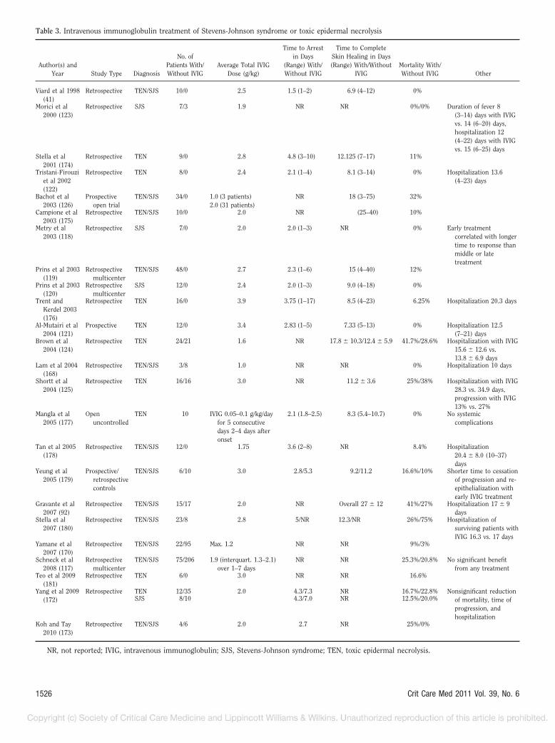
Table 3. Intravenous immunoglobulin treatment of Stevens-Johnson syndrome or toxic epidermal necrolysis
Author(s) andYear Study Type Diagnosis
No. ofPatients With/Without IVIG
Average Total IVIGDose (g/kg)
Time to Arrestin Days
(Range) With/Without IVIG
Time to CompleteSkin Healing in Days(Range) With/Without
IVIGMortality With/Without IVIG Other
Viard et al 1998(41)
Retrospective TEN/SJS 10/0 2.5 1.5 (1–2) 6.9 (4–12) 0%
Morici et al2000 (123)
Retrospective SJS 7/3 1.9 NR NR 0%/0% Duration of fever 8(3–14) days with IVIGvs. 14 (6–20) days,hospitalization 12(4–22) days with IVIGvs. 15 (6–25) days
Stella et al2001 (174)
Retrospective TEN 9/0 2.8 4.8 (3–10) 12.125 (7–17) 11%
Tristani-Firouziet al 2002(122)
Retrospective TEN 8/0 2.4 2.1 (1–4) 8.1 (3–14) 0% Hospitalization 13.6(4–23) days
Bachot et al2003 (126)
Prospectiveopen trial
TEN/SJS 34/0 1.0 (3 patients)2.0 (31 patients)
NR 18 (3–75) 32%
Campione et al2003 (175)
Retrospective TEN/SJS 10/0 2.0 NR (25–40) 10%
Metry et al2003 (118)
Retrospective SJS 7/0 2.0 2.0 (1–3) NR 0% Early treatmentcorrelated with longertime to response thanmiddle or latetreatment
Prins et al 2003(119)
Retrospectivemulticenter
TEN/SJS 48/0 2.7 2.3 (1–6) 15 (4–40) 12%
Prins et al 2003(120)
Retrospectivemulticenter
SJS 12/0 2.4 2.0 (1–3) 9.0 (4–18) 0%
Trent andKerdel 2003(176)
Retrospective TEN 16/0 3.9 3.75 (1–17) 8.5 (4–23) 6.25% Hospitalization 20.3 days
Al-Mutairi et al2004 (121)
Prospective TEN 12/0 3.4 2.83 (1–5) 7.33 (5–13) 0% Hospitalization 12.5(7–21) days
Brown et al2004 (124)
Retrospective TEN 24/21 1.6 NR 17.8 � 10.3/12.4 � 5.9 41.7%/28.6% Hospitalization with IVIG15.6 � 12.6 vs.13.8 � 6.9 days
Lam et al 2004(168)
Retrospective TEN/SJS 3/8 1.0 NR NR 0% Hospitalization 10 days
Shortt et al2004 (125)
Retrospective TEN 16/16 3.0 NR 11.2 � 3.6 25%/38% Hospitalization with IVIG28.3 vs. 34.9 days,progression with IVIG13% vs. 27%
Mangla et al2005 (177)
Openuncontrolled
TEN 10 IVIG 0.05–0.1 g/kg/dayfor 5 consecutivedays 2–4 days afteronset
2.1 (1.8–2.5) 8.3 (5.4–10.7) 0% No systemiccomplications
Tan et al 2005(178)
Retrospective TEN/SJS 12/0 1.75 3.6 (2–8) NR 8.4% Hospitalization20.4 � 8.0 (10–37)days
Yeung et al2005 (179)
Prospective/retrospectivecontrols
TEN/SJS 6/10 3.0 2.8/5.3 9.2/11.2 16.6%/10% Shorter time to cessationof progression and re-epithelialization withearly IVIG treatment
Gravante et al2007 (92)
Retrospective TEN/SJS 15/17 2.0 NR Overall 27 � 12 41%/27% Hospitalization 17 � 9days
Stella et al2007 (180)
Retrospective TEN/SJS 23/8 2.8 5/NR 12.3/NR 26%/75% Hospitalization ofsurviving patients withIVIG 16.3 vs. 17 days
Yamane et al2007 (170)
Retrospective TEN/SJS 22/95 Max. 1.2 NR NR 9%/3%
Schneck et al2008 (117)
Retrospectivemulticenter
TEN/SJS 75/206 1.9 (interquart. 1.3–2.1)over 1–7 days
NR NR 25.3%/20.8% No significant benefitfrom any treatment
Teo et al 2009(181)
Retrospective TEN 6/0 3.0 NR NR 16.6%
Yang et al 2009(172)
Retrospective TEN 12/35 2.0 4.3/7.3 NR 16.7%/22.8% Nonsignificant reductionof mortality, time ofprogression, andhospitalization
SJS 8/10 4.3/7.0 NR 12.5%/20.0%
Koh and Tay2010 (173)
Retrospective TEN/SJS 4/6 2.0 2.7 NR 25%/0%
NR, not reported; IVIG, intravenous immunoglobulin; SJS, Stevens-Johnson syndrome; TEN, toxic epidermal necrolysis.
1526 Crit Care Med 2011 Vol. 39, No. 6

Table 4. Studies of Stevens-Johnson syndrome or toxic epidermal necrolysis in pediatric patients
Author(s) andYear Type Diagnosis
No. ofPatients Treatment
Time to Arrest inDays (Range)
Time toComplete
Skin Healing inDays (Range)
Mortalitywith/without
SpecificTreatment Other
Rasmussen1976 (116)
Retrospective SJS 32 17 prednisone 40–80mg/m2/day15 supportive care
NR NR NR All ninecomplicationsin patientswith steroids;hospitalizationwith steroids21 dayswithout 13days
Rasmussen1980 (164)
Retrospective TENa 75 Antibiotics; 24patients: 60 mgprednisone/m2
1–3 7–12 NR
Ruiz-Maldonado1985 (102)
Retrospective TEN 60 Supportive NR 14.7 15%
Kakourou et al1997 (115)
Retrospective TEN/SJS 16 10 methylprednisolone4 mg/kg/day6 supportive care
7.0 � 3.3/9.8 � 3.0 NR 0%/0% Shorter period offever withsteroids
Morici et al2000 (123)
Retrospective SJS 12 7 IVIGs 1.5–2 g/kgsingle infusion onhospital day 3 (1–8)
NR NR 0%/0%/0% Duration of fever8 (3–14) dayswith IVIG vs.14 (6–20) days
2 corticosteroids Hospitalization12 (4–22) dayswith IVIG vs.15 (6–25) days
3 supportive care
Spies et al 2001(15)
Retrospective TEN 15 Supportive care 0% Hospitalization26 � 3 days
Forman et al2002 (167)
Retrospective SJS or TEN 28 11 patients treatedwithcorticosteroids
NR NR 3.6%overall
21%complications
Tristani-Firouziet al 2002(122)
Retrospective TEN 8 IVIG 0.5–0.75 g/dayfor 4 days onhospital day3.2 (2–5)
2.1 (1–4) 8.1 (3–14) 0% Hospitalization13.6 (4–23)days
Metry et al2003 (118)
Retrospective SJSPrevious
reportsSJS orTEN
728 previous
reports
IVIG 2.0 (1.2–4.0)g/kg distributedevenly over 4 days
3.75 (1–10) days afteronset of blistering
5 patientscorticosteroids
2 (1–3) NR NR Early treatmentcorrelatedwith longertime toresponse thanmiddle or latetreatment
Lam et al 2004(168)
Retrospective SJS or TEN 10 Corticosteroidsequivalent dose toprednisolone 1–2mg/kg/day 3–5days, if poorresponse IVIG 1g/kg/day
4 0% Hospitalization10 days
Mangla et al2005 (177)
Openuncontrolled
TEN 10 IVIG 0.05–0.1 g/kg/day for 5consecutive days
2.1 (1.8–2.5) 8.3 (5.4–10.7) 0% No systemiccomplications
2–4 days after onsetKoh and Tay
2010 (173)Retrospective SJS or TEN 15 4 patients: IVIG 2
mg/kg over 2–4days
NR NR 25%/0% Hospitalization22.7 days withIVIG vs. 7.7days withoutIVIG
5 patients:corticosteroidsequivalent to 0.5–1.5 mg/kg/dayprednisolonedecreasing doseover 2–4 wks
6 patients: supportivecare only
NR, not reported; IVIG, intravenous immunoglobulin; SJS, Stevens-Johnson syndrome; TEN, toxic epidermal necrolysis.aUnclear if all patients had TEN/SJS by today’s definition.
1527Crit Care Med 2011 Vol. 39, No. 6

Overall, cyclosporine treatment seemspromising, but larger trials are needed toconfirm the preliminary results.
Abnormal inherited metabolic pathwaysare presumed in some cases, which couldlead to a diminished detoxifying capacity.Administration of N-acetylcysteine en-hances the oxidant buffering capacity ofglutathione and inhibits nuclear factorkappa B, a transcription factor induced bytumor necrosis factor alpha and interleu-kin-6 (139). Treatment with N-acetylcyste-ine has been published with good results(140, 141), but numbers are small and con-trolled trials have not been performed.
Plasmapheresis has been used in sev-eral case reports and small studies,mostly in adult patients but also in chil-dren as young as 1 yr. The procedure canbe considered safe. One to eight plasmaexchange sessions have been used, pre-dominantly with good results (142–148).
Infliximab (149 –154), azathioprine(155), methotrexate (156), cyclophosph-amide (157–160), and recombinant gran-ulocyte colony-stimulating factor (161–163) were also used for the treatment ofTEN, but the data are limited and needfurther evaluation.
Since most studies have been per-formed in adult patients and treatment ofTEN and SJS in pediatric patients has notbeen studied extensively, we provide anoverview of all larger studies performedin pediatric patients in Table 4.
CONCLUSION
TEN is a life-threatening exfoliativeskin disease that shows blistering and ex-tensive shedding of the skin, and finallypresents with large denuded areas. Otherorgans are frequently affected, and an in-terdisciplinary team is needed to provideoptimal therapy. Although the incidenceis relatively low, it is important to iden-tify patients at risk to avoid delaying ther-apy. Treatment modalities vary widely be-tween supportive care alone, specifictreatment with immunosuppressivedrugs, IVIGs, and plasmapheresis. Todate, no treatment has been shown to besuperior, but in almost all cases no pro-spective randomized controlled trialshave been performed.
REFERENCES
1. Lyell A: Toxic epidermal necrolysis: Aneruption resembling scalding of the skin.Br J Dermatol 1956; 68:355–361
2. Stevens AM, Johnson F: A new eruptivefever associated with stomatitis and opthal-
mia: Report of two cases in children. Am JDis Child 1922; 24:526
3. Auquier-Dunant A, Mockenhaupt M, NaldiL, et al: Correlations between clinical pat-terns and causes of erythema multiformemajus, Stevens-Johnson syndrome, andtoxic epidermal necrolysis: Results of aninternational prospective study Arch Der-matol 2002; 138:1019–1024
4. Von Hebra F: Acute Exantheme undHautkrankheiten. In: Handbuch den spe-ziellen Pathologie. 3rd Edition. Virchow R(Ed), Ferdinand Enke, Erlangen, 1860
5. Rosenberg L, Rosenberg J: Erythema exsu-dativum multiforme with conjunctivitis andstomatitis. Arch Derm Syph 1940; 41:1066
6. Schopf E, Stuhmer A, Rzany B, et al: Toxicepidermal necrolysis and Stevens-Johnsonsyndrome. An epidemiologic study fromWest Germany. Arch Dermatol 1991; 127:839–842
7. Chan HL, Stern RS, Arndt KA, et al: Theincidence of erythema multiforme, Stevens-Johnson syndrome, and toxic epidermalnecrolysis: A population-based study withparticular reference to reactions caused bydrugs among outpatients Arch Dermatol1990; 126:43–47
8. Strom BL, Carson JL, Halpern AC, et al: Apopulation-based study of Stevens-Johnsonsyndrome. Incidence and antecedent drugexposures. Arch Dermatol 1991; 127:831–838
9. Naldi L, Locati F, Marchesi L, et al: Inci-dence of toxic epidermal necrolysis in Italy.Arch Dermatol 1990; 126:1103–1104
10. Roujeau JC, Guillaume JC, Fabre JP, et al:Toxic epidermal necrolysis (Lyell syn-drome). Incidence and drug etiology inFrance, 1981–1985. Arch Dermatol 1990;126:37–42
11. Prendiville JS, Hebert AA, Greenwald MJ, etal: Management of Stevens-Johnson syn-drome and toxic epidermal necrolysis inchildren. J Pediatr 1989; 115:881–887
12. Bastuji-Garin S, Rzany B, Stern RS, et al:Clinical classification of cases of toxic epi-dermal necrolysis, Stevens-Johnson syn-drome, and erythema multiforme. ArchDermatol 1993; 129:92–96
13. Roujeau JC, Chosidow O, Saiag P, et al:Toxic epidermal necrolysis (Lyell syn-drome). J Am Acad Dermatol 1990; 23:1039–1058
14. Roujeau JC, Stern RS: Severe adverse cuta-neous reactions to drugs. N Engl J Med1994; 331:1272–1285
15. Guillaume JC, Roujeau JC, Revuz J, et al:The culprit drugs in 87 cases of toxic epi-dermal necrolysis (Lyell’s syndrome). ArchDermatol 1987; 123:1166–1170
16. Spies M, Sanford AP, Aili Low JF, et al:Treatment of extensive toxic epidermalnecrolysis in children. Pediatrics 2001; 108:1162–1168
17. Roujeau JC, Kelly JP, Naldi L, et al: Medi-cation use and the risk of Stevens-Johnson
syndrome or toxic epidermal necrolysis.N Engl J Med 1995; 333:1600–1607
18. Mockenhaupt M, Viboud C, Dunant A, et al:Stevens-Johnson syndrome and toxic epi-dermal necrolysis: Assessment of medica-tion risks with emphasis on recently mar-keted drugs. The EuroSCAR-study. J InvestDermatol 2008; 128:35–44
19. Levi N, Bastuji-Garin S, Mockenhaupt M, etal: Medications as risk factors of Stevens-Johnson syndrome and toxic epidermalnecrolysis in children: A pooled analysis.Pediatrics 2009; 123:e297–e304
20. Paquet P, Jacob E, Damas P, et al: Recur-rent fatal drug-induced toxic epidermalnecrolysis (Lyell’s syndrome) after putativebeta-lactam cross-reactivity: Case reportand scrutiny of antibiotic imputability. CritCare Med 2002; 30:2580–2583
21. Belfort R Jr, de Smet M, Whitcup SM, et al:Ocular complications of Stevens-Johnsonsyndrome and toxic epidermal necrolysis inpatients with AIDS. Cornea 1991; 10:536–538
22. Fournier S, Bastuji-Garin S, Mentec H, etal: Toxic epidermal necrolysis associatedwith Mycoplasma pneumoniae infection.Eur J Clin Microbiol Infect Dis 1995; 14:558–559
23. Meseguer MA, de Rafael L, Vidal ML: Ste-vens-Johnson syndrome with isolation ofMycoplasma pneumoniae from skin lesions.Eur J Clin Microbiol 1986; 5:167–168
24. Stutman HR: Stevens-Johnson syndromeand Mycoplasma pneumoniae: Evidence forcutaneous infection. J Pediatr 1987; 111:845–847
25. Ravin KA, Rappaport LD, Zuckerbraun NS,et al: Mycoplasma pneumoniae and atypicalStevens-Johnson syndrome: A case series.Pediatrics 2007; 119:e1002–e1005
26. Werblowsky-Constantini N, Livshin R, Bur-stein M, et al: Toxic epidermal necrolysisassociated with acute cholestatic viral hep-atitis A. J Clin Gastroenterol 1989; 11:691–693
27. Ruggiero A, Buonuomo PS, Maurizi P, et al:Stevens-Johnson syndrome in children re-ceiving phenobarbital therapy and cranialradiotherapy. J Neurooncol 2007; 85:213–215
28. Maiche A, Teerenhovi L: Stevens-Johnsonsyndrome in patients receiving radiationtherapy. Lancet 1985; 2:45
29. Burge SM, Dawber RP: Stevens-Johnsonsyndrome and toxic epidermal necrolysis ina patient with systemic lupus erythemato-sus. J Am Acad Dermatol 1985; 13:665–666
30. Matsushita K, Ozaki A, Inoue H, et al: Ste-vens-Johnson syndrome induced by mizor-ibine in a patient with systemic lupus ery-thematosus. Mod Rheumatol 2006; 16:113–116
31. Nigen S, Knowles SR, Shear NH: Drugeruptions: Approaching the diagnosis ofdrug-induced skin diseases. J Drugs Derma-tol 2003; 2:278–299
32. Roujeau JC, Huynh TN, Bracq C, et al: Ge-
1528 Crit Care Med 2011 Vol. 39, No. 6

netic susceptibility to toxic epidermalnecrolysis. Arch Dermatol 1987; 123:1171–1173
33. Hung SI, Chung WH, Liou LB, et al: HLA-B*5801 allele as a genetic marker for severecutaneous adverse reactions caused by allo-purinol. Proc Nat Acad Sci U S A 2005;102:4134–4139
34. Chung WH, Hung SI, Hong HS, et al: Med-ical genetics: A marker for Stevens-Johnsonsyndrome. Nature 2004; 428:486
35. Tassaneeyakul W, Tiamkao S, Jantara-roungtong T, et al: Association betweenHLA-B*1502 and carbamazepine-inducedsevere cutaneous adverse drug reactions ina Thai population. Epilepsia 2010; 51:926–930
36. Mehta TY, Prajapati LM, Mittal B, et al:Association of HLA-B*1502 allele and car-bamazepine-induced Stevens-Johnson syn-drome among Indians. Indian J DermatolVenereol Leprol 2009; 75:579–582
37. Wu XT, Hu FY, An DM, et al: Associationbetween carbamazepine-induced cutaneousadverse drug reactions and the HLA-B*1502allele among patients in central China. Ep-ilepsy Behav 2010; 19:405–408
38. Lonjou C, Thomas L, Borot N, et al: Amarker for Stevens-Johnson syndrome . . . :Ethnicity matters. Pharmacogenomics J2006; 6:265–268
39. Locharernkul C, Shotelersuk V, HirankarnN: HLA-B* 1502 screening: Time to clinicalpractice. Epilepsia 2010; 51:936–938
40. Pereira FA, Mudgil AV, Rosmarin DM: Toxicepidermal necrolysis. J Am Acad Dermatol2007; 56:181–200
41. Viard I, Wehrli P, Bullani R, et al: Inhibitionof toxic epidermal necrolysis by blockade ofCD95 with human intravenous immuno-globulin. Science 1998; 282:490–493
42. Nassif A, Bensussan A, Boumsell L, et al:Toxic epidermal necrolysis: Effector cellsare drug-specific cytotoxic T cells. J AllergyClin Immunol 2004; 114:1209–1215
43. Abe R, Yoshioka N, Murata J, et al: Granu-lysin as a marker for early diagnosis of theStevens-Johnson syndrome. Ann InternMed 2009; 151:514–515
44. Chung WH, Hung SI, Yang JY, et al: Granu-lysin is a key mediator for disseminatedkeratinocyte death in Stevens-Johnson syn-drome and toxic epidermal necrolysis. NatMed 2008; 14:1343–1350
45. Revuz J, Penso D, Roujeau JC, et al: Toxicepidermal necrolysis. Clinical findings andprognosis factors in 87 patients. Arch Der-matol 1987; 123:1160–1165
46. Wong KC, Kennedy PJ, Lee S: Clinical man-ifestations and outcomes in 17 cases of Ste-vens-Johnson syndrome and toxic epider-mal necrolysis. Australas J Dermatol 1999;40:131–134
47. Jordan MH, Lewis MS, Jeng JG, et al: Treat-ment of toxic epidermal necrolysis by burnunits: Another market or another threat?J Burn Care Rehabil 1991; 12:579–581
48. Michel P, Joly P, Ducrotte P, et al: Ileal
involvement in toxic epidermal necrolysis(Lyell syndrome). Dig Dis Sci 1993; 38:1938–1941
49. Powell N, Munro JM, Rowbotham D: Co-lonic involvement in Stevens-Johnson syn-drome. Postgrad Med J 2006; 82:e10
50. Carter FM, Mitchell CK: Toxic epidermalnecrolysis–an unusual cause of colonic per-foration. Report of a case. Dis Colon Rec-tum 1993; 36:773–777
51. Pirrung MK: Management of toxic epider-mal necrolysis. J Intraven Nurs 2001; 24:107–112
52. Meneux E, Paniel BJ, Pouget F, et al: Vul-vovaginal sequelae in toxic epidermalnecrolysis. J Reprod Med 1997; 42:153–156
53. Rowan DM, Jones RW, Oakley A, et al: Vag-inal stenosis after toxic epidermal necroly-sis. J Low Genit Tract Dis 2010; 14:390–392
54. Kavanagh GM, Page P, Hanna MM: Siliconegel treatment of extensive hypertrophicscarring following toxic epidermal necroly-sis. Br J Dermatol 1994; 130:540–541
55. Lebargy F, Wolkenstein P, Gisselbrecht M,et al: Pulmonary complications in toxic epi-dermal necrolysis: A prospective clinicalstudy. Intensive Care Med 1997; 23:1237–1244
56. Namdar T, Stang FH, Siemers F, et al: Me-chanical ventilation in toxic epidermalnecrolysis. Handchir Mikrochir Plast Chir2010 Sep 2 [Epub ahead of print]
57. Sheridan RL, Kacmarek RM, McEttrick MM,et al: Permissive hypercapnia as a ventila-tory strategy in burned children: Effect onbarotrauma, pneumonia, and mortality.J Trauma 1995; 39:854–859
58. Sheridan RL, Zapol WM, Ritz RH, et al:Low-dose inhaled nitric oxide in acutelyburned children with profound respiratoryfailure. Surgery 1999; 126:856–862
59. McIvor RA, Zaidi J, Peters WJ, et al: Acuteand chronic respiratory complications oftoxic epidermal necrolysis. J Burn Care Re-habil 1996; 17:237–240
60. Gueudry J, Roujeau JC, Binaghi M, et al:Risk factors for the development of ocularcomplications of Stevens-Johnson syn-drome and toxic epidermal necrolysis. ArchDermatol 2009; 145:157–162
61. Power WJ, Ghoraishi M, Merayo-Lloves J, etal: Analysis of the acute ophthalmic mani-festations of the erythema multiforme/Stevens-Johnson syndrome/toxic epidermalnecrolysis disease spectrum. Ophthalmol-ogy 1995; 102:1669–1676
62. Lehman SS: Long-term ocular complica-tion of Stevens-Johnson syndrome. Clin Pe-diatr (Phila) 1999; 38:425–427
63. Lavker RM, Tseng SC, Sun TT: Corneal ep-ithelial stem cells at the limbus: Looking atsome old problems from a new angle. ExpEye Res 2004; 78:433–446
64. Dua HS, Azuara-Blanco A: Limbal stemcells of the corneal epithelium. Surv Oph-thalmol 2000; 44:415–425
65. Solomon A, Ellies P, Anderson DF, et al:Long-term outcome of keratolimbal allo-
graft with or without penetrating kerato-plasty for total limbal stem cell deficiency.Ophthalmology 2002; 109:1159–1166
66. Haus C, Paquet P, Marechal-Courtois C:Long-term corneal involvement followingdrug-induced toxic epidermal necrolysis(Lyell’s disease). Ophthalmologica 1993;206:115–118
67. Sotozono C, Ueta M, Koizumi N, et al: Di-agnosis and treatment of Stevens-Johnsonsyndrome and toxic epidermal necrolysiswith ocular complications. Ophthalmology2009; 116:685–690
68. Wilkins J, Morrison L, White CR Jr: Oculo-cutaneous manifestations of the erythemamultiforme/Stevens-Johnson syndrome/toxic epidermal necrolysis spectrum. Der-matol Clin 1992; 10:571–582
69. Shay E, Kheirkhah A, Liang L, et al: Amni-otic membrane transplantation as a newtherapy for the acute ocular manifestationsof Stevens-Johnson syndrome and toxic epi-dermal necrolysis. Surv Ophthalmol 2009;54:686–696
70. Coetzer M, van der Merwe AE, Warren BL:Toxic epidermal necrolysis in a burn patientcomplicated by acute pancreatitis. Burns1998; 24:181–183
71. Sahagun Flores JE, Soto Ortiz JA, TovarMendez CE, et al: [Stevens-Johnson syn-drome plus intrahepatic cholestasis causedby clindamycin or chlorpheniramine].Dermatol Online J 2009; 15:12
72. Masia M, Gutierrez F, Jimeno A, et al: Ful-minant hepatitis and fatal toxic epidermalnecrolysis (Lyell disease) coincident withclarithromycin administration in an alco-holic patient receiving disulfiram therapy.Arch Intern Med 2002; 162:474–476
73. Morelli MS, O’Brien FX: Stevens-Johnsonsyndrome and cholestatic hepatitis. Dig DisSci 2001; 46:2385–2388
74. Chan JC, Lai FM, Critchley JA: A case ofStevens-Johnson syndrome, cholestatichepatitis and haemolytic anaemia associ-ated with use of mefenamic acid. Drug Saf1991; 6:230–234
75. McArthur JE, Dyment PG: Stevens-Johnsonsyndrome with hepatitis following therapywith ampicillin and cephalexin. N Z Med J1975; 81:390–392
76. Fritsch PO, Sidoroff A: Drug-induced Ste-vens-Johnson syndrome/toxic epidermalnecrolysis. Am J Clin Dermatol 2000;1:349–360
77. Kelly JP, Auquier A, Rzany B, et al: Aninternational collaborative case-controlstudy of severe cutaneous adverse reactions(SCAR). Design and methods. J Clin Epide-miol 1995; 48:1099–1108
78. Mockenhaupt MNJ: Cutaneous adverse drugreactions: Stevens-Johnson syndrome andtoxic epidermal necrolysis. Allergy Clin Im-munol Int 2002; 14:143–150
79. Bastuji-Garin S, Fouchard N, Bertocchi M,et al: SCORTEN: A severity-of-illness scorefor toxic epidermal necrolysis. J Invest Der-matol 2000; 115:149–153
1529Crit Care Med 2011 Vol. 39, No. 6

80. Guegan S, Bastuji-Garin S, Poszepczynska-Guigne E, et al: Performance of theSCORTEN during the first five days of hos-pitalization to predict the prognosis of epi-dermal necrolysis. J Invest Dermatol 2006;126:272–276
81. Cartotto R, Mayich M, Nickerson D, et al:SCORTEN accurately predicts mortalityamong toxic epidermal necrolysis patientstreated in a burn center. J Burn Care Res2008; 29:141–146
82. Hague JS, Goulding JM, Long TM, et al:Respiratory involvement in toxic epidermalnecrolysis portends a poor prognosis thatmay not be reflected in SCORTEN. Br JDermatol 2007; 157:1294–1296
83. Imahara SD, Holmes JH IV, Heimbach DM,et al: SCORTEN overestimates mortality inthe setting of a standardized treatment pro-tocol. J Burn Care Res 2006; 27:270–275
84. Vaishampayan SS, Das AL, Verma R:SCORTEN: Does it need modification? In-dian J Dermatol Venereol Leprol 2008; 74:35–37
85. Gerdts B, Vloemans AF, Kreis RW: Toxicepidermal necrolysis: 15 years’ experiencein a Dutch burns centre. J Eur Acad Der-matol Venereol 2007; 21:781–788
86. Garcia-Doval I, LeCleach L, Bocquet H, etal: Toxic epidermal necrolysis and Stevens-Johnson syndrome: Does early withdrawalof causative drugs decrease the risk ofdeath? Arch Dermatol 2000; 136:323–327
87. Dreyfuss DA, Gottlieb LJ, Wilkerson DK, etal: Survival after a second episode of toxicepidermal necrolysis. Ann Plast Surg 1988;20:146–147
88. Kelemen JJ III, Cioffi WG, McManus WF, etal: Burn center care for patients with toxicepidermal necrolysis. J Am Coll Surg 1995;180:273–278
89. Palmieri TL, Greenhalgh DG, Saffle JR, etal: A multicenter review of toxic epidermalnecrolysis treated in U.S. burn centers atthe end of the twentieth century. J BurnCare Rehabil 2002; 23:87–96
90. Schulz JT, Sheridan RL, Ryan CM, et al: A10-year experience with toxic epidermalnecrolysis. J Burn Care Rehabil 2000; 21:199–204
91. McGee T, Munster A: Toxic epidermalnecrolysis syndrome: Mortality rate reducedwith early referral to regional burn center.Plast Reconstr Surg 1998; 102:1018–1022
92. Gravante G, Delogu D, Marianetti M, et al:Toxic epidermal necrolysis and StevenJohnson syndrome: 11-years experience andoutcome. Eur Rev Med Pharmacol Sci2007; 11:119–127
93. Engelhardt SL, Schurr MJ, Helgerson RB:Toxic epidermal necrolysis: An analysis ofreferral patterns and steroid usage. J BurnCare Rehabil 1997; 18:520–524
94. Shiga S, Cartotto R: What are the fluidrequirements in toxic epidermal necrolysis?J Burn Care Res 2010; 31:100–104
95. Coss-Bu JA, Jefferson LS, Levy ML, et al:Nutrition requirements in patients with
toxic epidermal necrolysis. Nutr Clin Pract1997; 12:81–84
96. Hildreth MA: Caloric needs of patients withtoxic epidermal necrolysis. J Am Diet Assoc1990; 90:A99
97. Starks LJ, Harrington D, Mozingo DW, et al:Extent of cutaneous exfoliation predictsmetabolic rate in patients with TENS. ProcAm Burn Assoc 1996; 28:115
98. Mayes T, Gottschlich M, Khoury J, et al:Energy requirements of pediatric patientswith Stevens-Johnson syndrome and toxicepidermal necrolysis. Nutr Clin Pract 2008;23:547–550
99. Prelack K, Cunningham JJ, Sheridan RL, etal: Energy and protein provisions for ther-mally injured children revisited: An out-come-based approach for determining re-quirements. J Burn Care Rehabil 1997; 18:177–181, discussion 176
100. Namdar T, von Wild T, Siemers F, et al:Does hypernatremia impact mortality intoxic epidermal necrolysis? Ger Med Sci2010; 8:Doc30
101. Westly ED, Wechsler HL: Toxic epidermalnecrolysis. Granulocytic leukopenia as aprognostic indicator. Arch Dermatol 1984;120:721–726
102. Ruiz-Maldonado R: Acute disseminated epi-dermal necrosis types 1, 2, and 3: Study ofsixty cases. J Am Acad Dermatol 1985; 13:623–635
103. Halebian PH, Corder VJ, Madden MR, et al:Improved burn center survival of patientswith toxic epidermal necrolysis managedwithout corticosteroids. Ann Surg 1986;204:503–512
104. Gore MA, Akolekar D: Evaluation of bananaleaf dressing for partial thickness burnwounds. Burns 2003; 29:487–492
105. Boorboor P, Vogt PM, Bechara FG, et al:Toxic epidermal necrolysis: Use of Biobraneor skin coverage reduces pain, improvesmobilisation and decreases infection in el-derly patients. Burns 2008; 34:487–492
106. Arevalo JM, Lorente JA: Skin coverage withBiobrane biomaterial for the treatment ofpatients with toxic epidermal necrolysis.J Burn Care Rehabil 1999; 20:406–410
107. Whitaker IS, Prowse S, Potokar TS: A crit-ical evaluation of the use of Biobrane as abiologic skin substitute: A versatile tool forthe plastic and reconstructive surgeon. AnnPlast Surg 2008; 60:333–337
108. Dalli RL, Kumar R, Kennedy P, et al: Toxicepidermal necrolysis/Stevens-Johnson syn-drome: Current trends in management.ANZ J Surg 2007; 77:671–676
109. Pfurtscheller K, Zobel G, Roedl S, et al: Useof Suprathel dressing in a young infant withTEN. Pediatr Dermatol 2008; 25:541–543
110. Asz J, Asz D, Moushey R, et al: Treatment oftoxic epidermal necrolysis in a pediatric pa-tient with a nanocrystalline silver dressing.J Pediatr Surg 2006; 41:e9–e12
111. Huang SH, Wu SH, Sun IF, et al: AQUACELAg in the treatment of toxic epidermalnecrolysis (TEN). Burns 2008; 34:63–66
112. Clennett S, Hosking G: Management oftoxic epidermal necrolysis in a 15-year-oldgirl. J Wound Care 2003; 12:151–154
113. Huang SH, Yang PS, Wu SH, et al: Aquacel((R))Ag with Vaseline gauze in the management oftoxic epidermal necrolysis (TEN). Burns 2009
114. Dorafshar AH, Dickie SR, Cohn AB, et al:Antishear therapy for toxic epidermalnecrolysis: An alternative treatment ap-proach. Plast Reconstr Surg 2008; 122:154–160
115. Kakourou T, Klontza D, Soteropoulou F, etal: Corticosteroid treatment of erythemamultiforme major (Stevens-Johnson syn-drome) in children. Eur J Pediatr 1997;156:90–93
116. Rasmussen JE: Erythema multiforme inchildren. Response to treatment with sys-temic corticosteroids. Br J Dermatol 1976;95:181–186
117. Schneck J, Fagot JP, Sekula P, et al: Effectsof treatments on the mortality of Stevens-Johnson syndrome and toxic epidermalnecrolysis: A retrospective study on patientsincluded in the prospective EuroSCARStudy. J Am Acad Dermatol 2008; 58:33–40
118. Metry DW, Jung P, Levy ML: Use of intra-venous immunoglobulin in children withstevens-johnson syndrome and toxic epider-mal necrolysis: Seven cases and review ofthe literature. Pediatrics 2003; 112:1430–1436
119. Prins C, Kerdel FA, Padilla RS, et al: Treat-ment of toxic epidermal necrolysis withhigh-dose intravenous immunoglobulins:Multicenter retrospective analysis of 48consecutive cases. Arch Dermatol 2003;139:26–32
120. Prins C, Vittorio C, Padilla RS, et al: Effectof high-dose intravenous immunoglobulintherapy in Stevens-Johnson syndrome: Aretrospective, multicenter study. Dermatol-ogy 2003; 207:96–99
121. Al-Mutairi N, Arun J, Osama NE, et al: Pro-spective, noncomparative open study fromKuwait of the role of intravenous immuno-globulin in the treatment of toxic epidermalnecrolysis. Int J Dermatol 2004; 43:847–851
122. Tristani-Firouzi P, Petersen MJ, Saffle JR, etal: Treatment of toxic epidermal necrolysiswith intravenous immunoglobulin in chil-dren. J Am Acad Dermatol 2002; 47:548–552
123. Morici MV, Galen WK, Shetty AK, et al:Intravenous immunoglobulin therapy forchildren with Stevens-Johnson syndrome.J Rheumatol 2000; 27:2494–2497
124. Brown KM, Silver GM, Halerz M, et al: Toxicepidermal necrolysis: Does immunoglobu-lin make a difference? J Burn Care Rehabil2004; 25:81–88
125. Shortt R, Gomez M, Mittman N, et al: In-travenous immunoglobulin does not im-prove outcome in toxic epidermal necroly-sis. J Burn Care Rehabil 2004; 25:246–255
126. Bachot N, Revuz J, Roujeau JC: Intravenousimmunoglobulin treatment for Stevens-
1530 Crit Care Med 2011 Vol. 39, No. 6

Johnson syndrome and toxic epidermalnecrolysis: A prospective noncomparativestudy showing no benefit on mortality orprogression. Arch Dermatol 2003; 139:33–36
127. Wolkenstein P, Latarjet J, Roujeau JC, et al:Randomised comparison of thalidomideversus placebo in toxic epidermal necroly-sis. Lancet 1998; 352:1586–1589
128. Valeyrie-Allanore L, Wolkenstein P, Bro-chard L, et al: Open trial of ciclosporintreatment for Stevens-Johnson syndromeand toxic epidermal necrolysis. Br J Derma-tol 2010
129. Hashim N, Bandara D, Tan E, et al: Earlycyclosporine treatment of incipient toxicepidermal necrolysis induced by concomi-tant use of lamotrigine and sodium val-proate. Acta Derm Venereol 2004; 84:90–91
130. Hewitt J, Ormerod AD: Toxic epidermalnecrolysis treated with cyclosporin. ClinExp Dermatol 1992; 17:264–265
131. Jarrett P, Ha T, Snow J: Toxic epidermalnecrolysis and cyclosporin. Clin Exp Der-matol 1997; 22:254
132. Rai R, Srinivas CR: Suprapharmacologicdoses of intravenous dexamethasone fol-lowed by cyclosporine in the treatment oftoxic epidermal necrolysis. Indian J Derma-tol Venereol Leprol 2008; 74:263–265
133. Renfro L, Grant-Kels JM, Daman LA: Drug-induced toxic epidermal necrolysis treatedwith cyclosporin. Int J Dermatol 1989; 28:441–444
134. Robak E, Robak T, Gora-Tybor J, et al: Toxicepidermal necrolysis in a patient with se-vere aplastic anemia treated with cyclo-sporin A and G-CSF. J Med 2001; 32:31–39
135. Sullivan JR, Watson A: Lamotrigine-inducedtoxic epidermal necrolysis treated with intra-venous cyclosporin: A discussion of pathogen-esis and immunosuppressive management.Australas J Dermatol 1996; 37:208–212
136. Yung A, Agnew K, Snow J, et al: Two un-usual cases of toxic epidermal necrolysis.Australas J Dermatol 2002; 43:35–38
137. Zaki I, Patel S, Reed R, et al: Toxic epider-mal necrolysis associated with severe hy-pocalcaemia, and treated with cyclosporin.Br J Dermatol 1995; 133:337–338
138. Arevalo JM, Lorente JA, Gonzalez-HerradaC, et al: Treatment of toxic epidermalnecrolysis with cyclosporin A. J Trauma2000; 48:473–478
139. Mihm S, Ennen J, Pessara U, et al: Inhibi-tion of HIV-1 replication and NF-kappa Bactivity by cysteine and cysteine derivatives.AIDS 1991; 5:497–503
140. Claes P, Wintzen M, Allard S, et al: Nevirap-ine-induced toxic epidermal necrolysis andtoxic hepatitis treated successfully with acombination of intravenous immunoglobu-lins and N-acetylcysteine. Eur J Intern Med2004; 15:255–258
141. Velez A, Moreno JC: Toxic epidermalnecrolysis treated with N-acetylcysteine.J Am Acad Dermatol 2002; 46:469–470
142. Chaidemenos GC, Chrysomallis F, Sombo-
los K, et al: Plasmapheresis in toxic epider-mal necrolysis. Int J Dermatol 1997; 36:218–221
143. Egan CA, Grant WJ, Morris SE, et al: Plas-mapheresis as an adjunct treatment in toxicepidermal necrolysis. J Am Acad Dermatol1999; 40:458–461
144. Furubacke A, Berlin G, Anderson C, et al:Lack of significant treatment effect ofplasma exchange in the treatment of drug-induced toxic epidermal necrolysis? Inten-sive Care Med 1999; 25:1307–1310
145. Kamanabroo D, Schmitz-Landgraf W, Czar-netzki BM: Plasmapheresis in severe drug-induced toxic epidermal necrolysis. ArchDermatol 1985; 121:1548–1549
146. Sakellariou G, Koukoudis P, Karpouzas J, etal: Plasma exchange (PE) treatment indrug-induced toxic epidermal necrolysis(TEN). Int J Artif Organs 1991; 14:634–638
147. Yamada H, Takamori K: Status of plas-mapheresis for the treatment of toxic epi-dermal necrolysis in Japan. Ther Apher Dial2008; 12:355–359
148. Szczeklik W, Nowak I, Seczynska B, et al:Beneficial therapeutic effect of plas-mapheresis after unsuccessful treatmentwith corticosteroids in two patients withsevere toxic epidermal necrolysis. TherApher Dial 2010; 14:354–357
149. Al-Shouli S, Abouchala N, Bogusz MJ, et al:Toxic epidermal necrolysis associated withhigh intake of sildenafil and its response toinfliximab. Acta Derm Venereol 2005; 85:534–535
150. Hunger RE, Hunziker T, Buettiker U, et al:Rapid resolution of toxic epidermal necroly-sis with anti-TNF-alpha treatment. J AllergyClin Immunol 2005; 116:923–924
151. Fischer M, Fiedler E, Marsch WC, et al:Antitumour necrosis factor-alpha antibod-ies (infliximab) in the treatment of a patientwith toxic epidermal necrolysis. Br J Der-matol 2002; 146:707–709
152. Meiss F, Helmbold P, Meykadeh N, et al:Overlap of acute generalized exanthema-tous pustulosis and toxic epidermalnecrolysis: Response to antitumour necro-sis factor-alpha antibody infliximab: Reportof three cases. J Eur Acad Dermatol Vene-reol 2007; 21:717–719
153. Wojtkiewicz A, Wysocki M, Fortuna J, et al:Beneficial and rapid effect of infliximab onthe course of toxic epidermal necrolysis.Acta Derm Venereol 2008; 88:420–421
154. Kreft B, Wohlrab J, Bramsiepe I, et al: Etori-coxib-induced toxic epidermal necrolysis:Successful treatment with infliximab.J Dermatol 2010; 37:904–906
155. Bunger P, Delventhal G: [Azathioprine ther-apy in a severe case of “Lyell’s syndrome”].Z Haut Geschlechtskr 1968; 43:853–860
156. Cuthbert RJ, Craig JI, Ludlam CA: Stevens-Johnson syndrome associated with metho-trexate treatment for non-Hodgkin’s lym-phoma. Ulster Med J 1993; 62:95–97
157. Heng MC, Allen SG: Efficacy of cyclophos-phamide in toxic epidermal necrolysis.
Clinical and pathophysiologic aspects. J AmAcad Dermatol 1991; 25:778–786
158. Eastham JH, Segal JL, Gomez MF, et al:Reversal of erythema multiforme majorwith cyclophosphamide and prednisone.Ann Pharmacother 1996; 30:606–607
159. Frangogiannis NG, Boridy I, Mazhar M, etal: Cyclophosphamide in the treatment oftoxic epidermal necrolysis. South Med J1996; 89:1001–1003
160. Hertl M, Bohlen H, Merk HF: Efficacy of cy-clophosphamide in toxic epidermal necroly-sis. J Am Acad Dermatol 1993; 28:511
161. Jarrett P, Rademaker M, Havill J, et al: Toxicepidermal necrolysis treated with cyclosporinand granulocyte colony stimulating factor.Clin Exp Dermatol 1997; 22:146–147
162. Goulden V, Goodfield MJ: Recombinantgranulocyte colony-stimulating factor inthe management of toxic epidermalnecrolysis. Br J Dermatol 1996; 135:305–306
163. Winfred RI, Nanda S, Horvath G, et al: Cap-topril-induced toxic epidermal necrolysisand agranulocytosis successfully treatedwith granulocyte colony-stimulating factor.South Med J 1999; 92:918–920
164. Rasmussen J: Toxic epidermal necrolysis.Med Clin North Am 1980; 64:901–920
165. Pasricha JS, Khaitan BK, Shantharaman R,et al: Toxic epidermal necrolysis. Int J Der-matol 1996; 35:523–527
166. Leaute-Labreze C, Lamireau T, Chawki D,et al: Diagnosis, classification, and manage-ment of erythema multiforme and Stevens-Johnson syndrome. Arch Dis Child 2000;83:347–352
167. Forman R, Koren G, Shear NH: Erythemamultiforme, Stevens-Johnson syndromeand toxic epidermal necrolysis in children:A review of 10 years’ experience. Drug Saf2002; 25:965–972
168. Lam NS, Yang YH, Wang LC, et al: Clinicalcharacteristics of childhood erythema mul-tiforme, Stevens-Johnson syndrome andtoxic epidermal necrolysis in Taiwanesechildren. J Microbiol Immunol Infect 2004;37:366–370
169. Kardaun SH, Jonkman MF: Dexamethasonepulse therapy for Stevens-Johnson syn-drome/toxic epidermal necrolysis. ActaDerm Venereol 2007; 87:144–148
170. Yamane Y, Aihara M, Ikezawa Z: Analysis ofStevens-Johnson syndrome and toxic epi-dermal necrolysis in Japan from 2000 to2006. Allergol Int 2007; 56:419–425
171. Hanken I, Schimmer M, Sander CA: Basicmeasures and systemic medical treatmentof patients with toxic epidermal necrolysis.J Dtsch Dermatol Ges 2009
172. Yang Y, Xu J, Li F, et al: Combination ther-apy of intravenous immunoglobulin andcorticosteroid in the treatment of toxic epi-dermal necrolysis and Stevens-Johnsonsyndrome: A retrospective comparativestudy in China. Int J Dermatol 2009; 48:1122–1128
1531Crit Care Med 2011 Vol. 39, No. 6

173. Koh MJ, Tay YK: Stevens-Johnson syn-drome and toxic epidermal necrolysis inAsian children. J Am Acad Dermatol 2010;62:54–60
174. Stella M, Cassano P, Bollero D, et al: Toxicepidermal necrolysis treated with intrave-nous high-dose immunoglobulins: Our ex-perience. Dermatology 2001; 203:45–49
175. Campione E, Marulli GC, Carrozzo AM, etal: High-dose intravenous immunoglobulinfor severe drug reactions: Efficacy in toxicepidermal necrolysis. Acta Derm Venereol2003; 83:430–432
176. Trent JT, Kerdel FA: Intravenous immuno-
globulin for the treatment of toxic epidermalnecrolysis. Arch Dermatol 2003; 139:1081
177. Mangla K, Rastogi S, Goyal P, et al: Efficacyof low dose intravenous immunoglobulinsin children with toxic epidermal necrolysis:An open uncontrolled study. Indian J Der-matol Venereol Leprol 2005; 71:398–400
178. Tan AW, Thong BY, Yip LW, et al: High-doseintravenous immunoglobulins in the treat-ment of toxic epidermal necrolysis: AnAsian series. J Dermatol 2005; 32:1–6
179. Yeung CK, Lam LK, Chan HH: The timingof intravenous immunoglobulin therapy inStevens-Johnson syndrome and toxic epi-
dermal necrolysis. Clin Exp Dermatol 2005;30:600–602
180. Stella M, Clemente A, Bollero D, et al:Toxic epidermal necrolysis (TEN) and Ste-vens-Johnson syndrome (SJS): Experi-ence with high-dose intravenous immu-noglobulins and topical conservativeapproach. A retrospective analysis. Burns2007; 33:452– 459
181. Teo L, Tay YK, Liu TT, et al: Stevens-Johnson syndrome and toxic epidermalnecrolysis: Efficacy of intravenous immu-noglobulin and a review of treatment op-tions. Singapore Med J 2009; 50:29–33
1532 Crit Care Med 2011 Vol. 39, No. 6