topics: 15. b-lymphocytes - ontogenesis, surface markers, function. ontogenesis of antibody...
TRANSCRIPT
Topics:
15. B-lymphocytes - ontogenesis, surface markers, function. Ontogenesis of antibody production. 16. Immunoglobulins (structure, classes). Genetic background of immunoglobulin production. Anti-idiotypes.17. Immunoglobulins (functions of different isotypes)18. Antibodies based immune reaction (primary, secondary).19. Mucous and cutaneous immune system (Barrier functions of the human body and defence mechanisms).20. External regulation of immune response (possibilities, purposes).
B lymphocytes
B-lymphocytes
B lymphocytes are a type of lymphocytes in the humoral immunity of the specific (adaptive) immune system
B-cells recognize native antigen through BCR (B cell receptor)
B-lymphocytes which bind Ag with BCR are stimulated to proliferate and differentiate into effector plasma cells which produce large quantities of antibodies of the same specificity as the BCR (it is actually the same protein in soluble form). Part of stimulated B-cells differentiate to memory cells.
APC (antigen presenting cell)
Surface markers of B lymphocytes CD 10 - immature B cells
CD 19 - characteristic surface marker of B cells (present on B cells from earliest recognizable B-lineage cells during development to B-cell blasts but is lost on maturation to plasma cells)
CD 20 - on the surface of Ig-positive B cells (expressed on all stages of B cell development except the first and last; it is present from late pro B cells through memory cells, but not on either early pro-B cells or plasma cells)
IgM, IgD - BCR
MHC gp II - Ag presenting molecules
CD 40 – costimulating receptor
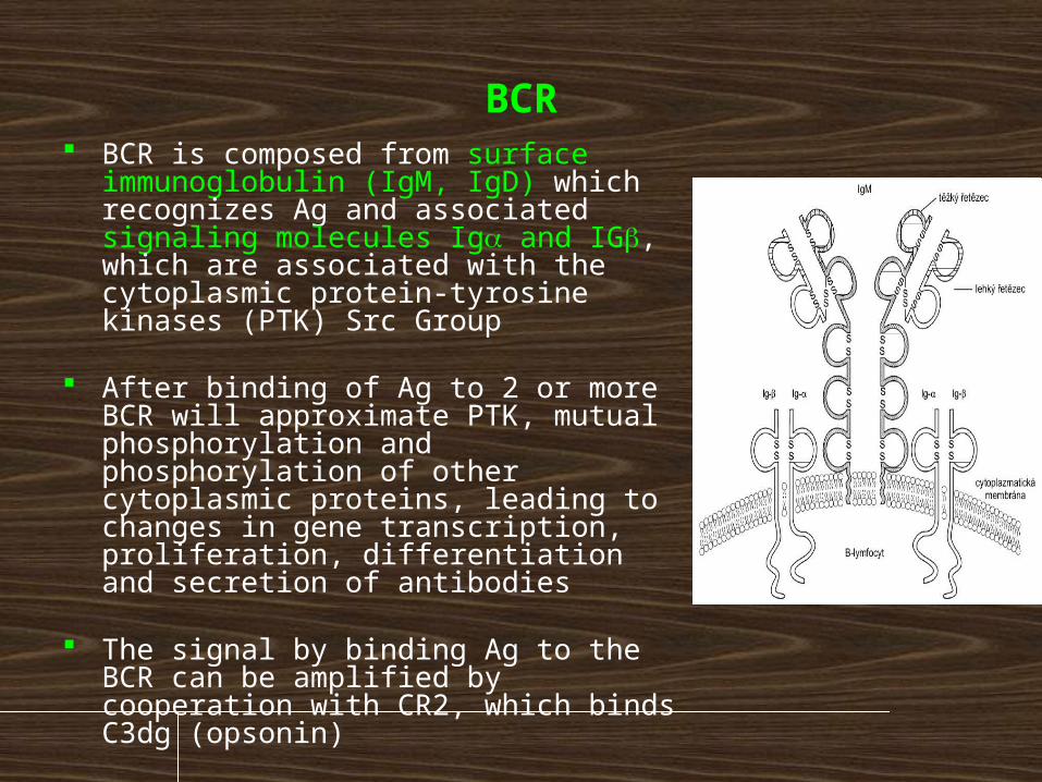
BCR BCR is composed from surface
immunoglobulin (IgM, IgD) which recognizes Ag and associated signaling molecules Iga and IGb, which are associated with the cytoplasmic protein-tyrosine kinases (PTK) Src Group
After binding of Ag to 2 or more BCR will approximate PTK, mutual phosphorylation and phosphorylation of other cytoplasmic proteins, leading to changes in gene transcription, proliferation, differentiation and secretion of antibodies
The signal by binding Ag to the BCR can be amplified by cooperation with CR2, which binds C3dg (opsonin)
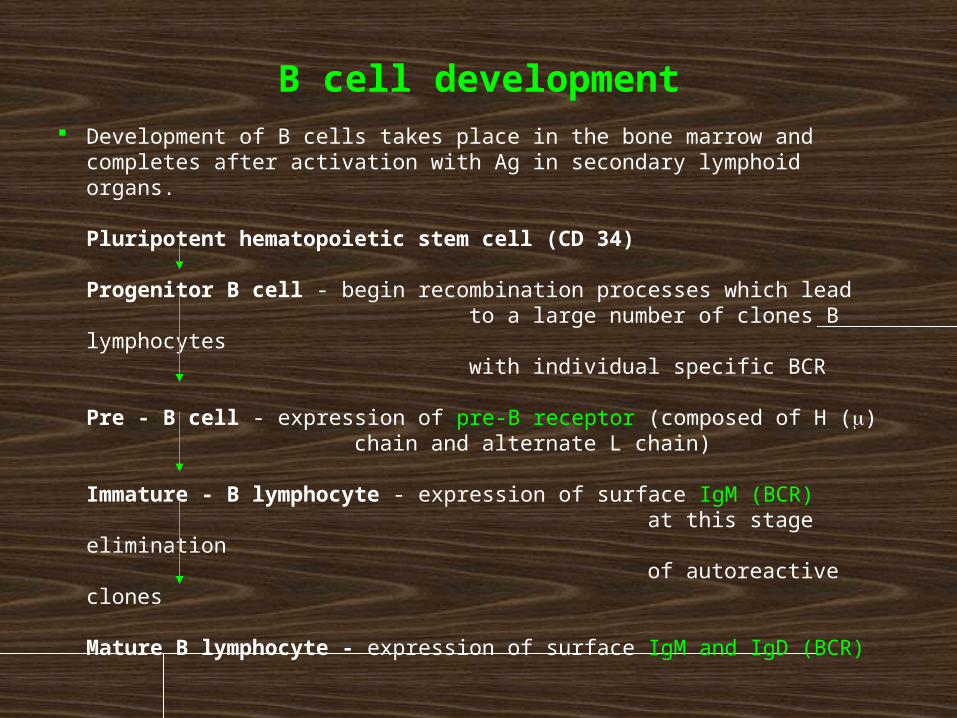
B cell development Development of B cells takes place in the bone marrow and
completes after activation with Ag in secondary lymphoid organs.
Pluripotent hematopoietic stem cell (CD 34)
Progenitor B cell - begin recombination processes which lead to a large number of clones B lymphocytes with individual specific BCR
Pre - B cell - expression of pre-B receptor (composed of H (m) chain and alternate L chain)
Immature - B lymphocyte - expression of surface IgM (BCR) at this stage elimination of autoreactive clones
Mature B lymphocyte - expression of surface IgM and IgD (BCR)
Elimination of autoreactive B
lymphocytes By random rearrangement of genes, connecting inaccuracy,
H-L pairing and somatic mutations may also arise clones of B cells bearing autoreactive receptors and produce autoreactive antibodies.
Majority of autoreactive B lymphocytes are eliminated as the immature B lymphocytes in the bone marrow, if its BCR bind autoantigen with sufficient affinity, receives a signal leading to apoptotic death (clonal deletion).
If some of the autoreactive clones pass this elimination, their autoreaktivity usually do not come because lack of TH lymphocytes for their activation, many autoantigens are cryptic, or occur in low concentrations and are ignored by the immune system. Tolerance to self-antigens is critical in preventing autoimmunity in the organism.
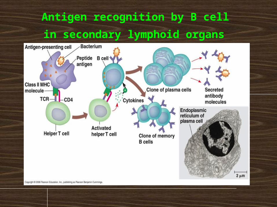
Antigen recognition by B cell
in secondary lymphoid organs
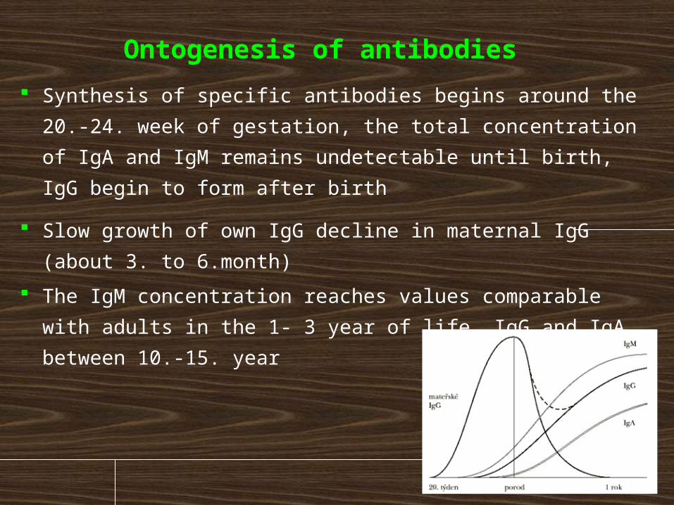
Ontogenesis of antibodies Synthesis of specific antibodies begins around the 20.-24.
week of gestation, the total concentration of IgA and IgM
remains undetectable until birth, IgG begin to form after
birth
Slow growth of own IgG decline in maternal IgG (about 3.
to 6.month)
The IgM concentration reaches values comparable with
adults in the 1- 3 year of life, IgG and IgA between 10.-15.
year
B lymphocytes respond to immunization predominantly by IgM formation, switching to other isotype is slower
Antibody response to polysaccharide antigens appears around 2. year of life
In old age is a lower antibody response to new stimuli and increased autoantibodies production
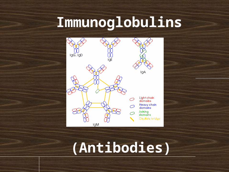
Immunoglobulins
(Antibodies)
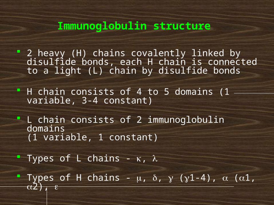
Immunoglobulin structure
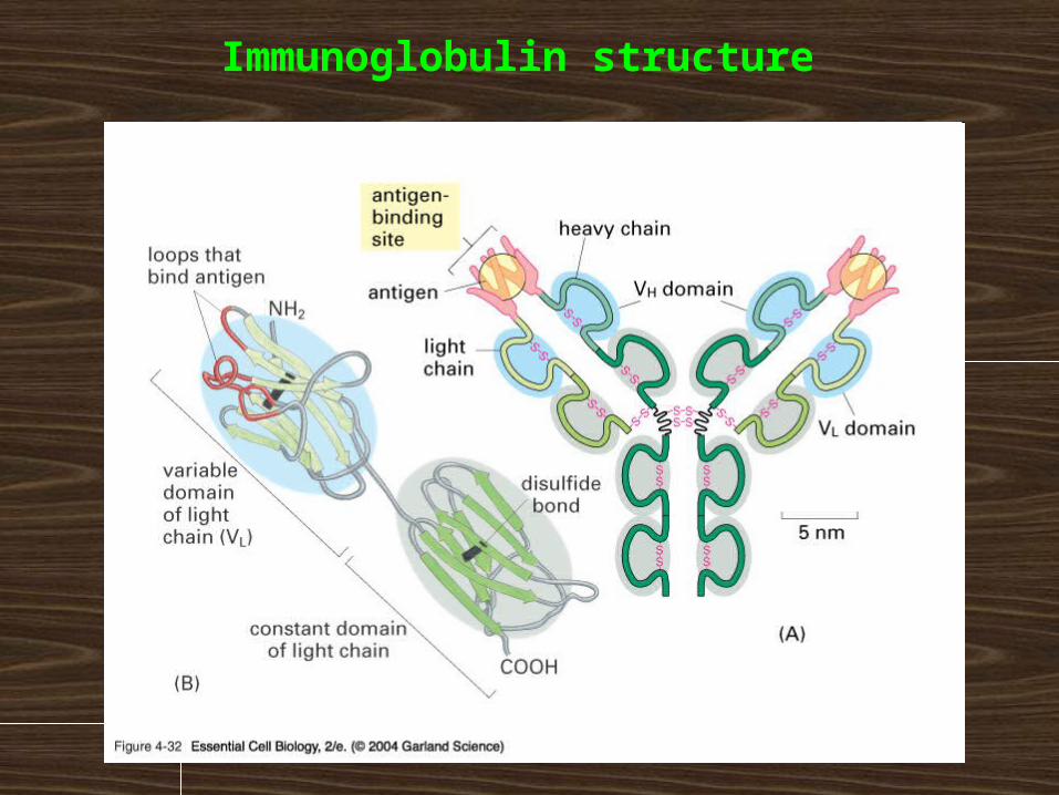
2 heavy (H) chains covalently linked by disulfide bonds, each H chain is connected to a light (L) chain by disulfide bonds
H chain consists of 4 to 5 domains (1 variable, 3-4 constant)
L chain consists of 2 immunoglobulin domains (1 variable, 1 constant)
Types of L chains - k, l
Types of H chains - m, d, g (g1-4), a (a1, a2), e
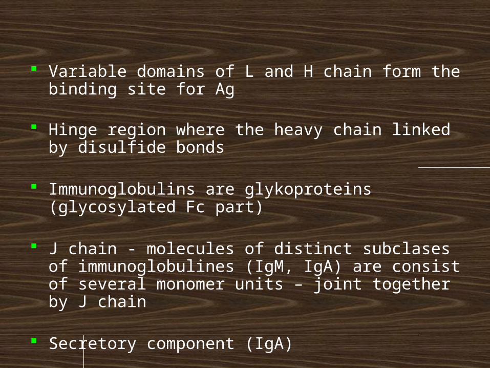
Variable domains of L and H chain form the binding site for Ag
Hinge region where the heavy chain linked by disulfide bonds
Immunoglobulins are glykoproteins (glycosylated Fc part)
J chain - molecules of distinct subclases of immunoglobulines (IgM, IgA) are consist of several monomer units – joint together by J chain
Secretory component (IgA)
Immunoglobulin structure
Immunoglobulins functions Antigen neutralization Antibodies prevents bacterial adherence and also
inhibit activity of toxins, viruses and other microorganisms by binding to their important epitopes for toxic activities
Complement activation (IgM, IgG) Antibody activates complement, which enhances opsonization and lyses some bacteria
Opsonization (IgA, IgG) Antibodies promotes phagocytosis by APC
Mast cell activation using IgE
ADCC (antibody-dependent cellular cytotoxicity) NK cells recognize cell opsonized with IgG antibodies by the Fc receptor CD16, this leads to the activation of cytotoxic mechanisms (NK degranulation)
Classes of immunoglobulins and their
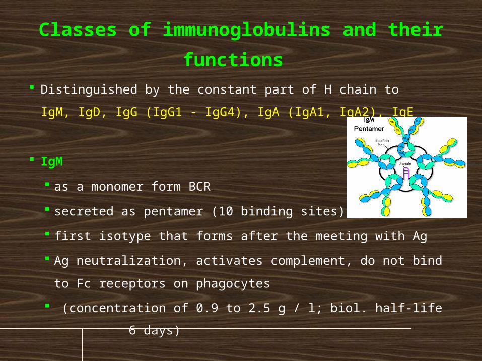
functions Distinguished by the constant part of H chain to
IgM, IgD, IgG (IgG1 - IgG4), IgA (IgA1, IgA2), IgE
IgM
as a monomer form BCR
secreted as pentamer (10 binding sites)
first isotype that forms after the meeting with Ag
Ag neutralization, activates complement, do not bind to Fc
receptors on phagocytes
(concentration of 0.9 to 2.5 g / l; biol. half-life
6 days)
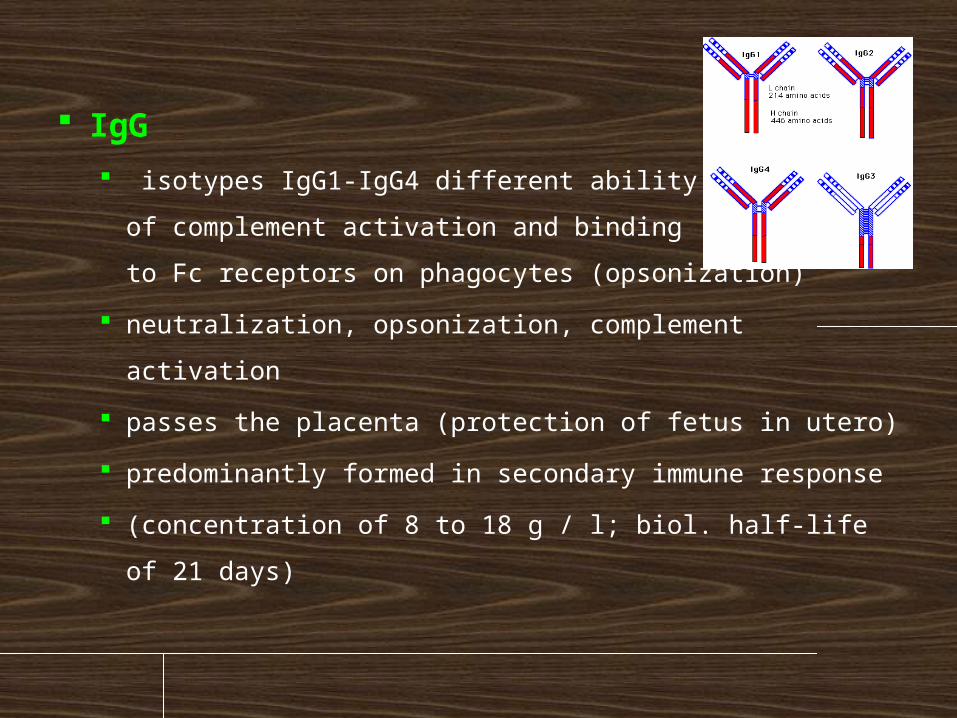
IgG
isotypes IgG1-IgG4 different ability
of complement activation and binding
to Fc receptors on phagocytes (opsonization)
neutralization, opsonization, complement activation
passes the placenta (protection of fetus in utero)
predominantly formed in secondary immune
response
(concentration of 8 to 18 g / l; biol. half-life of 21
days)
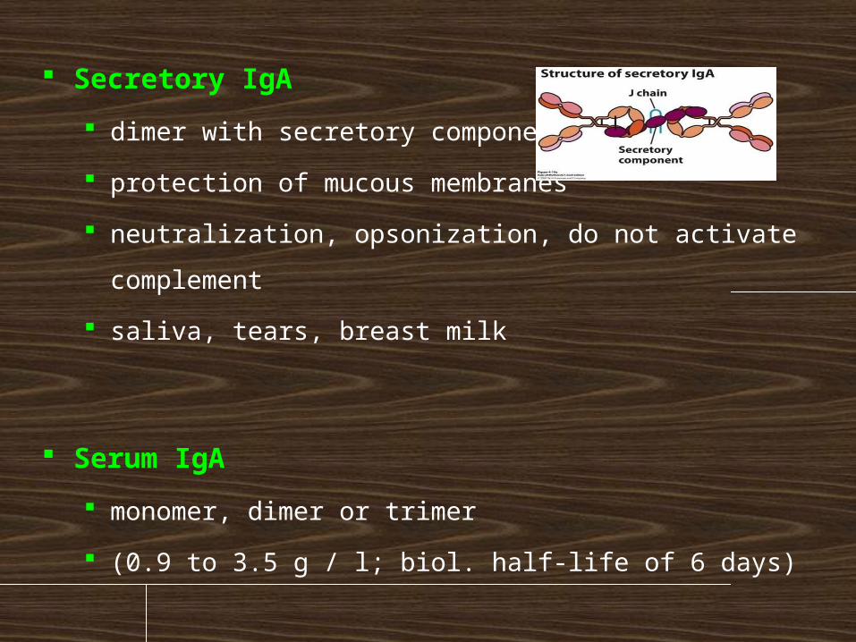
Secretory IgA
dimer with secretory component
protection of mucous membranes
neutralization, opsonization, do not activate
complement
saliva, tears, breast milk
Serum IgA
monomer, dimer or trimer
(0.9 to 3.5 g / l; biol. half-life of 6 days)
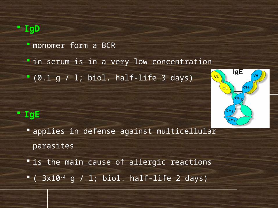
IgD
monomer form a BCR
in serum is in a very low concentration
(0.1 g / l; biol. half-life 3 days)
IgE
applies in defense against multicellular parasites
is the main cause of allergic reactions
( 3x10⁻⁴ g / l; biol. half-life 2 days)
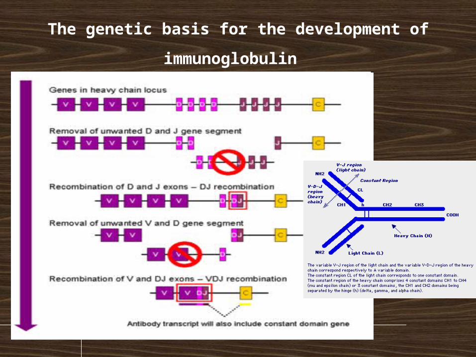
The genetic basis for the development of
immunoglobulin
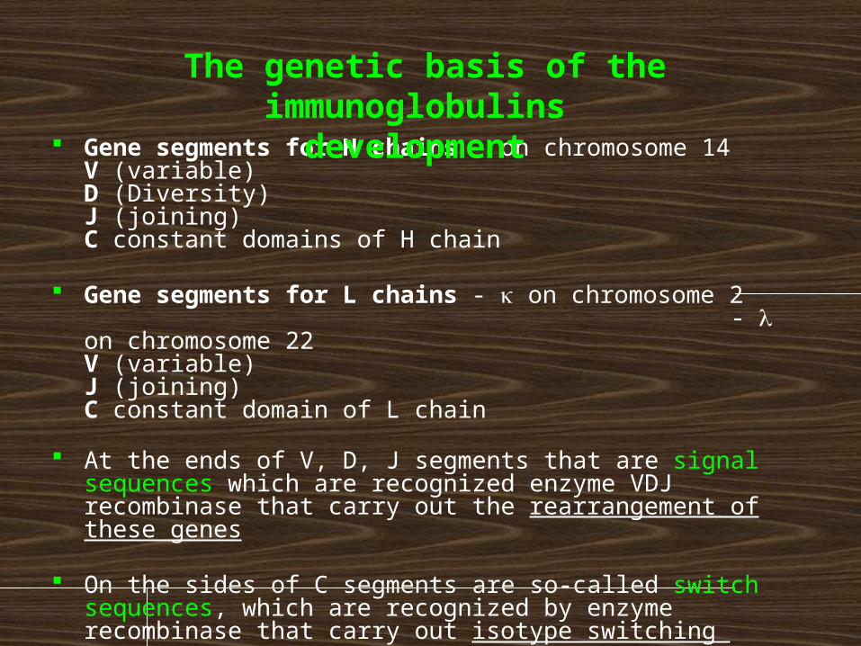
Gene segments for H chains – on chromosome 14 V (variable) D (Diversity) J (joining) C constant domains of H chain
Gene segments for L chains - k on chromosome 2
- l on chromosome 22 V (variable) J (joining) C constant domain of L chain
At the ends of V, D, J segments that are signal sequences which are recognized enzyme VDJ recombinase that carry out the rearrangement of these genes
On the sides of C segments are so-called switch sequences, which are recognized by enzyme recombinase that carry out isotype switching
The genetic basis of the immunoglobulins development
The rearrangement of genes coding H
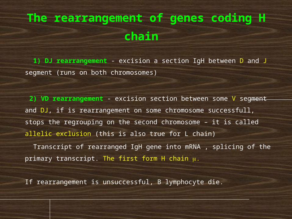
chain 1) DJ rearrangement - excision a section IgH between D and J
segment (runs on both chromosomes)
2) VD rearrangement - excision section between some V segment
and DJ, if is rearrangement on some chromosome successfull, stops
the regrouping on the second chromosome – it is called allelic
exclusion (this is also true for L chain)
Transcript of rearranged IgH gene into mRNA , splicing of the primary
transcript. The first form H chain m.
If rearrangement is unsuccessful, B lymphocyte die.
The rearrangement of genes coding L chain
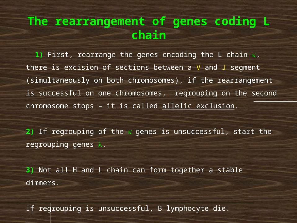
1) First, rearrange the genes encoding the L chain k, there is
excision of sections between a V and J segment
(simultaneously on both chromosomes), if the rearrangement
is successful on one chromosomes, regrouping on the second
chromosome stops – it is called allelic exclusion.
2) If regrouping of the k genes is unsuccessful, start the
regrouping genes l.
3) Not all H and L chain can form together a stable dimmers.
If regrouping is unsuccessful, B lymphocyte die.
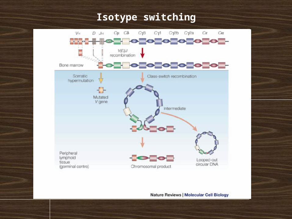
Isotype (class) switching
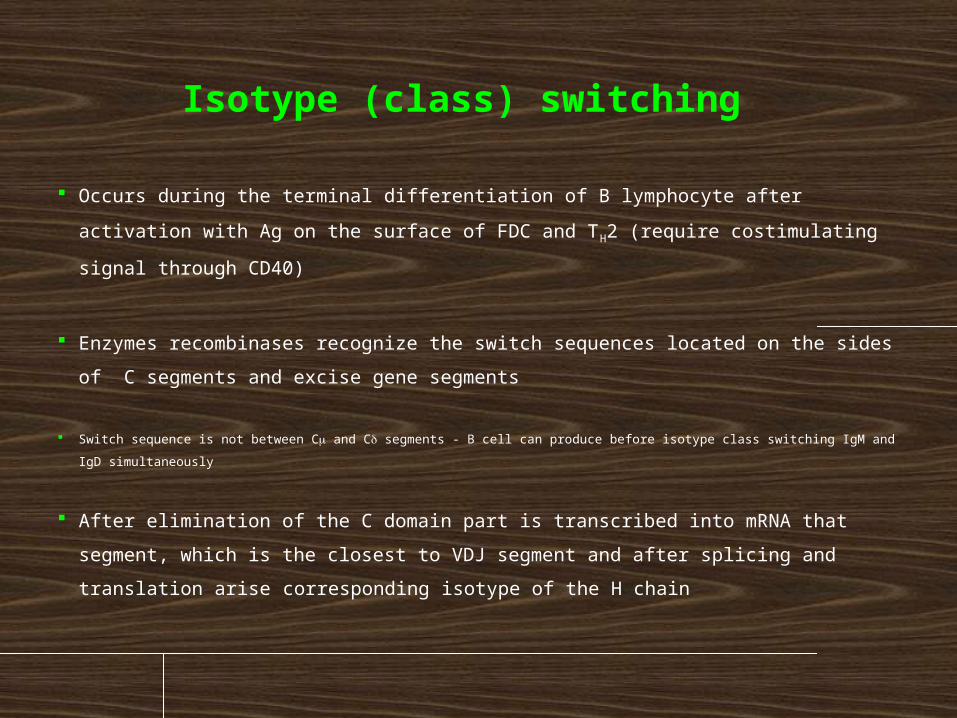
Occurs during the terminal differentiation of B lymphocyte after activation
with Ag on the surface of FDC and TH2 (require costimulating signal through
CD40)
Enzymes recombinases recognize the switch sequences located on the sides
of C segments and excise gene segments
Switch sequence is not between Cm and Cd segments - B cell can produce before isotype class switching IgM and
IgD simultaneously
After elimination of the C domain part is transcribed into mRNA that
segment, which is the closest to VDJ segment and after splicing and
translation arise corresponding isotype of the H chain
Isotype switching
Isotype switching
Cytokines regulate which isotype occurs:
IL-4 stimulates switching to IgE and IgG4
TGFb stimulates switching to IgG2 and IgA
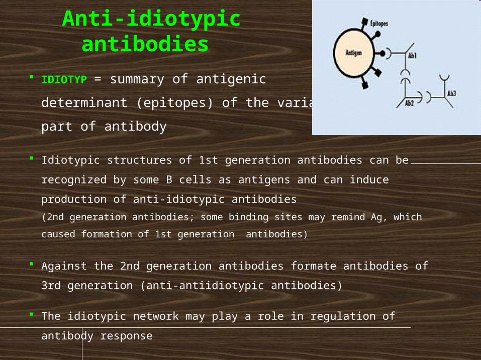
Anti-idiotypic antibodies
IDIOTYP = summary of antigenic
determinant (epitopes) of the variable
part of antibody
Idiotypic structures of 1st generation antibodies can be recognized by
some B cells as antigens and can induce production of anti-idiotypic
antibodies
(2nd generation antibodies; some binding sites may remind Ag, which caused
formation of 1st generation antibodies)
Against the 2nd generation antibodies formate antibodies of 3rd
generation (anti-antiidiotypic antibodies)
The idiotypic network may play a role in regulation of antibody
response
Humoral immune response
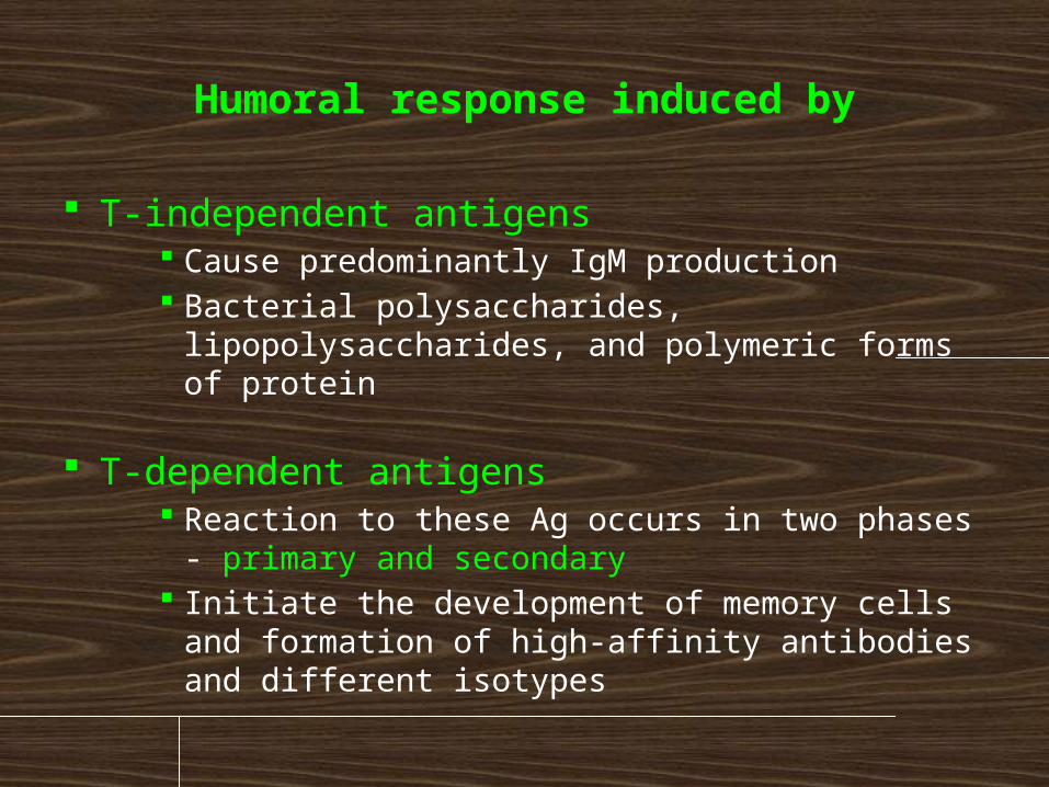
Humoral response induced by
T-independent antigens Cause predominantly IgM production Bacterial polysaccharides, lipopolysaccharides,
and polymeric forms of protein
T-dependent antigens Reaction to these Ag occurs in two phases -
primary and secondary Initiate the development of memory cells and
formation of high-affinity antibodies and different isotypes
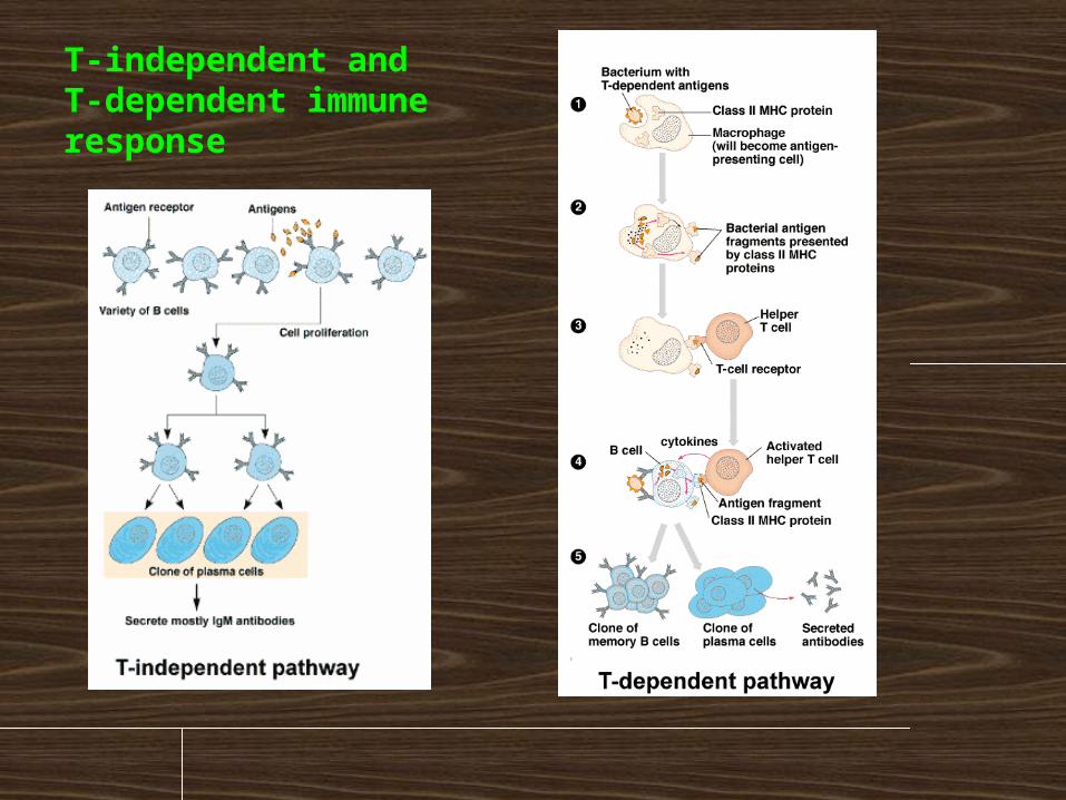
T-independent and T-dependent immune response
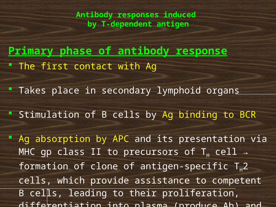
Antibody responses induced by T-dependent antigen
Primary phase of antibody response The first contact with Ag
Takes place in secondary lymphoid organs
Stimulation of B cells by Ag binding to BCR
Ag absorption by APC and its presentation via MHC gp class II to precursors of TH cell → formation of clone of
antigen-specific TH2 cells, which provide assistance to
competent B cells, leading to their proliferation, differentiation into plasma (produce Ab) and memory cells
Plasma cells are spread by bloodstream into the organism (particularly bone marrow)
Antibodies produced in the primary stage (3-4 days) are IgM and have a low affinity for Ag, create with Ag immune complexes
Immune complexes are captured in the secondary lymphoid organs on the surface of FDC (follicular dendritic cells) - Ag presenting cells to B lymphocytes
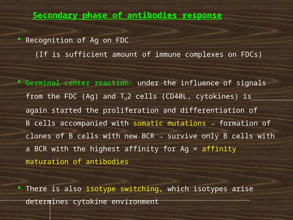
Secondary phase of antibodies response
Recognition of Ag on FDC
(If is sufficient amount of immune complexes on FDCs)
Germinal center reaction: under the influence of signals from the
FDC (Ag) and TH2 cells (CD40L, cytokines) is again started the
proliferation and differentiation of
B cells accompanied with somatic mutations → formation of
clones of B cells with new BCR → survive only B cells with a BCR
with the highest affinity for Ag = affinity maturation of antibodies
There is also isotype switching, which isotypes arise determines
cytokine environment
In the secondary phase of the immune response there are
generated antibodies with higher affinity to Ag and other
effector characteristics dependent on isotype, also formed a
memory cells for next meeting with the Ag
Antibodies in the body persist for a long time after primary
infection
Contact between CD40 (B lymphocytes) and CD40L (TH2
lymphocytes) is essential for the initiation of somatic
mutations, isotype switching and formation of memory cells
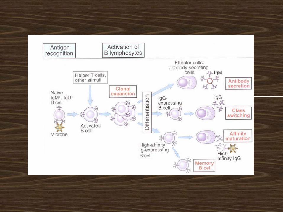
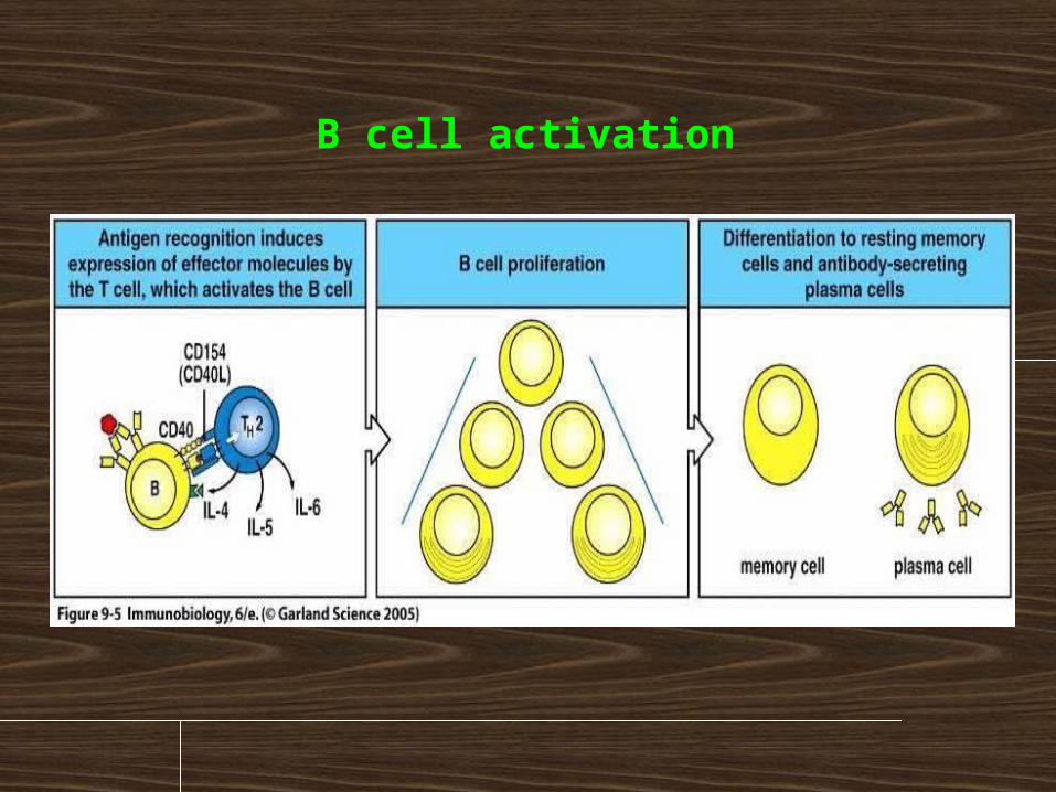
B cell activation
Primary and secondary immune response
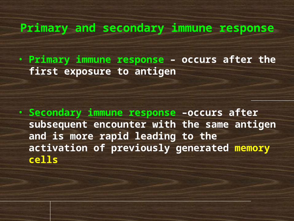
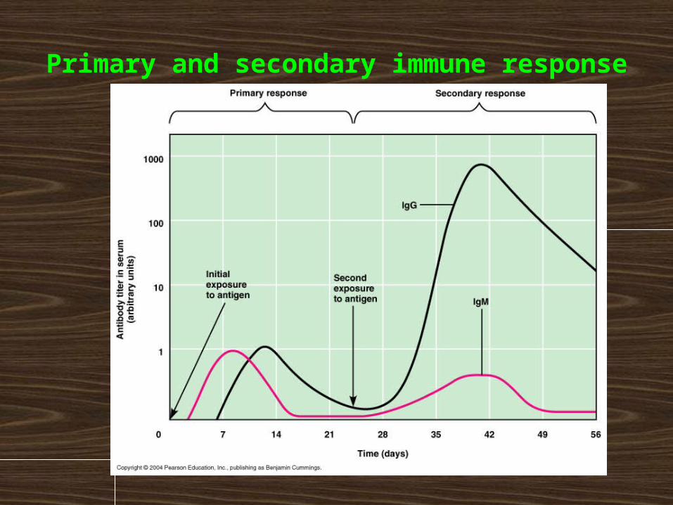
• Primary immune response – occurs after the first exposure to antigen
• Secondary immune response –occurs after subsequent encounter with the same antigen and is more rapid leading to the activation of previously generated memory cells
Primary and secondary immune response
Mucosal and skin
immune system
Function and structure of the mucosal and skin immune systemMucous membranes and skin are in constant contact with the outside environment, there is concentrated about 80% of immunocompetent cells.
Skin - barrier against mechanical, physical and chemical damage, and against the penetration of microorganisms, humans surface about 1,5 m2
Mucosal immune system - prevents the penetration of pathogenic microorganisms, prevents the development of self-harm inflammatory immune responses against pathogens and harmless antigens from the external environment, mucosal surface about 400 m2
Barrier functions of the human body and defence mechanismsMechanical bariers – intact skin and mucus, movement of cilia, coughing, sneezing,the flow of air and fluids, vomiting, diarhea
Chemical inhibitors - secrets of exocrine glands with bactericidal effects (fatty acids , lysozyme, pepsin, defensins, acidic pH of the stomach and urine)
Other factors – body temperature (37OC), tissue oxygen tension, age, stress
Physiological microflora
Structure of mucosal immune system
MALT (mucous associated lymphoid tissue) BALT (bronchus associated lymphoid tissue) GALT (gut associated lymphoid tissue) NALT (nasal associated lymphoid tissue)
o-MALT (organized) – consists of lymphoid follicles in the mucous membrane, tonsil and adenoids, appendix, Peyer patches
d-MALT (diffuse) – consist of leukocytes diffusely distributed in the lamina propria (T and B lymphocytes, macrophages, neutrophils, eosinophils and mast cells)
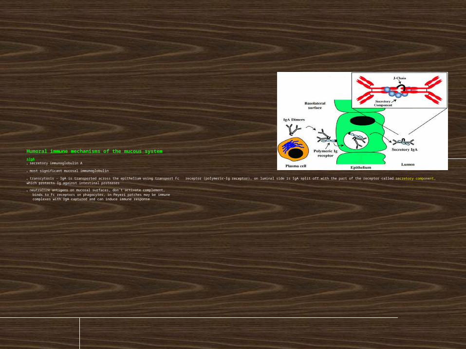
Humoral immune mechanisms of the mucous system
sIgA * secretory immunoglobulin A
* most significant mucosal immunoglobulin
* transcytosis - IgA is transported across the epithelium using transport Fc receptor (polymeric-Ig receptor), on luminal side is IgA split off with the part of the receptor called secretory component, which protects Ig against intestinal proteases
* neutralize antigens on mucosal surfaces, don´t activate complement, binds to Fc receptors on phagocytes, in Peyerś patches may be immune complexes with IgA captured and can induce immune response

sIgM
* secretory immunoglobulin M
* applied in newborns and in selective IgA deficiency
* more prone to intestinal protease degradation
* neutralize antigens on mucosal surfaces
IgG
* get on the mucous membrane by diffusion
* applies particularly in the lower airways
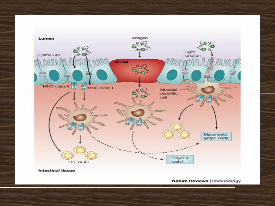
Induction of mucosal immune response
Oral tolerance
* majority of antigens given orally causes suppression of specific
immunity (critical is also the size of the antigenic particles)
* Tr lymphocytes (regulatory) - production of IL-10
Induction of mucosal immune response
* M cells - specialized enterocytes that provide transport of Ag
(endocyte Ag from the surroundings) - are in close contact with lymphocytes and APC
* mucosal immunization leads to stimulation of TH2 and TH3
lymphocytes and IgA production
External regulation of immune response
Substitution treatment
treatment with intravenous immunoglobulin (derived from plasma of blood donors)
substitution of C1 inhibitor for hereditary angioedema
substitution of erythropoietin in patients with chronic renal failure
substitution of G-CSF in agranulocytosis
Immunomodulation= medical procedure to adjust the disrupted immune
function
Non-specific immunosuppressive therapy
nonspecific = affects not only autoreactive and aloreactive lymphocytes, but also other components of immunity (risk of reduction antiinfectious and anti- tumor immunity)
used for treatment of autoimmune diseases, severe allergic conditions and for organ transplantation
Non-specific immunosuppressive therapy corticosteroids - anti-inflammatory, immunosuppressive
effects - blocking the activity of transcription factors (AP-1, NFkB) - suppress the expression of genes (IL-2, IL-1, phospholipase A, MHC gp II, adhesion molecules) - inhibition of histamine release from basophils - higher concentrations induce apoptosis of lymfocytes
immunosuppressants affecting the metabolism of DNA - cyclophosphamide,
azathioprine,methotrexate
immunosuppressant selectively inhibiting T lymphocytes - immunosuppressive ATB: cyclosporine A, tacrolimus, rapamycin (suppressing the expression of IL-2 and IL-2R in activated T lymphocytes) - monoclonal antibody anti-CD3 (Immunosuppression after transplantation, treatment of rejection crises)
immunoglobulins in the immunosuppressive indication - Polyspecific intravenous immunoglobulins (Inhibition of B lymphocytes, antiidiotype activity, inhibition of cytokines, neutralization of toxins, inhibition of complement activation ...)
Anti-inflammatory and antiallergic treatment
nonsteroidal anti-inflammatory drugs
antihistamines - blocking H1 receptor - reduce the expression of adhesion molecules - reduce the secretion of histamine ...
inhibitors of inflammatory cytokine - receptor antagonist for IL-1
- monoclonal antibodies against TNF
- thalidomide (TNF inhibitor)
Anti IgE antibodies (omalizumab)- - severe allergic astma
Non-specific immunostimulant therapy synthetic immunomodulators
Methisoprinol (Isoprinosine) - used in viral infections with more severe or relapsing course
bacterial extracts and lysates Broncho-Vaxom - prevention of recurrent respiratory tract infections Ribomunyl
products of the immune system IL-2 - renal adenocarcinoma IFNa, IFNb - viral hepatitis, some leukemia Erythropoietin – renal failure G-CSF, GM-CSF – neutropenia Transfer factor (blood donors leukocytes undergoing dialysis) Thymus hormones
Antigen-specific immunomodulatory therapy
specific immunomodulation = induce an
immune response or tolerance against a specific
antigen
a) active immunization
b) passive immunization
c) specific immunosuppression
d) vaccination against cancer
a) active immunization
= use of antigen to induce an immune response that can later
protect against a pathogen bearing the antigen
(or similar antigen)
immunization vaccines are made from inactivated or
attenuated microorganisms or their antigens (polysaccharide
capsule, toxins)
creates long-term immunity
activate cellular and antibody immunity
administration of antigen injectable
prophylaxis
risk of causing infection or anaphylactic reactions
b) passive immunization
natural - transfer of maternal antibodies in fetal blood
therapeutically - the use of animal antibodies against various toxins (snake toxins, tetanus toxin, botulinum toxin)
prophylaxis - the human immunoglobulin from immunized individuals (hepatitis A, rabies, tetanus) - Anti-RhD antibodies - preventing maternal immunization with RhD+ fetus
provides a temporary (3 weeks) specific humoral immunity
the risk of induction anaphylactic reactions
c) specific immunosuppression
= induction of tolerance against a specific antigen
ongoing clinical studies
induction of tolerance by oral administration of antigen (treatment of certain autoimmune diseases)
allergen immunotherapy (pollen, insect poisons)
d) vaccination against cancer
s a promising approach appears to immunization dendritic cells

• https://www.youtube.com/watch?v=HfU2z0TzBec
• http://www.youtube.com/watch?v=hEnvQGm6o00

Thank you for your attentionThank you for your attention