topical administration of anticancer drugs for skin cancer ...cdn.intechweb.org/pdfs/23038.pdf ·...
TRANSCRIPT
11
Topical Administration of Anticancer Drugs for Skin Cancer Treatment
Stephânia Fleury Taveira and Renata Fonseca Vianna Lopez School of Pharmaceutical Sciences of Ribeirão Preto – University of São Paulo,
Brazil
1. Introduction
Skin cancer is the most common type of cancer affecting Caucasian populations. It has a very high rate of incidence, exceeding the sum of all other cancers combined (Simonette et al., 2009). There are three forms of skin tumors that stand out: cutaneous malignant melanoma, basal cell carcinoma (BCC) and squamous cell carcinoma (SCC). BCC and SCC are classified as nonmelanoma skin cancers, with BCC being the most common and constituting 75% of cases (Anthony, 2000). The incidence of nonmelanoma skin cancers has steadily increased, making them a major challenge in terms of management of public health. Moreover, these cancers can have a huge impact on health care costs. In the United States, it is estimated that there are approximately 3 to 4 million cases annually of BCC and approximately 100,000 cases of SCC. Nonmelanoma skin cancers are not fatal but can destroy facial sensory organs such as the nose, ear and lips (Alam et al., 2011). Therefore, these lesions should preferably be treated using noninvasive techniques. In contrast, melanoma skin cancers are an aggressive type that can metastasize and cause death. These cancers originate from melanocytes, which are pigment-producing cells, and are associated with chronic exposure to sunlight (Einspahr et al., 2002). Because melanoma has a much higher mortality rate than nonmelanoma skin cancers, different treatments, including invasive interventions, are required (Martinez & Otley, 2001). There are some well-established treatments for nonmelanoma skin cancer, such as curettage, surgery, cryotherapy and chemotherapy. However, these conventional treatments lead to severe inflammation, pain and unappealing scars (Lopez et al., 2004). Treatments for melanoma, in turn, are primarily surgical because these tumors can be resistant to traditional chemo- and radiotherapies (Davids & Kleeman, 2010). Nonsurgical treatments for melanomas are limited to adjuvant therapies, such as immunotherapy, biochemotherapy, gene therapy and photodynamic therapy (Martinez & Otley, 2001; Davids & Kleeman, 2010). To increase patient compliance and to reduce surgical costs and undesirable scars, particularly in cases where the cancer has spread over large areas of the body, the topical administration of anticancer drugs has been investigated. The topical administration of anticancer drugs is an interesting alternative for reducing side effects and for increasing drug targeting and therapeutic benefits. The major challenge of this kind of treatment is to increase penetration of the antineoplastic tumor drug in sufficient levels to kill tumor cells.
www.intechopen.com
Skin Cancers – Risk Factors, Prevention and Therapy
248
Several techniques and formulations have therefore been developed to successfully overcome skin barriers and to reach skin malignancies by favoring drug penetration into the deep layers of the epidermis. The use of chemical penetration enhancers is the simplest strategy, causing temporary and reversible disruption of the stratum corneum bilayers and leading to increased anticancer drug penetration into the tumor. Moreover, great interest has been shown in nanoparticle delivery systems that can protect anticancer drugs against degradation and, combined with physical methods, significantly increase the tumor penetration of the drug. This chapter will briefly discuss skin anatomy, the primary barriers to topical anticancer
drugs’ skin penetration, and the most studied penetration enhancer methods for topical skin
cancer treatment. The aim of this chapter is to provide a basic understanding and
description of the strategies that can be used to overcome the skin barrier, such as
liposomes, polymeric and lipid nanoparticles, iontophoresis and electroporation. Each of
these modalities will be discussed in the context of their application for promoting and
targeting the delivery of skin tumor drugs following topical, noninvasive, administration.
2. The skin as a barrier against anticancer drug penetration
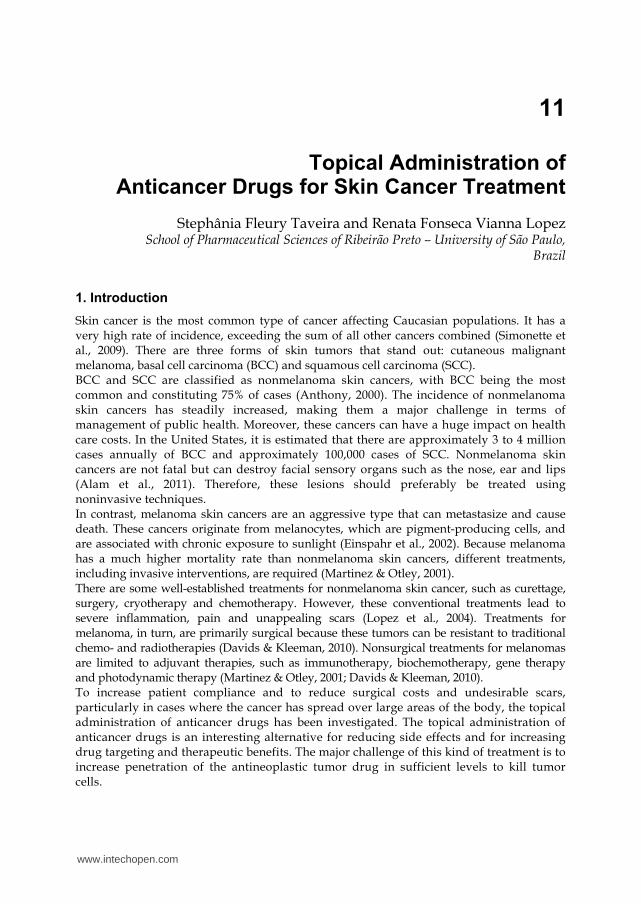
The skin is the largest organ of the body and is composed of three primary layers: the
epidermis, dermis and hypodermis. The epidermis plays an important role in the
penetration of substances into the skin. It is the outer avascular layer of the skin, primarily
composed of keratinocytes. Because of cellular differentiation, the epidermis is divided
into different layers, which are formed by the division of basal cells from the inner part of
the body toward the surface (Figure 1). Hence, basal cells undergo progressive
maturation, giving rise to the spinous layer or squamous cells. These cells also
differentiate, forming the granular layer and finally the stratum corneum, which is the
outermost layer of the skin.
The stratum corneum is the major barrier for the penetration of substances into the skin
because of its heterogeneous composition and packed organization of corneocytes and the
intracellular lipid matrix. The corneocytes are flat anucleated squamous cells packed
primarily with keratin filaments and surrounded by a lipid matrix composed primarily of
ceramides, cholesterol, and free fatty acids (Bouwstra et al., 2003).
Two other cell types that are important in the context of skin tumor composition,
melanocytes and Langerhans, are embedded between the basal keratinocytes. Melanocytes
are dendritic cells capable of melanin production, and Langerhans cells are antigen-
presenting cells that are responsible for the immune response in the skin (McGrath & Uitto,
2010) (Figure 1).
Because the skin is a heterogeneous organ, this wide variety of cell types can generate
several types of benign and malignant tumors. For instance, SCC and BCC originate from
keratinocytes. The development of these tumors is associated with many factors, but most of
these cancers are related to excess ultraviolet radiation (UV) exposure. Following sun
exposure-induced damage, the stratum corneum of tumor lesions usually presents with
hyperkeratinization (Neel & Sober et al., 2006), a factor known to hamper drug penetration.
Topical anticancer administration therefore requires a well-designed formulation to increase
drug penetration into the thicker stratum corneum and to favor drug penetration into the
deep skin layers, where tumors are usually located.
www.intechopen.com
Topical Administration of Anticancer Drugs for Skin Cancer Treatment
249
Fig. 1. Schematic representation of the epidermis/dermis and epidermis layers consisting of basal cells, squamous cells, the granular layer and the stratum corneum.
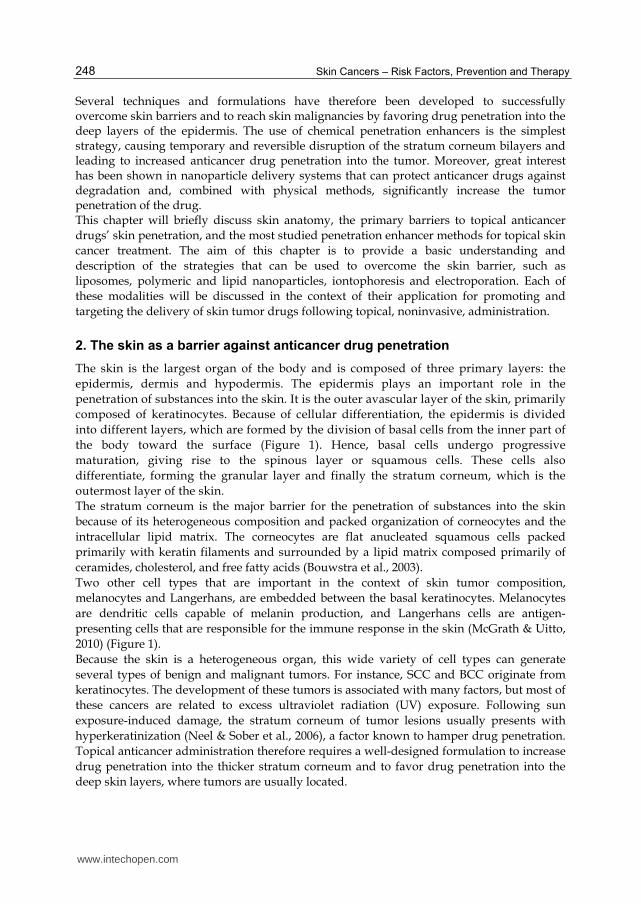
For example, the most superficial malignancy that develops in the epidermis is actinic keratosis, so-named because of the exaggerated production of keratin in the stratum corneum, which causes it to become thicker (Figure 2A and 2B). These lesions can develop into tumors, usually SCCs, which may be nodular (invasive) and hyperkeratotic (Figure 2C). For topical treatment of both actinic keratosis and SCC, anticancer drugs should penetrate the stratum corneum to reach the tumor cells.
Fig. 2. Schematic representation of the skin layers, especially the stratum corneum, of (A) Normal skin, (B) Actinic Keratosis: (1) hyperkeratosis, (2) atypia of cells, (3) Langerhans cells; (C) Invasive SCC: (1) hyperkeratosis and (2) squamous cells with atypical nuclei (enlarged and hyperchromatic).
www.intechopen.com
Skin Cancers – Risk Factors, Prevention and Therapy
250
Another example for the necessity of drug penetration through the stratum corneum is the use of immunoregulator drugs for the topical treatment of actinic keratosis. To be effective, these drugs need to act in the Langerhans cells to induce the release of proinflammatory cytokines, which stimulate an immune response. To reach Langerhans cells, anticancer drugs must cross the hyperkeratotic stratum corneum (Figure 2B). Within this context, it is important to understand the mechanisms of drug penetration through the stratum corneum to determine methods or strategies that can increase drug penetration such that the drug reaches sufficient concentrations to kill tumor cells.
2.1 Drug penetration in the skin
Over the past two decades, significant attention has been paid to understanding the mechanisms by which drugs penetrate the skin. It is well known that substances usually penetrate the skin by three different routes: through the stratum corneum between the corneocytes (intercellular route); through these cells and the intervening lipids (intracellular route); or through the skin appendages, such as hair follicles and sweat glands (Moser et al., 2001). Molecules with adequate solubility in water and oil, with a log of oil/water partition coefficients between 1 and 3 (Hadgraft & Lane, 2005) and a molecular weight lower than 0.6 kDa (Schäfer-Korting et al., 2007; Barry, 2001), may penetrate the skin. Therefore, topical administration is limited to hydrophobic and low-molecular weight drugs. Because most anticancer drugs are hydrophilic, have low oil/water partition coefficients, high molecular weights and ionic characters (Souza et al., 2011), they do not easily penetrate the stratum corneum. Drug permeation through the stratum corneum can be described with Ficks’s second law (Williams & Barry, 2004) (Equation 1).
Dm Cv
L
PJ , (1)
where J is the flux, Dm is the diffusion coefficient of the drug in the membrane, Cv is the drug concentration in the vehicle, P is the drug partition coefficient and L is the stratum corneum thickness. It can be seen in Equation I that the flux of a drug through the skin is governed by the diffusion coefficient of the drug in the stratum corneum, the concentration of the drug in the vehicle, the partition coefficient between the formulation and the stratum corneum and the membrane thickness. Using this equation, it is a simple matter to determine which parameters can be manipulated to increase drug flux through the stratum corneum. Formulations containing chemical penetration enhancers or the use of physical penetration methods, such as iontophoresis and electroporation, may alter one or more of these parameters to increase drug penetration in the skin. For instance, chemical enhancers can disrupt the stratum corneum barrier and increase the diffusion coefficient of the drug through the altered membranes. Alternatively, enhancers can alter the solvent nature of the skin and improve partitioning between the formulation and the stratum corneum. Nanocarriers can increase drug concentration in the vehicle and so increase drug flux. Physical penetration methods can modify drug penetration routes through the stratum corneum, making it less tortuous, facilitating drug penetration (Williams & Barry, 2004). In this context, several manuscripts have reported the use of anticancer drugs in combination with penetration-enhancing methods or nanocarriers aimed at obtaining high penetration of the drug through the skin for tumor elimination.
www.intechopen.com
Topical Administration of Anticancer Drugs for Skin Cancer Treatment
251
3. Current topical therapies for skin cancer treatment
Current topical treatments for skin cancer include semi-solid formulations of 5-fluorouracil, diclofenac and imiquimod. Another topical treatment also used and approved by the US Food and Drug Administration (FDA) is photodynamic therapy (PDT). These therapies are used to treat nonmelanoma skin cancers and its precursor lesions, such as actinic keratosis. Several studies have discussed the preferred schedules for topical treatment to avoid tumor recurrence. Advantages and disadvantages of each topical treatment will be discussed to give a better understanding of treatment limitations and to propose different approaches that may improve topical skin cancer treatment. 5-fluorouracil has been considered the topical treatment of choice for actinic keratosis since its approval in 1970 by the US FDA (Barrera & Herrera, 2007). It is a structural analogue of thyamine and inhibits the enzyme thymidylate synthetase, blocking DNA synthesis and preventing cell proliferation (Galiczynski & Vidimos, 2011). There are many 5-fluorouracil preparations, and it is available through a variety of trademarks both as creams (5%, 1% and 0.5%) and in solution (5%, 2% or 1%) (Barrera & Herrera, 2007). This medication has some side effects, such as an intense local inflammatory reaction, that result in a lack of patient compliance. Other disadvantages are the relative long treatment period and partial inefficacy of the treatment in the deep layers of skin, such as in cases of hyperkeratotic actinic keratosis (Barrera & Herrera, 2007). These drawbacks emphasize the need for alternative methods or techniques to improve the skin penetration of antineoplastic drugs. Imiquimod is an immune response modifier that directly and indirectly interacts with the immune system (Perrotta et al., 2011). It was initially approved by the FDA in 1997 for genital and perianal wart treatment but has been used off-label for neoplastic skin treatments (Burns & Brown, 2005). In 2004, the FDA approved the use of imiquimod 5% cream for the treatment of actinic keratosis and superficial BCC in patients for whom surgery is not an option (Perrota et al., 2011). Still, without FDA approval, it has been commonly used for many other cutaneous disorders, such as cutaneous melanoma metastases, BCC, Bowen’s disease, SCC and lentigo maligna. Studies have revealed that almost all patients treated with imiquimod exhibit some degree of local inflammation at the application site. In FDA studies, patients presented some degree of erythema, edema, ulceration or erosion (Burns & Brown, 2005). The formulation of 3% diclofenac gel has been used for the topical treatment of actinic keratosis. At present, it is approved by the FDA and is used only in the US (Berrera & Herrera, 2007). Diclofenac is a nonsteroidal anti-inflammatory drug and has been show to have an antitumor effect by inhibiting arachidonic acid metabolism (Galiczynski & Vidimos, 2011). The examined treatment schedules have been two applications daily for 60 or 90 days, with patients showing complete resolution of actinic keratosis lesions in 47% of cases (Berrera & Herreara, 2007). PDT is also approved by the FDA for the treatment of nonhypertrophic actinic keratosis of the head and scalp (Galiczynski & Vidimos, 2011). Nevertheless, it has been used off-label to treat various dermatoses, such as superficial and nodular BCC, SCC in situ and others (Galiczynski & Vidimos, 2011). PDT involves the administration of a photosensitizing drug or a pro-drug that is converted in a photosensitizer intracellularly (usually 5-aminolevulinic acid, 5-ALA). Subsequent activation by light of a specific wavelength leads to the formation of highly reactive singlet oxygen (1O2), destroying the cells via chemical, biological and physiological reactions (Araújo et al., 2010). To specifically kill tumor cells, it is therefore
www.intechopen.com
Skin Cancers – Risk Factors, Prevention and Therapy
252
important that the photosensitizing drug target the tumor cells at a high concentration. Great efforts have been made to design topical nanocarriers and techniques that can increase the penetration of photosensitizers into the deep skin layers (Souza et al., 2011; Araújo et al, 2010; Gelfuso et al., 2008; Gelfuso et al., 2011) to make topical PDT more effective. Other clinical trials are underway. The retinoids, or vitamin A analogs, which are commonly used in cosmetic products, have been considered to have certain chemopreventive effects (Berrera & Herrera, 2007). Resiquimod, which is structurally similar to imiquimod but 10–100 times more potent, is another new drug that modulates the immune system (Perrotta et al., 2011) when topically applied. Adequate formulations for both of these drugs still need to be designed for successful skin penetration. It is interesting to note that the current topical medications approved by the FDA for skin cancer treatment are used primarily to treat superficial skin cancers. This is due to the fact that some skin cancers, such as SCC, can metastasize, and topical therapy is only used for invasive tumors if the patient cannot receive surgical treatments. Moreover, response rates differ for superficial and invasive cancers. For example, Burns (2005) reported that imiquimod was more effective in treating superficial BCC than for nodular BCC. This is likely because nodular BCC occurs deep in the dermis and the drug may not reach the full depth of the tumor invasion. Again, these limitations highlight the importance of developing new formulations or methods that improve drug penetration of the skin for the treatment of different skin cancers. Several chemical/physical methods and nanocarriers have been studied with the aim of overcoming the above limitations of current treatments and are discussed further.
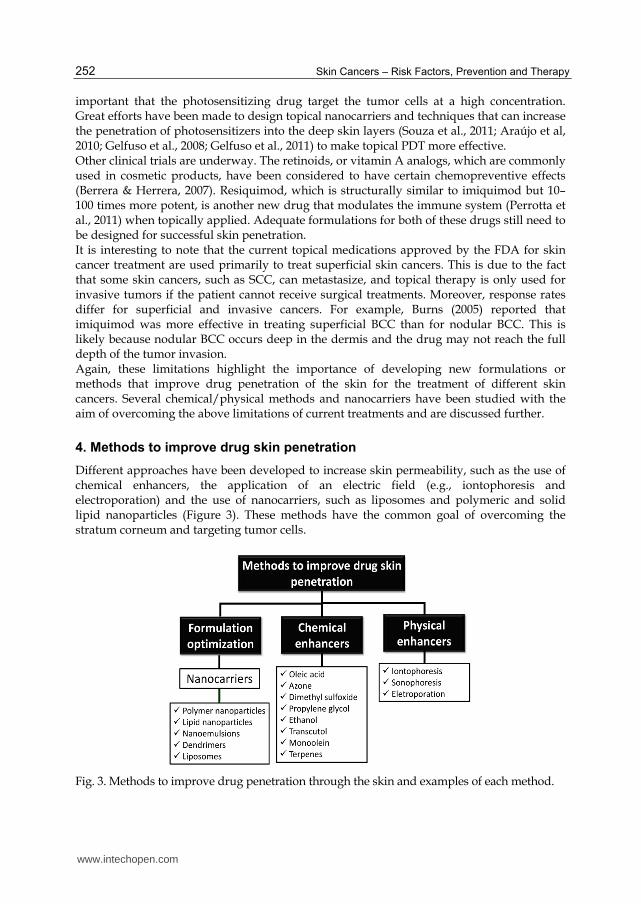
4. Methods to improve drug skin penetration
Different approaches have been developed to increase skin permeability, such as the use of chemical enhancers, the application of an electric field (e.g., iontophoresis and electroporation) and the use of nanocarriers, such as liposomes and polymeric and solid lipid nanoparticles (Figure 3). These methods have the common goal of overcoming the stratum corneum and targeting tumor cells.
Fig. 3. Methods to improve drug penetration through the skin and examples of each method.
www.intechopen.com
Topical Administration of Anticancer Drugs for Skin Cancer Treatment
253
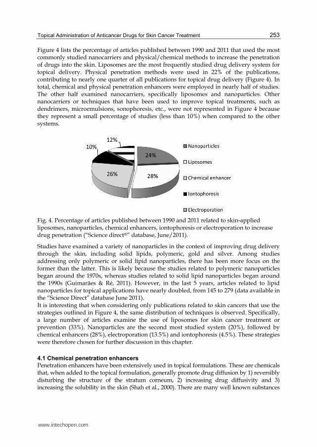
Figure 4 lists the percentage of articles published between 1990 and 2011 that used the most commonly studied nanocarriers and physical/chemical methods to increase the penetration of drugs into the skin. Liposomes are the most frequently studied drug delivery system for topical delivery. Physical penetration methods were used in 22% of the publications, contributing to nearly one quarter of all publications for topical drug delivery (Figure 4). In total, chemical and physical penetration enhancers were employed in nearly half of studies. The other half examined nanocarriers, specifically liposomes and nanoparticles. Other nanocarriers or techniques that have been used to improve topical treatments, such as dendrimers, microemulsions, sonophoresis, etc., were not represented in Figure 4 because they represent a small percentage of studies (less than 10%) when compared to the other systems.
Fig. 4. Percentage of articles published between 1990 and 2011 related to skin-applied liposomes, nanoparticles, chemical enhancers, iontophoresis or electroporation to increase drug penetration (“Science direct®” database, June/2011).
Studies have examined a variety of nanoparticles in the context of improving drug delivery through the skin, including solid lipids, polymeric, gold and silver. Among studies addressing only polymeric or solid lipid nanoparticles, there has been more focus on the former than the latter. This is likely because the studies related to polymeric nanoparticles began around the 1970s, whereas studies related to solid lipid nanoparticles began around the 1990s (Guimarães & Ré, 2011). However, in the last 5 years, articles related to lipid nanoparticles for topical applications have nearly doubled, from 145 to 279 (data available in the “Science Direct” database June 2011). It is interesting that when considering only publications related to skin cancers that use the strategies outlined in Figure 4, the same distribution of techniques is observed. Specifically, a large number of articles examine the use of liposomes for skin cancer treatment or prevention (33%). Nanoparticles are the second most studied system (20%), followed by chemical enhancers (28%), electroporation (13.5%) and iontophoresis (4.5%). These strategies were therefore chosen for further discussion in this chapter.
4.1 Chemical penetration enhancers
Penetration enhancers have been extensively used in topical formulations. These are chemicals that, when added to the topical formulation, generally promote drug diffusion by 1) reversibly disturbing the structure of the stratum corneum, 2) increasing drug diffusivity and 3) increasing the solubility in the skin (Shah et al., 2000). There are many well known substances
www.intechopen.com
Skin Cancers – Risk Factors, Prevention and Therapy
254
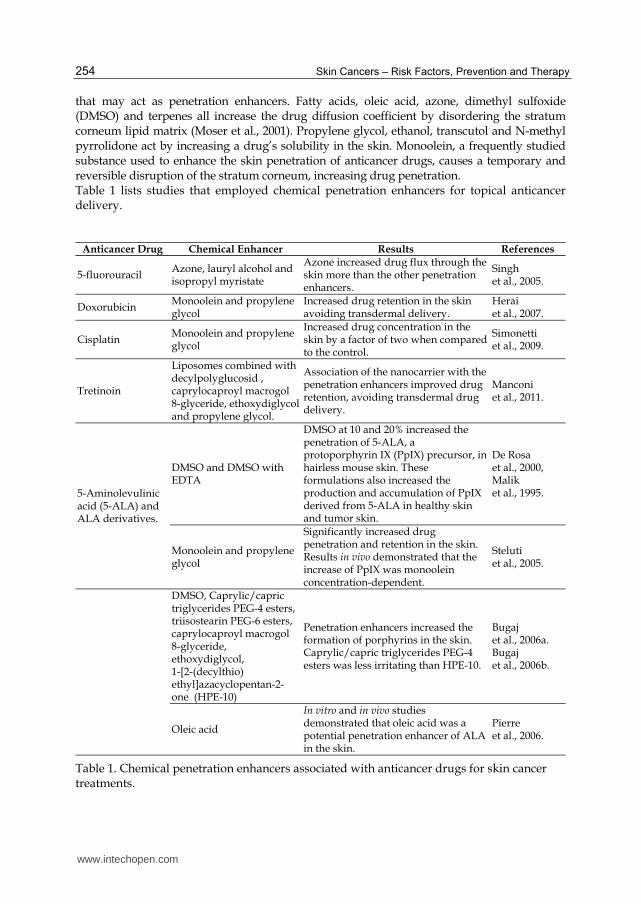
that may act as penetration enhancers. Fatty acids, oleic acid, azone, dimethyl sulfoxide (DMSO) and terpenes all increase the drug diffusion coefficient by disordering the stratum corneum lipid matrix (Moser et al., 2001). Propylene glycol, ethanol, transcutol and N-methyl pyrrolidone act by increasing a drug’s solubility in the skin. Monoolein, a frequently studied substance used to enhance the skin penetration of anticancer drugs, causes a temporary and reversible disruption of the stratum corneum, increasing drug penetration. Table 1 lists studies that employed chemical penetration enhancers for topical anticancer delivery.
Anticancer Drug Chemical Enhancer Results References
5-fluorouracil Azone, lauryl alcohol and isopropyl myristate
Azone increased drug flux through the skin more than the other penetration enhancers.
Singh et al., 2005.
Doxorubicin Monoolein and propylene glycol
Increased drug retention in the skin avoiding transdermal delivery.
Herai et al., 2007.
Cisplatin Monoolein and propylene glycol
Increased drug concentration in the skin by a factor of two when compared to the control.
Simonetti et al., 2009.
Tretinoin
Liposomes combined with decylpolyglucosid , caprylocaproyl macrogol 8-glyceride, ethoxydiglycol and propylene glycol.
Association of the nanocarrier with the penetration enhancers improved drug retention, avoiding transdermal drug delivery.
Manconi et al., 2011.
5-Aminolevulinic acid (5-ALA) and ALA derivatives.
DMSO and DMSO with EDTA
DMSO at 10 and 20% increased the penetration of 5-ALA, a protoporphyrin IX (PpIX) precursor, in hairless mouse skin. These formulations also increased the production and accumulation of PpIX derived from 5-ALA in healthy skin and tumor skin.
De Rosa et al., 2000, Malik et al., 1995.
Monoolein and propylene glycol
Significantly increased drug penetration and retention in the skin. Results in vivo demonstrated that the increase of PpIX was monoolein concentration-dependent.
Steluti et al., 2005.
DMSO, Caprylic/capric triglycerides PEG-4 esters, triisostearin PEG-6 esters, caprylocaproyl macrogol 8-glyceride, ethoxydiglycol, 1-[2-(decylthio) ethyl]azacyclopentan-2-one (HPE-10)
Penetration enhancers increased the formation of porphyrins in the skin. Caprylic/capric triglycerides PEG-4 esters was less irritating than HPE-10.
Bugaj et al., 2006a. Bugaj et al., 2006b.
Oleic acid
In vitro and in vivo studies demonstrated that oleic acid was a potential penetration enhancer of ALA in the skin.
Pierre et al., 2006.
Table 1. Chemical penetration enhancers associated with anticancer drugs for skin cancer treatments.
www.intechopen.com
Topical Administration of Anticancer Drugs for Skin Cancer Treatment
255
It can be seen in Table 1 that penetration enhancers generally increase drug penetration and retention into the skin. The most common and extensively studied chemical enhancers for topical chemotherapy are DMSO and monoolein. The lack of toxicity of monoolein compared to DMSO increases the potential use of this penetration enhancer in clinical trials. In addition, monoolein is biodegradable, safe and has been used in different formulations in the pharmaceutical field. The use of a chemical enhancer to increase the penetration of 5-ALA for topical PDT is another frequently studied strategy. 5-ALA is a prodrug, converted in situ by the heme biosynthetic pathway into a highly fluorescent substance, protoporphyrin IX (PpIX), an effective photosensitizer. Thus, for successful PDT therapy, it is important that high concentrations of 5-ALA penetrate the skin for its conversion to PpIX and to facilitate the death of tumor cells when light is applied. Although the FDA has already approved a topical application of ALA for actinic keratosis, extensive efforts have been made to increase 5-ALA penetration into the skin in an appropriate semi-solid formulation. Thus, chemical enhancers appear to be promising for PDT treatment in combination with ALA topical delivery.
4.2 Physical penetration methods
Iontophoresis and electroporation are the most frequently studied physical methods used to improve antineoplastic topical delivery. Both techniques employ an electrical current to overcome the stratum corneum and to increase drug penetration into the skin. In the next sections, the basic principles of iontophoresis and electroporation will be described, and some of the studies that have employed these modalities in the context of skin cancer treatment will be discussed.
4.2.1 Iontophoresis
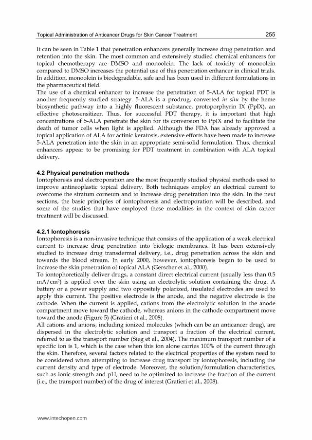
Iontophoresis is a non-invasive technique that consists of the application of a weak electrical current to increase drug penetration into biologic membranes. It has been extensively studied to increase drug transdermal delivery, i.e., drug penetration across the skin and towards the blood stream. In early 2000, however, iontophoresis began to be used to increase the skin penetration of topical ALA (Gerscher et al., 2000). To iontophoretically deliver drugs, a constant direct electrical current (usually less than 0.5 mA/cm2) is applied over the skin using an electrolytic solution containing the drug. A battery or a power supply and two oppositely polarized, insulated electrodes are used to apply this current. The positive electrode is the anode, and the negative electrode is the cathode. When the current is applied, cations from the electrolytic solution in the anode compartment move toward the cathode, whereas anions in the cathode compartment move toward the anode (Figure 5) (Gratieri et al., 2008). All cations and anions, including ionized molecules (which can be an anticancer drug), are dispersed in the electrolytic solution and transport a fraction of the electrical current, referred to as the transport number (Sieg et al., 2004). The maximum transport number of a specific ion is 1, which is the case when this ion alone carries 100% of the current through the skin. Therefore, several factors related to the electrical properties of the system need to be considered when attempting to increase drug transport by iontophoresis, including the current density and type of electrode. Moreover, the solution/formulation characteristics, such as ionic strength and pH, need to be optimized to increase the fraction of the current (i.e., the transport number) of the drug of interest (Gratieri et al., 2008).
www.intechopen.com
Skin Cancers – Risk Factors, Prevention and Therapy
256
Fig. 5. Schematic representation of an iontophoretic device: cations and anions migrate through the skin during the application of a low-strength electrical current.
The electrode choice has an important influence on the stability of the driving electric current, which is necessary to control drug delivery. Electrodes must guarantee the electroneutrality of the system without changing the pH of the electrolytic solution or altering the drug’s properties (Cullander et al., 1993). The most commonly used electrodes are the reversible Ag/AgCl electrodes. This choice is primarily because their electrochemical reactions are rapid and occur at a voltage lower than that required for the water to undergo electrolysis, thereby avoiding variations in the pH of the formulation (Kalia et al., 2004). During the application of the electrical current, Cl- ions in the electrolyte solution react with the silver electrode, i.e., the anode, donating one electron to the electric circuit (Ago + Cl- AgCl + e-). This electron arrives at the cathode, reducing the AgCl electrode (AgCl + e- Ago + Cl-). To ensure the system’s electroneutrality, a cation in the anode moves to the skin or an anion moves from the skin toward the anode. In the cathode, the opposite occurs, i.e., an anion moves to the skin or a cation moves from the skin toward the cathode (Kalia et al., 2004; Chang et al., 2000). Therefore, the electric circuit is completed by the inorganic ions of the skin, primarily Na+ and Cl-. Antineoplastic drugs can be delivered to the skin by iontophoresis through two mechanisms: electromigration and electroosmosis. These mechanisms can act in combination to increase drug skin penetration. Electromigration refers to the orderly movement of ions in the presence of an electric current. For instance, positively charged antineoplastic drugs, when placed in the positive electrode compartment (anode), migrate away from the electrode with the same polarity into the skin (Tesselaar & Sjöberg, 2011). The same occurs when a negative drug is placed in contact with the cathode compartment. The electromigration contribution to a drug’s skin penetration depends on the concentration and the electrical mobility of the drug (ion) (Gratieri et al., 2008). High-molecular weight drugs, which include most antineoplastic drugs, generally have a low electric mobility, decreasing the electromigration contribution for their permeating. Electroosmosis refers to the solvent flow when an electric potential is applied to the skin. Under physiologic conditions, this flow occurs from the anode toward the cathode due to the skin’s cation permselectivity (Figure 6). Specifically, the skin is twice more permeable to cations than to anions (Burnette & Ongpipattanakul, 1987). This is because the skin is
www.intechopen.com
Topical Administration of Anticancer Drugs for Skin Cancer Treatment
257
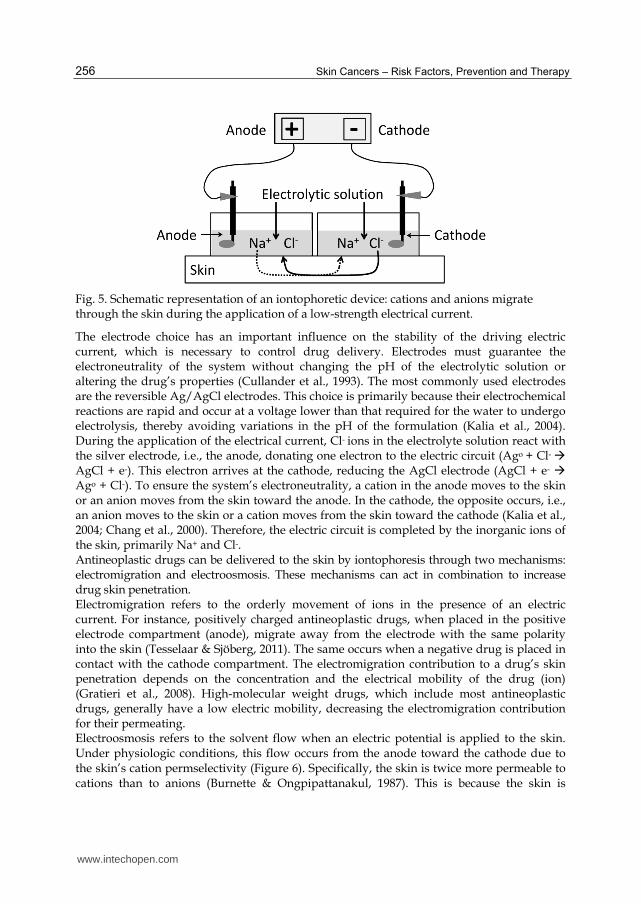
negatively charged when in contact with a solution at physiological pH (Merino et al., 1999). As a result, solvent flow occurs in the direction of cation flux, enhancing the transport of cations and slowing the transport of anions (Singh & Mabach, 1996). Electroosmotic flux is the dominant mechanism for macromolecule’s skin permeation. This is because the solvent flux pushes the macromolecule and takes advantage of the low-resistance of the skin when iontophoresis is applied (Abla et al., 2005; Pikal, 2001). Hence, neutral and high molecular weight antineoplastic drugs can take advantage of solvent flow and penetrate the skin by iontophoresis. Furthermore, positively charged drugs can penetrate the skin by both electromigration and electroosmotic contributions.
Fig. 6. Schematic representation of the electroosmotic flow that accompanies the electromigration of cations, which is due to the negative charge of the skin at pH 7.
The use of iontophoresis in the topical administration of antineoplastic drugs offers important advantages for tumor drug delivery. Most of these advantages are related to the
precise control of drug delivery by electrical current adjustments and formulation characteristics. The applied current density and the short duration of this application,
combined with the components and formulation characteristics, may rapidly target the drug to the tumor in high concentrations, avoiding the blood stream (Kalia et al., 2004). As an
example of the application of targeted drug delivery using iontophoresis and simple
modifications in drug formulation, Taveira et al. (2009) demonstrated that iontophoresis significantly increased skin permeability to doxorubicin. However, because this drug is
positively charged at a physiological pH, it interacted with the negatively charged stratum corneum, decreasing its permeation to the deep skin layers. Interestingly however, the
authors demonstrated that the incorporation of a cationic polymer (chitosan) in the drug formulation decreased the skin’s negatives charge when iontophoresis was applied,
releasing doxorubicin from ionic interactions with the skin and improving its diffusion into the deep skin layers. Despite the obvious benefits that iontophoresis may offer for topical skin tumor treatment, there have been limited studies using this technique to this purpose. Most studies have applied iontophoresis to increase the penetration of ALA and porphyrins for topical PDT (Table 2)
www.intechopen.com
Skin Cancers – Risk Factors, Prevention and Therapy
258
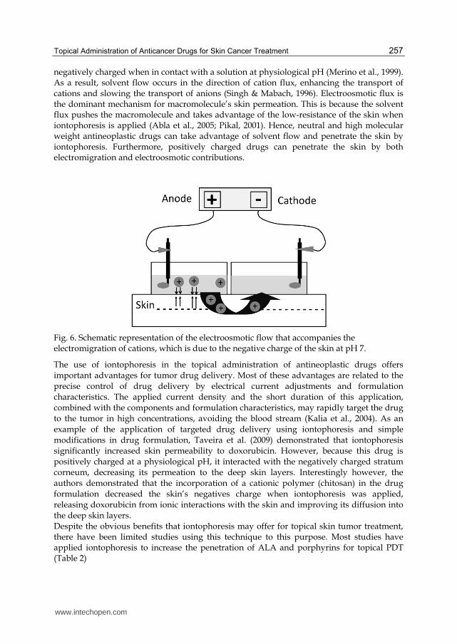
Drug Skin model Electrode polaritya
Current density
(mA/cm2) Formulationb References
ALA and ester In vivo: human Anode 0.25 Aqueous solution
Gerscher et al., 2000.
ALA and Ester In vivo: human Anode 0.25 Aqueous solution
Gerscher et al., 2001.
ALA In vitro: human SC Anode 0.15 - 0.78
isotonic phosphate buffer (pH 2.1)
Bodde et al., 2002.
ALA In vivo: human Anode 0.2 Aqueous solution and cream
Choudry et al., 2003.
ALA In vivo: rabbits – oral mucosa
Anode 0.5 Ointment Tanaka et al., 2003.
ALA In vitro: pig ear Anode and cathode
0.5 Physiological buffer
Lopez et al., 2003.
ALA and esters In vitro: pig ear Anode 0.5 Distilled waterLopez et al., 2003.
ALA and m-ALA In vitro: pig ear Anode 0.5
Lipid sponge phase and buffer with propylene glycol
Merclin et al., 2004.
ALA and m-ALA In vitro: pig ear Anode 0.5 Anionic Gel Merclin et al., 2004.
Meso-tetra-[4-sulfonatophenyl]-porphyrin
In vitro: pig ear Cathode 0.5 Electrolytic aqueous solution
Gelfuso et al., 2008.
ALA In vivo: human Anode 0.25 - 0.5 Distilled water Mizutani et al., 2009.
Doxorubicin In vitro: pig ear cathode 0.5 Non-ionic and cationic gel
Taveira et al., 2009.
zinc phthalocyanine tetrasulfonic acid
In vitro: pig ear Anode and cathode
0.5 Non-ionic gel Souza et al., 2011.
Meso-tetra-(N-methylpiridinium-4-yl)-porphyrin and meso-tetra-(4-sulfonatophenyl)-porphyrin
In vitro: pig ear Anode and cathode
0.5 Non-ionic Gel Gelfuso et al., 2011.
a Polarity of the electrode in contact with the formulation containing the drug. B Composition of the principal components/ions of the formulation/electrolytic solution that contains
the drug.
Table 2. Drugs that have been delivered into the skin using iontophoresis as a skin cancer
treatment.
The experiments shown in Table 2 demonstrate that iontophoresis significantly increases drug penetration into and through the skin much more rapidly than passive (no current)
www.intechopen.com
Topical Administration of Anticancer Drugs for Skin Cancer Treatment
259
administration. For examples, a 50-fold higher flux over passive transport was observed with iontophoretic delivery of methyl-ALA after 2 h (Lopez et al, 2003). Furthermore, the same amount of ALA delivered passively into the stratum corneum in several hours was delivered in only 10 minutes of iontophoresis application (Bodde et al., 2002). In vivo experiments performed with ALA and its esters generally show an increase in the depth and intensity of PpIX fluorescence following administration of the prodrug ALA. However, adjustments of electrical and formulation parameters are still required to improve the performance of iontophoretic delivery in vivo. In vitro, modifications in the pH and ionic strength of drug formulation have shown extensive improvements in drug delivery. For example, the simple elimination of Na+ from a gel formulation containing the porphyrin meso-tetra-(N-methylpyridinium-4-yl)-porphyrin at pH 5.5 increased anodal drug iontophoretic transport by approximately 30% (Gelfuso et al., 2011). In summary, iontophoresis has a huge potential for drug delivery in topical skin cancer therapy. Clearly, more studies should be performed in vivo with other antineoplastic drugs and with optimized iontophoretic parameters. Moreover, risks and toxicity for other organs should be evaluated to ensure that antineoplastic drugs accumulate in the tumor without entering the systemic circulation in significant quantities.
4.2.2 Electroporation
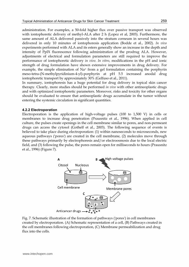
Electroporation is the application of high-voltage pulses (100 to 1,500 V) in cells or membranes to increase drug penetration (Prausnitz et al., 1996). When applied in cell culture, the pulses create openings in the cell membrane similar to pores, and non-permeant drugs can access the cytosol (Gothelf et al., 2003). The following sequence of events is believed to take place during electroporation: (1) within nanoseconds to microseconds, new aqueous pathways ('pores') are created in the cell membrane, (2) molecules move through these pathways primarily by electrophoresis and/or electroosmosis due to the local electric field, and (3) following the pulse, the pores remain open for milliseconds to hours (Prausnitz et al., 1996) (Figure 7).
Fig. 7. Schematic illustration of the formation of pathways ('pores') in cell membranes created by electroporation. (A) Schematic representation of a cell, (B) Pathways created in the cell membranes following electroporation, (C) Membrane permeabilization and drug flux into the cells.
www.intechopen.com
Skin Cancers – Risk Factors, Prevention and Therapy
260
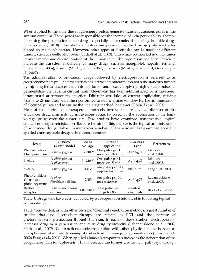
When applied to the skin, these high-energy pulses generate transient aqueous pores in the stratum corneum. These pores are responsible for the increase of skin permeability, thereby increasing the penetration of the drugs, especially macromolecules and hydrophilic drugs (Charoo et al., 2010). The electrical pulses are primarily applied using plate electrodes placed on the skin’s surface. However, other types of electrodes can be used for different tumors, such as needle electrodes (Gothelf et al., 2003). These may be inserted into the tumor to favor membrane electroporation of the tumor cells. Electroporation has been shown to increase the transdermal delivery of many drugs, such as metoprolol, heparin, fentanyl (Denet et al., 2004), insulin (Murthy et al., 2006), piroxican (Murthy et al., 2004; Guoqiang et al., 2007). The administration of anticancer drugs followed by electroporation is referred to as electrochemotherapy. The first studies of electrochemotherapy treated subcutaneous tumors by injecting the anticancer drug into the tumor and locally applying high voltage pulses to permeabilize the cells. In clinical trials, bleomycin has been administered by intravenous, intratumoral or intraarterial injection. Different schedules of current application, varying from 8 to 28 minutes, were then performed to define a time window for the administration of electrical pulses and to ensure that the drug reached the tumor (Gothelf et al., 2003). Most of the electrochemotherapeutic protocols involve the invasive application of the anticancer drug, primarily by intravenous route, followed by the application of the high-voltage pulse over the tumor site. Few studies have examined non-invasive, topical anticancer drug administration. Because the aim of this chapter is the topical administration of anticancer drugs, Table 3 summarizes a subset of the studies that examined topically applied antineoplastic drugs using electroporation.
Drug In vitro/
In vivo model Pulse
Voltage Time of
application Electrode
Type References
Photosensitizer Methylene blue
In vitro: pig ear 0 - 240 V One pulse per 1 msec for 10-30 min
Ag/AgCl Johnson et al., 1998.
5-ALA In vitro: pig ear
In vivo: mice 0 - 240 V
One pulse per 1 msec for 15 min
Ag/AgCl Johnson et al., 2002.
5-ALA In vitro: pig ear 300 V one pulse per 30 s, applied for 10 min
Platinum Fang et al., 2004.
Photosensitizer chlorin and phthalocyanine
In vitro: fibroblast cell line
1200V one pulse per 0.1 ms for 20 min
Ag/AgCl Labanauskiene et al., 2007.
Ruthenium complex
In vitro: melanoma cell line
80 – 240 V One pulse per 100 μs for 8 s
stainless steel plate
Bicek et al., 2007.
Table 3. Drugs that have been delivered by electroporation into the skin following topical administration.
Table 3 shows that, as with other physical/chemical penetration methods, a great number of studies that use electrochemotherapy are related to PDT and the increase of photosensitizer’s permeation through the skin. In each of these studies, electroporation increases drug skin penetration and even drug cytotoxicity (Labanauskiene et al., 2007; Bicek et al., 2007). Combinations of electroporation with other physical methods, such as iontophoresis, often lead to synergistic effects in increasing drug penetration (Johson et al., 2002; Fang et al., 2004). When applied alone, electroporation increases the penetration of the drugs more than iontophoresis. This is because the former creates new pathways through
www.intechopen.com
Topical Administration of Anticancer Drugs for Skin Cancer Treatment
261
the skin, whereas the later mostly takes advantage of the decreased electrical resistance in existing routes into the skin. In this context, electrochemotherapy is a potential strategy for topical anticancer drug administration. Nevertheless, more studies are required to investigate the side effects and to minimize patient discomfort during the application of the electroporation protocol.
4.3 Nanocarrier systems for topical anticancer drug delivery
Semi-solid conventional formulations, such as creams, ointments and gels, have been used for topical administration of drugs for many years. Simple application of the formulation on the skin’s surface, however, is not sufficient to allow the drug to reach the site of action. This means that it is important for the formulation to aid in drug penetration through the different skin layers to reach the tumor site (Schmid & Korting, 1996). Nanocarriers could improve skin targeting, improving the drug’s ability to reach and penetrate into tumor cells. Moreover, nanocarriers can improve drug stability and reduce skin irritation by avoiding direct contact of the drug with the skin’s surface (Schimid & Korting, 1996). Different nanocarriers have been used for topical application. This section will discuss the most frequently studied, topically applied carriers for the treatment of skin tumors.
4.3.1 Liposomes
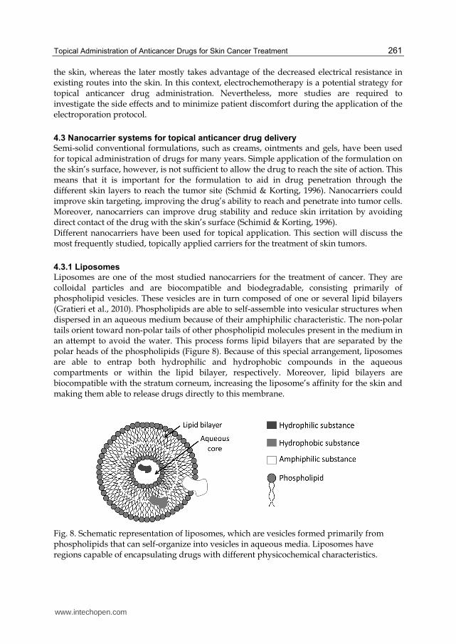
Liposomes are one of the most studied nanocarriers for the treatment of cancer. They are colloidal particles and are biocompatible and biodegradable, consisting primarily of phospholipid vesicles. These vesicles are in turn composed of one or several lipid bilayers (Gratieri et al., 2010). Phospholipids are able to self-assemble into vesicular structures when dispersed in an aqueous medium because of their amphiphilic characteristic. The non-polar tails orient toward non-polar tails of other phospholipid molecules present in the medium in an attempt to avoid the water. This process forms lipid bilayers that are separated by the polar heads of the phospholipids (Figure 8). Because of this special arrangement, liposomes are able to entrap both hydrophilic and hydrophobic compounds in the aqueous compartments or within the lipid bilayer, respectively. Moreover, lipid bilayers are biocompatible with the stratum corneum, increasing the liposome’s affinity for the skin and making them able to release drugs directly to this membrane.
Fig. 8. Schematic representation of liposomes, which are vesicles formed primarily from phospholipids that can self-organize into vesicles in aqueous media. Liposomes have regions capable of encapsulating drugs with different physicochemical characteristics.
www.intechopen.com
Skin Cancers – Risk Factors, Prevention and Therapy
262
Traditional liposomes are composed primarily of phospholipids. Liposomes formed with different components have been developed in an attempt to increase the stability of the vesicles and their ability to penetrate through different membranes, especially the stratum corneum. In this way, elastic liposomes, also called ultradeformable or ultraflexible liposomes, are the new generation of liposomes. These contain surfactants, other amphiphiles or ethanol in their composition, which improve the flexibility of the lipid bilayer. Transfersomes®, niosomes and ethosomes® were the names given to the first, second and third flexible liposome generations, respectively (Santana & Zancheta, 2011). Transfersomes® were introduced by Cevc and Blume (1992) and are composed of phosphatidylcholine and sodium cholate. Ethosomes® consist of a mixture of phosphatidilcoline and ethanol, and niosomes are non-ionic surfactant vesicles (Manconi et al., 2002). The ability of these vesicles to deform gives them the ability to pass through narrow pores, such as the pores present on the skin surface, possibly improving the penetration of drugs carried by these vesicles into the deep skin layers (Manconi et al., 2002, Santana & Zancheta, 2011). These new generations of liposomes have been well studied in the context of topical administration and have also been introduced into the field of topical skin cancer treatments. Most of the studies involving liposomes in the treatment of cancer have been performed using invasive administration, such as intravenous injections. Liposomes containing doxorubicin (Hosoda et al., 1995; Barenholz et al., 2001), cisplatin (Lasic et al., 1999; Krieger et al., 2010), oxaliplatin (Lila et al., 2010), camptothecin (Watanabe et al., 2008) and others have been shown to increase these drugs cytotoxicity and to reduce side effects because of direct targeting. Some of these liposomes, such as DOXIL®, are already commercially available. This liposomal formulation contains doxorubicin and was approved in the US in 1995 (Barenholz, 2001). The topical application of anticancer drugs is, once again, primarily related to the administration of the pro-drug ALA for topical PDT. Fang et al. (2008) performed an in vivo study of the influence of liposomes and ethosomes in ALA skin penetration. This study showed that the flexible liposomes (ethosomes) increased 5-ALA penetration to a greater degree than did the traditional liposomes, although both formulations increased ALA penetration when compared to the control treatment. Cationic ultradeformable liposomes have also been shown to increase ALA skin permeability in vitro. In vivo, these liposomes result in persistent ALA retention in the skin and induce the production of high levels of PpIX (Oh et al., 2011). ALA skin retention was also improved when a traditional ALA-containing liposome was examined in vitro (Pierre et al., 2001). In addition to these ALA studies, 5-fluorouracil-loaded niosomes showed an 8-fold improvement of this drug’s cytotoxicity and penetration when compared to the aqueous solution (Paolino et al., 2008). It is worth noting that liposomes in combination with other drugs not traditionally used in skin cancer treatments have also been studied. For instance, tretinoin and diclophenac-loaded liposomes (Kitagawa et al., 2006; Zaafarany et al., 2010) showed improvement in these drugs’ skin penetration over non-liposomal formulations. These studies, however, were aimed at treating acne, psoriasis and other inflammatory conditions but not skin tumors. These formulations, however, are currently proposed to treat skin cancer malignances. In summary, liposomes have been shown to increase drugs’ penetration into the skin, and it appears that ultradeformable liposomes may have an even stronger effect. However, some reports describe liposome instability and drug leakage during the storage period (Glavas-
www.intechopen.com
Topical Administration of Anticancer Drugs for Skin Cancer Treatment
263
Dodov et al., 2005). Therefore, more studies should be performed to develop more stable liposomes. In vivo experiments with humans should be performed to demonstrate the potential of flexible liposomes loaded with different anticancer drugs for topical skin cancer treatment.
4.3.2 Polymeric and lipid nanoparticles
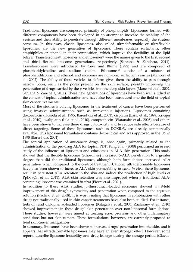
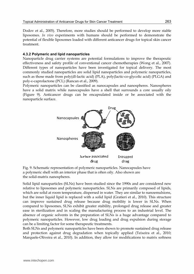
Nanoparticle drug carrier systems are potential formulations to improve the therapeutic effectiveness and safety profile of conventional cancer chemotherapies (Wong et al., 2007). Different types of nanoparticles have been investigated for topical delivery. The most commonly studied nanoparticles are solid lipid nanoparticles and polymeric nanoparticles, such as those made from poly(dl-lactic acid) (PLA), poly(lactic-co-glycolic acid) (PLGA) and poly-ε-caprolactone (PCL) (Rancan et al., 2009). Polymeric nanoparticles can be classified as nanocapsules and nanospheres. Nanospheres have a solid matrix while nanocapsules have a shell that surrounds a core usually oily (Figure 9). Anticancer drugs can be encapsulated inside or be associated with the nanoparticle surface.
Fig. 9. Schematic representation of polymeric nanoparticles. Nanocapsules have a polymeric shell with an interior phase that is often oily. Also shown are the solid-matrix nanospheres.
Solid lipid nanoparticles (SLNs) have been studied since the 1990s and are considered new relative to liposomes and polymeric nanoparticles. SLNs are primarily composed of lipids, which are solid at room temperature, dispersed in water. They are similar to nanoemulsions, but the inner liquid lipid is replaced with a solid lipid (Gratieri et al., 2010). This structure can improve sustained drug release because drug mobility is lower in SLNs. When compared to liposomes, SLNs exhibit greater stability, prolonged drug release and greater ease in sterilization and in scaling the manufacturing process to an industrial level. The absence of organic solvents in the preparation of SLNs is a huge advantage compared to polymeric nanoparticles. However, low drug loading and drug expulsion during storage can be a limiting factor for some therapeutic treatments. Both SLNs and polymeric nanoparticles have been shown to promote sustained drug release and protection against drug degradation when topically applied (Teixeira et al., 2010; Marquele-Oliveira et al., 2010). In addition, they allow for modifications to matrix softness
www.intechopen.com
Skin Cancers – Risk Factors, Prevention and Therapy
264
and superficial charges, adjustments that may improve skin targeting. The exact mechanism by which these particles increase drug penetration through the skin is not completely understood, but efforts to understand this property have been made by developing and characterizing different nanoparticles (Lopez et al., 2011). It appears that nanoparticles can closely contact the superficial junctions of corneocyte clusters and furrows, possibly favoring drug accumulation for several hours. This would allow for the sustained release of anticancer drugs. However, there are controversies regarding the ideal mean diameter, flexibility and superficial charge of nanoparticles to contribute to skin penetration. Studies of nanoparticles have not been limited to the examination of cytotoxic cancer drugs; numerous studies have also been performed that use these systems to deliver anti-proliferative drugs. Of studies of the topical administration of anticancer drugs, nanoparticles containing the 5-ALA, 5-fluorouracil and tretinoin are the most common (Prow et al., 2011). Some examples of recent results obtained with these and other drugs encapsulated in nanoparticles are described below. SLNs have been shown to increase tretinoin stability and to decrease drug irritation (Kumar et al., 2007, Mandawgade et al., 2008 ). Microparticles containing ALA were shown in vitro to be capable of temperature-triggered ALA release, enhanced drug stability and improved penetration through keratinized skin (Kassas et al., 2009). In vivo, high levels of PpIX were observed in mouse skin treated with ALA-loaded microparticles. This study also demonstrated a reduction in skin tumor growth rate (Donelly et al., 2009). Polymeric micro- and nanocapsules increased porphyrin-induced phototoxicity by a factor of 4 in cultured HeLa cells when compared to a liposomal emulsion of phosphatidylcholine loaded with an equivalent amount of porphyrin (Deda et al., 2009). PLA nanoparticles containing the prodrug 5-fluorouracil demonstrated linear release of this drug for 6 h with no evidence of a burst effect (McCarron et al., 2008). Promising results were found for resveratrol encapsulated in SLNs, a formulation that showed increased cellular uptake (Teskac et al., 2010). Nitrosyl ruthenium complex-loaded SLNs performed well at releasing and protecting the complex against degradation in vitro, showing this formulation to be a promising carrier for the topical delivery of nitric oxide (Marquele-Oliveira et al., 2010). Podophyllotoxin-SLNs were demonstrated in vitro to increase drug retention on the skin’s surface, avoiding transdermal penetration (Prow et al., 2011). In summary, most studies have described the advantages of drug encapsulation in nanoparticles by demonstrating increased drug stability, sustained release and improved skin penetration and cytotoxicity. Despite such promising results, more studies should be performed to elucidate the mechanisms by which nanoparticles increase the ability of anticancer drugs to penetrate the skin.
5. Conclusion
The penetration of drugs into the skin through the keratinized stratum corneum is a major obstacle to the delivery of high concentrations of anticancer drugs into tumor cells. The use of chemical and physical methods and the development of nanoparticle-based drug delivery systems are very important strategies to improve the ability of drugs to penetrate the skin. Nanocarriers appear to be promising systems because they offer several advantages, such as low skin irritation and increased protection of encapsulated drug. An especially important advantage of these formulations is that they often increase anticancer drug penetration through the skin. Iontophoresis also appears to be a promising technique for improving
www.intechopen.com
Topical Administration of Anticancer Drugs for Skin Cancer Treatment
265
drug delivery through the skin, especially for PDT, for which high concentrations of the photosensitizer are required for effective treatment. The use of physical methods to improve the penetration of nanocarriers should be considered to increase the anticancer drug’s penetration into the skin and to provide for targeted drug release inside tumor cells.
6. Acknowledgements
The authors thank the financial support of FAPESP, CAPES and CNPq.
7. References
Abla, N., Naik, A., Guy, R. H., Kalia, Y. N. (2005). Contributions of electromigration and electroosmosis to peptide iontophoresis across intact and impaired skin. Journal of Controlled Release, Vol.108, No.2-3, (November 2005), pp. 319-330, ISSN 0168-3659.
Alam, M., Goldber, L. H., Silapunt, S., Gardner, E. S., Strom, S. S., Rademaker, A. W., Margolis, D. J. (2011). Delayed treatment and continued growth of nonmelanoma skin cancer. Journal of American Academy Dermatolology, Vol.64, No.5, (May 2011), pp. 839-848, ISSN 0190-9622.
Anthony, M. L. (2000). Surgical treatment of nonmelanoma skin cancer. AORN, Vol.71, No.3, (March 2000), pp. 550-564, ISSN 0001-2092.
Araújo, L. M. P. C., Thomazine, J. A., Lopez, R. F. V. (2010). Development of microemulsions to topically deliver 5-aminolevulinic acid in photodynamic therapy. European Journal of Pharmaceutics and Biopharmaceutics, Vol.75, No.1, (May 2010), pp. 48-55, ISSN 0939-6411.
Barenholz, Y. (2001). Liposome application: problems and prospects. Current Opinion in Colloid & Interface Science, Vol.6, No.1, (February 2001), pp. 66-77, ISSN 1359-0294.
Barrera, M. V., Herrera, E. (2007). Topical chemotherapy of actinic keratosis and nonmelanoma skin cancer: current options and future perspectives. Actas Dermo-Sifiliográficas, Vol.98, No.8, (October 2007), pp. 556-562, ISSN 0001-7310.
Barry, B. W. (2001). Novel mechanisms and devices to enable successful transdermal drug delivery. European Journal of Pharmaceutical Science. Vol.14, No.2, (September 2001), pp. 101-114, ISSN 0928-0987.
Bicek, A., Turel, I., Kanduser, M., Miklavcic, D. (2007). Combined therapy of the antimetastatic compound NAMI-A and electroporation on B16F1 tumor cells in vitro. Bioelectrochemistry, Vol.71, No.2, (November 2007), pp. 113-117, ISSN 1567-5394.
Boddé, H. E., Roemelé, P. E., Star, W. M. (2002). Quantification of topically deliverd 5-aminolevulinic acid by iontophoresis across ex vivo human stratum corneum. Photochemistry Photobiology, Vol.75, No.4, (April 2002), pp. 418-423, ISSN 0031-8655.
Bouwstra, J. A., Ponec, M. (2006). The skin barrier in healthy and diseased state. Biochimica et Biophysica Acta (BBA) – Biomembranes, Vol.1758, No.12, (December 2006), pp. 2080-2095, ISSN 0005-2736.
Bugaj, A., Juzeniene, A., Juzenas, P., Iani, V., Ma, L. W., Moan, J. (2006a). The effect of skin permeation enhancers on the formation of porphyrins in mouse skin during topical application of the methyl ester of 5-aminolevulinic acid. Journal of Photochemistry and Photobiology B: Biology, Vol.83, No.2, (May 2006), pp. 94-97, ISSN 1011-1344.
www.intechopen.com
Skin Cancers – Risk Factors, Prevention and Therapy
266
Bugaj, A., Juzeniene, A., Juzenas, P., Iani, V., Ma, L. W., Moan, J. (2006b). The effect of dimethylsulfoxide, 1-[2-(decylthio)ethyl]azacyclopentan-2-one and Labrafac(R)CC on porphyrin formation in normal mouse skin during topical application of methyl 5-aminolevulinate: A fluorescence and extraction study. Photodiagnosis and Photodynamic Therapy, Vol.3, No.1, (March 2006), pp. 27-33, ISSN 1572-1000.
Burnette, R. R., Ongpipattanakul, B. (1987). Characterization of the permselective properties of excised human skin during iontophoresis. Journal of Pharmaceutical Science, Vol.76, No.10, (October 1987), pp. 765-773, ISSN 0022-3549.
Burns, C. A., Brown, M. D. (2005). Imiquimod for the treatment of skin cancer. Dermatologic Clinics, Vol.23, (January 2005), pp. 151-164, ISSN 0733-8635.
Chang, S.-L., Hofmann,G. A., Zhang, L., Deftos, L. J., Banga, A. K. (2000). The effect of electroporation on iontophoretic transdermal delivery of calcium regulating hormones. Journal of Controlled Release, Vol.66, No.2-3, (May 2000), pp. 127-133, ISSN 0168-3659.
Charoo, N. A., Rahman, Z., Repka, M. A., Murthy, S. N. (2010). Electroporation: An avenue for transdermal drug delivery. Current Drug Delivery, Vol.7, No.2, (April 2010), pp. 125-136, ISSN 1567-2018.
Choudry, K., Brooke, R. C. C., Farrar, W., Rhodes, L. E. (2003). The effect of an iron chelating agent on protoporphyrin IX levels and phototoxicity in topical 5-aminolaevulinic acid photodynamic therapy. British Journal of Dermatology, Vol.149, (July 2004), pp. 124-130, ISSN 0007-0963.
Cullander, C., Guy, R. H. Routes of delivery: Case studies: (6) Transdermal delivery of peptides and proteins. Advanced Drug Delivery Reviews, Vol.8, No.2-3, (March 1992), pp. 291-329, ISSN 0169-409X.
Davids, L. M., Kleemann, B. (2010). Combating melanoma: the use of photodynamic therapy as a novel, adjuvant therapeutic tool . Cancer Treatment Reviews, (December 2010), ISSN 0305-7372, doi 10.1016/j.ctrv.2010.11.007.
De Rosa, F. S., Marchetti, J. M., Thomazini, J. A., Tedesco, A. C., Bentley, M. V. L. B. (2000). A vehicle for photodynamic therapy of skin cancer: influence of dimethylsulphoxide on 5-aminolevulinic acid in vitro cutaneous permeation and in vivo protoporphyrin IX accumulation determined by confocal microscopy. Journal of Controlled Release, Vol.65, No.3, (April 2000), pp. 359-366, ISSN 0168-3659.
Deda, D. K., Uchoa, A. F., Carita, E., Baptista, M. S., Toma, H. E., Araki, K. (2009). A new micro/nanoencapsulated porphyrin formulation for PDT treatment. International Journal of Pharmaceutics, Vol.376, No.1-2, (July 2009), pp. 76-83, ISSN 0378-5173.
Denet, A. –R., Vanbever, R., Preat, V. (2003). Skin electroporation for transdermal and topical delivery. Advanced Drug Delivery Reviews, Vol.56, No.5, (March 2004), pp. 659-674, ISSN 0169-409X.
Donnelly, R. F., McCarron, P. a., Al-Kassas, R., Juzeniene, A., Juzenas, P., Iani, V., Woolfson, A. D., Moan, J. (2009). Influence of formulation factors on PpIX production and photodynamic action af novel ALA-loaded microparticles. Biopharm Drug Dispos, Vol.30, No.2, (March 2009), pp. 55-70, ISSN 0142-2782.
Einspahr, J. G., Stratton, S. P., Bowden, G. T., Alberts, D. S. (2002). Chemoprevention of human skin cancer. Critial Reviews in Oncolocy/Hematology, Vol.41, No.3, (March 2002), pp. 269-285, ISSN 1040-8428.
www.intechopen.com
Topical Administration of Anticancer Drugs for Skin Cancer Treatment
267
Fang, J. –Y, Lee, Y. –R., Shen, S-C., Fang, Y. –P., Hu, C, -H. (2004). Enhancement of topical 5-aminolaevulinic acid delivery by erbium: YAG laser and microdermobrasion: a comparison with iontophoresis and electroporation. British Journal of Dermatology, Vol.151, (July 2004), pp. 132-140, ISSN 0007-0963.
Fang, Y. –P., Tsai, Y. –H., Wu, P. –C., Huang, Y. –B. (2008). Comparison of 5-aminolevulinic acid-encapsulated liposome versus ethosome for skin delivery for photodynamic therapy. International Journal of Pharmaceutics, Vol.356, No.1-2, (May 2008), pp. 144-152, ISSN 0378-5173.
Galiczynski, E. M., Vidimos, A. T. (2011). Nonsurgical treatment of nonmelanoma skin cancer. Dermatologic Clinics, Vol.29, (April 2011), pp. 297-309, ISSN 0733-8635.
Gelfuso, G. M., Figueiredo, F. V., Gratieri, T., Lopez, R. F. V. (2008). The effect of pH and ionic strenght on topical delivery of a negatively charged porphyrin (TPPS4). Journal of Pharmaceutical Science, Vol.97, (October 2008), pp. 4249-4257, ISSN 0022-3549.
Gelfuso, G. M., Gratieri, T., Souza, J. G., Thomazine, J. A., Lopez, R. F. V. (2011). The influence of positive or negative charges in the passive and iontophoretic skin penetration of porphyrins used in photodynamic therapy. European Journal of Pharmaceutics and Biopharmaceutics. Vol. 77, No.2, (February 2011), pp. 249-256, ISSN 0939-6411.
Gerscher, S. Connelly, J. P., Griffiths, J., Brown, S. B., MacRobert, A. J., Wong, G., Rhodes, L. E. (2000). Comparison of the pharmacokinetics and phototoxicity of protoporphyrin IX metabolized from 5-amonolevulinic acid and two derivatives in human skin in vivo. Photochemistry and Photobiology, Vol.72, No.4, (October 2000), pp. 569-574, ISSN 0031-8655.
Gerscher, S., Connelly, J. P. (2001). A quantitative assessment of protoporphyrin IX metabolism and phototoxicity in human skin following dose-controlled delivery of the prodrugs 5-amonolaevulinic acid and 5-aminolaevulinic acid-n-pentylester. British Journal of Dermatology, Vol.144, No.5, (May 2001), pp. 983-990, ISSN 0007-0963.
Glavas-Dodov, M., Fredo-kumbaradzi, E., Goracinova, K., Simonoska, M., Calis, S., Trajkovic-Jolevska, S., Hincal, A. A. (2005). The effects of lyophilization on the stability of liposomes containing 5-FU. International Journal of Pharmaceutics, Vol. 291No. 1-2, ( March 2005), pp. 79-86, ISSN 0378-5173.
Gothelf, A., Mir, L. M., Gehl, J. (2000) Electrochemotherapy: results of cancer treatment using enhanced delivery of bleomycin by electroporation. Cancer Treatment Reviews, Vol.29, No.5, (October 2003), pp. 371-387, ISSN 0305-7372.
Gratieri, T., Gelfuso, G. M., Lopez, R. F. V. (2008). Basic principles and applications of iontophoresis for cutaneous penetration of drugs. Quimica Nova, Vol.31, No.6, (July 2008), pp. 1490-1498, ISSN 1678-7064.
Gratieri, T., Gelfuso, G. M., Lopez, R. F. V., Souto, E. B. (2010). Current efforts and the potential of nanomedicine in treating fungal keratitis. Expert Review of Ophthalmology, Vol.5, No.3 (June 2010), pp. 365-384, ISSN 1746-9899.
Guimarães, K. L., Ré, M. I. (2011). Lipid nanoparticles as carriers for cosmetic ingredients: The first (SLN) and the Second Generation (NLC). In: Nanocosmetics and Nanomedicines: New approaches for skin care. Ruy Beck, Silvia Guterres and Adriana Pohlmann, 101-122, Springer, ISBN 978-3-642-19791-8, Retrieved from
http://www.springerlink.com/openurl.asp?genre=book&isbn=978-3-642-19791-8.
www.intechopen.com
Skin Cancers – Risk Factors, Prevention and Therapy
268
Guoqiang, J., Dequan, Z., Jia, Z., Fuxin, D. (2007). Transdermal drug delivery of electroporation: the effects of surfactants on pathway lifetime and drug transport. Chinese Journal of Chemical Engineering, Vol.15, No.3, (June 2007), pp. 397-402, ISSN 1004-9541.
Hadgraft, J., Lane, M. E. (2005). Skin permeation: The years of enlightenment. International Journal of Pharmaceutics, Vol.305, No.1-2, (November 2005), pp. ISSN 0378-5173.
Herai, H., Gratieri, T., Thomazine, J. A., Bentley, M. V. L. B., Lopez, R. F. V. (2007). Doxorubicin skin penetration from monoolein-containing propylene glycol formulations. International Journal of Pharmaceutics, Vol.329, No.1-2, (February 2007), pp. 88-93, ISSN 0378-5173.
Hosoda, J., Unezaki, S., Maruyama, K., Tasuchiya, S., Iwatsuru, M. (1995). Antitumor activity of doxorubicin encapsulated in poly(ethylene glycol)-coated liposomes. Biological & Pharmaceutical Bulletin, Vol.18, No.9, (September 1995), pp. 1234-1237, ISSN 0918-6158.
Johnson, P. G., Gallo, S. A., Hui, S. W., Oseroff, A. R. (1998). A pulsed electric field enhances cutaneous delivery of methylene blue in excised full-thickness porcine skin. Journal of Investigative Dermatology, Vol.111, No.3, (September 1998), pp. 457-463, ISSN 0022-202X.
Johnson, P. G., Hui, S. W., Oseroff, A. R. (2002). Electrically enhanced percutaneous delivery of d-aminolevulinic acid using electric pulses and a DC potential. Photochemistry and Photobiology, Vol.75, No.5, (May 2002), pp. 534-540, ISSN 0031-8655.
Kalia, Y.N., Naik, A., Garrison, J., Guy, R. H. (2003). Iontophoretic drug delivery, Advanced Drug Delivery Reviews, Vol.56, No.5, (March 2004), pp. 619-658, ISSN 0169-409X.
Kitagawa, S., Kasamaki, M. (2006). Enhanced delivery of retinoic acid to skin by cationic liposomes. Chemical & Pharmaceutical Bulletin, Vol. 54, No.2, (February 2006), pp. 242-244, ISSN 0009-2363.
Krieger, M. L., Eckstein, N., Schneider, V., Koch, M., Royer, H. –D., Jaehde, U., Bendas, G. (2009). Overcoming cisplatin resistance of ovarian cancer cells by targeted liposomes in vitro. International Journal of Pharmaceutics, Vol.389, No.1-2, (April 2010), pp. 10-17, ISSN 0378-5173.
Labanauskiene, J., Gehl, J., Didziapetriene, J. (2007). Evaluation of cytotoxic effect of photodynamic therapy in combination with electroporation in vitro. Bioelectrochemistry, Vol.70, (January 2007), pp. 78-82, ISSN 1567-5394.
Lasic, D. D. (1999). Structure and Structure-Activity Relationships of Lipid-Based Gene Delivery Systems. In: Leaf Huang, Mien-Chie Hung and Ernst Wagner, Editor(s), Nonviral Vectors for Gene Therapy, Academic Press, San Diego, 1999, pp. 69-89, ISBN 978-0-12-358465-6.
Lila, A. S. A., Doi, Y., Nakamura, K., Ishida, T., Kiwada, H. (2010). Sequential administration with oxaliplatin-containing PEG-coated cationic liposomes promotes a significant delivery of subsequent dose into murine solid tumor. Journal of Controlled Release, Vol.142, No.2, (March 2010), pp. 167-173, ISSN 0168-3659.
Lopez, R ., Bentley, M. V. B., Delgado-Charro, M. B., Guy, R. H. (2003). Optimization of aminolevulinic acid delivery by iontophoresis. Journal of Controlled Release, Vol.88, (February 2003), p. 65-70, ISSN 0168-3659.
Lopez, R. F. V., Bentley, M. V. L. B., Delgado-Charro, M. B., Salomon, D., Bergh, H. D., Lange, N., Guy, R. H. (2003). Enhanced delivery of 5-Aminolevulinic Acid Esters by
www.intechopen.com
Topical Administration of Anticancer Drugs for Skin Cancer Treatment
269
iontophoresis in vitro. Photochemistry and Photobiology, Vol. 77, No. 3, (March 2003), pp. 304-308, ISSN 0031-8655.
Lopez, R. F. V., Lange, N., Guy, R., Bentley, M. V. L. B. (2004). Photodynamic therapy of skin cancer: controlled drug delivery of 5-ALA and its esters. Advanced Drug Delivery Reviews, Vol.56, No. 1, (January 2004), pp. 77-94, ISSN 0169-409X. .
Lopez, R. F. V., Seto, J. E., Blankschtein, D., Langer, R. (2011). Enhancing the transdermal delivery of rigid nanoparticles using the simultaneous application of ultrasound and sodium lauryl sulfate. Biomaterials, Vol.32, No.3, (January 2011), pp. 933-941, ISSN 0142-9612.
Malik, Z., Kostenich, G., Roitman, L., Ehrenberg, B., Orenstein, A. (1995). Topical application of 5-aminolevulinic acid, DMSO and EDTA: protoporphyrin IX accumulation in skin and tumours of mice. Journal of Photochemistry and Photobiology B: Biology, Vol.28, No.3, (June 1995), pp. 213-218, ISSN 1011-1344.
Manconi, M., Sinico, C., Caddeo, C., Vila, A. O., Valenti, D., Fadda, A. M. (2011). Penetration enhancer containing vesicles as carriers for dermal delivery of tretinoin. International Journal of Pharmaceutics, Vol.412, No.1-2, (June 2011), pp. 37-46, ISSN 0378-5173.
Manconi, M., Sinico, C., Valenti, D., Loy, G., Fadda, A. M. (2002). Niosomes as carriers for tretinoin. I. Preparation and properties. International Journal of Pharmaceutics, Vol.234, No.1-2, (March 2002), pp. 237-248, ISSN 0378-5173.
Mandawgade, S. D., Patravale, V. B. (2008). Development of SLNs from natural lipids: Application to topical delivery of tretinoin. International Journal of Pharmaceutics, Vol.363, No.1-2, (November 2008), pp. 132-138, ISSN 0378-5173.
Marquele-Oliveira, F., Santana, D. C. A., Taveira, S. F., Vermeulen, D. M., Oliveria, A. R. M., Silva, R. S., Lopez, R. F. V. (2010). Development of nitrosyl ruthenium complex-loaded lipid carriers for topical administration: improvement in skin stability and in nitric oxide release by visible light irradiation. Journal of Pharmaceutical and Biomedical Analysis, Vol.53, No.4, (December 2010), pp. 843-851, ISSN 0731-7085.
Martinez, J-C., Otley, C. C. (2001). The management of melanoma and nonmelanoma skin cancer: a review for the primary care physician. Mayo Clinic Proceedings, Vol.76, (December 2001), pp. 1253-1265, ISSN 0025-6196.
McCarron, P.A. , Hall, M. (2007). Incorporation of novel 1-alkylcarbonyloxymethyl prodrugs of 5-fluorouracil into poly(lactide-co-glycolide) nanoparticles. International Journal of Pharmaceutics, Vol.348, No.1-2, (February 2008), pp. 115-124, ISSN 0378-5173.
McGrath, J. A., Uitto, J. Anatomy and Organization of Human Skin. In: Rook’s Textbook of Dematology Volume 1. Tony Burns, Stephen Breathnach, Neil Cox and Christopher Griffiths, pp. 3.1-3.53, Wiley-Blackwell, ISBN 978-1-4051-6169-5, Singapore.
Merclin, N., Bender, J., Sparr, E., Guy, R. H., Ehrsson, H., Egstrom, S. (2004). Transdermal delivery from a lipid sponge phase—iontophoretic and passive transport in vitro of 5-aminolevulinic acid and its methyl ester. Journal of Controlled Release, Vol. 100, (November 2004), pp. 191-198, ISSN 0168-3659.
Merclin, N., Bramer, T., Edsman, K. (2004). Iontophoretic delivery of 5-aminolevulinic acid and its methyl ester using a carbopol gel as vehicle. Journal of Controlled Release, Vol.98, (July 2004), pp. 57-65, ISSN 0168-3659.
www.intechopen.com
Skin Cancers – Risk Factors, Prevention and Therapy
270
Merino, V., Lopez, A., Kalia, Y. N., Guy, R. H. (1999). Electrorepulsion versus electroosmosis: effect of pH on the iontophoretic flux of 5-fluorouracil. Pharmaceutical Research, Vol.16, No.5, (May 1999), pp. 758-761, ISSN 0724-8741.
Mizutani, K., Watanabe, D., Akita, Y., Akimoto, M., Tamada, Y., Matsumoto, Y. (2009). Photodynamic therapy using direct-current pulsed iontophoresis for 5-aminolevulinic acid application. Photodermatology, Photoimmunology & Potomedicine, Vol.25, (October 2009), pp. 280-282, ISSN 0905-4383.
Moser, K., Kriwet, K., Naik, A., Kalia, Y. N., Guy, R. H. (2001). Passive skin penetration enhancement and its qualification in vitro. European Journal of Pharmaceutics and Biopharmaceutics, Vol.52, No. 2, (September 2001), pp. 103-112, ISSN 0939-6411.
Murthy, S. N., Zhao, Y. –L., Hui, S. W., Sen, A. (2006). Synergistic effect of anionic lipid enhancer and electroosmosis for transcutaneous delivery of insulin. International Journal of Pharmaceutics, Vol.326, No.1-2, (December 2006), pp. 1-6, ISSN 0378-5173.
Murthy, S. N., Zhao, Y. –L., Sen, A., Hui, S. W. (2004). Cyclodextrin enhanced transdermal delivery of piroxicam and carboxyfluorescein by electroporation. Journal of Controlled Release, Vol.99, No.3, (October 2004), pp. 393-402, ISSN 0168-3659.
Neel, V. A., Sober, A. J. Other Skin Cancers. In: Cancer Medicine Volume 7. Donald W. Kufe, Raphael E. Pollock, Ralph R. Weichselbaum, Robert C. Bast, Ted S. Gansler, James F. Holland and Emil Frei, 1663-1674, DC Decker, ISBN 1-55009-307-X, Colombia.
Oh, E.k., Jin, S-E., Kim, J-K., Park, J-S., Park, Y., Kim, C-K. (2011). Retained topical delivery of 5-aminolevulinic acid using cationic ultradeformable liposomes for photodynamic therapy. European Journal of Pharmaceutical Sciences, (July 2011), ISSN 0928-0987, doi: 10.1016/j.ejps.2011.07.003
Paolino, D., Cosco, D., Muzzalupo, R., Trapasso, E., Picci, N., Freata, M. (2008). Innovative bola-surfactant niosomes as topical delivery systems of 5-fluorouracil for the treatment of skin cancer. International Journal of Pharmaceutics, Vol.353, No.1-2, (April 2008), pp. 233-242, ISSN 0378-5173.
Perrotta, E. R., Giordano, M., Malaguarnera, M. (2011). Non-melanoma skin cancers in elderly patients. Critical Reviews in Oncology/Hematology, (May 2011), ISSN 1040-8428,doi: 10.1016/j.critrevonc.2011.04.011.
Pierre, M. B., Ticci, E. Jr., Tedesco, A. C., Bentley, M. V. (2006). Oleic acid as optimizer of the skin delivery of 5-aminolevulinic acid in photodynamic therapy. Pharmaceutical Research, Vol.23, No.2, (February 2006), pp. 360-366, ISSN 0724-8741.
Pikal, M. J. (2001). The role of electroosmotic flow in transdermal iontophoresis. Advanced Drug Delivery Reviews, Vol. 46, No.1-3, (March 2001), pp. 281-305, ISSN 0169-409X.
Prausnitz, M. R., Lee, C. S., Liu, C. H., Pang, J. C., Singh, T., Langer, R., Weaver, J. C. (1996). Transdermal transport efficiency during skin electroporation and iontophoresis. Journal of Controlled Release, Vol.38, No.2-3, (February 1996), pp. 205-217, ISSN 0168-3659.
Prow, T. W., Grice, J. E., Lin, L. L., Faye, R., Butler, M., Becher, W., Wurm, E. M. T., Yoong, C., Robertson, T. A., Soher, H. P., Roberts, M. S. (2011). Nanoparticles and microparticles for skin drug delivery. Advanced Drug Delivery Reviews, Vol.63, No. 6, (May 2011), pp. 470-491, ISSN 0169-409X.
Rancan F, Papakostas D, Hadam S, Hackbarth S, Delair T, Primard C, Verrier B, Sterry W, Blume-Peytavi U, Vogt A (2009). Investigation of polylactic acid (PLA)
www.intechopen.com
Topical Administration of Anticancer Drugs for Skin Cancer Treatment
271
nanoparticles as drug delivery systems for local dermatotherapy. Pharmaceutical Research, Vol.26, No.8, pp. 2027-2036, ISSN 0724-8741.
Schäfer-Korting, M.A., Wolfgang, M.B., Korting, H. (2007). Lipid nanoparticles for improved topical application of drug for skin diseases. Advanced Drug Delivery Reviews, Vol.59, No. 6, (July 2007), pp. 427-443, ISSN 0169-409X.
Schmid, M. -H., Korting, H. C. (1996). Therapeutic progress with topical liposome drugs for skin disease. Advanced Drug Delivery Reviews, Vol.18, No.3, (February 1996), pp. 335-342, ISSN 0169-409X.
Shah, K. A., Date, A. A., Joshi, M. D., Patravale, V. B. (2007). Solid lipid nanoparticles (SLN) of tretinoin: potential in topical delivery. International Journal of Pharmaceutics, Vol.345, (December 2007) pp. 163-171, ISSN 0378-5173.
Shah, K. A., Date, A. A., Joshi, M. D., Patravale, V. B. (2007). Solid lipid nanoparticles (SLN) of tretinoin: Potential in topical delivery. International Journal of Pharmaceutics, Vol.345, No.1-2, (December 2007), pp. 163-171, ISSN 0378-5173.
Sieg, A., Guy, R. H., Delgado-Charro, M. B. (2004). Electroosmosis in Transdermal Iontophoresis: Implications for Noninvasive and Calibration-Free Glucose Monitoring. Biophysical Journal, Vol.87, No.5, (November 2004), pp. 3344-3350, ISSN 0006-3495.
Simonetti, L. D. D., Gelfuso, G. M., Barbosa, J. C. R., Lopez, R. F. V. (2009). Assessment of the percutaneous penetration of cisplatin: the effect of monoolein and the drug skin penetration pathway. European Journal of Pharmaceutics and Biopharmaceutics, Vol. 73,No. 1, (September 2009), pp. 90-94, ISSN 0939-6411.
Singh, B. N., Singh, R. B., Singh, J. (2005). Effects of ionization and penetration enhancers on the transdermal delivery of 5-fluorouracil through excised human stratum corneum. International Journal of Pharmaceutics, Vol.298, No.1, (July 2005), pp. 98-107, ISSN 0378-5173.
Singh, P., Maibach, H. I. (1996). Iontophoresis: an alternative to the use of carriers in cutaneous drug delivery. Advanced Drug Delivery Reviews, Vol.18, No.3, (February 1996), pp. 379-394, ISSN 0169-409X.
Souza, J. G., Gelfuso, G. M., Simão, P. S., Borges, A. C., Lopez, R. F. (2011). Iontophoretic transport of zinc phthalocyanine tetrasulfonic acid as a tool to improve drug topical delivery. Anticancer Drugs, Vol. 22, No. 8, (September 2011), pp. 783-93, ISSN 0959-4973.
Steluti, R., De Rosa, F. S., Collett, J., Tedesco, A. C., Bentley, M. V. L. B. (2005). Topical glycerol monooleate/propylene glycol formulations enhance 5-aminolevulinic acid in vitro skin delivery and in vivo protophorphyrin IX accumulation in hairless mouse skin. European Journal of Pharmaceutics and Biopharmaceutics, Vol.60, No.3, (August 2005), pp. 439-444, ISSN 0939-6411.
Tanaka, H., Fukuda, H., Kohno, E., Hashimoto, K. (2003). Experimental study on iontophoresis for topical application of 5-aminolevulinic acid to the oral mucosa. International Congress Series, Vol.1248, (May 2003), pp. 425-429, ISSN 0531-5131.
Taveira, S. F., Nomizo, A., Lopez, R. F. V. (2009). Effect of the iontophoresis of a chitosan gel on doxorubicin skin penetration and cytotoxicity. Journal of Controlled Release, Vol.134, No.1, (February 2009), pp. 35-40, ISSN 0168-3659.
Teixeira, Z., Zanchetta, B., Melo, B. A., Oliveria, L. L., Santana, M. H., Paredes-Gamero, E. J., Justo, G. Z., Nader, H. B., Guterres, S. S., Duran, N. (2010). Retinyl palmitate
www.intechopen.com
Skin Cancers – Risk Factors, Prevention and Therapy
272
flexible polymeric nanocapsules: characterization and permeation studies. Colloids and Surfaces B: Biointerfaces, Vol.81, No.1, (November 2010), pp. 374-380, ISSN 0927-7765.
Tesselaar, E., Sjoberg, F. (2011). Transdermal iontophoresis as an in-vivo technique for studying microvascular physiology. Microvascular Research, Vol.81, No.1, (January 2011), pp. 88-96, ISSN 0026-2862.
Venosa, G. D., Hermida, L., Fukuda, H., Defain, M. V., Rodriguez, L., Mamone, L., MacRobert, A., Casas, A., Batlle, A. (2009). Comparation of liposomal formulations of ALA Undecanoyl ester for its use in photodynamic therapy. Journal of Photochemistry and Photobiology B: Biology, Vol.96, No.2-3, (August 2009), pp. 152-158, ISSN 1011-1344.
Watanabe, M., Kawano, K., Toma, K., Hattori, Y., Maitani, Y. (2008). In vivo antitumor activity of camptothecin incorporated in liposomes formulated with an artificial lipid and human serum albumin. Journal of Controlled Release, Vol.127, No.3, (May 2008), pp. 231-238, ISSN 0168-3659.
Williams, A. C., Barry, B. W. (2004). Penetration enhancers. Advanced Drug Delivery Reviews, Vol.56, No.5 (March 2004), pp. 603-618, ISSN 0169-409X.
Wong, H. L., Bendayan, R., Rauth, A. M., Li, Y., Wu, X. Y. (2007). Chemotherapy with anticancer drugs encapsulated in solid lipid nanoparticles. Advanced Drug Delivery Reviews, Vol.59, No.6, (July 2007), pp. 491-504, ISSN 0169-409X.
Zaafarany, G. M. E., Awad, G. A. S., Holyel, S. M., Mortada, N. D. (2010). Role of edge activators and surface charge in developing ultradeformable vesicles with enhanced skin delivery. International Journal of Pharmaceutics, Vol.397, No.1-2, (September 2010), pp.164-172, ISSN 0378-5173.
www.intechopen.com
Skin Cancers - Risk Factors, Prevention and TherapyEdited by Prof. Caterina La Porta
ISBN 978-953-307-722-2Hard cover, 272 pagesPublisher InTechPublished online 14, November, 2011Published in print edition November, 2011
InTech EuropeUniversity Campus STeP Ri Slavka Krautzeka 83/A 51000 Rijeka, Croatia Phone: +385 (51) 770 447 Fax: +385 (51) 686 166www.intechopen.com
InTech ChinaUnit 405, Office Block, Hotel Equatorial Shanghai No.65, Yan An Road (West), Shanghai, 200040, China
Phone: +86-21-62489820 Fax: +86-21-62489821
Skin cancers are the fastest growing type of cancer in the United States and represent the most commonlydiagnosed malignancy, surpassing lung, breast, colorectal and prostate cancer. In Europe, the British Isleshave been the highest rates of skin cancer in children and adolescents. The overall idea of this book is toprovide the reader with up to date information on the possible tools to use for prevention, diagnosis andtreatment of skin cancer. Three main issues are discussed: risk factors, new diagnostic tools for preventionand strategies for prevention and treatment of skin cancer using natural compounds or nano-particle drugdelivery and photodynamic therapy.
How to referenceIn order to correctly reference this scholarly work, feel free to copy and paste the following:
Stepha ̂nia Fleury Taveira and Renata Fonseca Vianna Lopez (2011). Topical Administration of AnticancerDrugs for Skin Cancer Treatment, Skin Cancers - Risk Factors, Prevention and Therapy, Prof. Caterina LaPorta (Ed.), ISBN: 978-953-307-722-2, InTech, Available from: http://www.intechopen.com/books/skin-cancers-risk-factors-prevention-and-therapy/topical-administration-of-anticancer-drugs-for-skin-cancer-treatment

© 2011 The Author(s). Licensee IntechOpen. This is an open access articledistributed under the terms of the Creative Commons Attribution 3.0License, which permits unrestricted use, distribution, and reproduction inany medium, provided the original work is properly cited.