tools for endoscopic stricture dilation - asge
TRANSCRIPT

C00h
w
STATUS EVALUATION REPORT
opyright ª 2013 by the16-5107/$36.00ttp://dx.doi.org/10.1016
ww.giejournal.org
Tools for endoscopic stricture dilation
INTRODUCTION
To promote the appropriate use of new or emergingendoscopic technologies and those technologies thathave an impact on endoscopic practice, the ASGETechnology Committee presents relevant information topracticing physicians in the form of technology reviews.Evidence-based methodology is used whereby a MEDLINEliterature search is performed to identify pertinent clin-ical studies on the topic, a MAUDE (U.S. Food and DrugAdministration Center for Devices and RadiologicalHealth) database search is performed to identify thereported complications of a given technology, and bothare supplemented by accessing the “related articles”feature of PubMed and by scrutiny of pertinent referencescited in the identified studies. Controlled clinical trialsare emphasized, but in many cases, data from random-ized, controlled trials are lacking; in such cases, largecase series, preliminary clinical studies, and expertopinion are used. Technical data are gathered fromtraditional and Web-based publications, proprietarypublications, and informal communications with perti-nent vendors. Reviews are drafted by 1 or 2 committeemembers, reviewed in significant detail by the committeeas a whole, and approved by the Governing Board of theASGE. When financial guidance is appropriate, the mostrecent coding data and list prices at the time of publica-tion are provided. For this review, the MEDLINE databasewas searched through August 2012 for articles relatedto dilation by using the keywords “endoscopic dilation,”“bougie dilators,” “balloon dilators,” “esophageal stric-tures,” “anastomotic strictures,” “inflammatory bowelstrictures,” “pancreatic strictures,” “biliary strictures,”“colonic strictures,” “achalasia,” “pyloric stenosis,” and“self-expanding metal stents.” Practitioners should con-tinue to monitor the medical literature for subsequentdata about the efficacy, safety, and socioeconomic aspectsof these technologies.
BACKGROUND
Strictures may occur throughout the GI tract andcan occur from a variety of benign and malignant etiol-ogies. Stricture dilation may be indicated when there is
American Society for Gastrointestinal Endoscopy
/j.gie.2013.04.170
associated clinical impairment or a need to access beyondthe stricture for diagnosis or therapy. A variety of devicesand techniques are available for use in the GI lumen andpancreaticobiliary system. This status evaluation reportdescribes the dilating tools used in GI endoscopy.
TECHNOLOGY UNDER REVIEW
Dilation is accomplished by application of expansibleforces against a luminal stenosis. Dilators used in GIendoscopy can be organized into 2 categories: fixed-diameter push-type dilators (bougie dilators) and radialexpanding balloon dilators. Fixed-diameter push-type dila-tors exert radial forces and also cause a shearing effect thatexerts longitudinal forces as they are advanced througha stenosis.1 Balloon dilators only exert radial forces whenexpanded within a stenosis.
Dilators for the GI lumenBougie dilators. Bougie dilators come in a variety of
designs, calibers, and lengths (Table 1). They are usedprimarily in the treatment of esophageal strictures andcan be purchased individually or in sets of varyingcalibers. Most bougie dilators are designed to be reused.Users should refer to the manufacturer’s instructions forguidance on reprocessing.
Hurst and Maloney dilators (Medovations, Milwaukee,Wisc, and Teleflex Medical, Research Triangle Park, NC)are flexible push-type dilators that do not accommodatea guidewire.2-4 They are available in a variety of diameters.They are internally weighted with tungsten for gravity assis-tance when passed with the patient in the upright position.Hurst dilators have a blunt, rounded tip, whereas Maloneydilators have an elongated, tapered tip. Patients may beinstructed to use these devices for self-dilation. Olderbougies were internally weighted with mercury, but be-cause of concerns over exposure via ruptured dilators orimproper disposal, mercury has now been replaced withtungsten in newer bougies.
Wire-guided bougie dilators are flexible, tapered, polyvi-nyl chloride, latex-free cylindrical solid tubes with a centralchannel to accommodate a guidewire. Savary-Gilliard dila-tors (Cook Medical, Winston-Salem, NC) have a longtapered tip and a radiopaque marking at the base of thetaper designating the point of maximal dilating caliber.American Dilation System dilators (ConMed, Utica, NY)have a shorter tapered tip, and total radiopacity through-out their length. Bougie dilators have external markings
Volume 78, No. 3 : 2013 GASTROINTESTINAL ENDOSCOPY 391
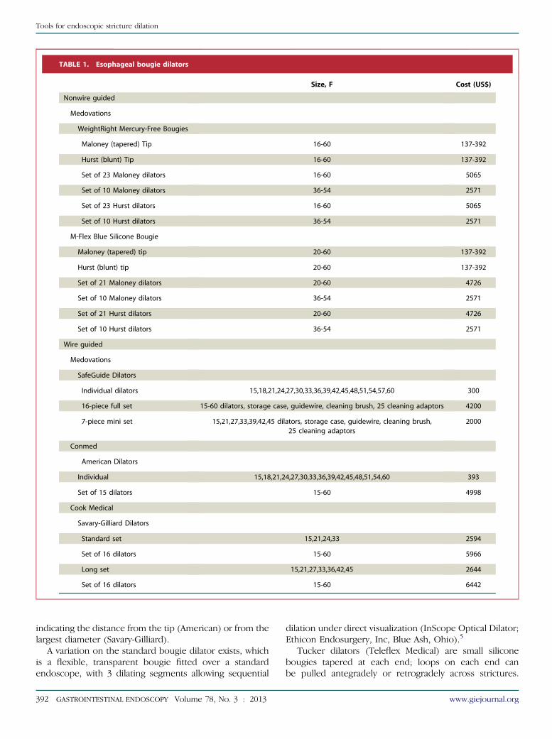
TABLE 1. Esophageal bougie dilators
Size, F Cost (US$)
Nonwire guided
Medovations
WeightRight Mercury-Free Bougies
Maloney (tapered) Tip 16-60 137-392
Hurst (blunt) Tip 16-60 137-392
Set of 23 Maloney dilators 16-60 5065
Set of 10 Maloney dilators 36-54 2571
Set of 23 Hurst dilators 16-60 5065
Set of 10 Hurst dilators 36-54 2571
M-Flex Blue Silicone Bougie
Maloney (tapered) tip 20-60 137-392
Hurst (blunt) tip 20-60 137-392
Set of 21 Maloney dilators 20-60 4726
Set of 10 Maloney dilators 36-54 2571
Set of 21 Hurst dilators 20-60 4726
Set of 10 Hurst dilators 36-54 2571
Wire guided
Medovations
SafeGuide Dilators
Individual dilators 15,18,21,24,27,30,33,36,39,42,45,48,51,54,57,60 300
16-piece full set 15-60 dilators, storage case, guidewire, cleaning brush, 25 cleaning adaptors 4200
7-piece mini set 15,21,27,33,39,42,45 dilators, storage case, guidewire, cleaning brush,25 cleaning adaptors
2000
Conmed
American Dilators
Individual 15,18,21,24,27,30,33,36,39,42,45,48,51,54,60 393
Set of 15 dilators 15-60 4998
Cook Medical
Savary-Gilliard Dilators
Standard set 15,21,24,33 2594
Set of 16 dilators 15-60 5966
Long set 15,21,27,33,36,42,45 2644
Set of 16 dilators 15-60 6442
Tools for endoscopic stricture dilation
indicating the distance from the tip (American) or from thelargest diameter (Savary-Gilliard).
A variation on the standard bougie dilator exists, whichis a flexible, transparent bougie fitted over a standardendoscope, with 3 dilating segments allowing sequential
392 GASTROINTESTINAL ENDOSCOPY Volume 78, No. 3 : 2013
dilation under direct visualization (InScope Optical Dilator;Ethicon Endosurgery, Inc, Blue Ash, Ohio).5
Tucker dilators (Teleflex Medical) are small siliconebougies tapered at each end; loops on each end canbe pulled antegradely or retrogradely across strictures.
www.giejournal.org

Tools for endoscopic stricture dilation
A gastrostomy is required for use. These may be useful inthe treatment of tortuous strictures secondary to causticsubstance ingestion.6,7 In very tight strictures in whichthere is the possibility of complete lumen occlusion, astring must be maintained across the stricture emergingfrom both the nose and gastrostomy site between dila-tions. Tucker dilators range in size from 4 to 13.3 mm(12F-40F).
Hegar’s dilators (Cooper Surgical, Trumball, Conn) arestainless steel bougies with rounded ends that can beused for dilation of benign anorectal strictures. Theyhave been used to dilate stenoses after transanal surgeryand in cases of perianal Crohn’s disease.8,9 In additionto being used by colorectal surgeons and gastroenterolo-gists, patients can also use them to self-dilate anal stric-tures. Hegar’s dilators range in size from 3 to 18 mm(9F-54F).
Balloon dilators. Radial expanding balloon dilatorsare available in an array of designs, lengths, and calibersfor various purposes in accessible strictures throughoutthe GI tract (Tables 2 and 3). They are designed to passthrough the endoscope with or without wire guidance sothat dilation can be observed. Balloon dilators are madeof low-compliance inflatable thermoplastic polymers thatallow uniform and reproducible expansion to their speci-fied diameter at maximum inflation. Some balloons have1 set diameter, whereas others allow for sequential expan-sion to multiple diameters. Dilating balloons are expandedby pressure injection of liquid (eg, water, radiopaque con-trast) by using a handheld accessory device. The hydraulicpressure of the balloon is monitored manometrically togauge radial expansion force. Inflation with radiopaquecontrast enhances fluoroscopic observation. Dilating bal-loons are marketed as single-use items.
Achalasia balloon dilators. Achalasia balloon dilatorsare large-diameter (30, 35, and 40 mm) polyethyleneballoon dilators that are disease specific for achalasia butalso have been used in other disease states.10-13 Availableachalasia balloons are listed in Table 4. All currently usedachalasia balloon dilators are wire guided and single useand do not pass through the endoscope. They arepositioned across the esophagogastric junction by usingfluoroscopic guidance with visualization aided by theradiopaque markers on the balloon. Balloon insufflationwith air is monitored manometrically.
StentsFully and partially covered self-expandable stents that
are potentially removable have been used to accomplishdilation of refractory benign strictures. This is accom-plished through the use of their radial expandable forces.A variety of covered self-expandable metal stents (SEMSs)and 1 self-expandable plastic stent (SEPSs) are availableas well as some that are biodegradable; biodegradablestents are not available in the United States at this time.A full ASGE Technology Committee review of enteral
www.giejournal.org
stents, including those used primarily to accomplish dila-tion, is available.14
Dilators for the pancreaticobiliary systemDilating catheters. Dilator catheters designed for
pancreaticobiliary use are tapered plastic cylindrical tubeswith a central channel (Table 5). They are passed over aguidewire through the accessory channel of the side-viewing duodenoscope. They are equipped with a radi-opaque band to indicate the point of maximal diameter.
Screw-tip stent retrievers (Soehendra Stent Retriever;Cook Medical) also have been used to dilate tight pancrea-ticobiliary strictures that otherwise only allow passage ofa guidewire.15-17 The wire-guided device is used to borethrough high-grade stenoses. A modified device is com-mercially available as a dilator (Soehendra rotary dilator;Cook Medical).
Balloon dilators. Balloon dilators are used in the bileduct and the pancreatic duct (Table 5). These balloons aresingle use and wire guided, come in a single-diameter size(4-10 mm diameter), and range from 2 to 4 cm in length.They are used during ERCP and inflated with dilutecontrast to facilitate visualization. If undiluted contrast isused, its increased viscosity may hinder proper inflationand deflation of the balloon. Radiopaque markers onboth ends of the balloon help to guide proper placementacross a stricture.
In addition to dilation of strictures, balloon dilation ofthe biliary and pancreatic sphincters can be performed.Sphincteroplasty with balloon dilators may be performedinstead of sphincterotomy when preferred by the endo-scopist by using smaller caliber biliary dilating balloons(eg, %10 mm). Larger caliber dilation of the sphincter(10-20 mm) may also be may be done after sphincterotomyto facilitate removal of large caliber stones. Currently, thereis only 1 balloon dilator approved by the U.S. Food andDrug Administration for this specific use (CRE; BostonScientific, Natick, Mass).
TECHNIQUE
Dilation can be performed with or without endoscopic,fluoroscopic, and/or wire guidance. Selection of differenttypes of dilators depends on operator preference and thecharacteristics of the site needing dilation. Dilator diame-ters are measured in millimeters or French. Size in millime-ters can be converted to French at a ratio of 1:3 (eg,15 mm Z 45F). Selection of the appropriate size is criticalfor safe and effective dilation. Techniques may need to bemodified for complex strictures (eg, length O2 cm, lumendiameter!12 mm, tortuous) and/or specific disease statesand locations in the GI tract.
Wire-guided bougie dilators (Savary and American dila-tors) are passed over a guidewire after initial endoscopicguidewire placement and subsequent endoscope removal.
Volume 78, No. 3 : 2013 GASTROINTESTINAL ENDOSCOPY 393

TABLE 2. Esophageal balloon dilators
Sizes (some dilators have sequential sizes), mm)Balloon
length, cmRequired
channel, mmUsable
length, cm Cost, US$
Cook Medical
Hercules balloon dilators
Nonwire guided, esophageal: 8,9,10; 10,11,12;12,13.5,15; 15,16.5,18; 18,19,20
8 2.8 180 248 per dilator
Wire-guided, esophageal/pyloric/colonic: 8,9,10;10,11,12; 12,13.5,15; 15,16.5,18; 18,19,20
5.5 2.8 240 289 per dilator
Quantum TTC balloon dilators, esophageal:6,8,10,12,14,16,18,20
8 2.8 195 189
Eclipse balloon dilators, wire-guided, esophageal:6,8,10,12, 14,16,18
8 2.8 240 262
19 8 3.2 240 262
Boston Scientific
CRE balloon dilation catheters, fixed-wire(nonwire guided), esophageal: 6,7,8; 8,9,10;10,11,12; 12,13.5,15; 15,16.5,18; 18,19,20
8 2.8 180 235
CRE balloon dilation catheters: wire guided,esophageal/pyloric/biliary: 6,7,8; 8,9,10;10,11,12; 12,13.5,15; 15,16.5,18; 18,19,20
8 2.8 180 314
CRE balloon dilation catheters: wire guidedesophageal/pyloric/colonic/biliary: 6,7,8; 8,9,10;10,11,12; 12,13.5,15; 15,16.5,18 18,19,20
5.5 2.8 240 260
Maxforce esophageal balloon dilators:6,8,10,12,14,15,16,18
6 2.8 180 180
Conmed
Eliminator PET esophageal balloon dilator:6,8,10,12,15,18,20
8 2.8 180 214
Hobbs Medical
Flex-Ez over-the-wire balloon dilators,esophageal/pyloric/colonic
8,10,12 3 2.8 240 155
14,16,18,20 3 3.7 240 165
6,8,10 4 2.8 240 155
16,20 4 3.7 240 165
8,10,12,14 8 3.7 240 155
16,18,20 8 3.7 240 165
Stylet wire balloon dilators, esophageal/pyloric/colonic
12,14 8 2.8 240 155
16,18,20 8 2.8 240 165
8,10,12,14 5.5 2.8 240 155
16,18,20 5.5 2.8 240 165
8,10,12,14 3 2.8 240 155
16,18,20 3 2.8 240 165
394 GASTROINTESTINAL ENDOSCOPY Volume 78, No. 3 : 2013 www.giejournal.org
Tools for endoscopic stricture dilation

TABLE 3. Pyloric/colonic balloon dilators*
Size, mm Balloon length, cm Required channel, mm Usable length, cm Cost, US$
Cook Medical
Quantum TTC balloon dilators
Pyloric: 6,8,10, 12,14,18,20 5.5 2.8 195 189
Colonic: 6,8,10, 12,14,16,20 5.5 2.8 240 189
Eclipse TTC balloon dilators, wire guided
Pyloric/colonic: 6,8,10,12,14,16,18 5.5 2.8 240 262
20 5.5 3.7 240 262
Conmed
Eliminator PET pyloric/colonic balloon dilator
6,8,10,12,15,18, 20 4 2.8 230 214
*See also esophageal balloon dilators in Table 2 also used for pyloric/colonic dilation.
TABLE 4. Achalasia balloon dilators
Description Balloon length, cm Usable length, cm Cost, US$
Cook Medical
Inflated balloon diameter 30 mm, 16F catheter 8 75 422
Inflated balloon diameter 35 mm, 16F catheter 8 75 422
Boston Scientific
Rigiflex balloon, inflated balloon diameter 30, 35, 40 mm; 14F catheter 10 90 650
Hobbs Medical
Inflated balloon diameter 30, 35, 40 mm; 16F catheter 10 100 335
Tools for endoscopic stricture dilation
Nonwire-guided bougies (Hurst and Maloney) are passedblindly into the esophagus. These may have a higher rateof perforation in the presence of large hiatal hernias orcomplex strictures.
Balloon dilators in the GI tract may be passed with orwithout wire guidance. They are expanded with liquid(water and/or contrast) by using a handheld inflationdevice. Wire-guided through-the-scope (TTS) balloon dila-tors are passed over a guidewire that has been placedthrough the endoscope accessory channel. The balloon ispositioned across the stenosis and inflated under directendoscopic visualization. Nonwire-guided balloons areused in a similar fashion but are passed across the stenosisby using endoscopic visualization only. The optimal dura-tion of balloon inflation is not known.
Initial selection of a specific size of dilator is based onan estimation of the diameter of the stenosis. The ruleof 3 has been used when deciding how much to dilate a
www.giejournal.org
stricture with a bougie dilator in 1 session. This rule statesthat after moderate resistance is encountered, no morethan 3 dilators of progressively increasing diameter shouldbe passed in that session.18 However, no studies havedemonstrated that this technique improves safety andefficacy.19 When dilating a symptomatic Schatzki ring ofthe esophagus, passage of a single large-diameter dilator(eg, 16-20 mm) has been advocated to allow for disruptionof the ring.20,21
Modifications to standard dilation are sometimes used,especially in the case of refractory benign strictures. Theseinclude injection of steroids immediately before or afterdilation.22-24 Disruption of strictures (eg, esophagealwebs, Schatzki rings) with biopsy forceps or needle-knifeelectrocautery, either as the sole treatment or in conjunc-tion with dilation, has been successfully demonstrated.25,26
Electroincision therapy has also been used in the treatmentof refractory benign esophageal strictures.27,28
Volume 78, No. 3 : 2013 GASTROINTESTINAL ENDOSCOPY 395

TABLE 5. Biliary and pancreatic dilators*
Dilation catheters
Description Catheter size, FTapered tip length, cm/tip
catheter size, FUsable
length, cm Cost, US$
Cook Medical
Soehendra biliary dilation catheters(wire guided 0.035-inch)
6 3/4 200 82
7 3/4 200 82
8.5 3/5 200 82
9 3/6 200 82
10 3/6 200 82
11.5 3/7 200 82
Cotton dilation catheters (used to dilatethe papilla or biliary strictures); wireguided 0.035-inch
8.5 4/7-5 200 82
10 4/7-5 200 82
7 2.5/7-4.5 200 82
7 4.5/7-4.5 200 82
Geenen graduated dilation catheter(for pancreatic strictures); wire guided0.025-inch
7 2/5-4 200 82
Biliary balloon dilators
Size, mmBalloon
length, cmRequired
channel, mmUsable
length, cm Cost, US$
Cook Medical
Quantum TTC biliary balloon dilators
4,6,8,10 3 2.8 180 189
Fusion Titan biliary balloon dilators:wire-guided
4,6,8,10 4 3.2 190 372
Boston Scientific
Hurricane Rx biliary balloon dilators
4,6,8,10 2 3.2 180 279
4,6,8,10 4 3.2 180 279
Olympus
MaxPass biliary balloon dilators
4,6 2 2.8 180 382
4,6 4 2.8 180 382
8 3 2.8 180 382
396 GASTROINTESTINAL ENDOSCOPY Volume 78, No. 3 : 2013 www.giejournal.org
Tools for endoscopic stricture dilation

TABLE 5. Continued
Biliary balloon dilators
Size, mmBalloon
length, cmRequired
channel, mmUsable
length, cm Cost, US$
Conmed
Eliminator PET biliary balloon dilators
4,6 2 2.8 180 272
8 3 2.8 180 272
Hobbs Medical
Biliary balloon dilators
4,6 2 2.8 180 155
8 3 2.8 180 155
*See also Boston Scientific CRE Balloon Dilators: Wire-Guided Balloon Dilators used for biliary dilation in Table 2.
Tools for endoscopic stricture dilation
OUTCOMES AND COMPARATIVE DATA
EsophagusBenign esophageal strictures. The majority of
benign esophageal strictures are caused by long-standingGERD.29 Other stricture etiologies include webs, Schatzkirings, anastomotic strictures, and inflammatory-type stric-tures caused by eosinophilic esophagitis, radiation expo-sure, and caustic substance ingestion. Multiple studieshave shown that benign peptic strictures may be dilatedwith bougie or balloon dilators with technical successand decrease in dysphagia in the majority of cases.30-33
Studies of use of balloon dilators in patients with a varietyof benign esophageal strictures have demonstrated an excel-lent or good symptomatic response in 7% to 100% patientsimmediately after dilation.3,34,35 However, a sustainedresponse may be difficult to achieve in nonpeptic strictures,with symptom recurrence often occurring within 1 year ofinitial dilation.36 The degree and duration of the effect andthe need for repeat dilation are often dependent on thestricture etiology and the length and degree of stenosis.Durable success appears greatest when a luminal diameterof larger than 12 mm is achieved.37 Complex strictures aredescribed as being long (O2 cm), tortuous, or having asmall lumen diameter that precludes passage of a standardendoscope.38 These have been shown to have a poorresponse to dilation compared with simple strictures (ie,shorter length, larger caliber predilation diameter).36
Two randomized, controlled trials compare bougie dila-tors with TTS balloons for benign strictures of the esoph-agus. These trials, including a total of 379 patients, foundno differences in efficacy at dysphagia relief or safety at1 year.30,32 A randomized prospective study of 26 patientscompared a single dilation with 52F Maloney dilator versusendoscopic rupture of a Schatzki ring by using biopsy
www.giejournal.org
forceps. There was similar improvement in dysphagiascores at 3 and 12 months in both groups (both groupswith 91% improvement at 3 months and 84% to 85% at12 months). There were no significant differences in H2
blocker or PPI use in either arm.25
Two randomized trials compared electrosurgical incisionand bougie dilation (52-54F Maloney dilators) of Schatzki’srings. One study of 11 patients showed no difference interms of dysphagia improvement or recurrence at 1 year.39
A larger trial of 50 patients showed that the electrosurgicalincision group had a longer symptom free time comparedwith the bougie group (7.99 vs 5.86 months; P Z .03) andalso had a greater improvement in dysphagia scores at1 month (P Z .05).26 Another randomized study of 62patients with anastomotic strictures after esophagectomyshowed no difference in clinical success or complicationrates between electrocautery incision and bougie dilation.40
Dilation has been shown to be effective in controllingdysphagia caused by eosinophilic esophagitis in uncon-trolled case series with or without concomitant medicaltherapy. Type of dilator used in the studies was not stan-dardized, but the majority had long-term relief (up to2 years) after dilation.41-44
There are no randomized trials comparing the use ofstents with other methods of dilation for the treatmentof refractory benign esophageal strictures. Nonrandomizedstudies examining the use of SEPS in the treatment ofrefractory benign esophageal strictures have shown highcomplication rates including migration (22%-81%), chestpain (11%), bleeding (8%), and perforation (5.5%).45-47
Additionally, only 6% to 40% were dysphagia free afterremoval. Although not approved by the U.S. Food andDrug Administration for removability in benign esophagealstrictures, the use of fully covered SEMSs use has been re-ported. Two recent small case series included 24 patients
Volume 78, No. 3 : 2013 GASTROINTESTINAL ENDOSCOPY 397

Tools for endoscopic stricture dilation
with benign esophageal strictures and showed low rates oflong-term response (!20%) and high rates of stent migra-tion (29%-34%).48,49 A recent small prospective study of 30patients examined 3 different types of stents (fully coveredSEMSs, SEPSs, biodegradable stents) in the treatment ofrefractory benign esophageal strictures. There was long-term symptom improvement at 8 months in 30% to 40%of patients with fully covered SEMSs and biodegradablestents but only a 10% improvement with SEPSs.50 Tissueingrowth may occur at the ends of the stent, potentiallyaffecting its removability.51
Achalasia. Pneumatic balloon dilation of the loweresophageal sphincter with a large diameter (O30 mm)cylindrical, wire-guided balloon has long been the mainstayof endoscopic therapy for achalasia. A brief 6-seconddilation, sufficient to obliterate the balloon’s waist, wasshown to be as effective as the standard 60-seconddilation.52 The first prospective, randomized study tocompare open surgical myotomy with pneumatic balloondilation showed that surgery was significantly superiorat 5-year follow-up, with excellent results in 95% of surgi-cally treated patients compared with good results in only65% of patients undergoing balloon dilation.53 A morerecent larger randomized, controlled trial comparingpneumatic dilation with laparoscopic myotomy showedno significant difference in clinical success with eitherapproach at 2-year follow-up (86% vs 90%; P Z .46).54 Ameta-analysis of 36 studies published between 2001 and2011 found that laparoscopic myotomy had a more durablelong-term response (generally defined as good or excellentsymptom control) at 10 years compared with pneumaticdilation (79.6% vs 47.9%).55
Injections of botulin toxin into the lower esophagealsphincter were initially described in 1994 as an alternativeendoscopic approach to achalasia.56 Comparative trials ofbotulinum toxin injection and pneumatic balloon dilationfor treatment of achalasia have shown equivalent earlysuccess rates but shorter duration of efficacy in thebotulinum injection groups.57-59 There are no data compar-ing the success and safety of the new surgical technique ofperoral endoscopic myotomy with other endoscopic andsurgical achalasia treatments.
Stomach and small bowelBenign strictures of the stomach and small bowel (eg,
pyloric stenosis, nonsteroidal anti-inflammatory drug–induced strictures, surgical anastomoses, inflammatorybowel disease) may be amenable to dilation for symptomcontrol. Because of their location, these are typicallymanaged by using TTS balloon dilators.
Multiple studies describe dilation of the pylorus to treatgastric outlet obstruction caused by benign conditions (eg,peptic ulcer disease), with short-term success rates ofapproximately 70% to 80%.60-64 Perforation rates in somestudies were fairly high (4%-7%),60,63,65 and long-termsuccess was poor, with 30% to 50% ultimately requiring
398 GASTROINTESTINAL ENDOSCOPY Volume 78, No. 3 : 2013
surgery.60,62 A more recent smaller study found long-term success was achieved in 100% at follow-up of 43months; most patients were maintained on antisecretorytherapy in this study.65 Gastric outlet obstruction relatedto chronic pancreatitis responds poorly to endoscopictherapy.66,67 Several uncontrolled studies evaluating bal-loon dilation in gastric bypass patients with gastroentericanastomotic strictures have demonstrated high short- andlong-term success rates. Dilation appears safe in thissetting, with only a single study reporting a higher (4.9%)perforation rate.68-72 Wire-guided TTS balloons are typicallyused for anastomotic strictures after gastric bypass surgery,but bougie dilators were used successfully in a singlestudy.73 Several uncontrolled case series suggest thatballoon dilation is an efficacious treatment of upper andlower GI tract fibrotic strictures in patients with Crohn’sdisease, allowing long-term avoidance of surgery for thestricture in 56% to 75% of dilated patients.74-78 There area few reports of successful dilation of Crohn’s stricturesof the small bowel by using temporary SEMS placement.However, high rates of stent migration and other complica-tions were noted.79,80 Distal small-bowel strictures ofvarious etiologies, inaccessible to standard endoscopes,have been accessed by using double-balloon enteroscopeswith successful stricture dilation with TTS balloons.81-84
Pancreaticobiliary systemBenign biliary strictures associated with primary scleros-
ing cholangitis (PSC), postoperative bile duct injury, andduct-to-duct anastomoses after orthotopic liver transplan-tation are amenable to endoscopic dilation therapy.85-87
Except for strictures associated with PSC, dilation alone islargely ineffective and should be accompanied by stentplacement. No additional benefit from stenting afterballoon dilation of dominant strictures in PSC was seenin 2 retrospective studies that included 81 patients withlong-term follow-up of more than a decade.88,89
Multiple plastic stents have been used to successfullydilate and treat benign biliary strictures and have beenshown to be superior to single stents for stricture resolu-tion.85,90,91 Recent uncontrolled series demonstrated suc-cessful use of temporary fully covered SEMSs in thetreatment of benign biliary strictures of various causes,with resolution rates ranging from 83% to 90%.92,93 Studiesof benign biliary stricture caused by anastomotic strictureafter liver transplantation have shown mixed results, withlong-term resolution seen in 53% to 95% of patients andmigration in as many as 46%.94-97 One recently publishedprospective study randomized 31 patients with postopera-tive biliary strictures to treatment with either partiallycovered SEMSs or multiple plastic stents with long-termfollow-up of more than 5 years. The SEMS group hadhigher long-term patency rates (81% vs 71%; P Z .02)and similar complication rates (40% vs 25%; P Z .37).98
Biliary strictures related to chronic pancreatitis respondmore poorly to endoscopic therapy, with long-term
www.giejournal.org

Tools for endoscopic stricture dilation
resolution after plastic stenting in 44% to 60%.91 Somewhatbetter resolution of strictures caused by chronicpancreatitis was seen in 2 studies with use of fully andpartially covered SEMS, with a 65% to 90% strictureresolution rate.93,99 However, follow-up was short (3.8-6months) and complications were noted in 20% to 30%.
Sphincter dilation. Dilation of the biliary sphincterhas been investigated as a potential alternative to sphinc-terotomy to facilitate biliary stone removal, but thistechnique has been associated with higher rates of pan-creatitis.100-102 Two meta-analyses comparing endoscopicsphincterotomy with endoscopic papillary balloon dilation(!10 mm) showed similar outcomes with regard to overallstone removal but a higher rate of post-ERCP pancreatitiswith papillary balloon dilation (7.4% vs 4.3%).103,104 A recentmeta-analysis of 7 randomized, controlled trials (790patients) comparing endoscopic papillary large balloon dila-tion alone with biliary sphincterotomy alone found similaroverall bile duct clearance rates (97% vs 96%; P Z .54).105
Balloon dilation was associated with decreased use ofmechanical lithotripsy and less hemorrhage, but otherwiseno difference in other complication rates (eg, post-ERCPpancreatitis, perforation, cholangitis).
Large-diameter (O10 mm) papillary balloon dilationafter sphincterotomy has been shown to be effective inthe removal of large bile duct stones without an increasedrate of pancreatitis.106 A technical review of 21 publishedstudies of 1292 patients undergoing sphincterotomy withlarge-diameter papillary balloon dilation showed a 91%initial success rate and 98% final success rate.107 Therewere low rates of reported complications includingbleeding (2.8%) and post-ERCP pancreatitis (1.2%). Dilationof pancreatic duct strictures or sphincters is primarily used inconcert with stone removal or stent placement.108-110
ColonDilation of benign colorectal strictures of varying etiol-
ogies (eg, anastomotic, nonsteroidal anti-inflammatorydrug–induced, diverticulitis, radiation, and inflammatorybowel disease) by using balloon or bougie-type dilatorshas been demonstrated to be effective in multiple uncon-trolled series.111-118
A prospective trial randomized 30 patients with symp-tomatic benign postoperative anastomotic colorectal stric-tures to dilation with either an 18-mm TTS balloondilator or an over-the-wire 35-mm pneumatic balloondilator designed for achalasia. Dilation was successful inall patients, and no complications occurred in either, butthe over-the-wire group required fewer sessions (1.6 vs2.6, P Z .009) and had a longer duration of response(560.8 days vs 245.2 days, P Z .016).119
SAFETY
All dilation is intended to displace tissue. Therefore,some disruption of mural elements, including mucosal
www.giejournal.org
tears and minor bleeding, is expected. Complications ofendoscopic stricture dilation include chest pain, clinicallysignificant bleeding, bacteremia, and perforation.120-122
Types of strictures and the degree of dilation under-taken may influence the rate of major complications.Perforation, estimated to occur in 0.4% of cases, isthe most clinically significant complication and occursbecause of transmural disruption or the creation of afalse track.123 Transmural disruption may occur whenaxial or radial forces exceed the structural integritylimits of the wall. A false track occurs when the dilatordirectly penetrates the wall. Guidewire use may reducethis risk of perforation.124 There are insufficient data tosubstantiate a difference in perforation rates with bougieversus balloon dilators. A retrospective analysis reportedan increased perforation rate associated with blindpassage of Maloney dilators versus Savary-Gilliard andballoon dilators in patients with complex esophageal stric-tures.123 It has also been demonstrated that endoscopicexperience influences perforation rates with esophagealdilation, with a 4 times higher rate noted when fewerthan 500 diagnostic upper endoscopies had beenperformed.125 Although endoscopic dilation has beenassociated with bacteremia,122,126 it is rarely clinically signif-icant, and current ASGE guidelines do not recommendroutine antibiotic prophylaxis at the time of endoscopicdilation.127
Regarding achalasia dilation, pooled data indicate an ap-proximately 2% to 4% cumulative perforation rate whenusing graded balloon dilation.128 Other complicationsassociated with achalasia dilation include prolonged painand intramural hematomas.129
Multiple studies have demonstrated the safety of largeballoon dilation after sphincterotomy for difficult biliarystones.107 No increased rates of pancreatitis, perforation,cholangitis, or bleeding rates were shown.
Small case series have described the occurrence of largetears, chest pain, and a high rate of perforations afterdilation in patients with eosinophilic esophagitis.130,131
Larger retrospective studies have not found an increasedrisk of perforation. A recent retrospective review of 54patients with eosinophilic esophagitis who underwentendoscopic dilation with Maloney, Savary-Gilliard, andTTS balloon dilators over a 5-year period did not reportany major complications.132 However, other studiesreporting safety data for balloon versus bougie dilators inthis population have shown disparate results, with somereporting higher complication rates with balloons andothers with bougies.41-43 Prospective data are not availableat this time.
Complications with stent placement for stricture man-agement include perforation, hemorrhage, and airway com-pression when placed in the proximal esophagus.133-135
The risk of perforation after placement of colonic SEMSsfor malignant obstruction is increased with predeploymentdilation.136-138
Volume 78, No. 3 : 2013 GASTROINTESTINAL ENDOSCOPY 399

TABLE 6. CPT codes and reimbursement for endoscopic dilation procedures, CMS national average unadjusted 2012
CPT* Description APCHospital outpatient
payment, US$
Ambulatory surgerycenter payment
(2012 averages 56%of HOPD), US$
Professional fee(facility), US$
43220 Esophageal endoscopy, dilation balloon 0419 887 497 129
43226 Esophageal endoscopy, dilation guidewire 0419 887 497 144
43248 EGD, dilation guidewire 0141 592 331 192
43249 EGD, balloon dilation of the esophagus 0419 887 497 177
43458 Dilation of the esophagus with a balloonO30 mm for achalasia
0419 887 497 184
43245 EGD with dilation of gastric outletobstruction, any method
0419 887 497 191
45340 Flexible sigmoidoscopy, balloondilation stricture
0147 773 432 118
45386 Colonoscopy, balloon dilation 0143 656 367 270
45303 Proctosigmoidoscopy, dilation any method 0147 773 432 91
43456 Dilation of the esophagus, retrograde 0140 460 258 158
43271 ERCP, balloon dilation ducts 0151 1729 968 434
43450 Esophagus dilation, no endoscopy (bougie) 0140 461 258 91 (office, $161)
APC, Ambulatory payment classification; HOPD, hospital outpatient department.*CPT codes, descriptions, and other data are copyright 2012 American Medical Association (AMA). All rights reserved.
Tools for endoscopic stricture dilation
FINANCIAL CONSIDERATIONS
The list prices of the available dilators and stents foruse in GI endoscopy are detailed in Tables 1 through 5.Reusable dilators have potential cost advantages oversingle-use devices, even when accounting for reprocessingcosts. Costs of single-use and reusable guidewires, contrastagents, manometry gauges, inflation devices, and fluoros-copy are also cost considerations. Costs of stents farexceed those for dilating catheters and balloons.
Codes for dilation during endoscopy and reimburse-ment rates are listed in Table 6. For services performedafter January 1, 2003, Medicare reimbursement no longerincludes pass-through codes for dilators or ancillaryequipment.
AREAS FOR FUTURE RESEARCH
Published studies have demonstrated the safety andefficacy of the various types of dilating catheters andballoons in the treatment of strictures throughout the GItract. The optimal treatment of refractory benign GI tractstrictures needs to be clarified. Prospective trials of dilationtherapy versus other treatments in eosinophilic esopaghitiswould be helpful. Previously limited to use in malignantstrictures, new variations of SEMSs are being used in
400 GASTROINTESTINAL ENDOSCOPY Volume 78, No. 3 : 2013
refractory benign strictures in the esophagus, colon, andvarious surgical anastomoses (including after Roux-en-Ygastric bypass surgery). Prospective, randomized studiesshould be performed comparing dilation with SEMS place-ment of various types to determine safety, efficacy, ease ofuse, and cost benefits.
CONCLUSIONS
Fixed-diameter push-type dilators and radial expansionballoon dilators are safe and effective for endoscopic man-agement of benign and malignant strictures throughoutthe digestive tract. Both types have comparable efficacyand safety, although wire-guided balloon dilators are usu-ally required for locations other than the esophagus. Lowercosts are associated with the multiuse, fixed-diameter,push-type dilators. Fully covered SEMSs are increasinglybeing used for the management of refractory benign GIstrictures. Further study is needed to determine the safety,efficacy, and cost advantages of stents for this expandedindication.
DISCLOSURE
The authors disclosed no financial relationships rele-vant to this publication.
www.giejournal.org

Tools for endoscopic stricture dilation
Abbreviations: PSC, primary sclerosing cholangitis; SEMS, self-expandable metal stent; SEPS, self-expandable plastic stent; TTS,through-the-scope.
REFERENCES
1. Abele JE. The physics of esophageal dilatation. Hepatogastro-enterology 1992;39:486-9.
2. Patterson DJ, Graham DY, Smith JL, et al. Natural history of benignesophageal stricture treated by dilatation. Gastroenterol 1983;85:346-50.
3. Cox JG, Winter RK, Maslin SC, et al. Balloon or bougie for dilatation ofbenign esophageal stricture? Dig Dis Sci 1994;39:776-81.
4. Wesdorf IC, Bartelsman JF, den Hartog Jager FC, et al. Results of con-servative treatment of benign esophageal strictures: a follow-upstudy in 100 patients. Gastroenterology 1982;82:487-93.
5. Jones MP, Bratten JR, McClave SA. The Optical Dilator: a clear, over-the-scope bougie with sequential dilating segments. GastrointestEndosc 2006;63:840-5.
6. Saleem MM. Acquired oesophageal strictures in children: emphasison the use of string-guided dilatations. Singapore Med J 2009;50:82-6.
7. Cicatricial stenosis of the esophagus with particular reference totreatment by continuous string retrograde bouginage with theauthor’s bougie. Ann Otol Rhinol Laryngol 1924;33:1180-223.
8. Barker JA, Hill J. Incidence, treatment and outcome of rectal stenosisfollowing transanal endoscopic microsurgery. Tech Coloproctol2011;15:281-4.
9. Galandiuk S, Kimberling J, Al-Mishlab TG, et al. Perianal Crohn disease.Ann Surg 2005;241:796-802.
10. Vaezi MF, Richter JE. Current therapies for achalasia: comparison andefficacy. J Clin Gastroenterol 1998;27:21-35.
11. Vaezi MF, Richter JE. Practice guidelines: diagnosis and managementof achalasia. Am J Gastroenterol 1999;12:3406-12.
12. Virgilio C, Cosentino S, Favara C, et al. Endoscopic treatment of post-operative colonic strictures using an achalasia dilator: short-term andlong-term results. Endosc 1995;27:219-22.
13. Tuset JA, Luján M, Huguet JM, et al. Endoscopic pneumatic balloondilation in primary achalasia: predictive factors, complications, andlong-term follow-up. Dis Esophagus 2009;22:74-9.
14. Varadarajulu S, Banerjee S, Barth B, ASGE Technology Committee.Enteral stents. Gastrointest Endosc 2011;74:455-64.
15. Faigel DO, Ginsberg GG, Kochman ML. Innovative use of the Soehen-dra stent retriever for biliary stricture recanalization [letter]. Gastro-intest Endosc 1996;44:635.
16. Ziebert JJ, Disario JA. Dilation of refractory pancreatic duct strictures:the turn of the screw. Gastrointest Endosc 1999;49:632-5.
17. Parasher VK. A novel approach to facilitate dilation of complex non-traversable esophageal strictures by efficient wire exchange using astent pusher. Gastrointest Endosc 2000;51:730-1.
18. Langdon DF. The rule of three in esophageal dilation. GastrointestEndosc 1997;45:111.
19. Spechler SJ. AGA technical review on treatment of patients withdysphagia caused by benign disorders of the distal esophagus.Gastroenterology 1999;117:233-54.
20. Jalil S, Castell DO. Schatzki’s ring: a benign cause of dysphagia inadults. J Clin Gastroenterol 2002;35:295-8.
21. Egan J, Baron T, Adler D, ASGE Standards of Practice Committee.Esophageal dilation. Gastrointest Endosc 2006;63:755-60.
22. Ramage JI Jr, Rumalla A, Baron TH, et al. Prospective, randomizeddouble-blind, placebo-controlled trial of endoscopic steroid injectiontherapy for recalcitrant esophageal peptic strictures. Am J Gastro-enterol 2005;100:2419-25.
23. Altintas E, Kacar S, Tunc B, et al. Intralesional steroid injection inbenign esophageal strictures resistant to bougie dilation.J Gastroenterol Hepatol 2004;19:1388-91.
www.giejournal.org
24. Kochhar R, Makharia GK. Usefulness of intralesional triamcinolonein treatment of benign esophageal strictures. Gastrointest Endosc2002;56:829-34.
25. Chotiprasidhi P, Minocha A. Effectiveness of single dilation withMaloney dilator versus endoscopic rupture of Schatzki’s ring usingbiopsy forceps. Dig Dis Sci 2000;45:281-4.
26. Wills JC, Hilden K, Disario JA, et al. A randomized, prospective trial ofelectrosurgical incision followed by rabeprazole versus bougie dila-tion followed by rabeprazole of symptomatic esophageal (Schatzki’s)rings. Gastrointest Endosc 2008;67:808-13.
27. Hordijk ML, Siersema PD, Tilanus HW, et al. Electrocautery therapy forrefractory anastomotic strictures of the esophagus. GastrointestEndosc 2006;63:157-63.
28. Canhoto M, Arroja B, Silva F, et al. Needle-knife incisional treatmentof refractory esophagic caustic stenosis. Endosc 2011;43(Suppl 2):E386.
29. Kuo WH, Kalloo AN. Reflux strictures of the esophagus. GastrointestEndosc Clin N Am 1998;8:273-81.
30. Saeed ZA, Winchester CB, Ferro PS, et al. Prospective randomizedcomparison of poly-vinyl bougies and through the scope balloonsfor dilation of peptic strictures of the esophagus. Gastrointest Endosc1995;41:189-95.
31. Marshall JB, Afridi SA, King PD, et al. Esophageal dilation withpolyvinyl (American) dilators over a marked guidewire: practice andsafety at one center over a 5-yr period. Am J Gastroenterol1996;91:1503.
32. Scolapio JS, Pasha TM, Gostout CJ, et al. A randomized prospectivestudy comparing rigid to balloon dilators for benign esophageal stric-tures and rings. Gastrointest Endosc 1999;50:13-7.
33. Pereira-lima JC, Ramires RP, Zamin I Jr, et al. Endoscopic dilation ofbenign esophageal strictures: report on 1043 procedures. Am J Gas-troenterol 1999;94:1497-501.
34. Ikeya T, Ohwada S, Ogawa T, et al. Endoscopic balloon dilation forbenign esophageal anastomotic stricture: factors influencing its effec-tiveness. Hepatogastroenterology 1999;46:959-66.
35. Honkoop P, Siersema PD, Tilanus HW, et al. Benign anastomoticstrictures after transhiatal esophagectomy and cervical esophagogas-trostomy: risk factors and management. J Thorac Cardiovasc Surg1996;111:1141-8.
36. Chiu YC, Hsu CC, Chiu KW, et al. Factors influencing clinical applica-tions of endoscopic balloon dilation for benign esophageal strictures.Endoscopy 2004;36:595-600.
37. Saeed ZA, Ramirez FC, Hepps KS, et al. An objective end point for dila-tion improves outcome of peptic esophageal strictures: a prospectiverandomized trial. Gastrointest Endosc 1997;45:354-9.
38. Lew RJ, Kochman ML. A review of endoscopic methods of esophagealdilation. J Clin Gastroenterol 2002;35:117-26.
39. Ibrahim A, Cole RA, Qureshi WA, et al. Schatzki’s rung: to cut or breakan unresolved problem. Dig Dis Sci 2004;49:379-83.
40. Hordijk ML, van Hooft JE, Hansen BE, et al. A randomized comparisonof electrocautery incision with Savary bougienage for relief of anasto-motic gastroesophageal strictures. Gastrointest Endosc 2009;70:849-55.
41. Schoepfer A, Gonsalves N, Bussmaan N, et al. Esophageal dilation ineosinophilic esophagitis: effectiveness, safety, and impact on the un-derlying inflammation. Am J Gastroenterol 2010;105:1062-70.
42. Dellon E, Gibbs W, Rubinas T, et al. Esophageal dilation in eosinophilicesophagitis: safety and predictors of clinical response and complica-tions. Gastrointest Endosc 2010;71:706-12.
43. Jung K, Gundersen N, Kopacova J, et al. Occurrence of and risk factorsfor complications after endoscopic dilation in eosinophilic esophagi-tis. Gastrointest Endosc 2011;73:15-21.
44. Bohm M, Richter J, Kelsen S, et al. Esophageal dilation: simple andeffective treatment for adults with eosinophilic esophagitis andesophageal rings and narrowing. Dis Esoph 2010;23:377-85.
45. Holm AN, de la Mora Levy JG, Gostout CJ, et al. Self-expanding plasticstents in treatment of benign esophageal conditions. GastrointestEndosc 2008;67:20-5.
Volume 78, No. 3 : 2013 GASTROINTESTINAL ENDOSCOPY 401

Tools for endoscopic stricture dilation
46. Dua KS, Vleggaar FP, Santharam R, et al. Removable self-expandingplastic esophageal stent as a continuous, non-permanent dilator intreating refractory benign esophageal strictures: a prospective two-center study. Am J Gastroenterol 2008;103:2988-94.
47. Oh YS, Kochman ML, Ahmad NA, et al. Clinical outcomes after self-expanding plastic stent placement for refractory benign esophagealstrictures. Dig Dis Sci 2010;55:1344-8.
48. Eloubeidi MA, Talreja JP, Lopes TL, et al. Success and complicationsassociated with placement of fully covered removable self-expandable metal stents for benign esophageal diseases. Gastroint-est Endosc 2001;73:673-81.
49. Wagh MS, Forsmark CE, Chauhan S, et al. Efficacy and safety of a fullycovered esophageal stent: a prospective study. Gastrointest Endosc2012;75:678-82.
50. Canena JM, Liberato MJ, Rio-Tinto RA, et al. A comparison of thetemporary placement of 3 different self-expanding stents for thetreatment of refractory benign esophageal strictures: a prospectivemulti-centre study. BMC Gastroenterol 2012;12:70.
51. Siersema P, Wijkerslooth L. Dilation of refractory benign esophagealstricture. Gastrointest Endosc 2009;1000-12.
52. Khan AA, Shah SW, Alam A, et al. Pneumatic balloon dilation in acha-lasia: a prospective comparison of balloon distention time. Am JGastroenterol 1998;93:1064-7.
53. Csendes A, Braghetto I, Henriquez A, et al. Late results of a prospec-tive randomized study comparing forceful dilatation and oesophago-myotomy in patients with achalasia. Gut 1989;30:299-304.
54. Boeckxstaens G, Annese V, des Varanes S, et al. Pneumatic dilationversus Laparoscopic Heller’s myotomy for idiopathic achalasia.N Engl J Med 2011;364:1807-16.
55. Weber CE, Davis CS, Kramer HJ, et al. Medium and long-term out-comes after pneumatic dilation or laparoscopic Heller myotomy forachalasia: a meta-analysis. Surg Laparosc Endosc Percutan Tech2012;22:289-96.
56. Pasricha P, Ravich W, Hendrix T, et al. Treatment of achalasia withintrasphincteric injection of botulinum toxin. A pilot trial. Ann InternMed 1994;121:590-1.
57. Ghoshal UC, Chaudhuri S, Pal BB, et al. Randomized controlled trial ofintrasphincteric botulinum toxin A injection versus balloon dilatationin treatment of achalasia cardia. Dis Esophagus 2001;14:227-31.
58. Allescher HD, Storr M, Seige M, et al. Treatment of achalasia: botuli-num toxin injection vs. pneumatic balloon dilation. A prospectivestudy with long-term follow-up. Endoscopy 2001;33:1007-17.
59. Mikaeli J, Fazel A, Montazeri G, et al. Randomized controlled trialcomparing botulinum toxin injection to pneumatic dilation for thetreatment of achalasia. Aliment Pharmacol Ther 2001;15:1389-96.
60. Kuwada SK, Alexander GL. Long-term outcome of endoscopic dilationof nonmalignant pyloric stenosis. Gastrointest Endosc 1995;41:15-7.
61. Kozarek RA. Endotherapy for gastric outlet obstruction. GastrointestEndosc 1996;43:173-4.
62. Lau J, Chung S, Sung J, et al. Through-the-scope balloon dilationfor pyloric stenosis: long-term results. Gastrointest Endosc 1996;43:98-101.
63. Solt J, Bajor J, Szabo M, et al. Long-term results of balloon catheterdilation for benign gastric outlet stenosis. Endoscopy 2003;35:490-5.
64. DiSario JA, Fennerty MB, Tietze CC, et al. Endoscopic balloon dilationfor ulcer-induced gastric outlet obstruction. Am J Gastroenterol1994;89:868-71.
65. Cherian P, Cherian S, Singh P. Long-term follow-up of patients withgastric outlet obstruction related to peptic ulcer disease treatedwith endoscopic balloon dilatation and drug therapy. GastrointestEndosc 2007;266:491-7.
66. Kochhar R, Sethy PK, Nagi B, Wig JD. Endoscopic balloon dilatation ofbenign gastric outlet obstruction. J Gastroenterol Hepatol 2004;19:418-22.
67. Rana S, Bhasin DK, Chandail VS, et al. Endoscopic balloon dilatationwithout fluoroscopy for treating gastric outlet obstruction becauseof benign etiologies. Surg Endosc 2011;25:1579-84.
402 GASTROINTESTINAL ENDOSCOPY Volume 78, No. 3 : 2013
68. Ukleja A, Afonso BB, Pimentel R, et al. Outcome of endoscopicballoon dilation of strictures after laparoscopic gastric bypass. SurgEndosc 2008;22:1746-50.
69. Ahmad J, Martin J, Ikramuddin S, et al. Endoscopic balloon dilation ofgastroenteric anastomotic stricture after laparoscopic gastric bypass.Endoscopy 2003;35:725-8.
70. Carrodeguas L, Szomstein S, Zundel N, et al. Gastrojejunal anasto-motic strictures following laparoscopic Roux-en-Y gastric bypass sur-gery: analysis of 1291 patients. Surg Obes Relat Dis 2006;2:92-7.
71. Mathew A, Veliuona MA, DePalma FJ, et al. Gastrojejunal strictureafter gastric bypass and efficacy of endoscopic intervention. DigDis Sci 2009;54:1971-8.
72. Peifer KJ, Shiels AJ, Azar R, et al. Successful endoscopic managementof gastrojejunal anastomotic strictures after Roux-en-Y gastric bypass.Gastrointest Endosc 2007;66:248-52.
73. Escalona A, Devaud N, Boza C, et al. Gastrojejunal anastomotic stric-ture after Roux-en-Y gastric bypass: ambulatory management withthe Savary-Gilliard dilator. Surg Endosc 2007;21:765-8.
74. Gustavsson A, Magnuson A, Blomberg B, et al. Endoscopic dilation isan efficacious and safe treatment of intestinal strictures in Crohn’sdisease. Aliment Pharmacol Ther 2012;36:151-8.
75. Singh VV, Dragnanov P, Valentine J. Efficacy and safety of endoscopicballoon dilation of symptomatic upper and lower GI Crohn’s diseasestrictures. J Clin Gastroenterol 2005;39:284-90.
76. Ferlitsch A, Reinisch W, Püspök A, et al. Safety and efficacy of endo-scopic balloon dilation for treatment of Crohn’s disease strictures.Endoscopy 2006;38:483-7.
77. Foster EN, Quiros JA, Prindiville TP. Long-term follow-up of the endo-scopic treatment of strictures in pediatric and adult patients withinflammatory bowel disease. J Clin Gastroenterol 2008;42:880-5.
78. Pohl J, May A, Nachbar L, et al. Diagnostic and therapeutic yield ofpush-and-pull enteroscopy for symptomatic small bowel Crohn’s dis-ease strictures. Eur J Gastroenterol Hepatol 2007;19:529-34.
79. Loras C, Pérez-Roldan F, Gornals JB, et al. Endoscopic treatment withself-expanding metal stents for Crohn’s disease strictures. AlimentPharmacol Ther 2012;36:833-9.
80. Attar A, Maunoury V, Vahedi K, et al. Safety and efficacy of extractibleself-expandable metal stents in the treatment of Crohn’s diseaseintestinal strictures: a prospective pilot study. Inflamm Bowel Dis2012;18:1849-54.
81. Hayashi Y, Yamamoto H, Taguchi H, et al. Nonsteroidal anti-inflammatory drug-induced small-bowel lesions identified bydouble-balloon endoscopy: endoscopic features of the lesions andendoscopic treatments for diaphragm disease. J Gastroenterol2009;44(Suppl 19):57-63.
82. May A, Nachbar L, Pohl J, Ell C. Endoscopic interventions in the smallbowel using double balloon enteroscopy: feasibility and limitations.Am J Gastroenterol 2007;102:527-35.
83. Jovanovic I, Vormbrock K, Zimmermann L, et al. Therapeutic double-balloon enteroscopy: a binational, three-center experience. Dig Dis2011;29(Suppl 1):27-31.
84. Despott EJ, Gupta A, Burling D, et al. Effective dilation of small-bowel strictures by double-balloon enteroscopy in patients withsymptomatic Crohn’s disease (with video). Gastrointest Endosc2009;70:1030-6.
85. Costamagna G, Pandolfi M, Mutignani M, et al. Long-term results ofendoscopic management of postoperative bile duct strictures withincreasing numbers of stents. Gastrointest Endosc 2001;54:162-8.
86. Schwartz DA, Petersen BT, Poterucha JJ, et al. Endoscopic therapy ofanastomotic bile duct strictures occurring after liver transplantation.Gastrointest Endosc 2000;51:169-74.
87. Baluyut AR, Sherman SS, Lehman GL, et al. Impact of endoscopic ther-apy on the survival of patients with primary sclerosing cholangitis.Gastrointest Endosc 2001;53:308-12.
88. Johnson GK, Saeian K, Geenen JE. Primary sclerosing cholangitistreated by endoscopic biliary dilation: a review and long termfollow-up evaluation. Curr Gastroenterol Rep 2006;8:147-55.
www.giejournal.org

Tools for endoscopic stricture dilation
89. Kaya M, Peterson BT, Angulo P, et al. Balloon dilation compared tostenting of dominant strictures in primary sclerosing cholangitis.Am J Gastroenterol 2001;96:1059-66.
90. Pozsar J, Sahin P, Laszio F, et al. Medium term results of endoscopictreatment of common bile duct strictures in chronic calcifyingpancreatitis with increasing number of stents. J Clin Gastroenterol2004;38:118-23.
91. Draganov P, Hoffman B, Marsh W, et al. Long-term outcome inpatients with benign biliary strictures treated endoscopically withmultiple stents. Gastrointest Endosc 2002;55:680-6.
92. Yasuda I, Mukai T, Doi S, et al. Temporary placement of covered self-expandable metallic stents in the management of benign biliary stric-ture. Dig Endosc 2012;24(Suppl 1):28-33.
93. Mahajan A, Ho H, Sauer B, et al. Temporary placement of fullycovered self-expandable metal stents in benign biliary strictures: amidterm evaluation (with video). Gastrointest Endosc 2009;70:303-9.
94. Traina M, Tarantino I, Barresi, et al. Efficacy and safety of fully coveredself-expandablemetallic stents in biliary complications after liver trans-plantation: a preliminary study. Liver transplantation 2009;15:1493-8.
95. Garcia-Pajares F, Sanchez-Antoloin G, Pelayo SL, et al. Covered metalstents for the treatment of biliary complications after orthotopic livertransplantation. Transplant Proc 2010;42:2966-9.
96. Tarantino I, Mangiavillano B, Di Mitri R, et al. Fully covered self-expandable metallic stents in benign biliary strictures: a multicenterstudy on efficacy and safety. Endoscopy 2012;44:923-7.
97. Chaput U, Scatton O, Bichard P, et al. Temporary placement ofpartially covered self-expandable metal stents for anastomotic biliarystrictures after liver transplantation: a prospective, multicenter study.Gastrointest Endosc 2010;72:1167-74.
98. Artifon EL, Coelho F, Frazao M, et al. A prospective randomized studycomparing partially covered metal stent vs plastic multistent in theendoscopic management of patients with postoperative benignbile duct strictures: a follow-up above 5 years. Rev GastroenterolPeru 2012;32:26-31.
99. Behm M, Brock A, Clarke BW, et al. partially covered self-expandablemetallic stents for benign biliary strictures due to chronic pancreatitis.Endoscopy 2009;41:547-51.
100. Mathuna P, White P, Clarke E, et al. Endoscopic balloon sphinctero-plasty (papillary dilation) for bile duct stones: efficacy, safety, andfollow-up in 100 patients. Gastrointest Endosc 1995;42:468-74.
101. Fujita N, Maguchi H, Komatsu Y, et al. Endoscopic sphincterotomyand endoscopic papillary balloon dilatation for bile duct stones: aprospective randomized controlled multicenter trial. GastrointestEndosc 2003;57:151-5.
102. Ersoz G, Tekesin O, Ozutemiz AO, et al. Biliary sphincterotomy plusdilation with a large balloon for bile duct stones that are difficultto extract. Gastrointest Endosc 2003;57:156-9.
103. Baron TH, Harewood GC. Endoscopic balloon dilation of the biliarysphincter compared to endoscopic biliary sphincterotomy forremoval of common bile duct stones during ERCP: a meta-analysisof randomized, controlled trials. Am J Gastroenterol 2004;99:1455-60.
104. Weinberg BM, Shindy W, Lo S. Endoscopic balloon sphincter dilation(sphincteroplasty) versus sphincterotomy for common bile ductstones. Cochrane Database Syst Rev 2006;4:CD004890.
105. Feng Y, Zhu H, Chen X, et al. Comparison of endoscopic papillarylarge balloon dilation and endoscopic sphincterotomy for retrievalof choledocholithiasis: a meta-analysis of randomized controlledtrials. J Gastroenterol 2012;47:655-63.
106. Attasaranya S, Cheon YK, Howell DA, et al. Large-diameter biliaryorifice balloon dilation to aid in endoscopic bile duct stone removal:a multicenter series. Gastrointest Endosc 2008;67:1046-52.
107. Meine GC, Baron TH. Endoscopic papillary large-balloon dilation com-bined with endoscopic biliary sphincterotomy for the removal of bileduct stones (with video). Gastrointest Endosc 2011;74:119-26.
108. Freeman ML, Cass OW, Dailey J. Dilation of high-grade pancreaticand biliary ductal strictures with small-caliber angioplasty balloons.Gastrointest Endosc 2001;54:89-92.
www.giejournal.org
109. Maydeo A, Bhandari S, Bapat M. Endoscopic balloon sphincteroplastyfor extraction of large radiolucent pancreatic duct stones (withvideos). Gastrointest Endosc 2009;74:1294-9.
110. Ueno N, Hasimoto M, Ozawa Y, et al. Treatment of pancreatic ductstones with the use of endoscopic balloon sphincter dilation. Gastro-intest Endosc 1998;47:309-10.
111. Gopal DV, Katon RM. Endoscopic balloon dilation of multipleNSAID-induced colonic strictures: case report and review of litera-ture on NSAID-related colopathy. Gastrointest Endosc 1999;50:120-3.
112. Sabate JM, Villarejo J, Bouhnik Y, et al. Hydrostatic balloon dilatationof Crohn’s strictures. Aliment Pharmacol Ther 2003;18:409-13.
113. Thomas-Gibson S, Broker JC, Hayward CM, et al. Colonoscopic balloondilation of Crohn’s strictures: a review of long-term outcomes. Eur JGastroenterol Hepatol 2003;15:485-8.
114. Araujo SE, Costa AF. Efficacy and safety of endoscopic balloon dila-tion of benign anastomotic strictures after oncologic rectal resection:report on 24 cases. Surg Laparosc Endosc Percutan Tech 2008;18:565-8.
115. Morini S, Hassan C, Cerro P, et al. Management of an ileocolic anas-tomotic stricture using polyvinyl over-the- guidewire dilators inCrohn’s disease. Gastrointest Endosc 2001;53:384-6.
116. Oz MC, Forde KA. Endoscopic alternatives in the management ofcolonic strictures. Surgery 1990;108:513-9.
117. Venkatesh KS, Ramanujam PS, McGee S. Hydrostatic balloon dilata-tion of benign colonic anastomotic strictures. Dis Colon Rectum1992;35:789-91.
118. Werre A, Mulder C, van Heteren C, et al. Dilation of benign stricturesfollowing low anterior resection using Savary-Gilliard bougies. Endos-copy 2000;32:385-8.
119. DiGiorgio P, De Luca L, Rivellini G, et al. Endoscopic dilation of benigncolorectal anastomotic stricture after low anterior resection: a pro-spective comparison study of two balloon types. Gastrointest Endosc2004;60:347-50.
120. Eisen GM, Baron TH, Dominitz JA, et al. ASGE guidelines: complica-tions of upper GI endoscopy. Gastrointest Endosc 2002;55:785-93.
121. Nelson DB, Sanderson SJ, Azar MM. Bacteremia with esophageal dila-tion. Gastrointest Endosc 1998;48:563-7.
122. Zuccaro G, Richter J, Rice TW, et al. Viridans streptococcal bacteremiaafter esophageal stricture dilation. Gastrointest Endosc 1998;48:463-8.
123. Hernandez LV, Jacobson JW, Harris MS. Comparison among theperforation rates of Maloney, balloon, and Savary dilation of esopha-geal strictures. Gastrointest Endosc 2000;51:460-2.
124. Carr-Locke DL, Branch MS, Byrne WJ, et al. ASGE technology assess-ment status evaluation: guidewires in GI endoscopy. GastrointestEndosc 47;579-583.
125. Quine MA, Bell GD, McCloy RF, et al. Prospective audit of perforationrates following upper gastrointestinal endoscopy in two regions ofEn- gland. Br J Surg 1995;82:530-3.
126. Nelson DB, Sanderson SJ, Azar MM. Bacteremia with esophageal dila-tion. Gastrointest Endosc 1998;48:563-7.
127. Antibiotic prophylaxis for GI endoscopy. American Society for Gastro-intestinal Endoscopy. Gastrointest Endosc 2008;67:791-8.
128. Katzka DA, Castell DO. Review article: an analysis of the efficacy,perforation rates and methods used in pneumatic dilation for acha-lasia. Aliment Pharmacol Ther 2011;34:832-9.
129. Eckhardt VF, Kanzler G, Westermeier T. Complications and theirimpact after pneumatic dilation for achalasia: prospective long-termfollow-up study. Gastrointest Endosc 1997;45:349-53.
130. Cohen MS, Kaufman AB, Palazzo JP, et al. An audit of endoscopiccomplications in adult eosinophilic esophagitis. Clin GastroenterolHepatol 2007;5:1149-53.
131. Kaplan M, Mutlu EA, Jakate S, et al. Endoscopy in eosinophilic esoph-agitis: “feline” esophagus and perforation risk. Clin GastroenterolHepatol 2003;1:433-7.
132. Ally MR, Dias J, Veerappan GR, et al. Safety of dilation in adults witheosinophilic esophagitis. Dis Esophagus 2013;26:241-5.
Volume 78, No. 3 : 2013 GASTROINTESTINAL ENDOSCOPY 403

Tools for endoscopic stricture dilation
133. Baron TH. Expandable metal stents for the treatment of cancerousobstruction of the GI tract. N Engl J Med 2001;344:1681-7.
134. Ramirez FC, Dennert B, Zierer ST, et al. Esophageal self-expandingmetallic stents-indications, practice, techniques, and complica-tions: results of a national survey. Gastrointest Endosc 1997;45:360-4.
135. van Boeckel PG, Repici A, Vleggaar FP, et al. A new metal stent witha controlled-release system for palliation of malignant dysphagia: aprospective, multicenter study. Gastrointest Endosc 2010;71:455-60.
136. Sebastian S, Johnston S, Geoghegan T, et al. Pooled analysis of theefficacy and safety of self-expanding metal stenting in malignantcolorectal obstruction. Am J Gastroenterol 2004;99:2051-7.
137. Small AJ, Coelho-Prabhu N, Baron TH. Endoscopic placement of self-expandable metal stents for malignant colonic obstruction: long-temoutcomes and complication factors. Gastrointest Endosc 2010;71:560-72.
138. Baron TH, Dean PA, Yates MR, et al. Expandable metal stents for thetreatment of colonic obstruction: techniques and outcomes. Gastro-intest Endosc 1998;47:277-85.
404 GASTROINTESTINAL ENDOSCOPY Volume 78, No. 3 : 2013
Prepared by:ASGE TECHNOLOGY COMMITTEEUzma D. Siddiqui, MDSubhas Banerjee, MDBradley Barth, MD, NASPGHAN RepresentativeShailendra S. Chauhan, MDKlaus T. Gottlieb, MDVani Konda, MDJohn T. Maple, DOFaris M. Murad, DOPatrick R. Pfau, MDDouglas K. Pleskow, MDJeffrey L. Tokar, MDAmy Wang, MDSarah A. Rodriguez, MD, Committee Chair
This document is a product of the ASGE Technology Committee. Thisdocument was reviewed and approved by the governing board of theAmerican Society for Gastrointestinal Endoscopy.
www.giejournal.org