toller color genetics - nsdtrc-usansdtrc-usa.org/pdf_forms/2007/toller color geneticsonline.pdf ·...
TRANSCRIPT
INTRODUCTION: This article has been written to offer breeders of the Nova Scotia Duck TollingRetriever a "hands-on" approach to coat color genetics and to acquaint readers with the more recentadvances in coat color research, especially as they apply to the Toller. We are not "there" yet: wedon't have all the answers today, but we are now on the road to answering completely and accurate-ly all these old and persistent questions of how coat color is inherited. Breeders should keep abreastof current research so that they can breed knowledgeably. This article will start with needed termi-nology and then quickly move into a discussion of the various color genes, outlining known facts andrecent advances. Some notes on the tradition and history of color in the Toller and a few commentson the relevance of color to a breeding program are offered in conclusion.
The Basics:Let's begin with a bit of basic terminology. We must first define a few terms just to put them in placeand get them out of the way. A LOCUS is simply the location of a gene; the place it lives on a partic-ular chromosome-its street address if you will. LOCI is the plural. An ALLELE is one particular formof a gene at any given locus. Essentially you have a wild type allele (the "standard" gene), and anyvariation is then defined as a mutation. A mutation can be an improvement in some way, a simplevariation, or can sometimes be detrimental, but is not necessarily so. Some mutations are "domi-nant" and many mutations (and more typically in purebred dogs) are "recessive." A dominant allelewill change the dog's color when the dog has just one copy while a recessive allele requires twocopies to change the dog's color. Some alleles are somewhere in between and are called "incom-plete dominants" or "co-dominants." This means that the trait is seen with just one copy but that it ismore pronounced if you have two copies. Some genes have a specific effect all on their own, butmany are altered by the affects of genes at other locations. A phenotype, (or what you see) is theresult of their combined action (and various non-heritable conditions as well).
Mutations for pigment produce the variations in coat color that we see, as well as some defects indogs. Genes named under the old "Little" system typically were identified by what sort of effect themutation had. The M Locus, for example, was named for the "merle" gene, as here was a dominantmutation that caused a solid coat to appear patched or dappled. The C Locus was named for the"chinchilla" gene, which is a recessive mutation that washes out a rich gold coat to a pale creamcolor. Under the new system, now emerging, genes are being named in an even more cryptic man-ner, but the newer names are more accurate and reflect new molecular data. So we shall all have toget used to them. The old Extension or "E" locus, for example, is now referred to as MC1R. Thissounds rather fearsome to most of us already a bit bemused by the "E locus," but is simply "genespeak" for the actual gene (the melanocortin receptor 1) that has been found to cause the effects weassociate with the E Locus. So this is a step forward really, and when you see these sorts of namesthat means the actual gene has been found and the action of the gene is known. Knowing this mightmake it easier to try to remember the new names. This is real progress, as it represents the firsttime we have real knowledge of the genes involved, and each discovery is quickly producing a DNAtest to identify the dogs that carry that mutation. That translates into breeder power, as you will thenbe able to select the dog you want at the direct, genetic level.
COAT COLOR IN THE TOLLER:Breed history and current genetics. By JP Yousha, Danika Bannasch DVM,PhD, & Phyllis McDonald(February 2007)
One more quick note on nomenclature: pigment in dogs comes in two basic forms. Eumelanin isthe dark pigment that is black, brown (i.e. chocolate) and blue by our breeder terminology. The otherpigment is phaeomelanin and this is the bright pigment that produces red, yellow, and cream colorsin canine coats. Each type of pigment is affected by different genes. Some genes function to affectthe intensity of the pigment and so change jet black eumelanin of the hair coat to the warm browntone called "chocolate" in breeds like the Labrador Retriever, and causes the nose on Tollers tochange from black to a brown that is referred to as "self." Other genes are "pattern" genes and alterthe distribution (not the color) of the pigment; like sable, which gives you a darker overlay (theeumelanin) over a brighter coat (the phaeomelanin). When you have more than one mutation, youcan get more than one effect on the same dog. For example, you can have a normal sable Tollerwith either a black overlay and a black nose or a brown sable Toller with a brown or "self" nose. Inthe latter case both the brown pigment mutation and the sable pattern mutation are acting to pro-duce the final effect. Note also that "white" genes are actually genes which disable the body's abilityto make pigment, as areas of white actually LACK pigment (i.e. white is not a "third" form ofmelanin). All white genes in the Toller are actually spotting genes.
Melanocytes are the pigment producing cells of the body. The coat color genes can be divided intotwo groups; those that control the development, differentiation and distribution of melanocytes andthose that directly affect pigment synthesis. The mature melanocytes are located at the base of thehair follicles where they synthesize pigment and release it into the hair shaft. Absence of themelanocytes leads to white spotting. As you can imagine there are lots of different genes that canmess up the melanocytes being in the right place There is a separate set of genes whose proteinproducts are important for proper deposition of pigment into the hair follicle. If the pigment is notevenly distributed along the hair shaft then it changes the color of the dog's coat. Dilute coat coloris caused by pigment clumping in the hair shaft.
When thinking of a gene, try to not think of the dog you see so much (i.e. try to forget the phenotypefor a moment), and start to think about the biology of the thing-what the gene does to make thecolor or pattern we see. For each gene there is a specific molecular action taking place. Thinkingthis way (rather than thinking "there is no buff in my pedigree!") will help you understand not only thenew information on coat color, but help you learn about genetics and how disease and inheritedtraits in general actually occur. Each functioning gene is preserved DNA code that results in the for-mation of a protein. Proteins literally build bodies. Mutations alter the protein formed, so the changesyou see are a result of the gene acting differently due to the alterations that occurred to it on a(sub)-molecular level. Isolating this change is the first step to identifying and controlling the gene inquestion. Traits that are "genetic" (that is heritable) are ultimately under our control given the righttools, so defining a trait as heritable is less dooming an individual to its fate than offering us all ahopeful message that we are going to be able to select just what we want in our dogs. That's anoth-er change we need to make in our mentality about genes: knowing a trait is inherited is GOODnews, even if the trait is a disease or unwanted color! So be glad a trait is inherited, as that meanswe can ultimately choose to have it or not have it in our dogs.
Variations in hair color can arise from either differences in pigment synthesis, or in the deposition ofpigment. Hair shafts can contain black, brown, gray, red, yellow, or colorless pigment granules,where the color of the granule is dependent on the kind and amount of pigment inside it. Each hairshaft can contain greater or lesser amounts of granules, thus producing variation in shade and inten-sity of color. The color and amount of granules can vary between hairs and along the length of thehair shaft in a single hair.
Now on to the specific genes associated with coat color: but please one quick note first. We wishto acknowledge that we have simply ignored various canine loci postulated (e.g. G Locus) that arenot relevant to the Toller. The complete list of known canine coat color gene loci is so widely pub-lished it's easily found if needed. However, be warned that much of the tradition repeated so widelyon coat color in dogs is so outdated and/or so inaccurate, any such source (especially internet lay-man sources) should be treated with some caution.
OUTLINE OF RED COAT COLOR GENES (as applies to the Toller):
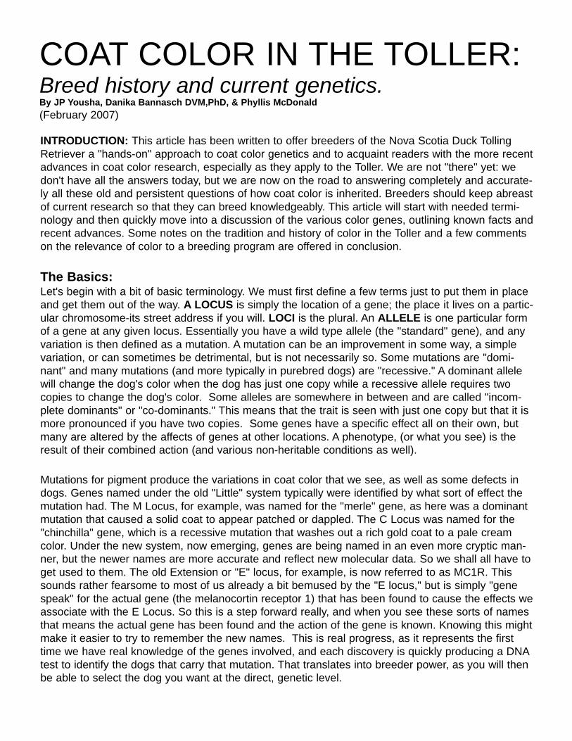
Illustration of how sable fades from infancy to the adult dog.
Sable pup at around one week Same pup at three months
This is the same pup at 12 months
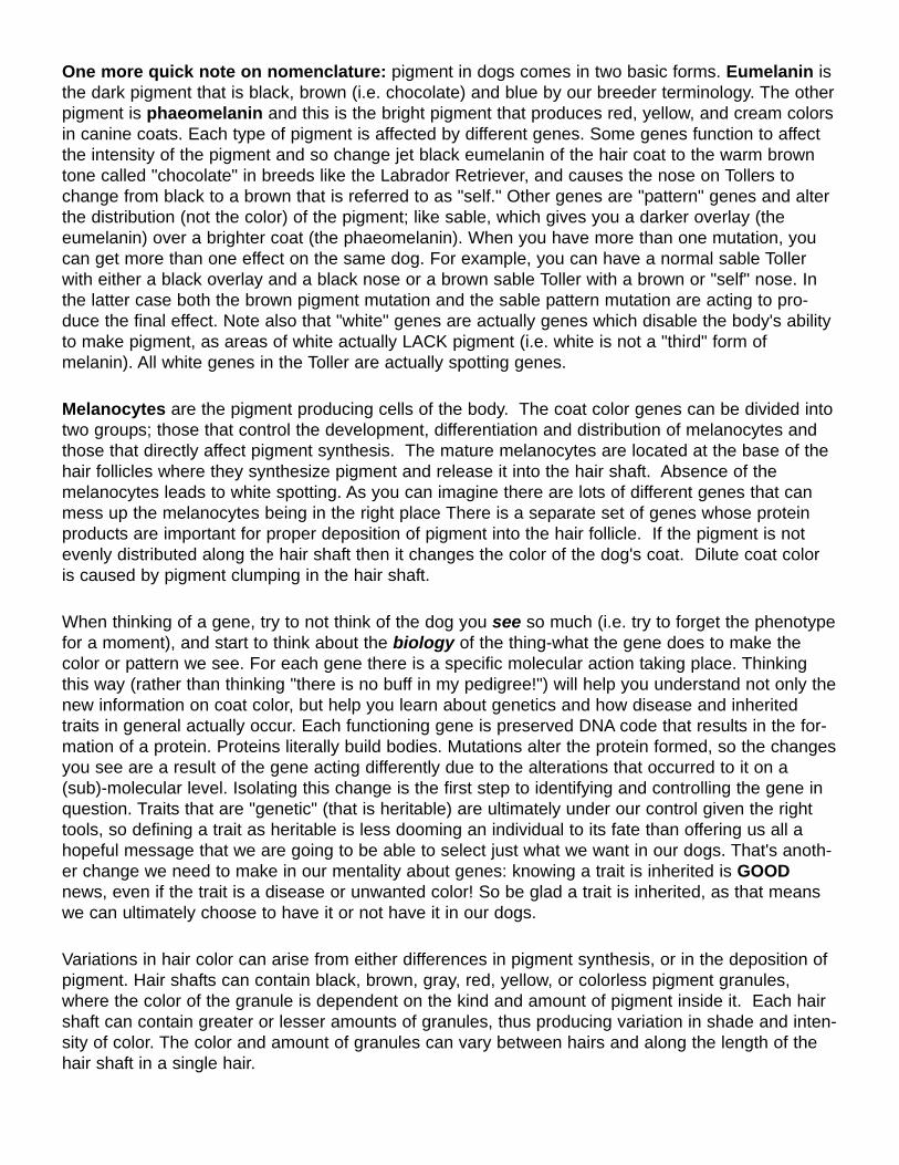
AGOUTI LOCUS: Most all Tollers arehomozygous for the sable agouti allelecalled ay. The Bannasch Laboratory test-ed 16 Tollers and found that they were allhomozygous for the agouti allele sable.Anecdote suggests that there are sometan-point (at)Tollers (think Dobermanmarkings), so this recessive allele maywell exist in this breed. Tan-point puppiesare the result of a mating with two tan-point carriers. In Tollers, in order to see thetan points the dogs must also have a spe-cific allele at MC1R- more in the next sec-tion!The agouti series works by modifying theamount of black versus yellow hair in the coat.One extreme is an all black dog and the otherextreme is an all yellow dog. In between thesetwo extremes are the sable color, black and tanand the normal allele which gives a strong band-ed pattern like you see in wolves.
a^y = Dominant allele that restricts dark pig-ment distribution; produces fawn/sable.a^w = agouti "wild-type" allele: gives wolf-grey coloration. Also called a^g.a^t = tan point allele: gives bicolored animal;dark body with tan points.
Red (mahogany dark red to bright red to even
pale yellow) animals with any black hairs in theircoats (even if only as puppies!) are a^y geneticsables. This "color"--which varies wildly in shadegiven its optics--has many names in dog breeds,but is traditionally called sable from the blackoverlay on a red coat. If the animal is a <bb>recessive "brown" dog, this is going to be lessobvious than with one with black hairs, butshading of any kind along the spine especiallysuggest the dog is a a^y sable. Sables tend tolighten as they age: many pups look "muddy" atbirth but are a bright clear color by adulthoodtypically, so bright reds that are genetic sablescould easily be confused as adults with the"clear red" described next. The distinctionbetweena sable and a clear red Toller wouldonly be noticed at a young age.
EXTENSION LOCUS: Clear red animals, or"gun dog reds," are recessive for an allele atMC1R called e. These animals are unable toexpress dark (eumelanin) pigment in their haircoat, but can still show it in their skin (includingeyes, as well as lips & noses). So, clear redTollers can have black (or brown) noses and eyerims, but never appear shaded even thoughthey are all also technically "sables" in thesense they are a^y agouti homozygotes. Thegenetic term for this is called epistasis andbasically it means that the action of one genecan mask the action of the other. If no dark pig-ment is produced then you can't see if it is dis-tributed into banded hair or tan points. It takesat least a single dominant <E> for any of the Alocus genes to express. So Tollers that areshaded reds, puppies that had masking orsabling when young, these dogs have at least
one dominant <E> gene at the extension locus.Labrador yellows, Irish Setters, and all Goldensare typically <ee> "recessive" reds. As Tollersapparently have both reds present in the breed,and both reds can interact with the "3rd" dog"red"--which is actually the B recessive browndescribed below-it's important to recognize allthese reds do not come from exactly the samegenetics, even if they look a lot alike. So, withso many "reds", the situation in this breed islikely subtle as to variation and requires a care-ful eye.
The MC1R gene has two known alleles that arenow well defined:E = allows for self colored dog; i.e. theactions of alleles of the Agouti locus can beseen. e = restricts pigment to red/yellow (no dark
Here is an exampleof a sable pup with
light eyes andbrown nose pig-
ment. These pupsalso usually fade to
a nice rich red.
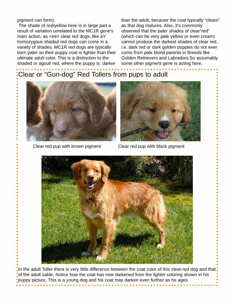
Clear or “Gun-dog” Red Tollers from pups to adult
Clear red pup with brown pigment Clear red pup with black pigment
In the adult Toller there is very little difference between the coat color of this clear-red dog and thatof the adult sable. Notice how the coat has now darkened from the lighter coloring shown in hispuppy picture. This is a young dog and his coat may darken even further as he ages.
pigment can form). The shade of red/yellow here is in large part a
result of variation unrelated to the MC1R gene'smain action, as <ee> clear red dogs, like aYhomozygous shaded red dogs can come in avariety of shades. MC1R red dogs are typicallyborn paler so their puppy coat is lighter than theirultimate adult color. This is a distinction to theshaded or agouti red, where the puppy is darker
than the adult, because the coat typically “clears”as that dog matures. Also, it's commonlyobserved that the paler shades of clear"red"(which can be very pale yellow or even cream)cannot produce the darkest shades of clear red,i.e. dark red or dark golden puppies do not evercome from pale blond parents in Breeds likeGolden Retrievers and Labradors.So assumablysome other pigment gene is acting here.
According to what we know of the history of theToller, the initial focus in the creation of thisbreed was in creating a red dog, somethingresembling the coloring of a fox. In order toachieve this, red from a variety of differentcanine sources was used. This has given Tollersmutations in three different red causing genes.Since most Tollers tend to either darken or light-en toward a similar tone of red as adults, nomatter which color they looked like as pups,Toller breeders generally don't distinguishbetween clear red or sable red when defining thecolors of their pups on registration paperwork. The AKC choices for Tollers when defining thecolor of Toller puppies are "Red", "Golden-Red","Fawn" and "Buff". "Red" is the default color on
the paperwork, but really, genetically what differ-entiates a "red" from a "golden-red" from a"fawn"? Sometimes, it's nothing, just shades of aclear-red dog, but sometimes it's a sabling genehidden in that rich red coat. There really is noth-ing in the registration paperwork color choiceswhich clearly indicates whether the dog is asable or clear red. In the adult Toller this is asubtle difference, but for those who want to knowwhat colors they are mixing into the "paint" oftheir puppies there are genetic tests availablethat can give breeders an exact picture of thecolor genetics he or she is dealing with. Colorgenetics testing is available through companieslike Healthgene and Vetgen for a reasonableprice.
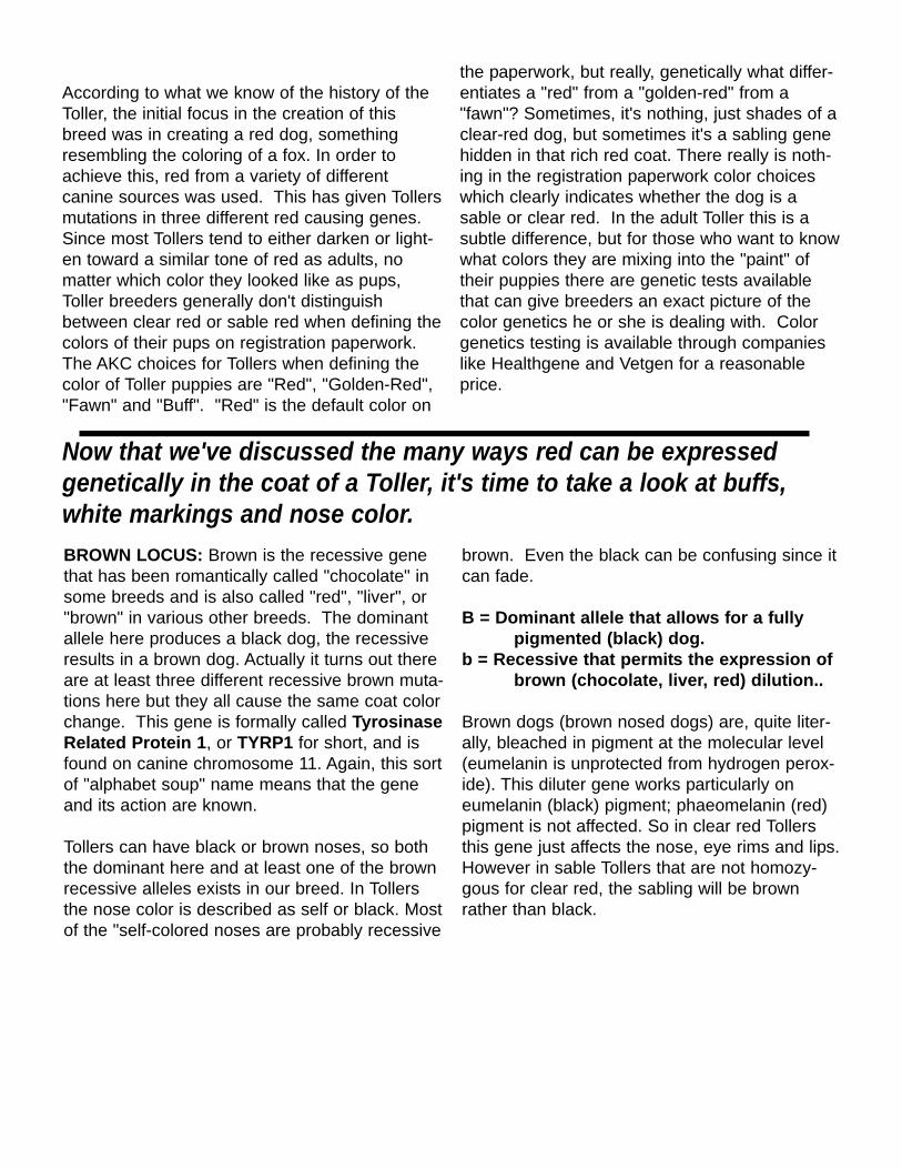
BROWN LOCUS: Brown is the recessive genethat has been romantically called "chocolate" insome breeds and is also called "red", "liver", or"brown" in various other breeds. The dominantallele here produces a black dog, the recessiveresults in a brown dog. Actually it turns out thereare at least three different recessive brown muta-tions here but they all cause the same coat colorchange. This gene is formally called TyrosinaseRelated Protein 1, or TYRP1 for short, and isfound on canine chromosome 11. Again, this sortof "alphabet soup" name means that the geneand its action are known.
Tollers can have black or brown noses, so boththe dominant here and at least one of the brownrecessive alleles exists in our breed. In Tollersthe nose color is described as self or black. Mostof the "self-colored noses are probably recessive
brown. Even the black can be confusing since itcan fade.
B = Dominant allele that allows for a fully pigmented (black) dog.
b = Recessive that permits the expression ofbrown (chocolate, liver, red) dilution..
Brown dogs (brown nosed dogs) are, quite liter-ally, bleached in pigment at the molecular level(eumelanin is unprotected from hydrogen perox-ide). This diluter gene works particularly oneumelanin (black) pigment; phaeomelanin (red)pigment is not affected. So in clear red Tollersthis gene just affects the nose, eye rims and lips.However in sable Tollers that are not homozy-gous for clear red, the sabling will be brownrather than black.
Now that we've discussed the many ways red can be expressedgenetically in the coat of a Toller, it's time to take a look at buffs,white markings and nose color.
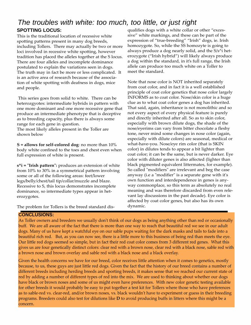
Adult Buff Tollers
Here is that same pup as an adult. While hiseyes are no longer a bright blue, notice how lightthey still are. Also notice his fur. It's almost as ifhe has been given a silvery overlay.
When pictured alongside of a non-dilute Toller, a TollerBuff appears to be faded or bleached.
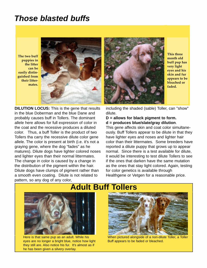
Those blasted buffs
The two buffpuppies in
the litter can be
easily distin-guished from
their litter-mates.
This threemonth oldbuff pup hasvery lighteyes and hisskin and furappears to bebleached orfaded.
DILUTION LOCUS: This is the gene that resultsin the blue Doberman and the blue Dane andprobably causes buff in Tollers. The dominantallele here allows for full expression of color inthe coat and the recessive produces a dilutedcolor. Thus, a buff Toller is the product of twoTollers tha carry the recessive dilute color geneallele. The color is present at birth (i.e. it's not agraying gene, where the dog “fades” as hematures). Dilute dogs have lighter colored nosesand lighter eyes than their normal littermates.The change in color is caused by a change inthe distribution of the pigment within the hair.Dilute dogs have clumps of pigment rather thana smooth even coating. Dilute is not related topattern, so any dog of any color,
including the shaded (sable) Toller, can "show"dilute. D = allows for black pigment to form.d = produces blue/slate/gray dilution.This gene affects skin and coat color simultane-ously. Buff Tollers appear to be dilute in that theyhave lighter eyes and noses and lighter haircolor than their littermates. Some breeders havereported a dilute puppy that grows up to appearnormal. Since there is a test available for dilute,it would be interesting to test dilute Tollers to seeif the ones that darken have the same mutationas the ones that stay light colored. Again, testingfor color genetics is available throughHealthgene or Vetgen for a reasonable price.
The troubles with white: too much, too little, or just rightSPOTTING LOCUS: This is the traditional location of recessive whitespotting patterns present in many dog breeds,including Tollers. There may actually be two or moreloci involved in recessive white spotting, howevertradition has placed the alleles together at the S locus.There are four alleles and incomplete dominancepostulated to explain the variations seen in dogs.The truth may in fact be more or less complicated. Itis an active area of research because of the associa-tion of white spotting with deafness in dogs, miceand people.
This series goes from solid to white. There can beheterozgyotes: intermediate hybrids in pattern withone more dominant and one more recessive gene thatproduce an intermediate phenotype that is deceptiveas to breeding capacity, plus there is always somerange for each gene in question. The most likely alleles present in the Toller areshown below
S = allows for self-colored dog: no more than 10%body white confined to the toes and chest even whenfull expression of white is present.
s^i = "Irish pattern": produces an extension of whitefrom 10% to 30% in a symmetrical pattern involvingsome or all of the following areas: feet/lowerlegs/belly/chest/tail tip/collar/muzzle and blaze.Recessive to S, this locus demonstrates incompletedominance, so intermediate types appear in het-erozygotes.
The problem for Tollers is the breed standard dis-
qualifies dogs with a white collar or other “exces-sive” white markings, and these can be part of theexpression of “true-breeding” “Irish” dogs, ie. Irishhomozygote. So, while the SS homozyte is going toalways produce a dog nearly solid, and the S/s^i het-erozygote (“Irish hybrid”) will likely always producea dog within the standard, in it’s full range, the Irishallele can produce too much white on a Toller tomeet the standard.
Note that nose color is NOT inherited separatelyfrom coat color, and in fact it is a well establishedprinciple of coat color genetics that nose color largelyINFORMS as to coat color. Nose/skin color is a hugeclue as to what coat color genes a dog has inherited.That said, again, inheritance is not monolithic and sonot every aspect of every physical feature is purelyand directly inherited after all. So as to skin color,especially with brown dilute dogs, the shade of thenose/eyerims can vary from bitter chocolate a fleshytone, never mind some changes in nose color (again,especially with dilute colors) are seasonal, medical orwhat-have-you. Nose/eye rim color (that is SKINcolor) in dilutes tends to appear a bit lighter thancoat color; it can be the same, but is never darker. Eyecolor with diluter genes is also affected (lighter thanblack pigmented equivalent littermates, for example).So called "modifiers" are irrelevant and beg the caseanyway (i.e a "modifier" is a separate gene with it'sown function and interdependence in genes is any-way commonplace, so this term as absolutely no realmeaning and was therefore discarded from even rele-vant lay discussions in the past decade). Eye color isaffected by coat color genes, but also has its owndynamic.
CONCLUSIONS: As Toller owners and breeders we usually don't think of our dogs as being anything other than red or occasionallybuff. We are all aware of the fact that there is more than one way to reach that beautiful red we see in our adultdogs. Many of us have kept a watchful eye on our sable pups waiting for the dark masks and tails to fade into abeautiful rich red. But, as you can now see, there is a little more to this business of being red than meets the eye.Our little red dogs seemed so simple, but in fact their red coat color comes from 3 different red genes. What thisgives us are four genetically distinct colors: clear red with a brown nose, clear red with a black nose, sable red witha brown nose and brown overlay and sable red with a black nose and a black overlay.
Given the health concerns we have for our breed, color receives little attention when it comes to genetics, mostlybecause, to us, these guys are just little red dogs. Given the fact that the history of our breed contains a number ofdifferent breeds including herding breeds and sporting breeds, it makes sense that we reached our current state ofred by adding a number of different types of red into the mix. We are used to thinking about whether our dogshave black or brown noses and some of us might even have preferences. With new color genetic testing availablefor other breeds it would probably be easy to put together a test kit for Tollers where those who have preferencesas to sable-red vs. clear-red, and/or brown noses, vs. black wouldn't have to experiment or guess in their breedingprograms. Breeders could also test for dilutions like D to avoid producing buffs in litters where this might be aconcern.

RESOURCES:
HEALTH GENE: Offers DNA-based technology including gene tests for coat color in dogs: blue, brown, both reds (agouti & extension). Website is: http://www.healthgene.com/
GENMARK: Offers DNA-based technology including gene tests for coat color in dogs: merle (and soon harl) gene(s). Website is: http://www.genmarkag.com/canine_faqs.php/
Vetgen: Offers DNA-based technology including gene tests for coat color in dogswww.vetgen.com
S. M. Schmutz, Ph.D. A current researcher on coat color in dogs who has worked extensively with coat color in the various breeds. She has a tutorial and informational website: GENETICS OF COAT COLOR IN DOGS: http://skyway.usask.ca/~schmutz/dogcolors.html
JP Yousha. Author of various articles on health and color issues in dogs, research liaison for coat color research in the Great Dane. Former Chairman, Health and Welfare Committee & member of Color Research Committee, GDCA. Current member (new) Health & Research committee. Email: [email protected] Phone (CT): 432-684-8940. Links page for color-related articles: http://www.chromadane.com/chlinx.htm.
BIBLIOGRAPHY:
• Bondurand N, Pingault V, Goerich DE, Lemort N, Sock E, Le Caignec C, Wegner M, & Goossens M. 2000. Interaction among SOX10, PAX3 and MITF, three genes altered in Waardenburg syndrome. Human Molecular Genetics 9:1907-1917. • Branis M, Burda H. 1985. Inner ear structure in the deaf and normally hearing Dalmatian dog. Journal of Comparative Pathology 95:295-299. •Breen M, Jouquand S, Renier C, Mellersh CS, Hitte C, Holmes NG, Cheron A, Suter N, Vignaux F, Bristow AE, Priat C, McCann E, Andre C, Boundy S, Gitsham P, Thomas R, Bridge WL, Spriggs HF, Ryder EJ, Curson A, Sampson J, Ostrander EA, Binns MM, Galibert F. 2001. Chromosome-specific single-locus FISH probes allow anchorage of an 1800-marker integrated radiation-hybrid/linkage map of the domestic dog genome to all chromosomes. Genome Research 11:1784-1795. • Brenig B, Pfeiffer I, Jaggy A, Kathmann I, Balzari M, Gaillard C, Dolf G. 2003. Analysis of the 5' region of the canine PAX3 gene and exclusion as a candidate for Dalmatian deafness. Animal Genetics 34:47-50. • Burns, M. and Fraser, M.N. 1966. Genetics of the Dog: The basis of successful breeding. Edinburgh: Oliver & Boyd. • Cattanach, B. (1999). The 'dalmatian dilemma': white coat colour and deafness. J. of Small Animal Practice 40: 193-+. • Clark, Ross D., DVM, and Joan R. Stainer. 1994. Medical and Genetic Aspects of Purebred Dogs. St. Simon Island, GA: Forum Publishing. • Coppens AG, Resibois A, & Poncelet L. 2000. Bilateral deafness in a Maltese terrier and a Great Pyrenean puppy: inner ear morphology. Journal of Comparative Pathology 122:223-228. • Coppens AG, Kiss R, Heizmann CW, Schafer BW, & Poncelet L. 2001. Immunolocalization of the calcium binding S100A1, S100A5 and S100A6 proteins in the dog cochlea during postnatal development. Brain Research Developmental Brain Reseach 28;126:191-9. • Coppens AG, Salmon I, Heizmann CW, Kiss R, & Poncelet L. 2003. Postnatal maturation of the dog stria vascularis - an immunohistochemical study. Anatomical Record Part A 270A:82-92. • Coppens AG, Steinberg SA, & Poncelet L. 2003. Inner ear morphology in a bilaterally deaf Dogo Argentino pup. Journal of Comparative Pathology 128:67-70. • Coppens AG, Salmon I, Heizmann CW, & Poncelet L. 2004. Dark-cell areas in the dog vestibular endorgans: an immunohistochemical study. Histology and Histopathology 19:1227-1235. • Cordaux R & Batzer MA. 2006. Teaching an old dog new tricks: SINEs of canine genomic diversity. Proceedings of the National Academy of Sciences, 9 January 2006, 103(5):1157-8. • Green, B.K. 1974. The Color of Horses. Flagstaff, AZ: Northland Press. • Greibrokk, T. 1994. Hereditary Deafness in the Dalmatian-Relationship to Eye and Coat Color. JAAHA 30: 170-176.
2
• Hayes, H.M., Wilson, G.P., Fenner, W.R. & Wyman, M. 1981. Canine congenital deafness: epidemiologic study of 272 cases. Journal of the American Animal Hospital Association 17:473-476. • Hoskins, J.D. 1990. Veterinary Pediatrics: Dogs and Cats from Birth to Six Months. Philadelphia, PA: W. B. Saunders Company. • Johnson, Di. 1994. Great Danes Today. New York: McMillian. • Juraschko K. 2000. Populationsgenetische Untersuchung der kongenitalen Taubheit beim Dalmatiner (Genetic analysis of congenital deafness in Dalmatians). Doctoral thesis, Tierarztliche Hochschule Hannover, Hanover, Germany. • Karlsson EK, Hillbertz NS, Wade CM, ANdersson G, von Euler H, Hedhammar Å, Zody MC, Biagi T, Lai J, Anderson N, Liu G, Jones K, Andersson L, Lindblad-Toh K. 2006. Two-stage association mapping in dogs identifies coat color locus. Third International Conference on Advances in Canine and Feline Genomics, Davis, CA, Aug, 2006. To be published in a future issue of the Journal of Heredity. • Kerns JA, Newton J, Berryere TG, Rubin EM, Cheng J-F, Schmutz SM & Barsh GS. 2004. Characterization of the dog Agouti gene and a nonagouti mutation in German Shepherd Dogs. Mammalian Genome 15:798-808. • Kerns, J.A., M. Olivier, G. Lust, & G. S. Barsh. 2003. Exclusion of Melanocortin-1 Receptor (Mc1r) and Agouti as candidates for dominant black in dogs. J. of Hered. 94:75-79. Kim JH, Kang KI, Sohn HJ, Woo GH, Jean YH, & EK Hwang. 2005. Color-dilution alopecia in dogs. J. Vet Sci Sep: 6(3):259-61. • Klein, E., Steinberg, S.A., Weiss, S.R.B., Matthews, D.M., and T.W. Uhde. 1988. The relationship between genetic deafness and fear- related behaviors in nervous pointer dogs. Physiology and Behavior 43: 307-312. • Krempler, A., Breen, M. and Brenig, B. 2000. Assignment of the canine paired-box 3 (PAX3) gene to chromosome 37q16->q17 by in situ hybridization. Cytogenet. Cell Genet. 90 (1-2), 66-67. • Langebaek, R. 1986. Variations of hair coat and skin texture in blue dogs. Nord Vet Med: Nov-Dec:: 38 (6): 383-7. • Laukner, A. 1998. [Coat color in dogs. 2: Clinical significance] Tierarztl Prax Ausg K Kleintiere: Feb;26(1):49-54. • Little, Clarence C. 1957. The Inheritane of Coat Color in Dogs. New York: Howell Publishing. • MacMillan, Gail 1998. A Breed Apart: Nova Scotia’s Duck Tolling Retriever. Nimbus Publishing Ltd. • Metallinos, D and Rine, J. 2000. Exclusion of EDNRB and KIT as the basis for white spotting in Border Collies. Genome Biology (a web based only journal) online article• Padgett, George A., DVM. (1998) "Control of Canine Genetic Diseases." New York: Howell Publishing. • Phillip U, Hanamm H, Mecklenburg L, Mishino S, Mignot E, Gunzel-Apel AR, Schutx SM & T Leeb 2005. Polymorphism within the canine MLPH gene are associated with dilute coat color in dogs. BMC Genet. Jun 16;6:34. • Rak SG, Drogemuller C, Leeb T, Quignon P, Andre C, Scott A, Breen M, & Distl O. 2003. Chromosomal assignment of 20 candidate genes for canine congenital
3
sensorineural deafness by FISH and RH mapping. Animal Cytogenetics and Comparative Mapping 101:130-135. • Rak SG & Distl O. 2005. Congenital sensorineural deafness in dogs: A molecular genetic approach toward unravelling the responsible genes. The Veterinary Journal 169:188-196. • Rawitz, B. 1896. Gehörorgan und Gehirn eines Weissen Hundes mit blauen Augen (Hearing and deafness in white dogs with blue eyes). Morphol. Arbeiten. 6, 545-553. • Reetz, I., Stecker M., and W. Wegner. 1977. Audiometrische befunde in einer Merlezucht. [Audiometric findings in dachshonds (merle gene carriers)]. DTW (Deustche Teirarztliche Wochenschrift). 84(7):273-7. • Robinson, R. 1982. Genetics for dog breeders. Oxford: Pergamon Press. • Schaible, R.H. and Brumbaugh, J.A. 1976. Electron microscopy of pigment cells in variegated and nonvariegated piebald spotted dogs. Pigment Cell. 3: 191-220. • Schmutz, S. M., T. G. Berryere, and C. A. Sharp. 2003. KITLG mapping to CFA15 and exclusion as a candidate gene for merle. Animal Genetics 34: 75-76. • Schmutz, S. M., T. G. Berryere, N. M. Ellinwood, J. A. Kerns, and G. S. Barsh, MC1R Studies in Dogs With Melanistic Mask or Brindle Patterns. J Hered 2003 94: 69-73. • Schmutz, S. M., T. G. Berryere, and A. D. Goldfinch. 2002. TYRP1 and MC1r genotypes and their effects on coat color in dogs. Mammalian Genome 13:380-387. • Schmutz S.M., Moker J.S, Yuzbasiyan-Gurkan V., Zemke D., Sampson J., Lingaas F., Susana Dunner S., and G Dolf. 2001. DCT and EDNRB map to DogMap Linkage Group L07. Animal Genetics 32:321. • Sorsby, A. 1970. Ophthalmic Genetics. London: Butterworths. • Sponenberg DP, Rothschild MF 2001. Genetics of coat colour and hair texture. In The Genetics of the Dog, eds. A. Ruvinsky, J. Sampson, pp. 61-85. Wallingford, Oxon, UK: CABI Publishing. • Steel KP, Kros CJ. 2001. A genetic approach to understanding auditory function. Nature Genetics 27:143-9. • Steel, K.P., and C. Barkway.1989. Another role for melanocytes: their importance for normal stria vascularis development in the inner ear. Development 107: 453-463. • Strain GM. 2004. Deafness prevalence and pigmentation and gender associations in dog breeds at risk. Veterinary Journal 167(1):23-32. Strain GM. 1992. Deafness in dogs and cats. Proceedings of the 10th American College of Veterinary Internal Medicine Forum 10, 275-278. • Strain GM. 1996. Aetiology, prevalence, and diagnosis of deafness in dogs and cats. British Veterinary Journal 152:17. • Strain GM. 1991. Congenital deafness in dogs and cats. Compendium on Continuing
Education for the Practicing Veterinarian 13:245. • Strang, A., and G. MacMillan The Nova Scotia Duck Tolling Retriever. Loveland,
Colo.: Alpine Publications, 1996. • Tsai KL, Guyon T, & Murphy KE. 2003. Identification of isoforms and RH mapping of canine KIT. Cytogenetic and Genome Research 102:261-263. • van Hagen MAE, van der Kolk J, Barendse MAM, Imholz S, Leegwater PAJ, Knol BW, & van Oost BA. 2004. Analysis of the inheritance of white spotting and the
4
evaluation of KIT and EDNRB as spotting loci in Dutch boxer dogs. Journal of Heredity 95(6): 526-531. • Watanabe K-I, Takeda R, Yasumoto K-I, Udono T, Saito H, Ikeda K, Takasaka T, Takahashi K, Kobayashi T, Tahibana M, & Shibahara S. 2002. Identification of a distal enhancer for the melanocyte-specific promoter of the MITF gene. Pigment Cell Research 15:201-211. • Wegner, W., and A. Akcan. 1980. Auswirkungen der Merlefactors auf die Area optica beim Hund. DTW (Deutsche Teirarztliche Wochenschrift). 87(9):342. Willis, Malcolm B. 1989. Genetics of the Dog. New York: Howell Publishing. • Wood JLN, Lakhani KH. 1997. Prevalence and prevention of deafness in the Dalmatian - Assessing the effect of parental hearing status and gender using ordinary logistic and generalized random litter effect models. Veterinary Journal 154:121-33. • Wood, JLN, Lakhani, KH, & Henley, WE. 2004. An epidemiological approach to prevention and control of three common heritable diseases in canine pedigree breeds in the United Kingdom. The Veterinary Journal 168:14-27. • Yousha, JP. Black, blue and fawn: Color gene interaction in solid colored Danes. Online publication: August 2006. Online article.• Yousha, JP. Control of Canine Genetic Disease. 2005. Dane World Nov-Dec: Vol 11, Issue 6, pp.97-99. GDCA publication: November 2005. Online article. • Zemke D, Cao Y, Yuzbasiyan-Gurkan V. 1999. Hereditary hearing loss in dogs: models for sensorineural deafness. Hereditary Deafness Newsletter 16:34 (www.ihr.mrc.ac.uk/ hereditary/newsletters/ index.shtml). • Zemke, D. and V. Yuzbasiyan-Gurkan. 1999. A single nucleotide polymorphism and a (GA)n microsatellite in intron 6 of the canine endothelin receptor B (EDNRB) gene. Anim. Genet. 30:390.
5