to 4d microscopy camera obscura of the twenty-first century
TRANSCRIPT

doi: 10.1098/rsta.2009.0265, 1191-1204368 2010 Phil. Trans. R. Soc. A
Ahmed H. Zewail
to 4D microscopycamera obscura of the twenty-first century: from Micrographia
Referencesl.html#ref-list-1http://rsta.royalsocietypublishing.org/content/368/1914/1191.ful
This article cites 38 articles, 10 of which can be accessed free
This article is free to access
Rapid response1914/1191http://rsta.royalsocietypublishing.org/letters/submit/roypta;368/
Respond to this article
Subject collections
(18 articles)electron microscopy � (1 articles)spectroscopy �
(17 articles)optics � collectionsArticles on similar topics can be found in the following
Email alerting service herein the box at the top right-hand corner of the article or click Receive free email alerts when new articles cite this article - sign up
http://rsta.royalsocietypublishing.org/subscriptions go to: Phil. Trans. R. Soc. ATo subscribe to
This journal is © 2010 The Royal Society
on February 26, 2010rsta.royalsocietypublishing.orgDownloaded from

on February 26, 2010rsta.royalsocietypublishing.orgDownloaded from
Phil. Trans. R. Soc. A (2010) 368, 1191–1204doi:10.1098/rsta.2009.0265
REVIEW
Micrographia of the twenty-first century:from camera obscura to 4D microscopy
BY AHMED H. ZEWAIL*
Physical Biology Center for Ultrafast Science and Technology, Arthur AmosNoyes Laboratory of Chemical Physics, California Institute of Technology,
Pasadena, CA 91125, USA
In this paper, the evolutionary and revolutionary developments of microscopic imagingare overviewed with a perspective on origins. From Alhazen’s camera obscura, toHooke and van Leeuwenhoek’s two-dimensional optical micrography, and on to three-and four-dimensional (4D) electron microscopy, these developments over a millenniumhave transformed humans’ scope of visualization. The changes in the length and timescales involved are unimaginable, beginning with the visible shadows of candles at thecentimetre and second scales, and ending with invisible atoms with space and timedimensions of sub-nanometre and femtosecond. With these advances it has becomepossible to determine the structures of matter and to observe their elementary dynamicsas they unfold in real time. Such observations provide the means for visualizing materialsbehaviour and biological function, with the aim of understanding emergent phenomenain complex systems.
Keywords: light; electrons; microscopy
1. Origins in light
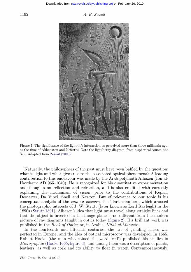
The ever-increasing progress made by humans in making the very small andthe very large visible and tangible is truly remarkable. The human eye is notdiffraction limited, but its spatial and temporal resolutions are limited to about100 μm and a fraction of a second, respectively. Today we are aided by toolsthat enable the visualization of objects that are below a nanometre in size andthat move in femtoseconds or attoseconds (Zewail & Thomas 2009, and referencestherein). How did it all begin? Surely the power of light for observation has beenwith humans since their creation. Stretching back over six millennia, one finds itsconnection to the science of time clocking (Zewail 2000) (first in calendars) andto the mighty monotheistic faiths and rituals (figure 1).
One contribution of 17 to a Theme Issue ‘Personal perspectives in the physical sciences for theRoyal Society’s 350th anniversary’.
This journal is © 2010 The Royal Society1191
1192 A. H. Zewail
on February 26, 2010rsta.royalsocietypublishing.orgDownloaded from
Figure 1. The significance of the light–life interaction as perceived more than three millennia ago,at the time of Akhenaton and Nefertiti. Note the light’s ‘ray diagram’ from a spherical source, theSun. Adapted from Zewail (2008).
Naturally, the philosophers of the past must have been baffled by the question:what is light and what gives rise to the associated optical phenomena? A leadingcontribution to this endeavour was made by the Arab polymath Alhazen (Ibn al-Haytham; AD 965–1040). He is recognized for his quantitative experimentationand thoughts on reflection and refraction, and is also credited with correctlyexplaining the mechanism of vision, prior to the contributions of Kepler,Descartes, Da Vinci, Snell and Newton. But of relevance to our topic is hisconceptual analysis of the camera obscura, the ‘dark chamber’, which arousedthe photographic interests of J. W. Strutt (later known as Lord Rayleigh) in the1890s (Strutt 1891). Alhazen’s idea that light must travel along straight lines andthat the object is inverted in the image plane is no different from the modernpicture of ray diagrams taught in optics today (figure 2). His brilliant work waspublished in the Book of Optics or, in Arabic, Kitab al-Manazir.
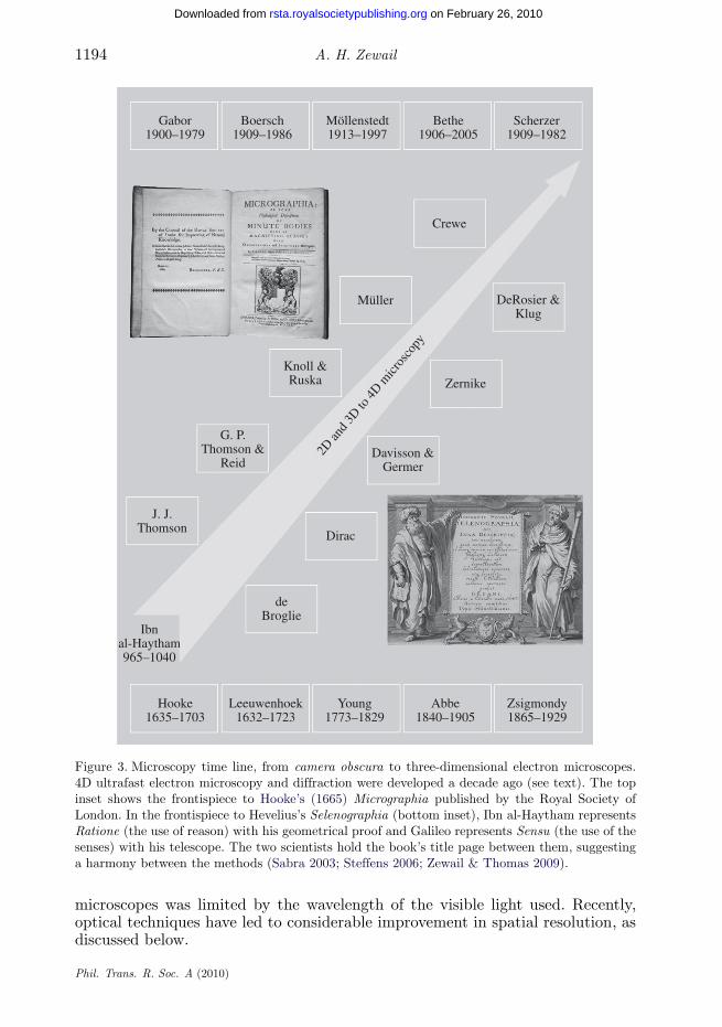
In the fourteenth and fifteenth centuries, the art of grinding lenses wasperfected in Europe, and the idea of optical microscopy was developed. In 1665,Robert Hooke (the man who coined the word ‘cell’) published his studies inMicrographia (Hooke 1665; figure 3), and among them was a description of plants,feathers, as well as cork and its ability to float in water. Contemporaneously,
Phil. Trans. R. Soc. A (2010)

Review. Micrographia of the 21st century 1193
on February 26, 2010rsta.royalsocietypublishing.orgDownloaded from
Alhazen’s Camera Obscura
Figure 2. The concept of the camera obscura as perceived a thousand years ago by Alhazen (Ibnal-Haytham), who coined the term (see text). Note the formation of the inverted image through aray diagram. Adapted from Al-Hassani et al. (2006).
Anton van Leeuwenhoek used a simple, one-lens microscope to examine blood,insects and other objects, and was the first to visualize bacteria, amongother microscopic objects. More than a hundred years later, an experiment bythe physicist, physician and Egyptologist, Thomas Young, demonstrated theinterference of light, an experiment that revolutionized our views on the natureof light. His double-slit experiment of 1801 performed at the Royal Institutionof Great Britain led to the demise of Newton’s corpuscular theory of light. Ofrelevance here is the phenomenon of diffraction due to interferences of waves(coherence). Much later, such diffraction was found to yield the (microscopic)interatomic distances characteristic of molecular and crystal structures, asdiscovered in 1912 by von Laue and elucidated later that year by W. L. Bragg.
Resolution in microscopic imaging was brought to a whole new level by twomajor developments in optical microscopy. In 1878, Ernst Abbe formulated amathematical theory correlating resolution to the wavelength of light (beyondwhat we now designate the empirical Rayleigh criterion for incoherent sources),and hence the optimum parameters for achieving higher resolution. At thebeginning of the twentieth century, Richard Zsigmondy, by extending the work ofFaraday and Tyndall, developed the ‘ultramicroscope’ to study colloidal particles;for this work, he received the Nobel Prize in Chemistry in 1925. Then camethe penetrating developments in the 1930s by Frits Zernike, who introducedthe phase-contrast concept in optical microscopy; he, too, received the NobelPrize, in Physics, in 1953. It was understood that the spatial resolution of optical
Phil. Trans. R. Soc. A (2010)
1194 A. H. Zewail
on February 26, 2010rsta.royalsocietypublishing.orgDownloaded from
Gabor1900–1979
Boersch1909–1986
Möllenstedt1913–1997
Bethe1906–2005
Scherzer1909–1982
Crewe
Müller
Knoll &Ruska
G. P.Thomson &
Reid
J. J.Thomson
Dirac
Davisson &Germer
Zernike
2D an
d 3D
to 4
D micr
osco
py
DeRosier &Klug
deBroglie
Ibnal-Haytham965–1040
Hooke1635–1703
Leeuwenhoek1632–1723
Young1773–1829
Abbe1840–1905
Zsigmondy1865–1929
Figure 3. Microscopy time line, from camera obscura to three-dimensional electron microscopes.4D ultrafast electron microscopy and diffraction were developed a decade ago (see text). The topinset shows the frontispiece to Hooke’s (1665) Micrographia published by the Royal Society ofLondon. In the frontispiece to Hevelius’s Selenographia (bottom inset), Ibn al-Haytham representsRatione (the use of reason) with his geometrical proof and Galileo represents Sensu (the use of thesenses) with his telescope. The two scientists hold the book’s title page between them, suggestinga harmony between the methods (Sabra 2003; Steffens 2006; Zewail & Thomas 2009).
microscopes was limited by the wavelength of the visible light used. Recently,optical techniques have led to considerable improvement in spatial resolution, asdiscussed below.
Phil. Trans. R. Soc. A (2010)

Review. Micrographia of the 21st century 1195
2. Electrons in microscopy
on February 26, 2010rsta.royalsocietypublishing.orgDownloaded from
Just before the dawn of the twentieth century, in 1897, electrons, or thecorpuscles of J. J. Thomson, were discovered, but they were not conceived asimaging rays until Louis de Broglie formulated the concept of particle–waveduality in 1924. The duality character of an electron, which is quantified inthe relationship λde Broglie = h/p, suggested the possibility of achieving waves ofpicometre wavelength, and became essential to the understanding of diffractionand imaging. The first experimental evidence of the wave character of the electronwas established in 1927 by Davisson and Germer (diffraction from a nickel surface)and, independently, by G. P. Thomson (the son of J. J. Thomson), who, with Reid,observed diffraction of electrons penetrating a thin foil. In 1923, Dirac postulatedthe concept of ‘single-particle interference’.
Later, Knoll & Ruska (1932) invented the electron microscope (EM) andimproved the resolution to the (sub)micrometre scale in the transmission electronmicroscope (TEM). Boersch introduced the diffraction lens in TEM in 1936, andlater (1940) he found the so-called Fresnel fringes as ‘diffraction at edges’ inthe microscope. Concurrently, Walther Kossel and Gottfried Möllenstedt in 1939combined in their EM the ability to record projected two-dimensional imagesand electron diffraction patterns, which contain information on the structure,the repeating lattice distances and other aspects pertaining to crystallographicsymmetry. These and other related developments in microscopy led to electroninterferometry and holography. The original proposal of electron holography byDenis Gabor in 1948 and the birth of electron biprism interference by Möllenstedtin 1953 laid the foundation (Silverman et al. 1995; Lichte 2002, and referencestherein; Spence 2009) for the impressive advances made by Tonomura (1998,1999) and others in the years to follow.
3. Imaging atoms, molecules and cells
The first images of individual atoms were obtained in 1951 by Müller (Müller1951; Tsong 2006; Thomas 2008), who introduced the technique of field-ionmicroscopy to visualize them at fine tips of metals and alloys, and to detectvacancies and atomic steps and kinks at the surfaces. With the invention offield-emission sources and scanning TEM, pioneered in 1970 by Crewe, isolatedheavy atoms became readily visible (Crewe et al. 1970; Thomas 1979). (Thescanning tunnelling microscope was developed in the 1980s and made possibleatomic-scale images of conducting surfaces.) Today, with aberration-correctedmicroscopes, imaging has reached a resolution of less than an ångström (Nellistet al. 2004). This history would be incomplete if I did not mention that the totalityof technical developments and applications in the investigations of inorganic andorganic materials have benefited enormously from the contributions of many otherscientists, and for more details I refer the reader to the books by Cowley (1995),Humphreys (2002), Gai & Boyes (2003), Spence (2003) and Hawkes & Spence(2007), and the most recent papers by Hawkes (2009) and Howie (2009).
Biological EM has been transformed by several major advances, includingelectron crystallography, single-particle tomography and cryo-microscopy, aidedby large-scale computational processing. Beginning with the 1968 electron
Phil. Trans. R. Soc. A (2010)

1196 A. H. Zewail
on February 26, 2010rsta.royalsocietypublishing.orgDownloaded from
crystallography work of DeRosier and Klug (see Klug 1982), three-dimensionaldensity maps became retrievable from EM images. Landmark experimentsrevealing the high-resolution (atomic-scale) structure from two-dimensionalcrystals, single-particle three-dimensional cryo-EM images of different butidentical particles (6 Å resolution) and three-dimensional cryo-EM images ofthe same particle (tomography with 6 Å resolution) represent the impressiveprogress made. With these methods, the first membrane protein structure wasdetermined, the first high-resolution density maps for the protein shell of anicosahedral virus were obtained, and the imaging of whole cells was accomplished.Minimizing radiation damage by embedding the biological macromolecules andmachines in vitreous ice affords a non-invasive, high-resolution imaging techniquefor visualizing the three-dimensional organization of eukaryotic cells, withtheir dynamic organelles, cytoskeletal structure and molecular machines in anunperturbed context, with a resolution of 6 Å to 2 nm, being limited by radiationdamage. I refer the reader to the papers by Henderson (1995), Sali et al. (2003),Crowther (2008) and Glaeser (2008), and the books by Glaeser et al. (2007) andby Frank (2006).
4. 4D electron microscopy
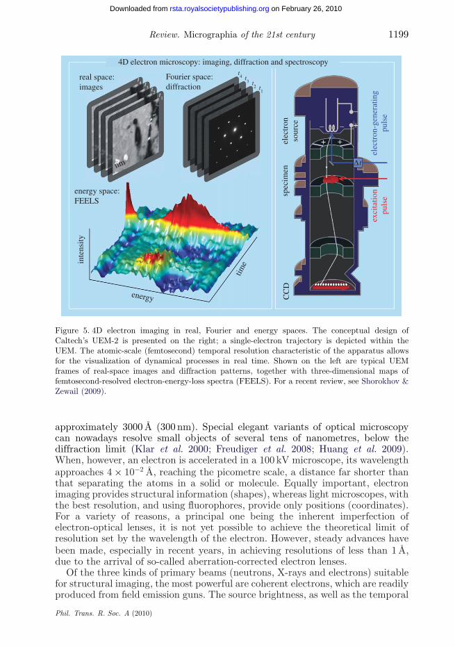
Whereas in all of the above methods the processes of imaging, diffraction andchemical analysis have been conducted in a static (time-averaged) manner,it has now become possible to unite the time domain with the spatial one,thereby creating 4D electron microscopy (Barwick et al. 2008; Carbone et al.2009; Yurtsever & Zewail 2009; Barwick et al. 2009); for a recent review,see Shorokhov & Zewail (2009). This development owes its success to theadvancement of the concept of coherent single-electron imaging, with theelectron packets being liberated from a photocathode using femtosecond opticalpulses. In such a mode of electron imaging, the repulsion between electronsis negligible, and thus atomic-scale spatiotemporal resolution can be achieved.Atomic motions, phase transitions, mechanical movements and the natureof fields at interfaces are examples of phenomena that can be charted inunprecedented structural detail at a rate that is ten orders of magnitudefaster than hitherto. Furthermore, because electrons are focusable and canbe pulsed at these very high rates, and because they have appreciableinelastic cross sections, the EM yields information in four distinct ways: inreal space, in reciprocal space, in energy space and in the time domain.Convergent beam imaging was also shown to provide nanoscale diffraction ofheterogeneous structures (Yurtsever & Zewail 2009), and near-field imaging canmap nanoscale electromagnetic fields of material structures (Barwick et al. 2009).Thus, besides structural imaging, the energy landscapes of macromolecules maybe explored; and, under optimal conditions, elemental compositions, valence-states bonding and three-dimensional information (from tomography) may alsobe retrieved.
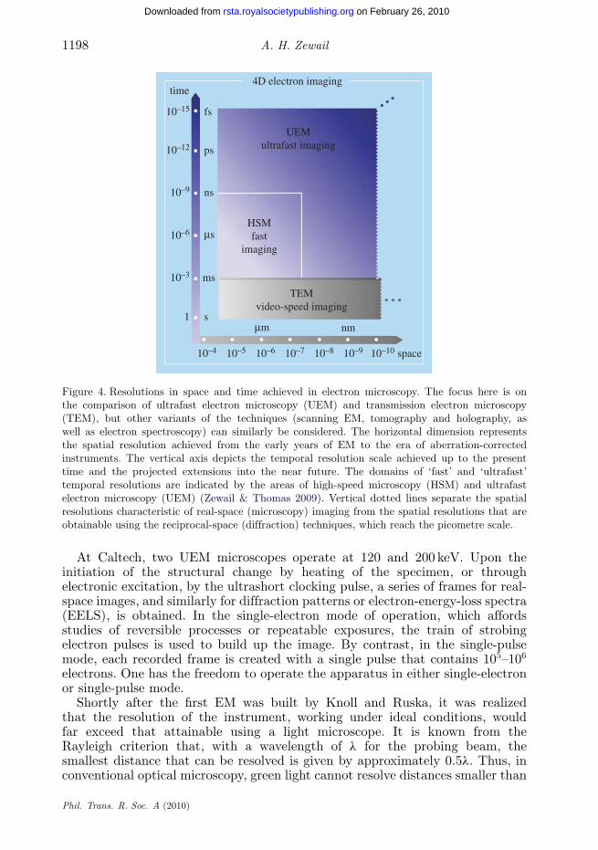
Figure 4 depicts the space and time dimensions of TEM and of ultrafastelectron microscopy (UEM). The boundaries of the time resolution arerepresentative of the transition from the millisecond video speed used in TEMimaging, to fast or high-speed (nanosecond to microsecond) imaging, and on to
Phil. Trans. R. Soc. A (2010)
Review. Micrographia of the 21st century 1197
on February 26, 2010rsta.royalsocietypublishing.orgDownloaded from
the ultrafast (femtosecond to picosecond) imaging regime. The spatial resolutionin the high-speed, nanosecond domain indicated in the figure is limited byelectron–electron (space–charge) repulsion in the nanosecond pulses of electrons.The UEM landscape is that of single-electron imaging, which, owing to theabsence of inter-electron repulsion, reaches the spatial resolution of the TEM.Examples of time-averaged EM and of UEM studies can be found in Zewail &Thomas (2009, and references therein). The key concepts involved in the UEMand some prototypical results are provided in figure 5.
The concept of single-electron imaging is based on the premise that thetrajectories of coherent and timed, single-electron packets can provide an imageequivalent to that obtained using many electrons in conventional microscopes.Unlike the random electron distribution of conventional microscopes, in UEMthe packets are timed with femtosecond precision, and each electron has aunique coherence volume. As such, each electron of finite de Broglie wavelengthis (transversely) coherent over the object length scale to be imaged, with alongitudinal coherence length that depends on its velocity. On the detector, theelectron produces a ‘click’ behaving as a classical particle, and when a sufficientnumber of such clicks is accumulated stroboscopically, the whole image emerges.Putting it in Dirac’s famous dictum: each electron interferes only with itself. In themicroscope, the electron pulse that produces the image is termed the probe pulse,and in ultrafast imaging with a train of such pulses, the number of frames in amovie could then be as high as 1012 per second; this ‘stop-motion photography’constitutes a real-time movie of the process.
To visualize the motion, the molecule or material must be launched on itspath using a femtosecond initiation pulse, the clocking or pump pulse, thusestablishing a temporal reference point (time zero) for the changes that occurin the motion. By sending the clocking pulse along an adjustable optical path,we can precisely fix each probe frame on the time axis—knowing the speed oflight, a typical optical path accuracy of 1μm corresponds to absolute timingof the snapshots of 3.3 fs. Because the clocking pulse is controlled to precedeeach electron pulse, the time axis is defined by the separation between them andis no longer limited by the response of the video detector in the microscope.Lastly, in order to synchronize the motion of many independent atoms ormolecules so that all of them have reached a similar point in the course oftheir structural evolution, the relative timing of clocking and probe pulses mustbe of femtosecond precision, and the launch configuration must be defined tosub-ångström resolution.
In imaging with electrons, unlike with photons, we must also consider theconsequences of the Pauli exclusion principle. The maximum number of electronsthat can be packed into a state (or a cell of phase space) is two, one for eachspin; in contrast, billions of photons can be condensed in a state of the laserradiation. This characteristic of electrons represents a fundamental difference inwhat is termed the ‘degeneracy’, or the mean number of electrons per cell inphase space. Typically it is about 10−4–10−6 but it is possible in UEM to increasethe degeneracy by orders of magnitude, a feature that could be exploited forstudies in quantum electron optics (Zewail & Thomas 2009). I note here thatthe definition of ‘single-electron packet’ is reserved for the case when each timedpacket contains one or a small number of electrons such that coulombic repulsionis effectively absent.
Phil. Trans. R. Soc. A (2010)
1198 A. H. Zewail
on February 26, 2010rsta.royalsocietypublishing.orgDownloaded from
time
10–15 fs
ps
ns
nm
ms
s
ms
mm
10–12
10–9
10–6
1
10–4 10–5 10–6 10–7 10–8 10–9 10–10 space
TEMvideo-speed imaging
UEMultrafast imaging
HSMfast
imaging
10–3
4D electron imaging
Figure 4. Resolutions in space and time achieved in electron microscopy. The focus here is onthe comparison of ultrafast electron microscopy (UEM) and transmission electron microscopy(TEM), but other variants of the techniques (scanning EM, tomography and holography, aswell as electron spectroscopy) can similarly be considered. The horizontal dimension representsthe spatial resolution achieved from the early years of EM to the era of aberration-correctedinstruments. The vertical axis depicts the temporal resolution scale achieved up to the presenttime and the projected extensions into the near future. The domains of ‘fast’ and ‘ultrafast’temporal resolutions are indicated by the areas of high-speed microscopy (HSM) and ultrafastelectron microscopy (UEM) (Zewail & Thomas 2009). Vertical dotted lines separate the spatialresolutions characteristic of real-space (microscopy) imaging from the spatial resolutions that areobtainable using the reciprocal-space (diffraction) techniques, which reach the picometre scale.
At Caltech, two UEM microscopes operate at 120 and 200 keV. Upon theinitiation of the structural change by heating of the specimen, or throughelectronic excitation, by the ultrashort clocking pulse, a series of frames for real-space images, and similarly for diffraction patterns or electron-energy-loss spectra(EELS), is obtained. In the single-electron mode of operation, which affordsstudies of reversible processes or repeatable exposures, the train of strobingelectron pulses is used to build up the image. By contrast, in the single-pulsemode, each recorded frame is created with a single pulse that contains 105–106
electrons. One has the freedom to operate the apparatus in either single-electronor single-pulse mode.
Shortly after the first EM was built by Knoll and Ruska, it was realizedthat the resolution of the instrument, working under ideal conditions, wouldfar exceed that attainable using a light microscope. It is known from theRayleigh criterion that, with a wavelength of λ for the probing beam, thesmallest distance that can be resolved is given by approximately 0.5λ. Thus, inconventional optical microscopy, green light cannot resolve distances smaller than
Phil. Trans. R. Soc. A (2010)
Review. Micrographia of the 21st century 1199
on February 26, 2010rsta.royalsocietypublishing.orgDownloaded from
real space:images
energy space:FEELS
elec
tron
sour
ce
Δt
elec
tron
-gen
erat
ing
puls
eex
cita
tion
puls
espec
imen
CC
D
nm
timein
tens
ityFourier space:diffraction
energy
– –
–
+
++
t1
t2
t3
t4
t1
t2
t3
t4
4D electron microscopy: imaging, diffraction and spectroscopy
Figure 5. 4D electron imaging in real, Fourier and energy spaces. The conceptual design ofCaltech’s UEM-2 is presented on the right; a single-electron trajectory is depicted within theUEM. The atomic-scale (femtosecond) temporal resolution characteristic of the apparatus allowsfor the visualization of dynamical processes in real time. Shown on the left are typical UEMframes of real-space images and diffraction patterns, together with three-dimensional maps offemtosecond-resolved electron-energy-loss spectra (FEELS). For a recent review, see Shorokhov &Zewail (2009).
approximately 3000 Å (300 nm). Special elegant variants of optical microscopycan nowadays resolve small objects of several tens of nanometres, below thediffraction limit (Klar et al. 2000; Freudiger et al. 2008; Huang et al. 2009).When, however, an electron is accelerated in a 100 kV microscope, its wavelengthapproaches 4 × 10⁻² Å, reaching the picometre scale, a distance far shorter thanthat separating the atoms in a solid or molecule. Equally important, electronimaging provides structural information (shapes), whereas light microscopes, withthe best resolution, and using fluorophores, provide only positions (coordinates).For a variety of reasons, a principal one being the inherent imperfection ofelectron-optical lenses, it is not yet possible to achieve the theoretical limit ofresolution set by the wavelength of the electron. However, steady advances havebeen made, especially in recent years, in achieving resolutions of less than 1 Å,due to the arrival of so-called aberration-corrected electron lenses.
Of the three kinds of primary beams (neutrons, X-rays and electrons) suitablefor structural imaging, the most powerful are coherent electrons, which are readilyproduced from field emission guns. The source brightness, as well as the temporal
Phil. Trans. R. Soc. A (2010)

1200 A. H. Zewail
on February 26, 2010rsta.royalsocietypublishing.orgDownloaded from
and spatial coherence of such electrons, significantly exceeds the values achievablefor neutrons and X-rays: moreover, the minimum probe diameter of an electronbeam is as small as 1 Å, and its elastic mean free path is approximately 100 Å(for carbon), much less than for neutrons and X-rays. For larger samples andfor those studied in liquids, X-ray absorption spectroscopy and diffraction, whentime-resolved, provide unprecedented details of energy pathways and molecularstructural changes (Bressler et al. 2009; Chergui & Zewail 2009; Kim et al.2009). It is significant to note that in large samples the precision is highbut it represents an average over micrometre-scale specimens. When electronmicroscopy is invoked, the high resolution in real space can reveal defects ofstructures at the atomic scale. Such defects were shown to be critical in Gai’sseminal studies of catalysis by environmental TEM (Gai & Boyes 2003), andthey are local in nature.
As a result of these developments and inventions, new fields of researchare now emerging. First, by combining energy-filtered electron imaging withelectron tomography, chemical compositions of sub-attogram (less than 10−18 g)quantities located at the interior of microscopic or mesoscopic objects may beretrieved non-destructively. Second, transmission electron microscopes fitted withfield-emission guns to provide coherent electron waves can be readily adaptedfor electron holography to record the magnetic fields within and surroundingnanoparticles or metal clusters, thereby yielding the lines of force of, for example,a nanoferromagnet encapsulated within a multi-walled carbon nanotube. Third,advances in the design of aberration-corrected high-resolution EMs have greatlyenhanced the quality of structural information pertaining to nanoparticlemetals, binary semiconductors, ceramics and complex oxides. Moreover, electrontomography sheds light on the shape, size and composition of materials. Finally,with convergent-beam and near-field 4D UEM (Yurtsever & Zewail 2009; Barwicket al. 2009), the structural dynamics of a nanoscale single site (particle), and ofnanoscale interface fields, can be visualized, reaching the atomic scale and beyond(Zewail & Thomas 2009).
5. Visualization and complexity
Realization of the importance of visualization and observation is evident in theexploration of natural phenomena, from the very small to the very large. Acentury ago, the atom appeared complex, a ‘raisin or plum pie of no structure’,until it was visualized on the appropriate length and time scales. Similarly,with telescopic observations, a central dogma of the cosmos was changed andcomplexity yielded to the simplicity of the heliocentric structure and motionin the entire Solar System. From the atom to the Universe, the length andtime scales span extremes of powers of 10. The electron in the first orbital ofa hydrogen atom has a ‘period’ of sub-femtoseconds, and the size of atoms ison the nanometre scale or less. The lifetime of our Universe is approximately 13billion years and, considering the light year (approx. 1016 m), its length scale isof the order of 1026 m. In between these scales lies the world of life processes,with scales varying from nanometres to centimetres and from femtosecondsto seconds.
Phil. Trans. R. Soc. A (2010)

Review. Micrographia of the 21st century 1201
on February 26, 2010rsta.royalsocietypublishing.orgDownloaded from
In the early days of DNA structural determination (1950s), a cardinal concept,in vogue at that time, was encapsulated in Francis Crick’s statement: If you wantto know the function, determine the structure. This view pervaded the thinkingat the time, and it was what drove Max Perutz and John Kendrew earlier intheir studies of proteins. But as we learn more about complexity, it becomesclear that the so-called ‘structure–function’ correlation is insufficient to establishthe mechanisms that determine the behaviour of complex systems. For example,the structures of many proteins have been determined, but we still do notunderstand how they fold, how they selectively recognize other molecules, how thematrix water assists folding and the role it plays in directionality, selectivity andrecognition. The proteins haemoglobin and myoglobin (a subunit of haemoglobin)have unique functions: the former is responsible for transporting oxygen in theblood of vertebrates, while the latter carries and stores oxygen in muscle cells.The three-dimensional structures of the two proteins have been determined (byPerutz and Kendrew), but we still do not understand the differences in behaviourin the oxygen uptake by these two related proteins, the role of hydration, andthe exact nature of the forces that control the dynamics of oxygen binding andliberation from the haem group. Visualization of the changing structures duringthe course of their functional operation is what is needed.
A supreme example of large-scale complexity is evident in correlated physicalsystems exhibiting phase transitions or self-assembly, and in biological systemswith emergent behaviour (Zewail 2008). For materials, an assembly of atoms ina lattice can undergo a change, which leads to a new structure with propertiesdifferent from the original ones. In other materials, the structural transformationleads to a whole new material phase, as in the case of metal–insulator phasetransitions. Questions of fundamental importance pertain to the time and lengthscales involved and to the elementary pathways that describe the mechanism.Recently, a number of such questions have been addressed by means of 4D electronimaging. Of significance are two regimes of structural transformation: the firstinvolves an initial (coherent) bond dilation that triggers unit-cell expansion andphase growth (Baum et al. 2007), and the second involves phase transformationsin a diffusionless (collective) process that emerges from an initial random motionof atoms (Park et al. 2009).
In biological transformations, the energy landscape involves very complicatedpathways, including those that lead to a multitude of conformations, with somethat are ‘active’ and others that are ‘inactive’ in the biological function. Moreover,the landscapes define ‘good’ and ‘bad’ regions, the latter being descriptive of theorigin of molecular diseases. It is remarkable that the robustness and function ofthese ‘molecules of life’ are the result of a balance of weak forces—hydrogenbonding, electrostatic forces, dispersion and hydrophobic interactions—all ofenergy of the order of a few kcal mol−1, or approximately 0.1 eV or less.Determination of time-averaged molecular structures is important and has ledto an impressive list of achievements, for which more than ten Nobel Prizes havebeen awarded, but the structures relevant to function are those that exist inthe non-equilibrium state. Understanding their behaviour requires an integrationof the trilogy: structure, dynamics and function. Experimental and theoreticalefforts (Lin et al. 2006, 2008, 2009a,b; Zewail & Thomas 2009, and referencestherein) have been launched to explore these areas of research pertaining tobiological structures and energy landscapes.
Phil. Trans. R. Soc. A (2010)
1202 A. H. Zewail
6. Epilogue
on February 26, 2010rsta.royalsocietypublishing.orgDownloaded from
The microscope is arguably one of the two most powerful human-madeinstruments of all time, the other being the telescope. To our vision they broughtthe very small and the very far. Robert Hooke, for his Micrographia, chose thesubtitle: or some physiological descriptions of minute bodies made by magnifyingglasses with observations and inquiries thereupon. These words were made inreference to conventional optical microscopes, the spatial resolution of which islimited by the wavelength of visible light, the Rayleigh criterion. The transmissionelectron microscope, since its invention in the 1930s, has provided the wavelengthof picometres, taking the field of imaging beyond the ‘minutes’ of the seventeenthcentury Micrographia—it has now become possible to image individual atoms,and the scope of applications spans essentially all of the physical sciences aswell as biology. With 4D microscopy, the structures determined are no longertime-averaged over seconds of recording. They can be seen as frames of a moviethat elucidates the nature of the processes involved. We have come a long wayfrom the epochs of the camera obscura and Hooke’s Micrographia, but I amconfident that new research frontiers will continue to emerge in the twenty-firstcentury, especially at the intersection of physical, chemical and biological sciences(Zewail 2009).
This perspective is based on a recent invited review article (Zewail in press) and the monographco-authored with Sir John Meurig Thomas (Zewail & Thomas 2009). The author wishesto acknowledge enjoyable scholarly discussions and collaboration with John throughout thedevelopment of the field of 4D electron microscopy, which has captured John’s attention sincethe inception of the concept (Thomas 1991, 2004, 2005, 2009). This research was carried out withsupport from the National Science Foundation and the Air Force Office of Scientific Research atthe Physical Biology Center for Ultrafast Science and Technology (UST) established at Caltech bythe Gordon and Betty Moore Foundation.
References
Al-Hassani, S. T. S., Woodcock, E. & Saoud, R. (eds) 2006 1001 inventions: Muslim heritage inour world. Manchester, UK: Foundation for Science, Technology and Civilisation.
Barwick, B., Park, H. S., Kwon, O.-H., Baskin, J. S. & Zewail, A. H. 2008 4D imaging oftransient structures and morphologies in ultrafast electron microscopy. Science 322, 1227.(doi:10.1126/science.1164000)
Barwick, B., Flannigan, D. J. & Zewail, A. H. 2009 Photon-induced near-field electron microscopy.Nature 462, 902. (doi:10.1038/nature08662)
Baum, P., Yang, D.-S. & Zewail, A. H. 2007 4D visualization of transitional structures in phasetransformations by electron diffraction. Science 318, 788. (doi:10.1126/science.1147724)
Bressler, C. et al. 2009 Femtosecond XANES study of the light-induced spin crossover dynamicsin an iron(II) complex. Science 323, 489. (doi:10.1126/science.1165733)
Carbone, F., Kwon, O.-H. & Zewail, A. H. 2009 Dynamics of chemical bonding mapped by energy-resolved 4D electron microscopy. Science 325, 181. (doi:10.1126/science.1175005)
Chergui, M. & Zewail, A. H. 2009 Electron and X-ray methods of ultrafast structural dynamics:advances and applications. Chem. Phys. Chem. 10, 28. (doi:10.1002/cphc.200800667)
Cowley, J. M. 1995 Diffraction physics, 3rd edn. Amsterdam, The Netherlands: Elsevier.Crewe, A. V., Wall, J. & Langmore, J. 1970 Visibility of single atoms. Science 168, 1338.
(doi:10.1126/science.168.3937.1338)Crowther, R. A. 2008 The Leeuwenhoek lecture 2006. Microscopy goes cold: frozen viruses reveal
their structural secrets. Phil. Trans. R. Soc. B 363, 2441. (doi:10.1098/rstb.2007.2150)
Phil. Trans. R. Soc. A (2010)

Review. Micrographia of the 21st century 1203
on February 26, 2010rsta.royalsocietypublishing.orgDownloaded from
Frank, J. 2006 Three-dimensional electron microscopy of macromolecular assemblies: visualizationof biological molecules in their native state. New York, NY: Oxford University Press.
Freudiger, C. W., Min, W., Saar, B. G., Lu, S., Holtom, G. R., He, C., Tsai, J. C., Kang, J. X. &Xie, X. S. 2008 Label-free biomedical imaging with high sensitivity by stimulated Ramanscattering microscopy. Science 322, 1857. (doi:10.1126/science.1165758)
Gai, P. L. & Boyes, E. D. 2003 Electron microscopy in heterogeneous catalysis. Bristol, UK: IOPPublishing.
Glaeser, R. M. 2008 Macromolecular structures without crystals. Proc. Natl Acad. Sci. USA 105,1779. (doi:10.1073/pnas.0800032105)
Glaeser, R. M., Downing, K., DeRosier, D., Chiu, W. & Frank, J. 2007 Electron crystallography ofbiological macromolecules. New York, NY: Oxford University Press.
Hawkes, P. W. 2009 Aberration correction past and present. Phil. Trans. R. Soc. A 367, 3637.(doi:10.1098/rsta.2009.0004)
Hawkes, P. W. & Spence, J. C. H. (eds) 2007 Science of microscopy. New York, NY: Springer.Henderson, R. 1995 The potential and limitations of neutrons, electrons and X-rays for atomic
resolution microscopy of unstained biological molecules. Q. Rev. Biophys. 28, 171. (doi:10.1017/S003358350000305X)
Hooke, R. 1665 Micrographia: or some physiological descriptions of minute bodies made bymagnifying glasses with observations and inquiries thereupon. London, UK: Royal Society.
Howie, A. 2009 Aberration correction: zooming out to overview. Phil. Trans. R. Soc. A 367, 3859.(doi:10.1098/rsta.2009.0104)
Huang, B., Bates, M. & Zhuang, X. 2009 Super-resolution fluorescence microscopy. Annu. Rev.Biochem. 78, 993. (doi:10.1146/annurev.biochem.77.061906.092014)
Humphreys, C. J. (ed.) 2002 Understanding materials: a festschrift for Sir Peter Hirsch. London,UK: Maney.
Kim, T. K., Lee, J. H., Wulff, M., Kong, Q. & Ihee, H. 2009 Spatiotemporal kinetics in solutionstudied by time-resolved X-ray liquidography (solution scattering). Chem. Phys. Chem. 10, 1958.(doi:10.1002/cphc.200900154)
Klar, T. A., Jakobs, S., Dyba, M., Egner, A. & Hell, S. W. 2000 Fluorescence microscopy withdiffraction resolution barrier broken by stimulated emission. Proc. Natl Acad. Sci. USA 97, 8206.(doi:10.1073/pnas.97.15.8206)
Klug, A. 1982 Nobel lecture.Knoll, M. & Ruska, E. 1932 Das Elektronenmikroskop. Z. Phys. 78, 318. (doi:10.1007/BF01342199)Lichte, H. 2002 Electron interference: mystery and reality. Phil. Trans. R. Soc. Lond. A 360, 897.
(doi:10.1098/rsta.2001.0973)Lin, M. M., Shorokhov, D. & Zewail, A. H. 2006 Helix-to-coil transitions in proteins: helicity
resonance in ultrafast electron diffraction. Chem. Phys. Lett. 420, 1. (doi:10.1016/j.cplett.2005.11.088)
Lin, M. M., Meinhold, L., Shorokhov, D. & Zewail, A. H. 2008 Unfolding and melting of DNA(RNA) hairpins: the concept of structure-specific 2D dynamic landscapes. Phys. Chem. Chem.Phys. 10, 4227. (doi:10.1039/b804675c)
Lin, M. M., Shorokhov, D. & Zewail, A. H. 2009a Structural ultrafast dynamics of macromolecules:diffraction of free DNA and effect of hydration. Phys. Chem. Chem. Phys. 11, 10 619.(doi:10.1039/b910794k).
Lin, M. M., Shorokhov, D. & Zewail, A. H. 2009b Conformations and coherences in structuredetermination by ultrafast electron diffraction. J. Phys. Chem. A 113, 4075. (doi:10.1021/jp8104425)
Müller, E. W. 1951 Das Feldionenmikroskop. Z. Phys. 131, 136. (doi:10.1007/BF01329651)Nellist, P. D. et al. 2004 Direct sub-angstrom imaging of a crystal lattice. Science 305, 1741.
(doi:10.1126/science.1100965)Park, H. S., Kwon, O.-H., Baskin, J. S., Barwick, B. & Zewail, A. H. 2009 Direct observation
of martensitic phase-transformation dynamics in iron by 4D single-pulse electron microscopy.Nano Lett. 9, 3954. (doi:10.1021/nl9032704)
Sabra, A. I. 2003 Ibn al-Haytham. Harvard Mag. IX–X, 54.
Phil. Trans. R. Soc. A (2010)

1204 A. H. Zewail
on February 26, 2010rsta.royalsocietypublishing.orgDownloaded from
Sali, A., Glaeser, R. M., Earnest, T. & Baumeister, W. 2003 From words to literature in structuralproteomics. Nature 422, 216. (doi:10.1038/nature01513)
Shorokhov, D. & Zewail, A. H. 2009 New light on molecular and materials complexity: 4D electronimaging. J. Am. Chem. Soc. 131, 17998. (doi:10.1021/ja907432p)
Silverman, M. P., Strange, W. & Spence, J. C. H. 1995 The brightest beam in science: new directionsin electron microscopy and interferometry. Am. J. Phys. 63, 800. (doi:10.1119/1.17804)
Spence, J. C. H. 2003 High-resolution electron microscopy, 3rd edn. New York, NY: OxfordUniversity Press.
Spence, J. C. H. 2009 In Compendium of quantum physics: concepts, experiments, history andphilosophy (eds D. Greenberger, K. Hentschel & F. Weinert). Berlin, Germany: Springer.
Steffens, B. 2006 Ibn al-Haytham: first scientist. Greensboro, NC: Morgan Reynolds.Strutt, J. W. 1891 On pinhole photography. Phil. Mag. 31, 87.Thomas, J. M. 1979 Direct imaging of atoms. Nature 281, 523. (doi:10.1038/281523a0)Thomas, J. M. 1991 Femtosecond diffraction. Nature 351, 694. (doi:10.1038/351694a0)Thomas, J. M. 2004. Ultrafast electron crystallography: the dawn of a new era. Angew. Chem.,
Int. Edn. 43, 2606. (doi:10.1002/anie.200301746)Thomas, J. M. 2005. A revolution in electron microscopy. Angew. Chem., Int. Edn. 44, 5563.
(doi:10.1002/anie.200501466)Thomas, J. M. 2008 In Physical biology: from atoms to medicine (ed. A. H. Zewail), p. 51. London,
UK: Imperial College Press.Thomas, J. M. 2009 The renaissance and promise of electron energy-loss spectroscopy. Angew.
Chem., Int. Edn. 48, 8824. (doi:10.1002/anie.200904052)Tonomura, A. 1998 The quantum world unveiled by electron waves. Singapore: World Scientific.Tonomura, A. 1999 Electron holography, 2nd edn. Berlin, Germany: Springer.Tsong, T. T. 2006 Fifty years of seeing atoms. Phys. Today 59, 31. (doi:10.1063/1.2195313)Yurtsever, A. & Zewail, A. H. 2009 4D nanoscale diffraction observed by convergent-beam ultrafast
electron microscopy. Science 326, 708. (doi:10.1126/science.1179314)Zewail, A. H. 2000 Femtochemistry: atomic-scale dynamics of the chemical bond using ultrafast
lasers. Angew. Chem., Int. Edn. 39, 2587. (doi:10.1002/1521-3773(20000804)39:15<2586::AID-ANIE2586>3.0.CO;2-O) (Originally published in: T. Frängsmyr (ed.) 2000 Les Prix Nobel:the Nobel Prizes 1999, p. 110. Stockholm, Sweden: Almqvist & Wiksell.)
Zewail, A. H. 2008 In Physical biology: from atoms to medicine (ed. A. H. Zewail), p. 23. London,UK: Imperial College Press.
Zewail, A. H. 2009 Chemistry at a historic crossroads. Chem. Phys. Chem. 10, 23. (doi:10.1002/cphc.200800778)
Zewail, A. H. To be published. 4D electron microscopy. Science.Zewail, A. H. & Thomas, J. M. 2009 4D electron microscopy: imaging in space and time. London,
UK: Imperial College Press.
Phil. Trans. R. Soc. A (2010)