tmj disorders
TRANSCRIPT
TEMPOROMANDIBULAR JOINT (TMJ) DISORDERS
Fadi Al-Zaitoun
Rana Al-Dous
Mohammad Al-Tari
Elias Azar
TMJ Disorders
Patients frequently consult a dentist
because of pain or dysfunction in the
temporomandibular region.
Etiology
The most common causes of
temporomandibular disorders (TMDs) are :
muscular disorders, which are commonly
referred to as myofascial pain and dysfunction:
These muscular disorders are generally
managed well with a variety of nonsurgical
treatment methods.
Could involve one or more of the TMJ elements.
Anatomy of TMJ.
TMJ as a functional joint consists of the
following elements :
1) Glenoid fossa of temporal bone.
2) Condyles of the mandible.
3) Joint capsule.
4) Disk of the joint.
5) Ligaments associated with the joint.
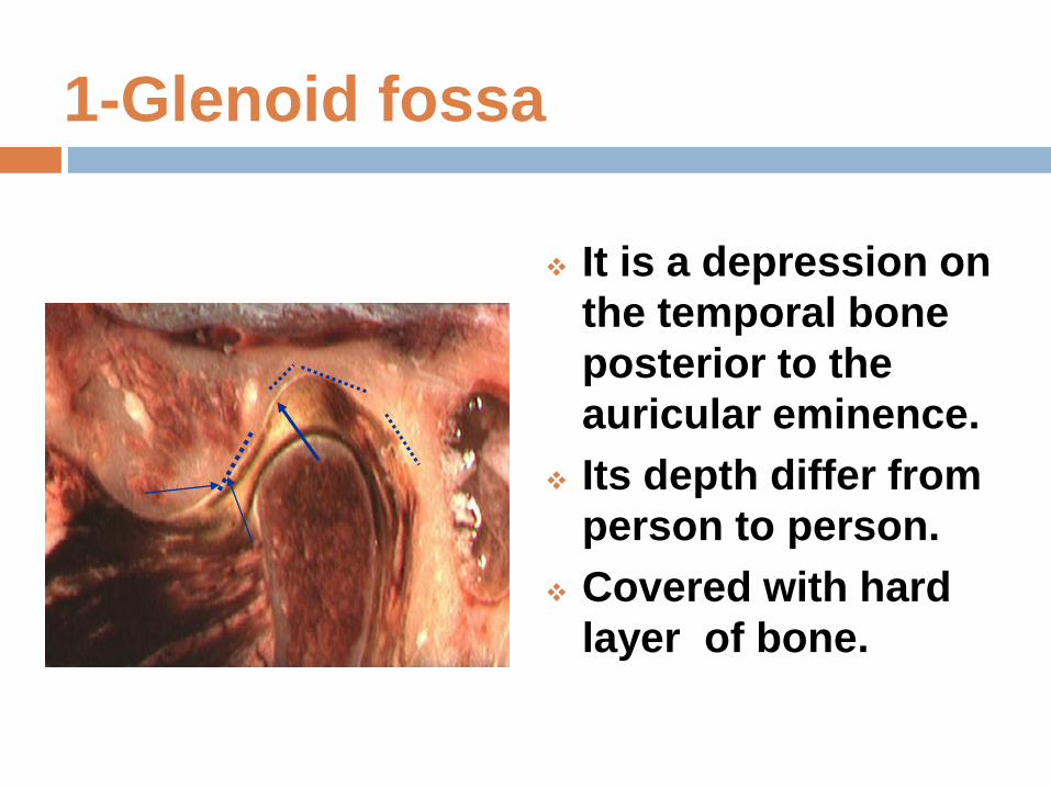
1-Glenoid fossa
It is a depression on
the temporal bone
posterior to the
auricular eminence.
Its depth differ from
person to person.
Covered with hard
layer of bone.
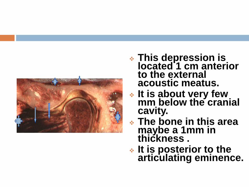
This depression is located 1 cm anterior to the external acoustic meatus.
It is about very few mm below the cranial cavity.
The bone in this area maybe a 1mm in thickness .
It is posterior to the articulating eminence.
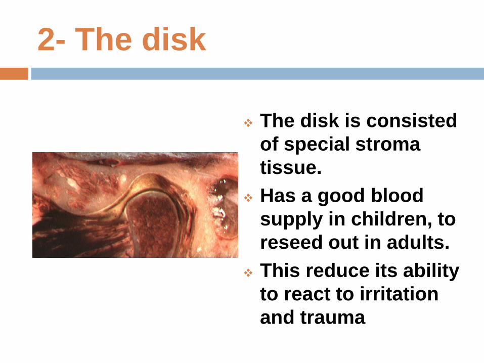
2- The disk
The disk is consisted
of special stroma
tissue.
Has a good blood
supply in children, to
reseed out in adults.
This reduce its ability
to react to irritation
and trauma
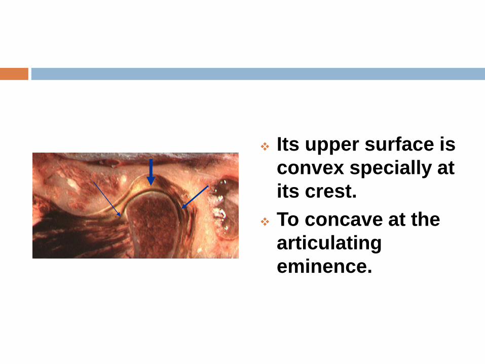
Its upper surface is
convex specially at
its crest.
To concave at the
articulating
eminence.
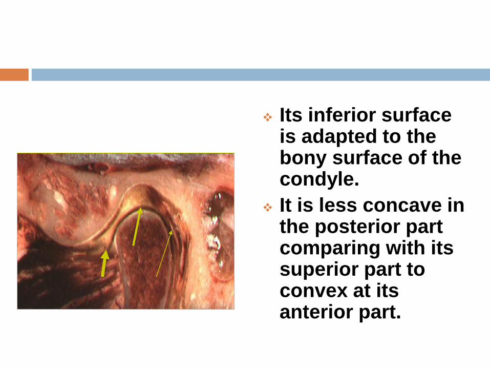
Its inferior surface is adapted to the bony surface of the condyle.
It is less concave in the posterior part comparing with its superior part to convex at its anterior part.
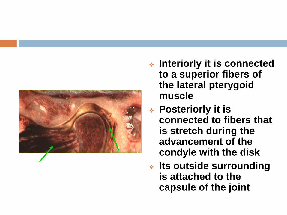
Interiorly it is connected to a superior fibers of the lateral pterygoid muscle
Posteriorly it is connected to fibers that is stretch during the advancement of the condyle with the disk
Its outside surrounding is attached to the capsule of the joint
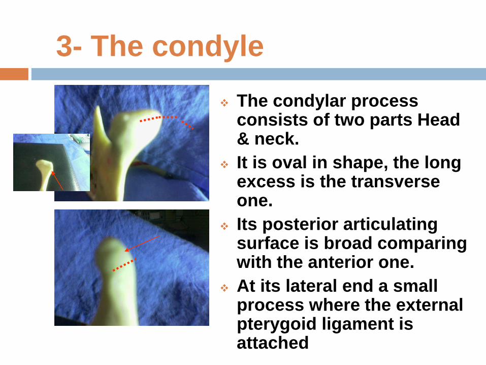
3- The condyle
The condylar process consists of two parts Head & neck.
It is oval in shape, the long excess is the transverse one.
Its posterior articulating surface is broad comparing with the anterior one.
At its lateral end a small process where the external pterygoid ligament is attached
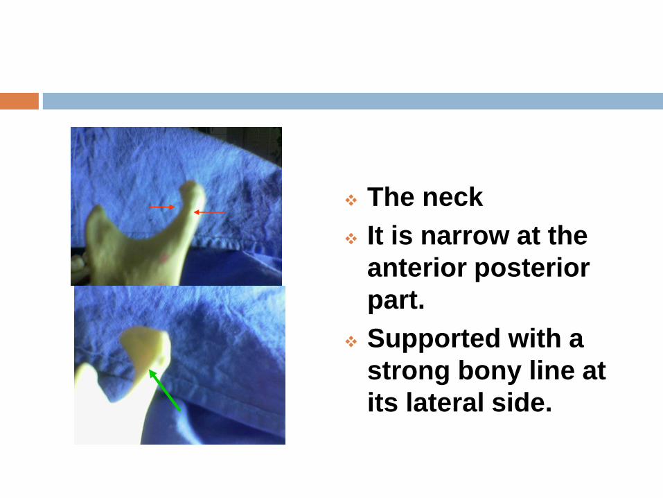
The neck
It is narrow at the
anterior posterior
part.
Supported with a
strong bony line at
its lateral side.
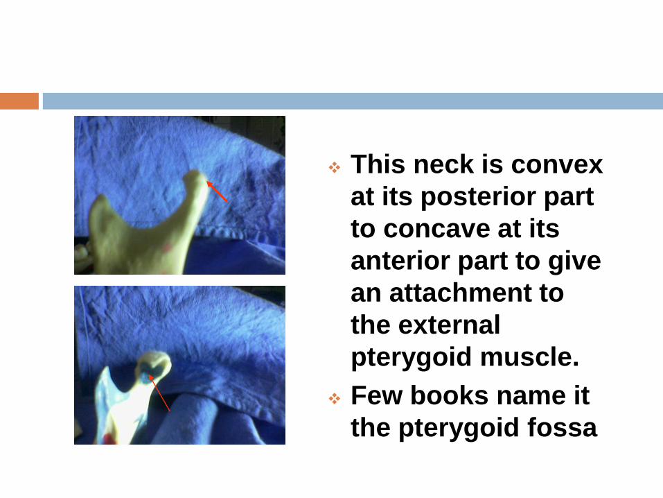
This neck is convex
at its posterior part
to concave at its
anterior part to give
an attachment to
the external
pterygoid muscle.
Few books name it
the pterygoid fossa
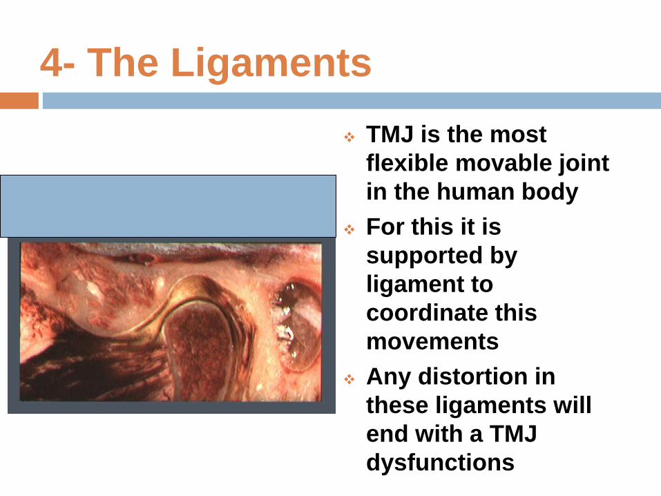
4- The Ligaments
TMJ is the most
flexible movable joint
in the human body
For this it is
supported by
ligament to
coordinate this
movements
Any distortion in
these ligaments will
end with a TMJ
dysfunctions
The Ligaments
There are three groups of them
1) Capsule
2) Intracapsular
3) Extra capsule
The superior fibers of the pterygoid muscle as it is attached to the disk plays an important part coordinating these elements during the joint movement.
Dysfunction of this muscle has its effects on the function of the joint.
The posterior ones will stretch under the effect of PT.M. during the advancement of the condyle keeping the disk covering the articulating surfaces.
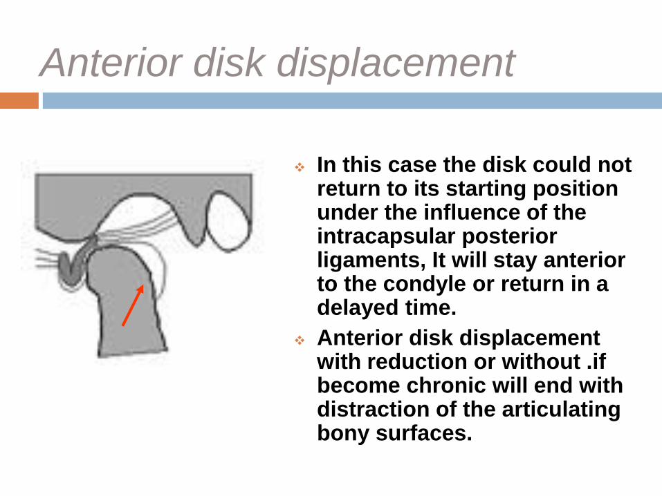
Anterior disk displacement
In this case the disk could not return to its starting position under the influence of the intracapsular posterior ligaments, It will stay anterior to the condyle or return in a delayed time.
Anterior disk displacement with reduction or without .if become chronic will end with distraction of the articulating bony surfaces.
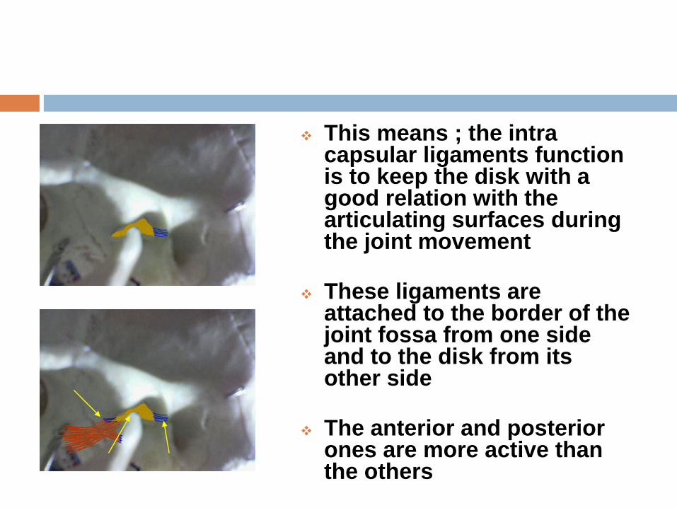
This means ; the intra capsular ligaments function is to keep the disk with a good relation with the articulating surfaces during the joint movement
These ligaments are attached to the border of the joint fossa from one side and to the disk from its other side
The anterior and posterior ones are more active than the others
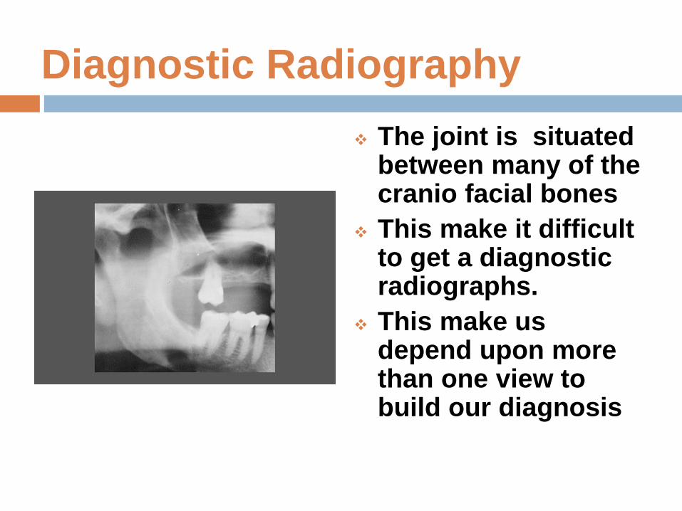
Diagnostic Radiography
The joint is situated between many of the cranio facial bones
This make it difficult to get a diagnostic radiographs.
This make us depend upon more than one view to build our diagnosis
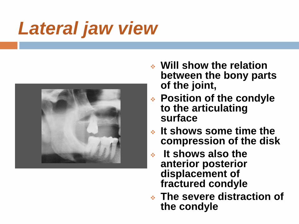
Lateral jaw view
Will show the relation between the bony parts of the joint,
Position of the condyle to the articulating surface
It shows some time the compression of the disk
It shows also the anterior posterior displacement of fractured condyle
The severe distraction of the condyle
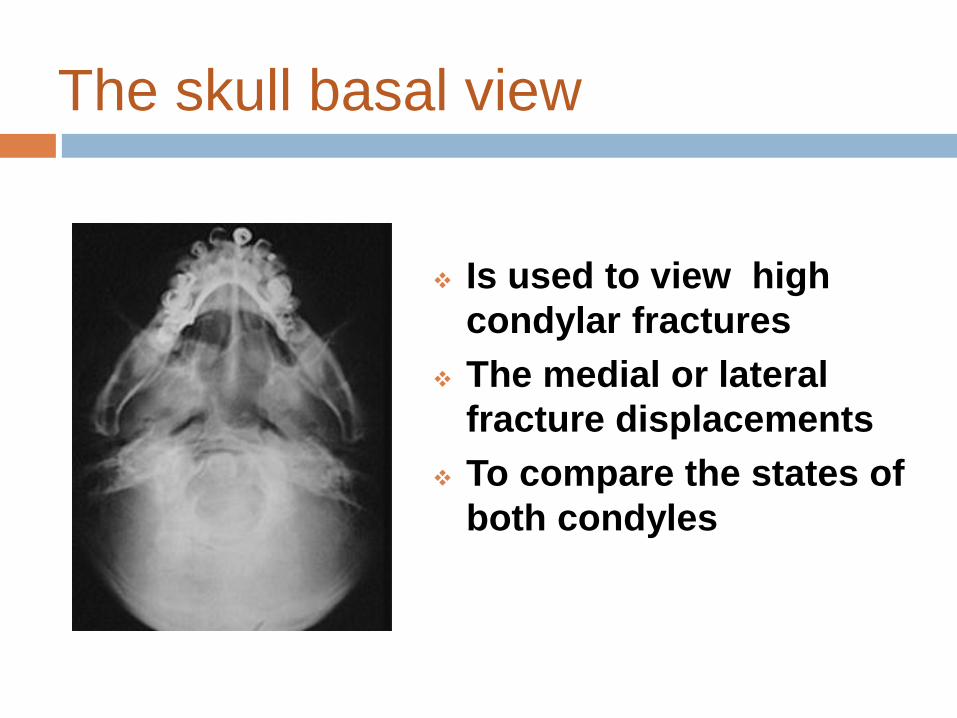
The skull basal view
Is used to view high
condylar fractures
The medial or lateral
fracture displacements
To compare the states of
both condyles

PA view
It is not a diagnostic
view because of the
overlapping specially
when it is taken and
the mouth is closed
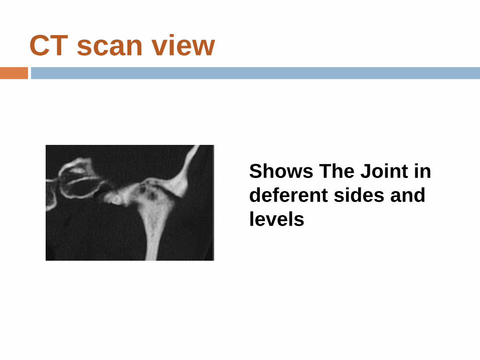
CT scan view
Shows The Joint in
deferent sides and
levels
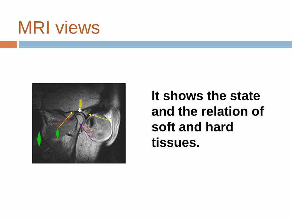
MRI views
It shows the state
and the relation of
soft and hard
tissues.
Management of TMJ disorders
Most cases of TMJ syndrome are
temporary; thus, treatment is usually
conservative.
Early therapy starts simply with resting the
jaw, using warm compresses (ice packs at
first if an injury is present), and pain
medication.

Management of TMJ disorders
Jaw rest can help heal temporomandibular
joints. Eat soft foods. Avoid chewing gum
and eating hard candy or chewy foods. Do
not open the mouth wide. Perform gentle
muscle stretching and relaxation
exercises. Stress-reduction techniques
may help manage stress and relax the jaw
along with the rest of the body.
Management of TMJ disorders
We may prescribe a splint or bite plate.
This is a plastic guard that fits over upper
and lower teeth, much like a mouth guard
in sports. The splint can help reduce
clenching and teeth grinding, especially if
worn at night. This will ease muscle
tension. The splint should not cause or
increase the pain. If it does, tell the patient
to stop using it.
Management of TMJ disorders
A more invasive procedure can be
performed in the doctor's office or clinic
under local anesthesia. This is carried out
by inserting two needles in the
temporomandibular joint to wash it out.
One needle is connected to a syringe filled
with a cleansing solution, and the fluid
exits via the other syringe.
Management of TMJ disorders
This procedure can be done in the office.
Most people find relief from the pain and
return to almost normal. Sometimes, pain
medication can be injected into the joint in
a similar procedure.

Management of TMJ disorders
Alternatively, a simple injection of
cortisone medication can be very helpful
in relieving inflammation and pain.
Management of TMJ disorders
A last option, surgery, is often irreversible
and should be avoided when possible. If
necessary, surgery can be used to replace
the jaw joints with artificial implants.