tlr x-ray structures harma brondijk bio-informatics course 2009
Post on 21-Dec-2015
215 views
TRANSCRIPT

TLR X-ray structures
Harma Brondijk
Bio-informatics course 2009

TIR domain structures
Protein-protein interaction surfaces:
1. Oligomerizationn interface
2. Interaction surface(s) with TIR domains of adapter molecules: MAL/MyD88 TRAM/TRIF SARM

TIR-domain structures
• TLR-TIR domains:– hTLR1 2.90 Å 1fyv (2000)– hTLR2 3.00 Å 1fyw (2000)– hTLR2-P681H 2.80 Å 1fyx (2000)– hTLR2-C713S 3.20 Å 1o77 (2002)– hTLR10 2.20 Å 2j67 (2006)
• Other TIR-domains– hMyD88 (NMR) 2js7/2z5v (2008)– IL1RAPL 2.30 Å 1t3g (2005)

TIR domains: similar structure, flexible regions
hTLR1
hTLR10
IL1RAPL
hTLR2
hTLR2_P681H
hTLR2_C713S

TIR domains: similar structure, flexible regions
BB-loop

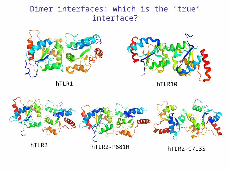
Dimer interfaces: which is the ‘true’ interface?
hTLR1725 Å2, 2 disulfide
hTLR1807 Å2, 2 saltbridges
hTLR1: - 1 molecule in the asymetric unit- protein-protein contacts -> two possible dimer interfaces
Dimer interfaces: which is the ‘true’ interface?
hTLR1 hTLR10
hTLR2 hTLR2-P681H hTLR2-C713S

TLR1-TLR2 TIR domain docking: putative dimer
GAUTAM et al. JBC 2006

hTLR10 TIR dimer: most likely ‘real’ signaling dimer
Nyman et al. JBC 2008
Molecule B
Molecule A
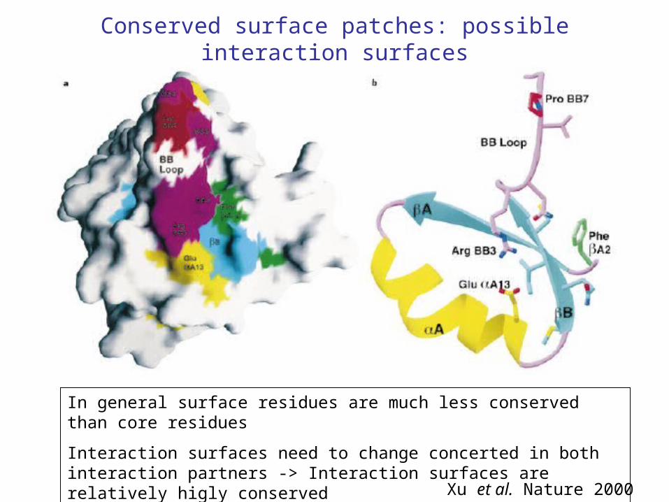
Conserved surface patches: possible interaction surfaces
In general surface residues are much less conserved than core residues
Interaction surfaces need to change concerted in both interaction partners -> Interaction surfaces are relatively higly conserved
Xu et al. Nature 2000

Modelling + Information driven docking programs -> Putative TLR4-Mal/TRAM interactions
Miguel et al. PLoS ONE 2007
LRR domain structures
Issues:
1. Overall structure
2. Interaction with ligands (agonist/antagonist)
3. Ligand induced dimerization? Conformational changes Interfaces
4. Interactions with co-factors

LRR-domain structures
• TLR ectodomains:– hTLR1/hTLR2 dimer + PAM3CSK4 (TLR1 aa 25-475/TLR2 aa 27-506)
2.10 Å 2z7x (2007)– hTLR2 aa 1-284 1.80 Å 2z80 (2007)– mTLR2 aa 27-506 1.80 Å/2.60 Å 2z81/2z82 (2007)– hTLR3 2.10 Å 1ziw (2005)– hTLR3 2.40 Å 1aoz (2005)– mTLR3 2.66 Å 3cig (2008)– mTLR3 + dsRNA 3.41 Å 3ciy (2008)– hTLR4 aa27-228 1.70 Å 2z62 (2007)– hTLR4 aa 27-527 2.00 Å 2z63 (2007)– hTLR4/MD2/Eritoran 2.70 Å 2z65 (2007)
- hTLR4/MD2/LPS 3.10 Å 3FXI (2009)– mTLR4/MD2 2.84 Å 2z64 (2007)– CD14 2.50 Å 1wwl (2005)

TLR ecto-domains consist ofleucine-rich-repeats
Choe et al. Science 2005Bell et al. PNAS 2005
hTLR3 hTLR3
convex
concave
lateral
Curvature may vary along the ectodomain: TLR1/2/4: divided in 3 distinct regions
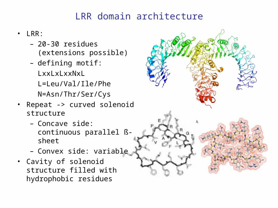
LRR domain architecture
• LRR: – 20-30 residues (extensions
possible)– defining motif:
LxxLxLxxNxL
L=Leu/Val/Ile/Phe
N=Asn/Thr/Ser/Cys• Repeat -> curved solenoid
structure– Concave side: continuous
parallel ß-sheet– Convex side: variable
• Cavity of solenoid structure filled with hydrophobic residues
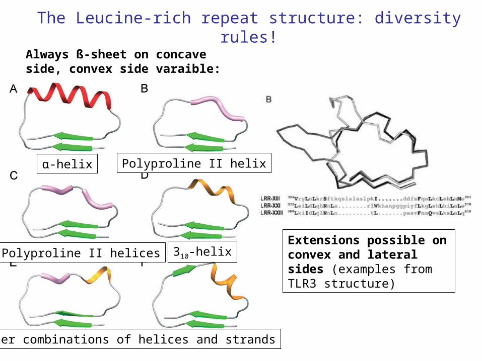
The Leucine-rich repeat structure: diversity rules!
α-helix
Always ß-sheet on concave side, convex side varaible:
Polyproline II helix
310-helix
Other combinations of helices and strands
2 Polyproline II helicesExtensions possible on convex and lateral sides (examples from TLR3 structure)

Ligand binding: TLRs recognize chemically diverse compounds
Flagellin(TLR5)
(TLR3) (TLR3)
Pam2CSK4(TLR6/TLR2 heterodimer)

How do TLR’s recognize/bind their ligands?
How does ligand binding induce TLR oligomerization?
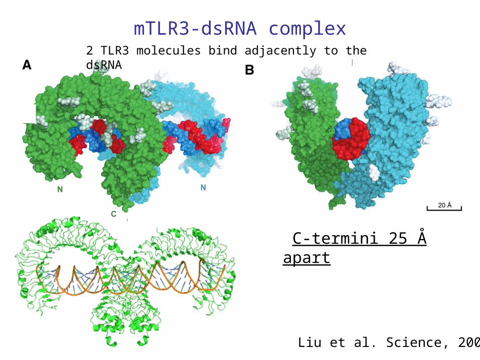
mTLR3-dsRNA complex
Liu et al. Science, 2008
C-termini 25 Å apart
2 TLR3 molecules bind adjacently to the dsRNA

• Two interaction sites close to N and C terminus
• Interactions with sugar-phosphate backbones only explains lack of sequence specificity
• Histidine involvement explains pH dependence
Liu et al. Science, 2008
dsRNA-mTRL3 interactions
N
NC
C
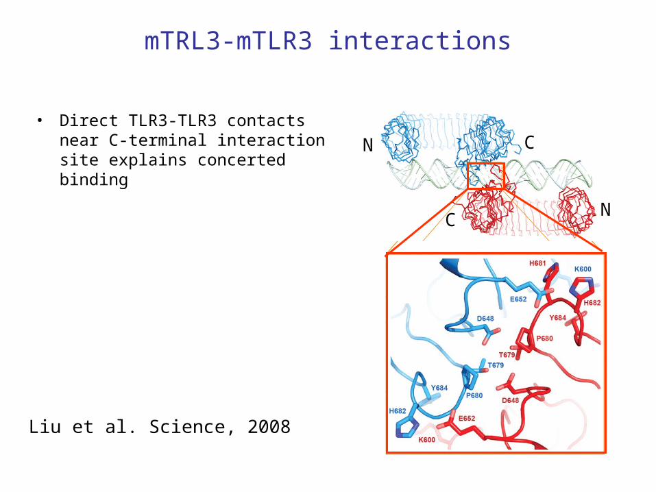
mTRL3-mTLR3 interactions
• Direct TLR3-TLR3 contacts near C-terminal interaction site explains concerted binding
Liu et al. Science, 2008
N
N
C
C

hTLR1-hTLR2 Pam3CSK4 complex
Jin et al. Cell 2007
C-termini <42 Å apart

hTLR1-hTLR2 Pam3CSK4 complex
Jin et al. Cell 2007
Top view
Ligand binds to both TLR1 and TLR2 -> heterodimerization
Question: How does the hTLR2-hTLR6 dimer form?? (ligand lacks the 3rd lipid chain)

mTLR4-mMD2 complex
Kim et al. Cell 2007
MD2 binds on the edge of the central and N-terminal region, at the lateral side of the molecule
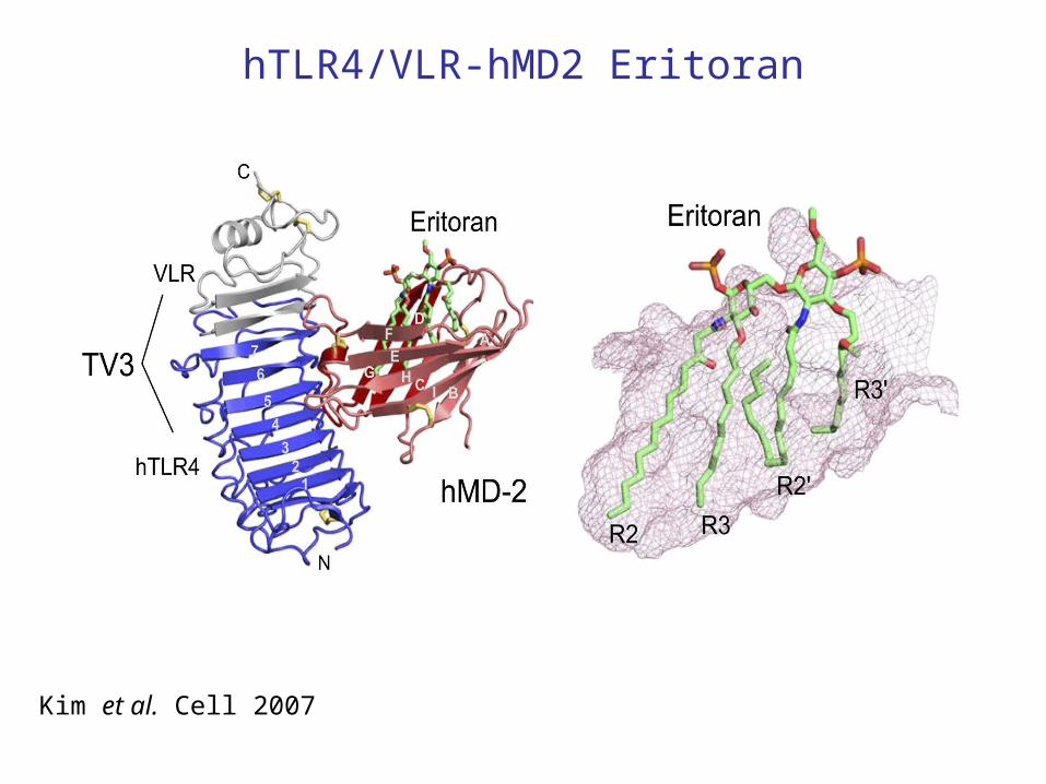
hTLR4/VLR-hMD2 Eritoran
Kim et al. Cell 2007

hTLR4-MD2-LPS homodimer
Park et al. Nature 2009

Dimerization through LPS and MD2 interactions
TLR4
TLR4*
MD2*
MD2

What makes LPS an agonist?
Lipid Iva and Eritoran have fewer lipid tails -> bind deeper in the MD2 binding pocket -> Phosphates not available for interactions
Park et al. Nature 2009

TLR4 dimerization appears to induce a conformational change
Central domains of TLR1/2/4 less rigid compared to standard LRR structures; needed to accommodate conformational changes???
Park et al. Nature 2009

Central theme: ligand interactions induce dimerization -> C-termini in close approximation
Park et al. Nature 2009

Thanks for your attention

Dimerization through LPS and MD2 interactions

TLR4-MD2-LPS dimerization interactions
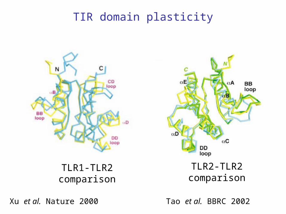
TIR domain plasticity
Xu et al. Nature 2000
TLR1-TLR2comparison
Tao et al. BBRC 2002
TLR2-TLR2comparison