title 脳血管外科に適した人工血管の開発 : 構造の設計 … is greater in the inner...
TRANSCRIPT

Title 脳血管外科に適した人工血管の開発 : 構造の設計と物理的特性の改善
Author(s) 宮本, 享
Citation 日本外科宝函 (1991), 60(1): 25-37
Issue Date 1991-01-01
URL http://hdl.handle.net/2433/203776
Right
Type Departmental Bulletin Paper
Textversion publisher
Kyoto University

Arch Jpn Chir 60(1), 25~37, Jan., 1991
脳血管外科に適した人工血管の開発
一一構造の設計と物理的特性の改善一一
京都大学医学部脳神経外科学教室(指導・菊池晴彦教授)
宮本 享
〔原稿受付平成2年10月25日〕
Development of Artificial Blood Vessel Suitable for Cerebrovascular Surgery:
Improvement in the Mechanical Properties
SusuMu恥1IYAMOTO
Department of Neurosur許可, Facultyof Medicine, Kyoto University
Saphenous vein interposition grafts have been commonly used for the reconstruction of occlusive le-
sions in the extracranial cerebral arteries, such as carotid or vertebral arteries. In contrast, cerebral
revascularization using an artificial blood vessel has not been so common. This is due to the fact that con-
ventional artificial blood vessels have been too stiff for use in the cerebrovascular surgery. Another reason
is that the patency rate of small-calibered artificial blood vessels has usually been inferior to that found in
autologous vein grafts. The purpose of this study was to develop a pliable and compliant artificial blood
vessel suitable for the reconstructive cerebrovascular surgery.
This new artificial blood vessel is made of polyurethane, porous in structure (porous polyurethane).
Thus, multiple small-sized pores exist both in the inner and outer surfaces, and in the wall of the graft.
To test its mechanical properties, stress-strain curves and compliance were evaluated. In com-
parison to expanded polytetrafluoroethylene gra氏, whichhas been one of the most commonly used ar-
tificial blood vessels in the cardiovascular surgery, the mechanical properties of the porous polyurethane
graft more closely resembled those of the common carotid artery in dogs. This means not only that it can
be maneuvered with technical ease for anastomosis but also that there is a reduction of compliance
mismatch between the host vessel and the artificial vessel. Compliance mismatch has been documented as
a m勾orfactor in the inducement of intimal hyperplasia, which causes a delayed occlusion of the grafts.
In the in vivo animal experiments, a porous polyurethane graft (2.3-3.2 mm in diameter, 40-50 mm
in length) was transplanted into the common carotid artery of dogs. The mean size of pore varied from 0
(group 1), 1. 7 (group 2), 4.4 (group 3), 5.5 (group 4), 7.4 (group 5), and 30.0 μm (group 6). In the
Key words: Artificial blood vessel, Polyurethane porous structure, Compliance, Cerebrovascular surgery.
索引語.人工血管,血行再建,ポリウレタ,コンブライアンス,脳血管外科
Present address: Department of Neurosurgery, Faculty of Medicine, Kyoto University, Sakyo -ku, Kyoto 606, Japan,
Kyoto University.

26 日外宝第60巻第 1号(平成3年 1月)
follow-up study conducted 2 months after the transplantation, all grafts in group 4, 5, and 6 were shown to be patent,
whereas no patency could be obtained in groups 1, 2 and 3. In addition, all grafts in group 6 were patent 6 months
after transplantation, though one showed remarkable stenosis due to anastomotic intimal hyperplasia. Histological ex-
amination showed that the luminal surface of the graft was covered with monolayer of endothelium-like cells.
Neoadventitia made of collagen developed around the external surface of the graft. Foreign body reactions were
minimal
In conclusion, a porous polyurethane graft could be developed, which has improved mechanical properties suitable
for the reconstructive surgeηr of the extracranial cerebral arteries.
I.はじめに
閉塞性脳血管障害に対する血行再建術はその病変の
部位や性状に応じていろいろな種類の術式が報告され
てきた.然しながら, interpositiongraftが必要とされ
る場合には頭蓋外脳血行再建術では主に自家静脈片移
植が用いられ,頭蓋内外 bypass手術においてはこの
他, radialartery graftを用いる報告もある.これに対
して,人工血管を用いた脳血行再建術の報告は少な
l ,6,13,31,36).この原因として,既存の人工血管が可
動性の大きい頚部血管や脆弱な脳血管に対して吻合す
るには硬過ぎること,脳血行再建術に応用可能な小口
径人工血管の開存率が自家血管移植には及ばないこと
が挙げられよう.
この研究では,脳血行再建術に応用可能な軟らかさ
を持つ小口径人工血管を開発するために,その物理学
的特性と構造上の設計につき検討を加えた.
Pore xlumen
Fig. 1 The schema of porous polyurethane graft
Porosity is greater in the inner layer than in the outer
layer.
Spandex • exists between these two layers as an internal
support.
Fig. 2 The photograph of porous polyurethane graft
脳血管外科に適した人工血管の開発 27
II.方 法
一人工血管の設計について
検討した人工血管は polyurethaneを素材とし,Fig.1
に示したように有孔性構造を持ち,その壁内に動脈圧
に耐えることができるように内部補強材として
polyurethane製弾性繊維の Spandex(直径 90μm)を
2重ラセン状に編み込んである.製造法としては
polyurethaneを有機溶媒に溶解した後,粒子サイズの
揃った無機物(CaCl2 または NaCl)を添加し,チュー
フ占状に成形する.最後に,この無機物を取り除くこと
により,その粒子のサイズに一致した孔径の poreが
形成される.入手可能であった無機物の粒子の平均サ
イズにより,有孔性構造の平均孔径(poresize)として
0即ち non-porousのものから, 1.7,4.4, 5.5, 7.4, 30.0
μmの合計 6種類の porouspolyurethane graftを製造し
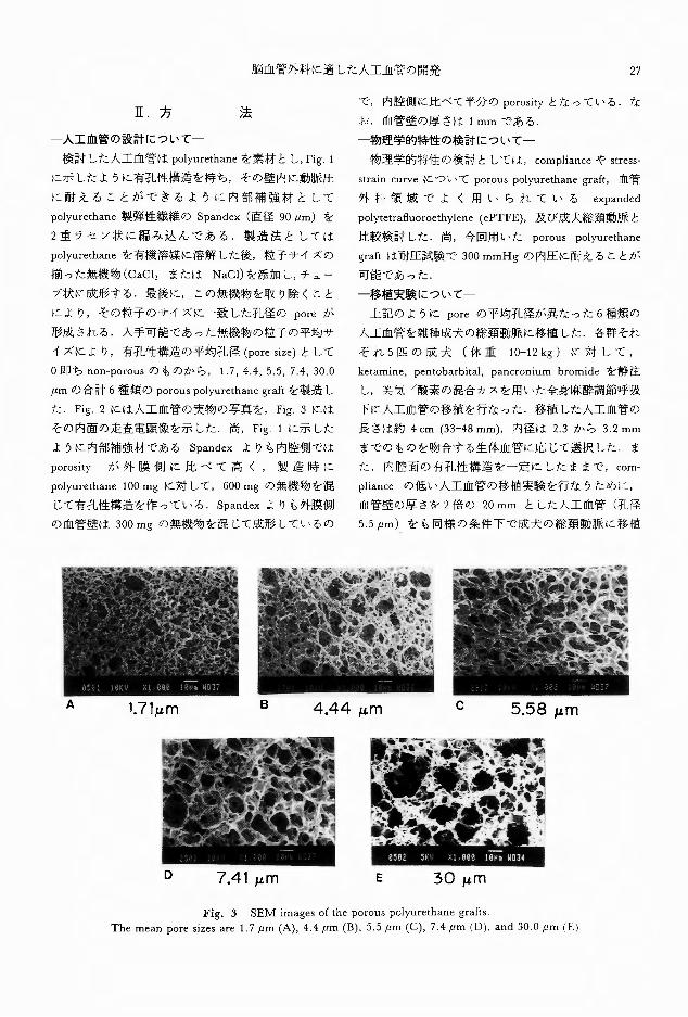
た. Fig. 2には人工血管の実物の写真を, Fig.3には
その内面の走査電顕像を示した 尚, Fig.lに示した
ように内部補強材である Spandex よりも内腔側では
porosity が外膜側に比べて高く,製造時に
polyurethane 100 mgに対して, 600mgの無機物を混
じて有孔性構造を作っている. Spandexよりも外膜側
の血管壁は 300mgの無機物を混じて成形しているの
γ ~i~
で,内腔倶IJに比べて半分の porosityとなっている.な
お,血管壁の厚さは lmmである.
物理学的特性の検討についてー
物理学的特性の検討としては, complianceや stress-
strain curveについて porouspolyurethane graft,血管
外科領域でよく用いられている expanded
polytetrafluoroethylene ( ePTFE),及び成犬総頚動脈と
比較検討した.尚,今回用いた porous polyurethane
graftは耐圧試験で 300mmHgの内圧に耐えることが
可能であった.
一移植実験についてー
上記のように poreの平均孔径が異なった 6種類の
人工血管を雑種成犬の総頚動脈に移植した 各群それ
ぞれ 5匹の成犬(体重 10-12kg)に対して,
ketamine, pentobarbital, pancronium bromideを静注
し,笑気/酸素の混合カスを用いた全身麻酔調節呼吸
下に人工血管の移植を行なった 移植した人工血管の
長さは約 4cm (33-48 mm),内径は 2.3から 3.2mm
までのものを吻合する生体血管に応じて選択した.ま
た,内腔面の有孔性構造を一定にしたままで, com・
pliance の低い人工血管の移植実験を行なうために,
血管壁の厚さを 2倍の 20mm とした人工血管 (干し径
5.5 μm)をも同様の条件下で成犬の総頚動脈に移植
A 1.71,um B 4.44 μm c 5.58 μm
。7.41 ,um
Fig. 3 SEM images of the porous polyurethane grafts.
The mean pore sizes are 1.7 μm (A), 4.4 μm (B), 5.5 μm (C), 7.4 μm (0). and 30.0 μm (El

監
第 l号(平成3年 1月)
Compliance (% mmHg-1〕
0.4
0.3
した.移植は手術用顕微鏡下に 7-0polypropylene糸
(Prolene •)を用い,片端10から 14針で結節縫合によ
りend-to-endanastomosisを行なった.抗凝固剤は術
前,術中,術後を通じて全く用いていない.
移植後 2,4, 8, 24週の時点で創部を再開創してその
開存を確認した後に,取り出して各々組織学的検索を
行なった.long-term follow up studyでは入手できた粒
子サイズが最も揃っていた(即ちサイズの分散が最も
小さし、)平均孔径 30.0μmの人工血管を用レた.
第60巻日外宝28
~,l 0.263 ±0.025
(02~
0.78)
1.71 4.48 5.58 7.41 μm μm μm μm
Fig. 5 Compliance of the vessels
Porous polyurethane grafts are proved to be more com-
pliant. Fi思Hesindicate mean± standard deviation
The reported values of the compliance of carotid artery
are between 0.2 and 0.78 [41].
Carotid artery
J~ 0.222 ±0.028
30.0 μm
円也
F09h
i
l
j
a
“守nH
T
’aa’人νEEEi勾
ιnu
。±
ePTFE
0.2
日1
果
一物理学的特性の検討
Fig. 4にporouspolyurethane graft,成犬総頚動脈,
ePTFE (Goretex R)各々の応力歪曲線を示した.
ePTFEに比べ, porouspolyurethane graftは明らかに
生体血管に近い curveを示した.
Fig. 5には ePTFE,平均孔径の異なった各種porous
polyurethane graft及び報告のある総頚動脈の各々の
complianceを示した.更に porouspolyurethane graft
製造時に添加する無機物の量を変化させることにより,
porosityとcomplianceとの関係を検討し, Fig.6に示
した ePTFEに比し, porouspolyurethane graftは
complianceが大きく,総頚動脈のそれにより近い値を
示した.porosityを上げることにより, complianceの
大きい,即ちより軟らかい人工血管を作ることが可能
であったが(Fig.6), polyurethane 100 mgに対して無
III.結
4 6
Solt I PU
The interrelationship between porosity and
compliance
0.6
@ u c: 0 ~ e 0.2 0 ιJ
O> 工E E 渓 0.4
100
(
e、』E E so \ 。、
80
40
20
的的由」↑
ω
8 rcitio
。Fig. 6
50
Stress-strain curves of ePTFE (1 ), porous
polyurethane graft (2), and of the common
carotid artery of dog (3)
The stress-strain curve of porous polyurethane graft
resembles that of the common carotid artery of dog,
in contrast with ePTFE.
40 30
(%)
20
Strain
10 。
Fig. 4

29
poreの平均孔径が5.5,7.4, 30.0 flmの各群では移植後
4.8週では人工血管はし、ずれも開存してレた.
6ヵ月後の follow-upを行なった poresizeが 30.0
pmの群では 5f7U1,、ずれもが開存していたが,内 1iYU
は吻合部を中心として著明な狭窄を呈していた
組織学的杉:宗ては,移植4および8週後に開存して
いた 3群の人工血管の内股面はいずれも両端の吻合部
を生体血管側から単層の扇平な内皮様細胞に覆われて
おり, graft の中央部ではζ く薄い疎性結合組織から
成る pseudointimaが内腔面を覆ってし、た(Fig.7, 8).
人工血管の外側には新生血管を伴ない糟生した coト
lagenからなる新生外膜が sheath状に graftを取り囲
脳血管外科に適した人工血管の開発
機物 BOOmgを添加して製造した人工血管は軟らかし、
ものの脆弱であり,吻合後血流を再開すると血圧に耐
えず perforate したため,方法の項で前述のように内
外2層から成る porosityをもっ人工血管を実際の移植
実験に用いた.実際の吻合操作においても用いた
porous polyurethane graftは十分な軟らかさを持ち,
生体血管どうしの吻合とほぼ同様の感触であった.
一構造の設計と移植実験についてー
有孔性構造の poreの平均孔径と移植実験との関係
をTablelに示した. non porous及びporeの平均孔径
が 1.7,4.4 flmの3群では移植した人工血管は移植後
4週の時点ですべて閉塞していた これに対して,
Table 1 Summary of the transplanted porous polyurethane graft
Figures indicate the number of patent graft. PPU: porous polyurethane graft
PU: non-porous polyurethane graft
No. of Patent Graft
6m
PU (non-porous)
PPU (pore size= !. 71)
PPU (pore size=4.44)
PPU (pore回ze=S.58)
PPU (pore size=7.47)
PPU (pore size=30.0)
PPU (thick wall)
2m
5 (100%)
5 (100%)
5 (100%)
lm
。(0%)
0 (0%)
0 (0%)
5 (100%)
5 (100%)
5 (100%)
l (20%)
Om
民J
R
d
k
J
R
J
R
J
F
3
R
d
Graft
5 (100%)
民主ー 戸-,一一-一一ー一一一一一- - .-
Histological evaluation of the porous polyurethane graft 8 weeks a氏erthe transplantation (Azan stain)
The luminal surface of the gra代(top)is smooth without thrombi. Neoadventitia composed of colla伊n
surrounds the outer surface of the graft
Arrows indicate the porous polyurethane graft
Fig. 7

30 日外宝第60巻第 l号 (平成 3年 1月)
んでいたが,人工血管とこの新生外膜との癒着は僅か
で,容易に剥離で‘きた(Fig.8)ー異物巨細胞や炎症細
胞浸潤などの異物反応は殆ど認めなかった. pore size
が 30.0μmの porouspolyurethane graftの移植6ヵ月
後の組織学的検索では 1例を除いて,人工血管の内面
は新生内膜に覆われ, graft の全長にわたって単層の
内皮様細胞がこの新生内膜の表面を覆っていた(Fig.9).
しかしながら,走査電子顕微鏡による観察では, 内皮
様細胞の形態は多様であり,核の明瞭な大きな幼若型
のものから敷石状配列を呈する成熟型のものまで様々
であった.また,光顕像では graftの全長が内皮様細
胞で覆われているよう に見えても,電顕で、は内腔面に
は内皮様細胞に覆われず,その下の結合組織が露呈し
ている部分もあることが観察された(Fig.10, 11, 12)・
Fig. 8 Histological evaluation of the porous polyurethane graft 8 weeks after the transplantation (Masson trichrome stain)
The luminal surface of the gra丘四 linedwith monolayer of endothelium-like cells (arrows).
Fig. 9 Histological evaluation of the porous polyurethane gra氏6months after the transplantation (hematoxyhn eosm stain)
The neointima developed on the luminal surface of the graft.

脳血管外科に適した人工血管の開発
Fig. 10 SEM image of the luminal surface of the porous polyurethane graft 6 months after the transplantation
The luminal surface is covered with endothelium-like cells showing cobble stone appearance.
Fig. 11 SEM image of the luminal surface of the porous polyurethane graft 6 months after the transplantation
Some immature endothelium-like cells are large in size and have a remarkable big nucleus.
31

32 日外宝第60巻第 1号(平成3年 1月)
新生内膜の深層には紡錘形の modifiedsmooth muscle
cellが増生していた(Fig 13). 人工血管に対する異物
反応や新生外膜の状態;士作植2ヵ月後のものとほほ同
様であった.著明な狭窄を呈した l例は吻合部を中心
として内膜が肥厚しており,その本態は mod凶ed
smooth muscle cell及びそれらが造成する collagenから
Fig. 12 SEl¥l image of the luminal surface of the porous polyurethane graft 6 months after the transplantat10n
Several areas of the luminal surface are not covered with endothelium-like cells. Subendothelial connec-
tive tissue is exposed
Fig. 13

脳血管外科に適した人工血管の開発 33
Fig. 14 Histological evaluation of the porous polyurethane graft 6 months after the transplantation (Azan stain)
Intimal hyperplasia composed of modified smooth muscle cells and collagen resulted in marked stenotic le-
SIOn.
構成されていた(Fig.14).
N.考 察
人工血管の開存性を向上させるためにこれまで種々
のアプローチが報告されてきた1&-21,27,28,33-35抑止4判噌川2).
heparin などの抗凝固剤を徐放させる薬理学的手法,
抗血栓性の優れた水溶性高分子を表面にコーティング
する医用高分子学的方法,表面の幾何学的構造により
偽内膜形成を促進させる物理的方法,物理学特性に注
目して吻合する生体血管への力学的負荷を減弱する生
体力学的方法など多彩である.
脳神経外科における人工血管の需要としては,まず
頭蓋内動脈血行再建術においての donararteryである
頭皮動脈の代用が挙げられる. recipient arteryとして
吻合される頭蓋内動脈は体動脈に比べて脆弱であり,
その口径は成人であっても 1~2mm と小さレ.この
ため既存の人工血管は硬過ぎ且小口径においての開存
率が芳しくないため使用に適さなレ.自家静脈片移植
を用いる際には静脈弁の存在により血流を!|慎行性に保
つためには細かい頭蓋内動脈に対して静脈片の中枢側
すなわち太い方の端を吻合することとなり caliber
discrepancy が問題となる.つまり軟らかL、小口径の
人工血管が必要とされる.また,頭蓋外脳血管の血行
再建術においては伸展屈曲回旋を行なう頚部という可
動性の大きい領域の特殊性を考えると,やはりその運
動に耐え得る軟らかで compliantな人工血管が理想と
される.脳血管外科におけるこれらの特殊性を考慮に
入れて本研究では人工血管の物理的特性をできるかぎ
り生体血管に類似させ, 吻合される血管への力学的負
荷を減弱させるというアプローチを採った
complianceが人工血管の開存に関しての大きな要素
であることはこれまでにも数多く報告されてき
た1,15,22,23,26,30-32,42,50) • compliance mismatch があ
ると血流に対する impedanceの増大, turbulenceの発
生,末梢への潅流低下などの招来が知られている 文
吻合部近傍の生体血管に力学的負荷が加わり,内膜過
形成を惹起し人工血管の閉塞をもたらすことが知られ
ている38). Fig. 4および5に示したように有孔性構造
を持たせることにより, porouspolyurethane graftは
既存の人工血管である ePTFEに比べてはるかに成犬
頚動脈に近い物理学的特性を示した.このことは吻合
後の生体血管に対する力学的負荷が既存の人工血管で
ある ePTFEに比しporouspol刊 rethanegra食では小さ
いことを意味し,前述のごとく軟らかで小口径の人工
血管が要求される脳血行再建術に適していると考えら
れる.本研究ではこれに加えて人工血管の開存性に対
して complianceの相違が与える影響を出来るだけ他
の因子を除外した条件で‘検討するため Tablelに示し
たように壁の厚い porouspolyure出anegraftを用いて
移植実験を行なった.Fig. 6のように porosityを変化

34 日外宝第60巻第 1号(平成 3年 1月)
させると complianceを変えることができるが,内腔
面の状態(例えば内腔面に存在する poreの密度)が
変わってしまうので他の因子を除外した compliance
固有の影響を検討することができないI),これは種類
の違う人工血管の聞で complianceの違いが開存性に
与える影響を考察しても同様である.用いた壁厚の
porous polyurethane graftのcomplianceは約70%に低
下したが, porosity や血流との接触面の状態は他の
porous polyurethane graftと同じである.移植4週後
の壁厚の porouspolyurethane graftの開存率は20%で
あり,同じく 5.5μmの poresizeを持つ通常の壁厚の
porous polyurethane graftの開存率(100%)と大きく異
なることから,やはり compliancemismatchが人工血
管の開存に与える影響は大きいと考えられる.
小口径ということに関しては本研究で用いた
porous polyurethane graftは内径が 2.33.2 mmのもの
を用いそれ以下の口径のものは試していなレか,これ
は成犬総頚動脈の内径に合わせて人工血管を選択した
結釈てある この内径は成人であれば十分脳血行再建
術に応用可能な口径である より小口径の人工血管に
ついての報告もあるが多くはラ y トを実験動物として
おり,移植した人工血管の長さが短いこと,また動物
の種差の問題もある43)ー本研究では人工血管の治癒過
程がヒトに近いとされる成犬を用いて行なった.
有JL性構造とし、う人工血管の幾何学的構造と移植実
験における開存性との関係に注目すると porous
polyurethane graftのporesizeが 0,1.7, 4.4 μmの各群
の graftはすべて移植後4週の時点で閉塞しており,
pore sizeが 5.5,7 .4, 30.0 μmの各群では移植後 8週経
ってもすべての graftは開存していた Fig. 5に示し
たように,これらの graftはその poresizeが異なるの
みで complianceは類似した値である目すなわち物理
学的特性がほぼ一定という条件下で, graft の開存の
ためにはある程度以上(本研究では 5.5pm以上)の
孔径の pore の存在カ過重要であることが示された.
porosityや poresizeが大きいほど人工血管の治癒過程
が進行しやすいとL、う過去の知見に対応する結果と思
われる4,5,24,29,31,47,51).
組織学的傾索では移植後数日では人工血管の内腔面
に疎性結合組織を主体とした薄い偽内膜が形成され,
時が経つとともに生体血管側より graftの中央部に向
かつて扇平な核を持つ内皮様細胞が人工血管の内腔面
を覆ってゆき,移植後6ヵ月では graftの全長にわた
って内皮様細胞が覆っていた日7,8,11,14,29,31).炎症細胞
浸潤や異物巨細胞なとeの異物反応はきわめて僅かで、あ
りporouspolyurethane graftが生体によく適合してし、
ると考えられた しかしながら,移植後6ヵ月の検索
では程度の差こそあれ内皮様細胞に覆われた新生内膜
の深層に紡錘形の modifiedsmooth muscle cellの増殖
を認め,これが著明に増殖した 1例では増生した coト
lagen とともに内膜過形成に依る狭窄の原因となって
いた.人工血管の吻合部周辺の内膜過形成についてi土
これまで‘fこも多く報告されている川11,町
常内皮細胞は抗血栓作用を持つためこJLに?宣われた人
工血管は優れた抗血栓作用を持つと考えられるが,
modi品edsmooth muscle cellは内皮細胞層下の内膜組
織内で増殖することが知られている l) modified
smooth muscle cellは動脈硬化性病変形成においての
関与が指摘されているが,移植後慢性期における人工
血管の閉塞性病変の原因としても人工血管素材の抗血
栓性という問題よりこの細胞の増殖性変化の関与が大
きいと考えられる9,凧39l. modified smooth muscle cell
の増殖に関する制御機構として血/上阪, macrophage
なとからの成長因子の重要性が指摘されている 10,39).
人工血管における上述の増殖性変化においても人工血
管の表面に粘着した血小板から成長因子が放出され
(platelet derived growth factor (PDGF)), mod凶ed
smooth muscle cellの増殖を促すことは十分に考えら
れる.また,人工血管吻合部周辺では内皮細胞の
turn overが冗進しており慢性的な内皮細胞障害が存
在しそれが慢性的に smoothmuscle cellの増殖を促進
させる可能性があることも報告されている 7,8).本研
究でも人工血管上の内皮様細胞は走査電子顕微鏡では
多彩な形態を示し一部には核が明瞭で、核/細胞質比
が大きく幼若型内皮細胞と思われる細胞が存在したこ
とは内皮細胞の turnoverが允進していることを示し
ていると考えられる smoothmuscle cellの増殖がよく
制御されている際には人工血管の表面を内皮細胞か覆
い,その深層にほ modifiedsmooth muscle cellが散在
する厚さの均一な新生内膜となり, PDGFその他の成
長因子に(より増殖性変化が促進されすぎた例て、は内膜
の過形成に至ると考えられる.現在の人工血管研究の
アプローチでは移植後早期の抗血栓性という問題はあ
る程度克服できても,慢性期における modified
smooth muscle cellの増殖性変化の病態や発生機構に
は動脈硬化性病変と同様いまだ解明されていない点も
多く,これを抑制するのはなかなか難しい しかし少
なくとも PDGFの関与は既に報告されており, PDGF

脳血管外科に適した人工血管の開発
放出には血小板凝集が先行し,抗血小板療法が内膜過
35
形成を減弱させたとの報告もある 12).
porous polyurethane graftは物理学的特性が生体血
管に極めて類似しているため吻合部における pulsatile
mechanical stressの程度が既存の人工血管に比べて軽
L、と考えられる.これは血小板凝集の原因となる吻合
部近傍における血流の turbulence,内皮損傷を減じて
いることとなり, complianceの小さな人工血管に比し
て慢性期における増殖性変化の予防にも役立っている
と考えられる 本研究では人工血管の物理学的特性に
注目しそれが移植実験に与える影響を検討したが,上
述のように慢性期の細胞増殖性変化を抑制するために
はその病態の解明が今後必要と考えられる.また光顕
上人工血管内陸面の全長を覆っているように見えた内
皮様細胞も走査電子顕微鏡に依る観察ではその形態は
多様であり, mature な内皮細胞としての正常の機能
を発現するにはより長い観察が必要と考えられる
v.結 三五回日
1) porous polyurethane graftはその有孔性構造により
生体血管に類似した物理学的特性を示し,吻合し
た生体血管への力学的負荷を減弱すると考えられ
た.
2) porous polyurethane graftの開存のため仁はその
pore size が一定以上の大きさ(本研究では 5.5
μm以上)であることが必要と考えられたー
3)組織学的検索では異物反応はわずかであり, graft
の内腔面は単層の内皮様細胞に覆われ生体によく
適合してし、た.
稿を終えるにあたり,本研究について多大なご指導
を頂きました京都大学医学部脳神経外科 菊池晴彦教
授,京都大学生体医療工学研究センター長筏義人教
授に深甚なる謝意を捧げます また,本研究の実施に
あたり惜しみなく御協力を頂レた京都大学生体医療工
学研究センタ一生体材料研究部門 湊立雄先生(現日
本メディカルサプライ中央研究所),藤本啓二先生(現
慶応義塾大学理工学部応用化学科助手),字部日東
化成岐阜研究所 阪井和彦先生,京都大学医学部脳神
経外科金子隆昭先生に心からお礼申し上げます最
後に本研究について大切なζ助言を幾度も頂いた京都
大学医学部脳神経外科永田泉講師仁川甚なる謝意を
捧げます.
引用文献
1) Abbott WM, MegermanJ, HassonJE, et al: Effect
of compliance mismatch on vascular graft patency.
J Vasc Surg 5: 376-382, 1987.
2) Absolom DR, Hawthorn LA, Chang G: En-
doth el凶 1zationof polymer surfaces. J Biomed
Mater Res 22: 271-285, 1988.
3) Berger K, Sauvage LR, Rao AM, et al: Heali暗 of
arterial prostheses in man: Its incompleteness. Ann
Surg 175: 118 127, 1972.
4) Boyd KL, Schm凶 S,Pippert TR, et al: The
effects of pore size and endothelial cell seeding upon
the performance of small-diameter e-PTFE vascular
grafts under controlled flow conditions. J Biomed
Mater Res 22: 163, 1988.
5) Campbell CD, Goldfarb D, Roe R: A small
arterial substitute: expanded micro-porous
polytetrafluoroethylene: patency versus porosity
Ann Surg 182: 138-143, 1975.
6) Cavanaugh DA, Story JL, Brown WE Jr, et al:
Polytetrafluoroethylene interposition grafts in
vertebral to carotid artery transposition. A long-
term follow-up study. J Neurosurg 70: 212-215,
1989.
7) Clowes AW, Gown AM, Hanson SR, et al:
Mechanisms of arterial graft failure: 1 Role of
cellular proliferation in early healing of PTFE pro-
stheses. Am J Pathol 118:・ 43-54,1985
8) Clowes AW. Kirkman LTR, Clowes 恥1M:
Mechanisms of arterial graft failure: 2. Chronic en-
dothelial and smooth muscle cell proliferation in
healing polytetrafluoroethylene prostheses. J Vasc
Surg 1: 525-535, 1984.
9) Davies PF: Biology of disease: Vascular cell interac
tions with special reference to the pathogenesis of
atherosclerosis. Laboratory Investigation 55: 5-24,
1986
10) DiCorleto PE, Bowen-Pope DF: Cultured en-
dothelial cells producぞaplatelet-derived grwoth fac-
tor-like protein. Proc Natl Acad Sci USA 80:
1919ー1923,1983
11) Echave V, Koornick AR, Haimov M, et al: In-
timal hyperplasia as a compli仁ationof the use of the
polytetrafluoroethylene graft for femoralpopliteal
bypass. Surgery 86: 791-798, 1979.
12) Hagen PO, v何angZG, Mil《atEM, et al: An
tiplatelet therapy reduces aortic intimal hyperplasia
distal to S口iall-diametervascular prostheses (PTFE)
in nonhuman primates. Ann Surg 195: 328-339,
1982
13) Hallett JW Jr, Chnrv KJ Jr, Pairolero PC, et al.

36 日外宝肋う{}を第 l号 (平成 3年 1月)
Reconstructive surgen・ for the aortic arch branches
and the vertebral arteries. in Sundt Tl¥.リr(ed): Oc-
elusive Cerebrovascular Diseases, Saunders,
Philadelphia, 1987, pp 355-384.
14) Hanel KC. i¥lcCabe C, Abbott WM, et al: Cur-
rent PTFE grafts. A biomechanical scanning el引
tron and light microscope evaluation. Ann Surg
195: 456-463、1982.
15) Hasson JE, i¥k"汀manJ, Abbott ¥¥"t¥I: Suture
technique and para-anastomotic compliance. J
Vasc Surg 3: 591-598, 1986
16) Hayashi K, Handa H, Nagasawa S. et al: Stiffness
and elastic behavior of human intracranial and ex-
tracranial arteries. J Biochem 13: 17 5, 1980
17) How T\’ Annis D: Viscoelastic behavior of
polyurethane vascular prosthes日 JBio med M川
Res 21: 1092 1108, 1987
18)井島宏,斎藤大,村井正,他: 人工血管の現状と問題点. 人工臓器 19:1060-1063, 1990
19)筏 義人.血絵を生成しない人工表面は可能かH.商 26:49可7,1988
20)筏義人 ;イオマテリ アル一人工臓器へのアプ
ロー チー.東京,日刊工業新聞社 1988, pp
118-127
21) Kambic HE: Polyurethane small artery
substitutes. Trans Am Soc Artif Intern Organs 34
1047-1050, 1988
22) Kidson IG, Abbot ¥¥'M Lo"' compliance and
arterial graft occlusion. Circulation 58 (suppl.)
1-4 1978
23) Kinley CE. i¥larble AE: Compliance. 人 rontmu-
ing problem with vascular grafts. J Cardiovas Surg
21: 163-170, 1980
24) Ku"1ba A, Fischer CR, Marulewski TJ: Ex-
perimental study of the influence of porosity on
development of neointima in Ge"" l円 grafts: a
method to increase long term patency rates. :¥rn
Surg 47: 347 354, 1981
25) LoGerfo FW、 Soncrant T, Teel T, et al
Downstream anastomotic hyperplasia: A
mechanism of failure in dacron arterial grafts. Ann
Surg 197: 479-483, 1983
26) Lyman DJ, Fazzio FJ. Voorhees H, et al: Com
pliance as a factor e町ecting the patency of "
copolyurethane vascular graft. J B問nedt¥ l."i..'
Res 12: 337-345, 1978.
27)松田武久,岩田博夫,高野久輝,他血管壁の再構成器官培養技術の開発と小口径人工血管への応用の可能性.人工臓器 17・651, 1988
28)総本博志,吉良一明,宮脇富土失,他新しい人工血管の開発 人工臓器 I7: 610-613, 1988
29)絵本博志,吉良一明,宮脇富士*..,他新しL、人
工血管の開発一病理組倣像と開存性ー.人工臓器18: 257-260 1989.
30 l J¥.kgermanj, Abbott ¥¥'M: Compliance in vascular
grafts. in C ¥¥'right (ed), Vascular Grafting
Boston, John ¥Vright-PSB, 1983, pp. 344-364
31)宮本亨,菊池晴彦,筏義人,他 ー脳血管外科
領域に適した人工血管の開発.構造の設計と物理的特性の改拾 脳外 18:253-258, 1990.
32)宮脇富士夫,松本博去、,斎藤寛文,他:低コンプ
ライアンス人工血管の流体力学的影響の検討.人
工臓器 18:264-268, 1989.
33) Noishiki Y, J¥.l1yata T: Successful animal study of
small caliber heparin-protamin collagen vascular
grafc Trans Am Soc Artif Intern Organs 31・
102-106, I 985
34) Noishiki、Mi下1ata T: A simple method to
heparinize biological materials. J Biomed Mater
Rれ 20:337-Hti. 1986
35)野尻知里・人工血管 この 1年の進歩.人工臓器
18: 159←1595, 1989.
36)大西英之: lnterposiion gra丘materialとして何が
最適か.Neurosurgeons 6・ 243-244, 1987
37) Phua SK, Castillo CE. Anderson JM, et al
Biodegradation of a polyurethane in vitro. J Biom-
ed Mater Res 21: 231-246, 1987.
38) Pomposelli F, Schoonト.Cohen R,川 al: Conforma-
tional stress and anastomotic hyperplas】a J ¥' asc
Surg 1: 525-535, 1984
39) Ross R, Glomset J,札 iri、aB, et al: A platelet-
dependent serum factor that stimulates the prolifera-
tion of artenal smooth muscle cells in vitro. Proc
Natl Acad Sci USA 71: 1207-1210, 1974.
40)佐久間まこと,西部俊哉,安田慶秀,他 ・理想的人工血管開発へのアプロ ーチ 抗血栓性と弾性特性から見た人工血管の性能評価 人工臓器 19・
1056-1059, 1990.
41 )笹島唯博,久f 'U~ 子、稲葉雅史,他代用血管の至適 compliance.人工臓器 12:179-182, 1983.
42)笹島唯博,久('1'.!':!彦, ’l、注正樹,他小口径人工
血管開発の動向.従来の問題点と今後の方向.人工臓器 19:1052-1055, 1けり()
43) Sauvage LR, Berger KE Wood SJ, et al: In-
terspecies healing of porous arterial prosthesis
Observations, 1960 to 1974 Arch Surg 109
698ー705,1974
44) Sottiurai ¥'S. Yao JST、Flinn¥¥'R, et al: Intimal
hyperplasia and neointima: An ultrastructural
analysis of thrombosed grafts in humans. Surgery
93: 809-817, 1983.
4うIStanley JC, LindenauerおM‘GrahamLM, et al:
Vascular grafts, in Moore ¥Vお(ed),九ascular
Surgery A comp町 hensive Review, 2nd edition,
Orlando, Grune and Stratton, 1986、pp.365-390.
47)田村康一, 河原崎茂孝,池 修,他:
Polyurethaneを素材とする人工血管の開発 内股表面の poresite'の膨 響 人工臓器 17:614-617,

脳血管外科に適した人工血管の開発 37
1988.
48)田辺達三 Synthetic gra丘 Neurosurgeons 6:
240-242, 1987.
49)内田波三,白井由行,寺本 滋・人工血管の新た
な動向ー当科における人工血管の選択についてー.人工臓器 19:1069-1073, 1990
50) Walden R, L'Italien GJ, Megerman J, et al
恥fatchedelastic properties and successful arterial
grafting. Arch Surg 115: 1166 1169, 1980.
51) Wesolowski SA, Fries CC, Karlson KE, et al:
Poro、1tv Primary determinant of ultimate fate of
synthetic vascular grafts. Surgery 59: 91-96, 1961
52) Yeager A, Callow AD: New graft materials and cur-
rent approaches to an acceptable small diameter
vascular graft. Trans Am Soc Artif Intern Organs
34: 88-94, 1988.