timing of surgery for cervical spine anomalies · 2018-11-05 · timing of surgery for cervical...
TRANSCRIPT
Timing of Surgery for Cervical Spine
Anomalies
Ilkka Helenius, MD, PhD
Professor and Chairman
Department of Pediatric Orthopedic Surgery
University of Turku and Turku University
Hospital, Finland
Disclosures
• IH: Consultant and Grants to Institution from
Medtronic
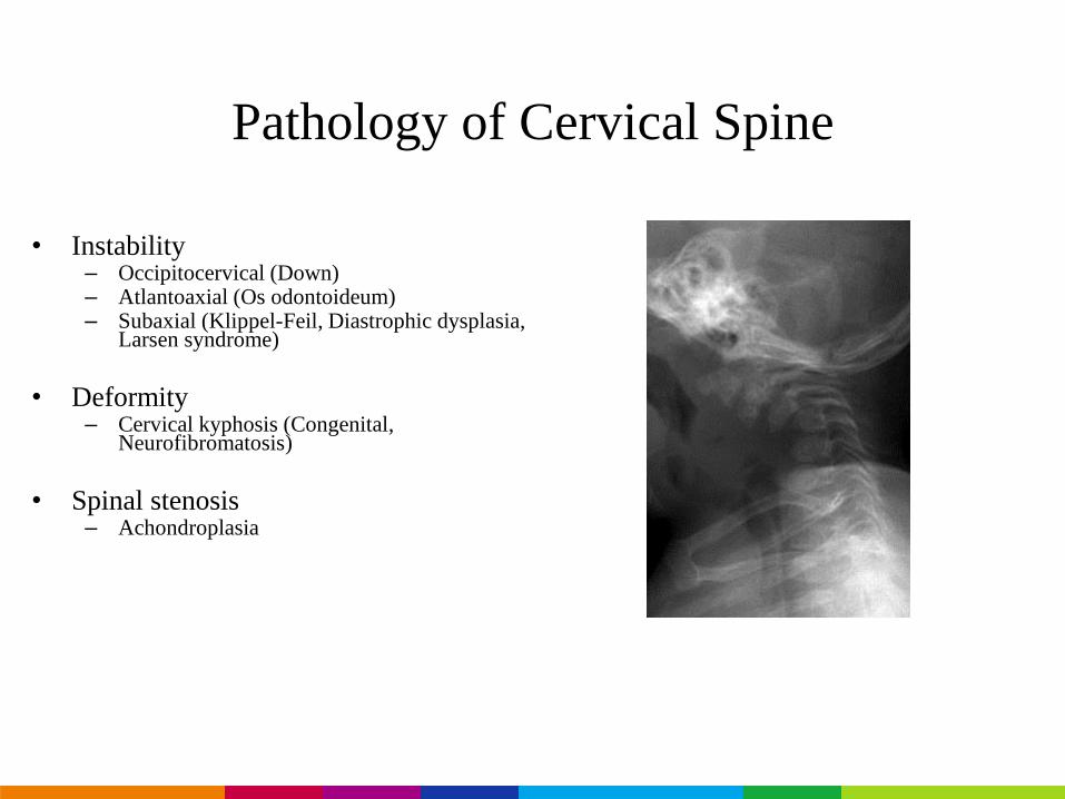
Pathology of Cervical Spine
• Instability– Occipitocervical (Down)– Atlantoaxial (Os odontoideum)– Subaxial (Klippel-Feil, Diastrophic dysplasia,
Larsen syndrome)
• Deformity– Cervical kyphosis (Congenital,
Neurofibromatosis)
• Spinal stenosis– Achondroplasia
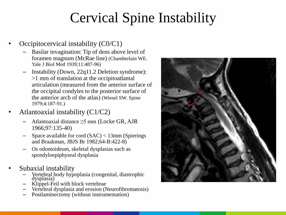
Cervical Spine Instability
• Occipitocervical instability (C0/C1)– Basilar invagination: Tip of dens above level of
foramen magnum (McRae line) (Chamberlain WE. Yale J Biol Med 1939;11:487-96)
– Instability (Down, 22q11.2 Deletion syndrome): >1 mm of translation at the occipitoatlantalarticulation (measured from the anterior surface of the occipital condyles to the posterior surface of the anterior arch of the atlas) (Wiesel SW. Spine 1979;4:187-91.)
• Atlantoaxial instability (C1/C2)
– Atlantoaxial distance ≥5 mm (Locke GR, AJR 1966;97:135-40)
– Space available for cord (SAC) < 13mm (Spieringsand Braakman, JBJS Br 1982;64-B:422-8)
– Os odontoideum, skeletal dysplasias such as spondyloepiphyseal dysplasia
• Subaxial instability– Vertebral body hypoplasia (congenital, diastrophic
dysplasia)– Klippel-Feil with block vertebrae– Vertebral dysplasia and erosion (Neurofibromatosis)– Postlaminectomy (without instrumentation)
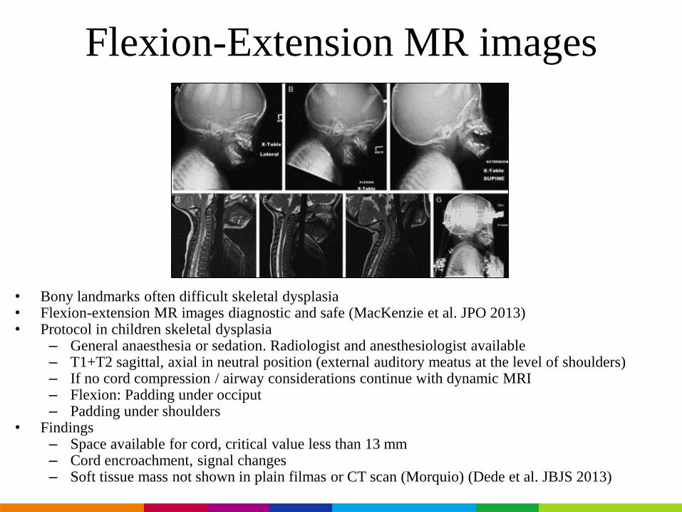
Flexion-Extension MR images
• Bony landmarks often difficult skeletal dysplasia• Flexion-extension MR images diagnostic and safe (MacKenzie et al. JPO 2013)• Protocol in children skeletal dysplasia
– General anaesthesia or sedation. Radiologist and anesthesiologist available– T1+T2 sagittal, axial in neutral position (external auditory meatus at the level of shoulders)– If no cord compression / airway considerations continue with dynamic MRI– Flexion: Padding under occiput– Padding under shoulders
• Findings– Space available for cord, critical value less than 13 mm– Cord encroachment, signal changes– Soft tissue mass not shown in plain filmas or CT scan (Morquio) (Dede et al. JBJS 2013)
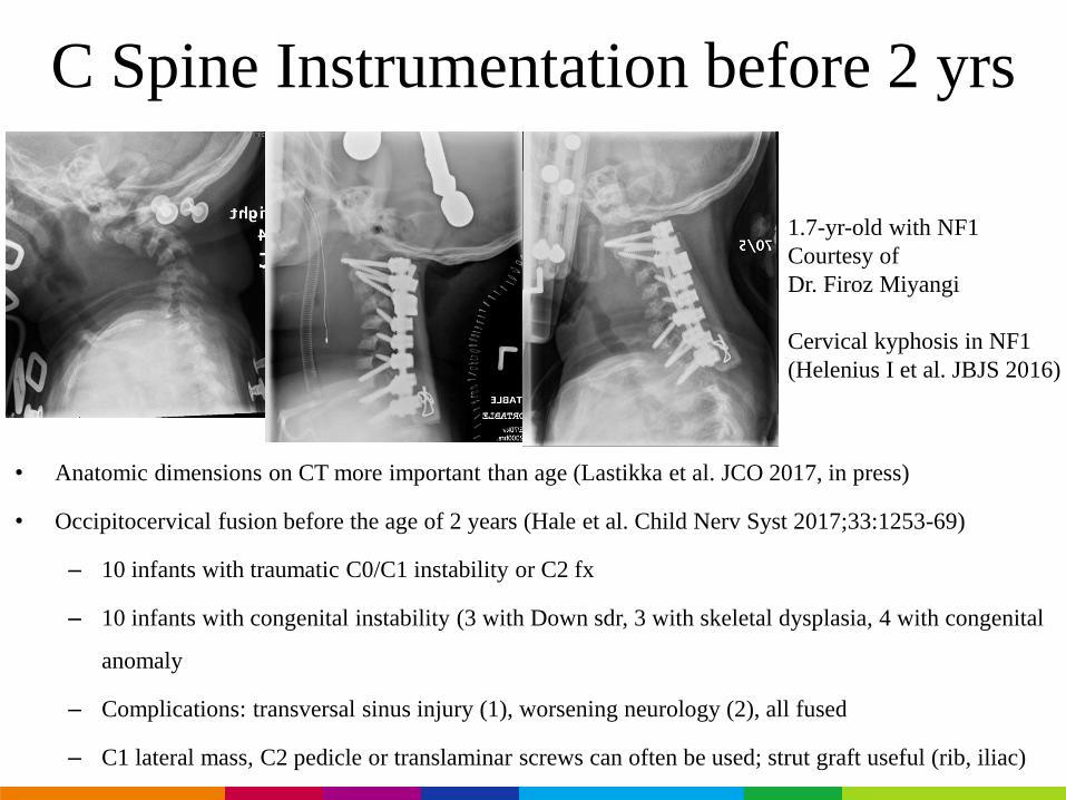
C Spine Instrumentation before 2 yrs
• Anatomic dimensions on CT more important than age (Lastikka et al. JCO 2017, in press)
• Occipitocervical fusion before the age of 2 years (Hale et al. Child Nerv Syst 2017;33:1253-69)
– 10 infants with traumatic C0/C1 instability or C2 fx
– 10 infants with congenital instability (3 with Down sdr, 3 with skeletal dysplasia, 4 with congenital
anomaly
– Complications: transversal sinus injury (1), worsening neurology (2), all fused
– C1 lateral mass, C2 pedicle or translaminar screws can often be used; strut graft useful (rib, iliac)
1.7-yr-old with NF1
Courtesy of
Dr. Firoz Miyangi
Cervical kyphosis in NF1
(Helenius I et al. JBJS 2016)
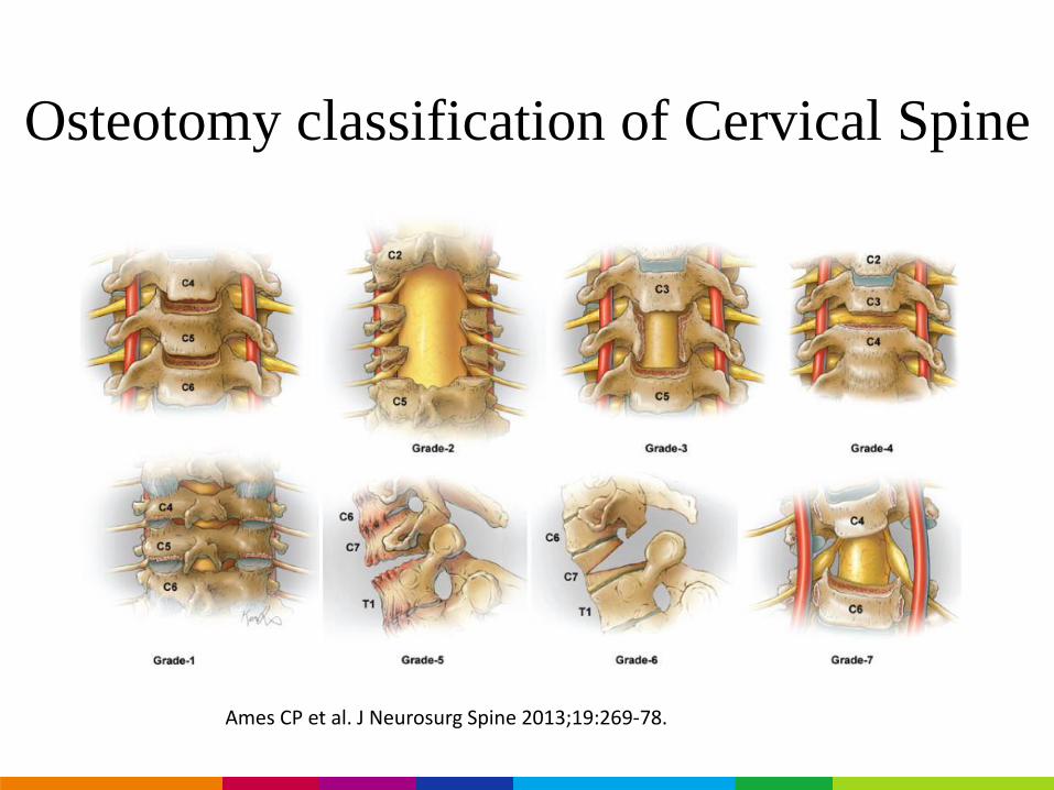
Osteotomy classification of Cervical Spine
Ames CP et al. J Neurosurg Spine 2013;19:269-78.
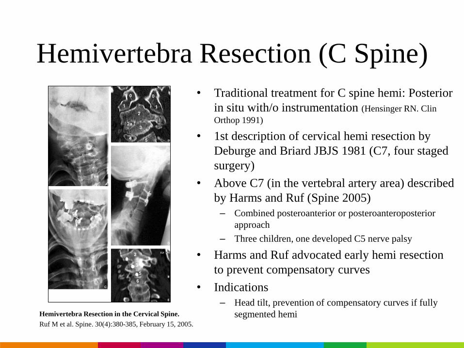
Hemivertebra Resection (C Spine)
Hemivertebra Resection in the Cervical Spine.
Ruf M et al. Spine. 30(4):380-385, February 15, 2005.
• Traditional treatment for C spine hemi: Posterior
in situ with/o instrumentation (Hensinger RN. Clin
Orthop 1991)
• 1st description of cervical hemi resection by
Deburge and Briard JBJS 1981 (C7, four staged
surgery)
• Above C7 (in the vertebral artery area) described
by Harms and Ruf (Spine 2005)
– Combined posteroanterior or posteroanteroposterior
approach
– Three children, one developed C5 nerve palsy
• Harms and Ruf advocated early hemi resection
to prevent compensatory curves
• Indications
– Head tilt, prevention of compensatory curves if fully
segmented hemi
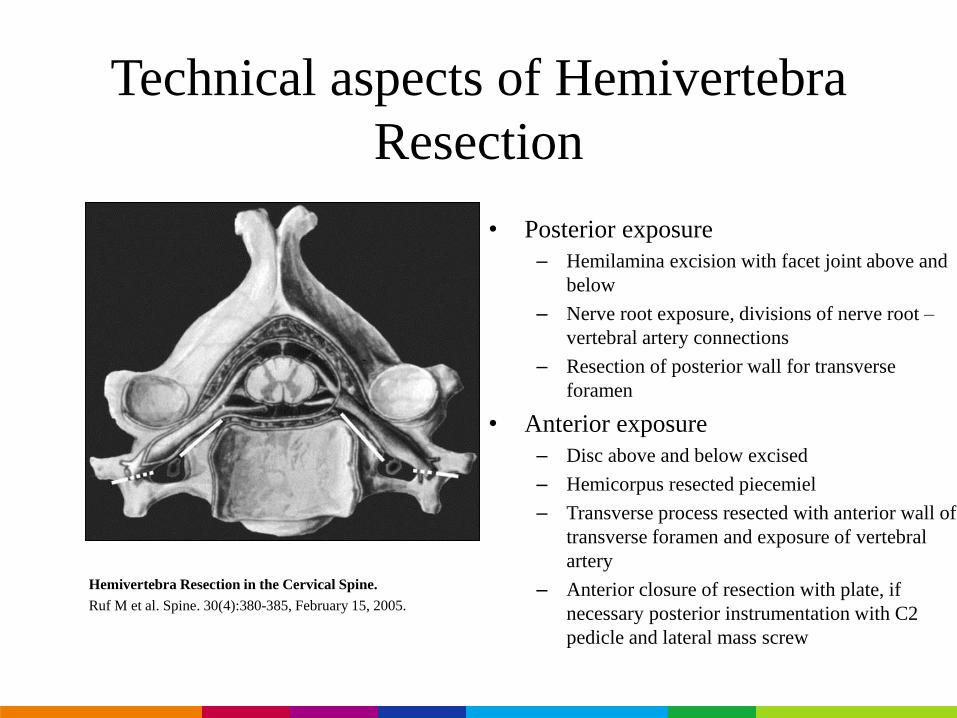
Technical aspects of Hemivertebra
Resection
• Posterior exposure
– Hemilamina excision with facet joint above and
below
– Nerve root exposure, divisions of nerve root –
vertebral artery connections
– Resection of posterior wall for transverse
foramen
• Anterior exposure
– Disc above and below excised
– Hemicorpus resected piecemiel
– Transverse process resected with anterior wall of
transverse foramen and exposure of vertebral
artery
– Anterior closure of resection with plate, if
necessary posterior instrumentation with C2
pedicle and lateral mass screw
Hemivertebra Resection in the Cervical Spine.
Ruf M et al. Spine. 30(4):380-385, February 15, 2005.
5
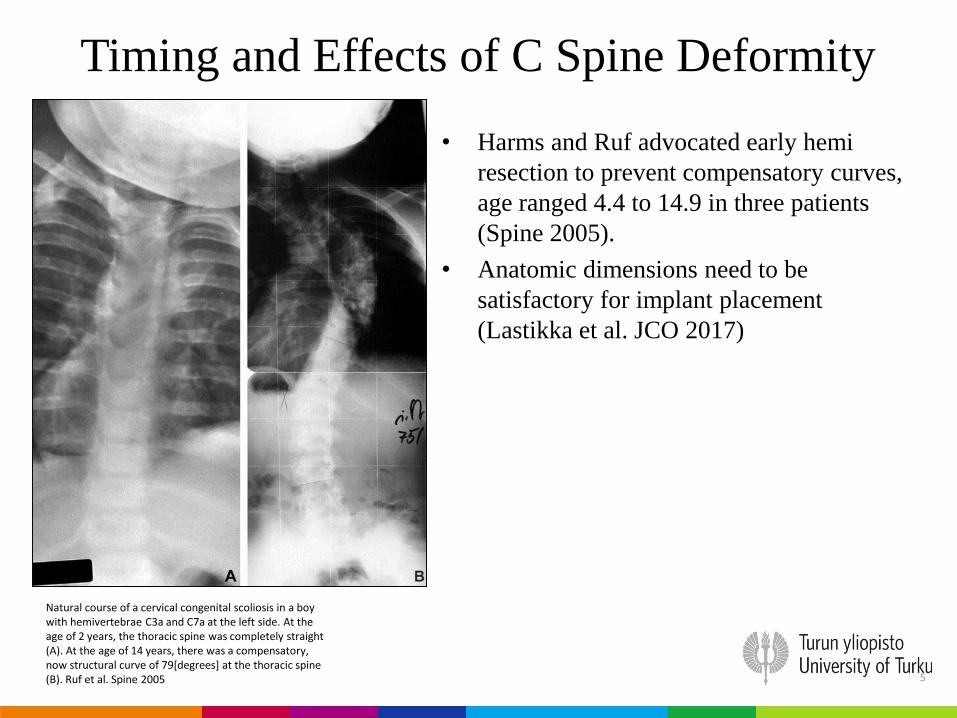
Timing and Effects of C Spine Deformity
Natural course of a cervical congenital scoliosis in a boy with hemivertebrae C3a and C7a at the left side. At the age of 2 years, the thoracic spine was completely straight (A). At the age of 14 years, there was a compensatory, now structural curve of 79[degrees] at the thoracic spine (B). Ruf et al. Spine 2005
• Harms and Ruf advocated early hemi
resection to prevent compensatory curves,
age ranged 4.4 to 14.9 in three patients
(Spine 2005).
• Anatomic dimensions need to be
satisfactory for implant placement
(Lastikka et al. JCO 2017)
5
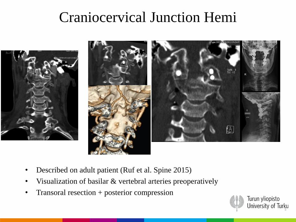
Craniocervical Junction Hemi
• Described on adult patient (Ruf et al. Spine 2015)
• Visualization of basilar & vertebral arteries preoperatively
• Transoral resection + posterior compression
5
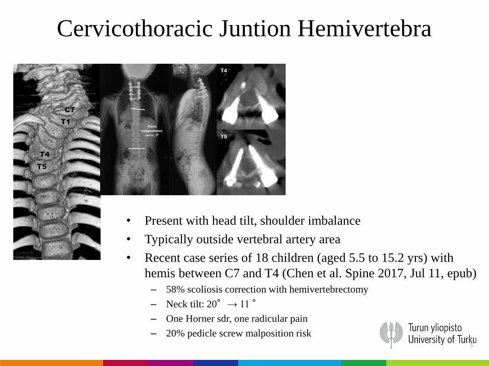
Cervicothoracic Juntion Hemivertebra
• Present with head tilt, shoulder imbalance
• Typically outside vertebral artery area
• Recent case series of 18 children (aged 5.5 to 15.2 yrs) with
hemis between C7 and T4 (Chen et al. Spine 2017, Jul 11, epub)
– 58% scoliosis correction with hemivertebrectomy
– Neck tilt: 20°→ 11°
– One Horner sdr, one radicular pain
– 20% pedicle screw malposition risk
5
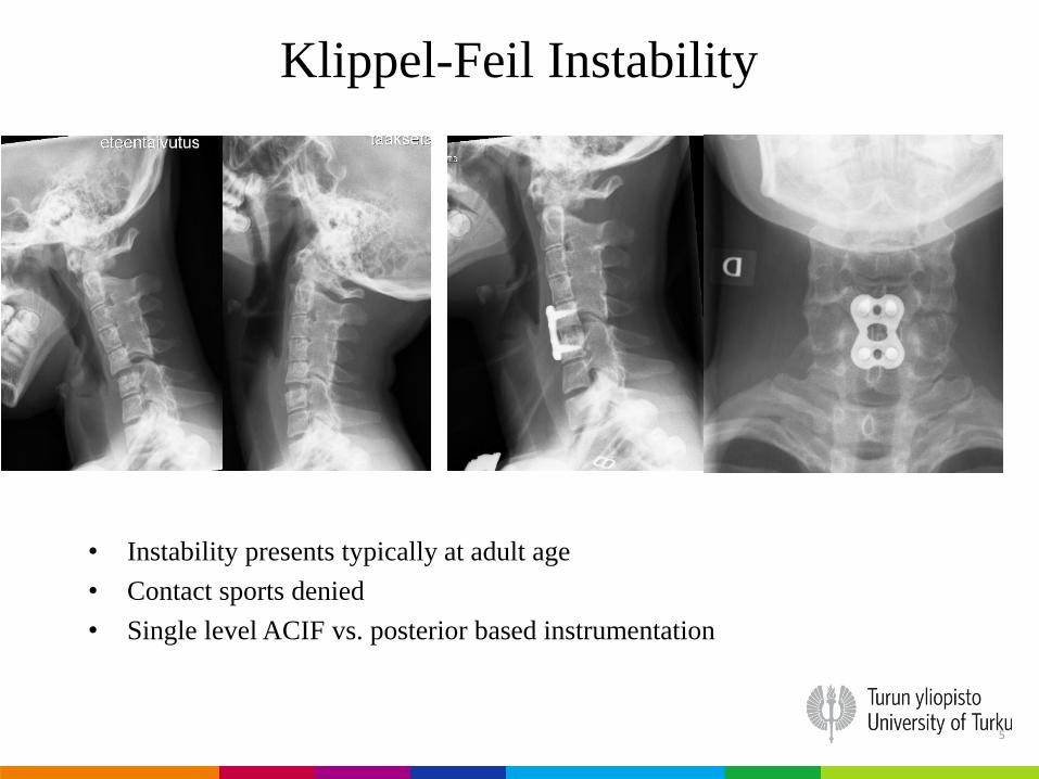
Klippel-Feil Instability
• Instability presents typically at adult age
• Contact sports denied
• Single level ACIF vs. posterior based instrumentation
5
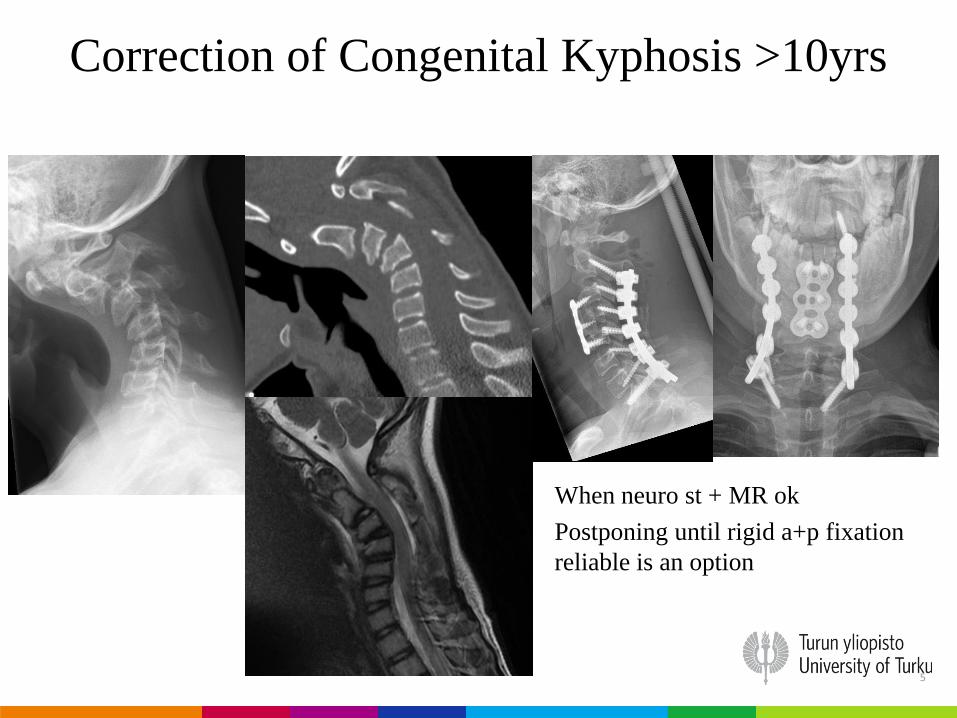
Correction of Congenital Kyphosis >10yrs
• When neuro st + MR ok
• Postponing until rigid a+p fixation
reliable is an option
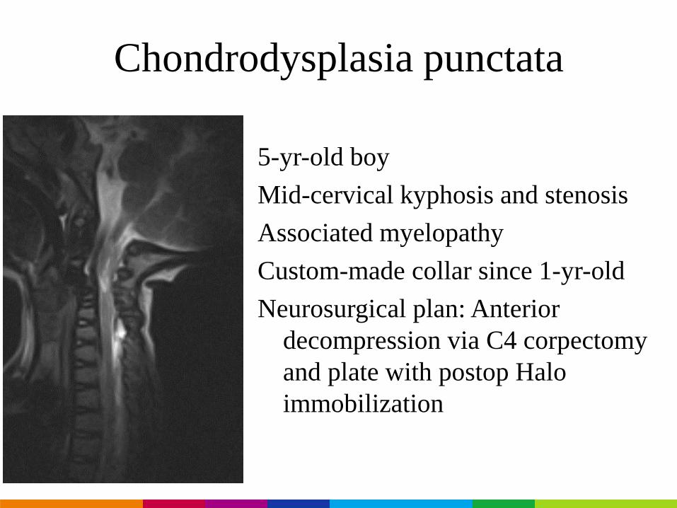
Chondrodysplasia punctata
5-yr-old boy
Mid-cervical kyphosis and stenosis
Associated myelopathy
Custom-made collar since 1-yr-old
Neurosurgical plan: Anterior
decompression via C4 corpectomy
and plate with postop Halo
immobilization
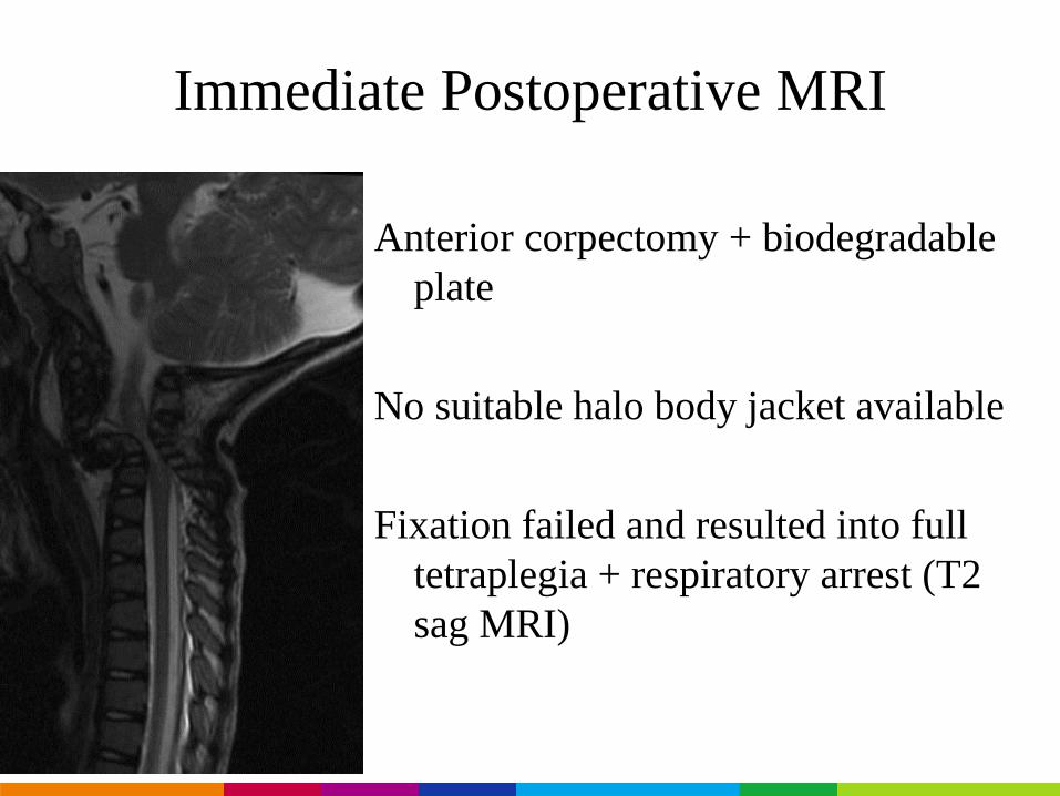
Immediate Postoperative MRI
Anterior corpectomy + biodegradable
plate
No suitable halo body jacket available
Fixation failed and resulted into full
tetraplegia + respiratory arrest (T2
sag MRI)
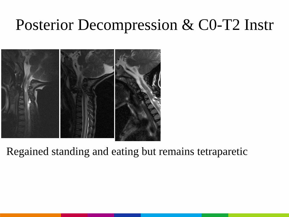
Posterior Decompression & C0-T2 Instr
Regained standing and eating but remains tetraparetic
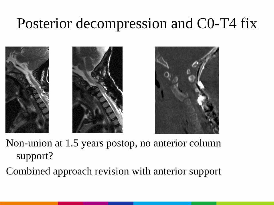
Posterior decompression and C0-T4 fix
Non-union at 1.5 years postop, no anterior column
support?
Combined approach revision with anterior support
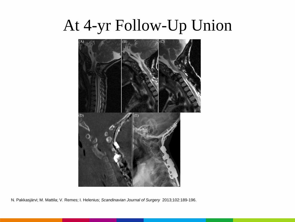
At 4-yr Follow-Up Union
N. Pakkasjärvi; M. Mattila; V. Remes; I. Helenius; Scandinavian Journal of Surgery 2013;102:189-196.
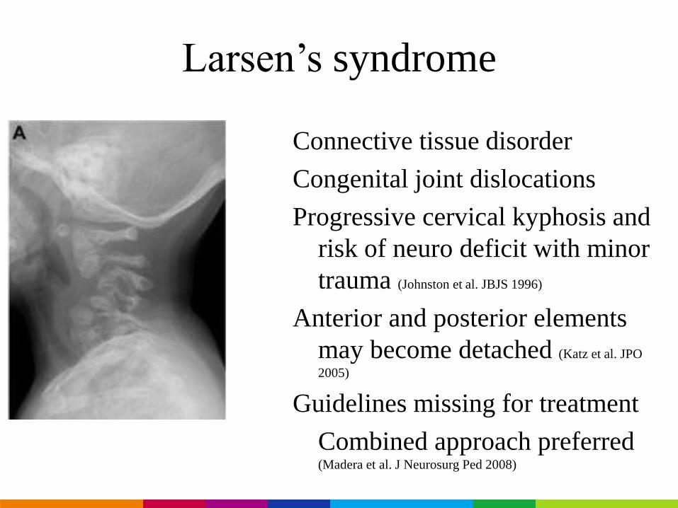
Larsen’s syndrome
Connective tissue disorder
Congenital joint dislocations
Progressive cervical kyphosis and
risk of neuro deficit with minor
trauma (Johnston et al. JBJS 1996)
Anterior and posterior elements
may become detached (Katz et al. JPO
2005)
Guidelines missing for treatment
Combined approach preferred(Madera et al. J Neurosurg Ped 2008)
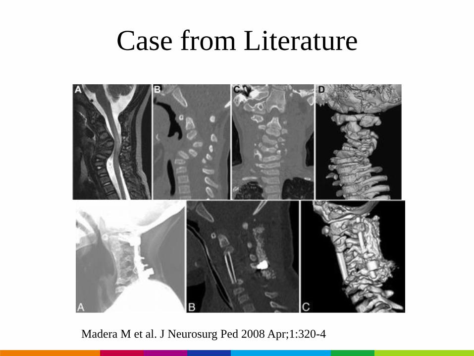
Case from Literature
Madera M et al. J Neurosurg Ped 2008 Apr;1:320-4
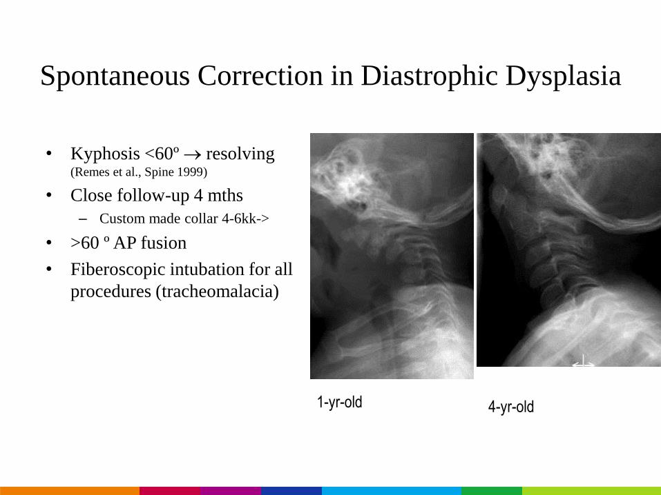
Spontaneous Correction in Diastrophic Dysplasia
• Kyphosis <60º resolving (Remes et al., Spine 1999)
• Close follow-up 4 mths
– Custom made collar 4-6kk->
• >60 º AP fusion
• Fiberoscopic intubation for all
procedures (tracheomalacia)
1-yr-old 4-yr-old
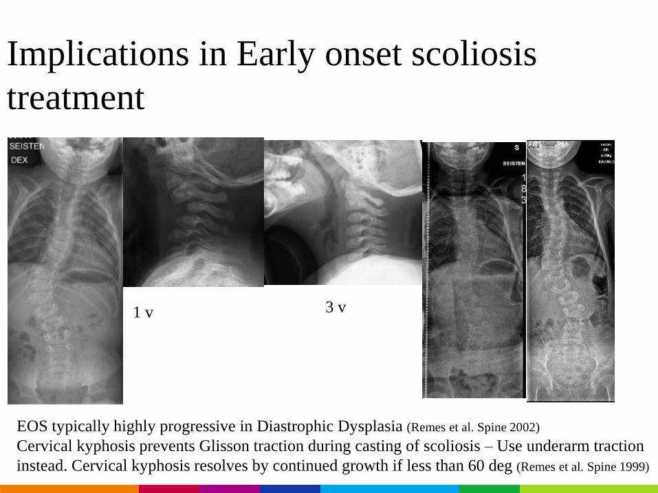
Implications in Early onset scoliosis
treatment
TractionEOS typically highly progressive in Diastrophic Dysplasia (Remes et al. Spine 2002)
Cervical kyphosis prevents Glisson traction during casting of scoliosis – Use underarm traction
instead. Cervical kyphosis resolves by continued growth if less than 60 deg (Remes et al. Spine 1999)
1 v 3 v
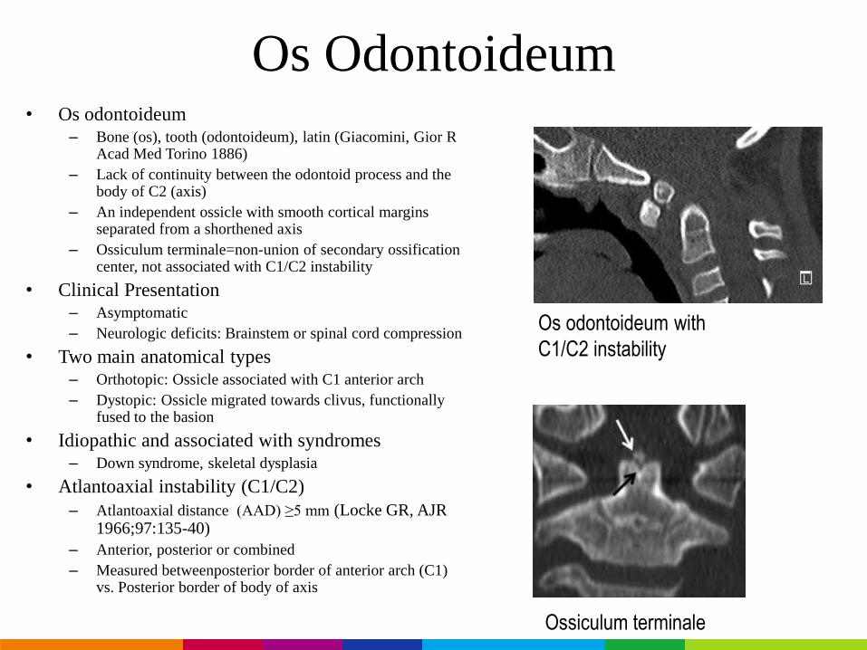
Os Odontoideum• Os odontoideum
– Bone (os), tooth (odontoideum), latin (Giacomini, Gior R Acad Med Torino 1886)
– Lack of continuity between the odontoid process and the body of C2 (axis)
– An independent ossicle with smooth cortical marginsseparated from a shorthened axis
– Ossiculum terminale=non-union of secondary ossificationcenter, not associated with C1/C2 instability
• Clinical Presentation– Asymptomatic
– Neurologic deficits: Brainstem or spinal cord compression
• Two main anatomical types– Orthotopic: Ossicle associated with C1 anterior arch
– Dystopic: Ossicle migrated towards clivus, functionallyfused to the basion
• Idiopathic and associated with syndromes– Down syndrome, skeletal dysplasia
• Atlantoaxial instability (C1/C2)
– Atlantoaxial distance (AAD) ≥5 mm (Locke GR, AJR 1966;97:135-40)
– Anterior, posterior or combined
– Measured betweenposterior border of anterior arch (C1) vs. Posterior border of body of axis
Os odontoideum with
C1/C2 instability
Ossiculum terminale
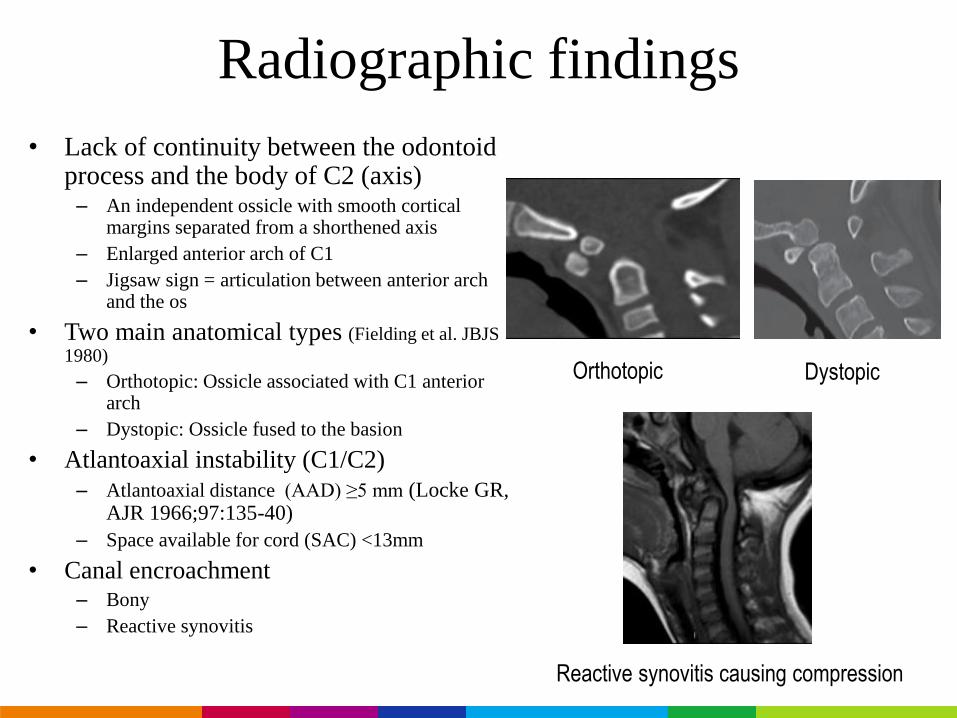
Radiographic findings
• Lack of continuity between the odontoidprocess and the body of C2 (axis)– An independent ossicle with smooth cortical
margins separated from a shorthened axis
– Enlarged anterior arch of C1
– Jigsaw sign = articulation between anterior archand the os
• Two main anatomical types (Fielding et al. JBJS
1980)
– Orthotopic: Ossicle associated with C1 anteriorarch
– Dystopic: Ossicle fused to the basion
• Atlantoaxial instability (C1/C2)
– Atlantoaxial distance (AAD) ≥5 mm (Locke GR, AJR 1966;97:135-40)
– Space available for cord (SAC) <13mm
• Canal encroachment– Bony
– Reactive synovitis
Orthotopic Dystopic
Reactive synovitis causing compression
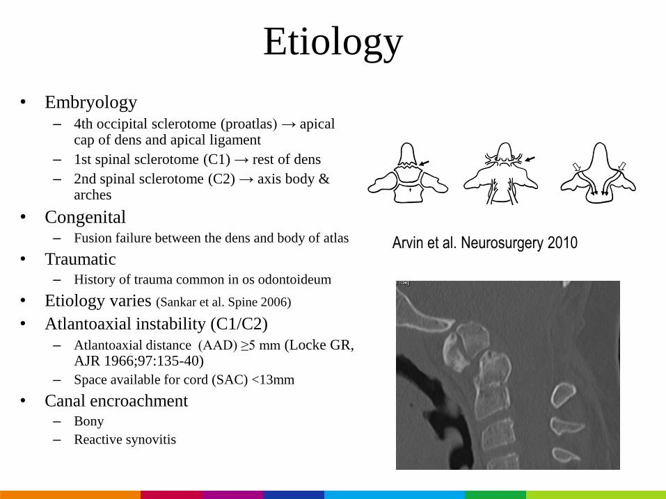
Etiology
• Embryology– 4th occipital sclerotome (proatlas) → apical
cap of dens and apical ligament
– 1st spinal sclerotome (C1) → rest of dens
– 2nd spinal sclerotome (C2) → axis body & arches
• Congenital– Fusion failure between the dens and body of atlas
• Traumatic– History of trauma common in os odontoideum
• Etiology varies (Sankar et al. Spine 2006)
• Atlantoaxial instability (C1/C2)
– Atlantoaxial distance (AAD) ≥5 mm (Locke GR, AJR 1966;97:135-40)
– Space available for cord (SAC) <13mm
• Canal encroachment– Bony
– Reactive synovitis
Arvin et al. Neurosurgery 2010
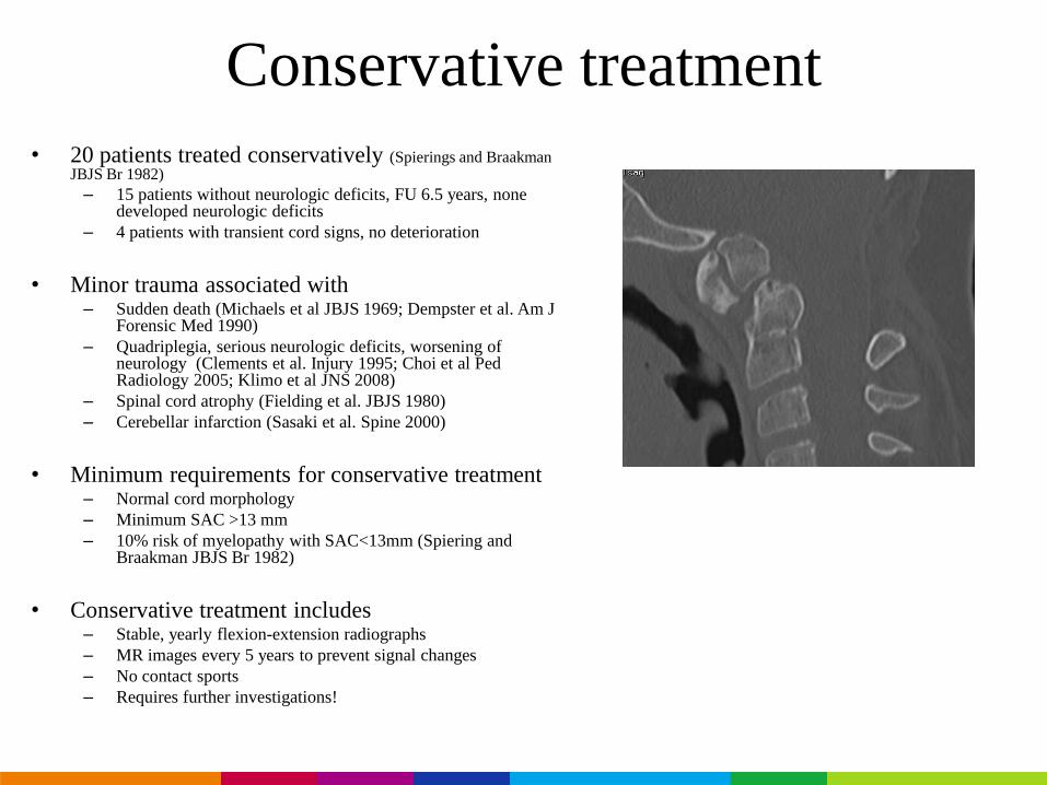
Conservative treatment
• 20 patients treated conservatively (Spierings and BraakmanJBJS Br 1982)
– 15 patients without neurologic deficits, FU 6.5 years, nonedeveloped neurologic deficits
– 4 patients with transient cord signs, no deterioration
• Minor trauma associated with – Sudden death (Michaels et al JBJS 1969; Dempster et al. Am J
Forensic Med 1990)
– Quadriplegia, serious neurologic deficits, worsening of neurology (Clements et al. Injury 1995; Choi et al PedRadiology 2005; Klimo et al JNS 2008)
– Spinal cord atrophy (Fielding et al. JBJS 1980)
– Cerebellar infarction (Sasaki et al. Spine 2000)
• Minimum requirements for conservative treatment– Normal cord morphology
– Minimum SAC >13 mm
– 10% risk of myelopathy with SAC<13mm (Spiering and Braakman JBJS Br 1982)
• Conservative treatment includes– Stable, yearly flexion-extension radiographs
– MR images every 5 years to prevent signal changes
– No contact sports
– Requires further investigations!
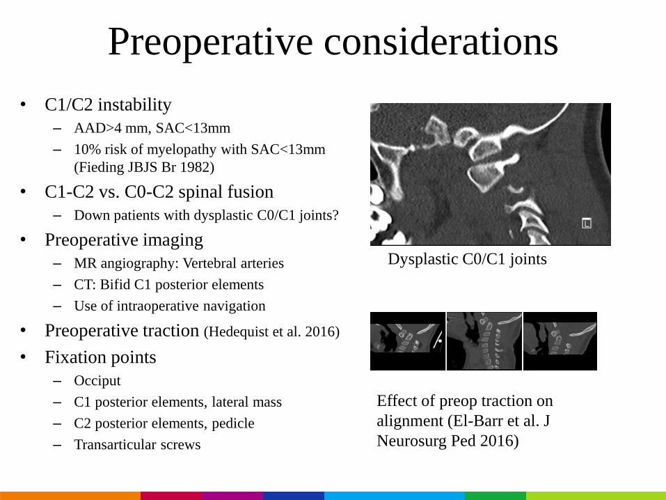
Preoperative considerations
• C1/C2 instability
– AAD>4 mm, SAC<13mm
– 10% risk of myelopathy with SAC<13mm
(Fieding JBJS Br 1982)
• C1-C2 vs. C0-C2 spinal fusion
– Down patients with dysplastic C0/C1 joints?
• Preoperative imaging
– MR angiography: Vertebral arteries
– CT: Bifid C1 posterior elements
– Use of intraoperative navigation
• Preoperative traction (Hedequist et al. 2016)
• Fixation points
– Occiput
– C1 posterior elements, lateral mass
– C2 posterior elements, pedicle
– Transarticular screws
Effect of preop traction on
alignment (El-Barr et al. J
Neurosurg Ped 2016)
Dysplastic C0/C1 joints
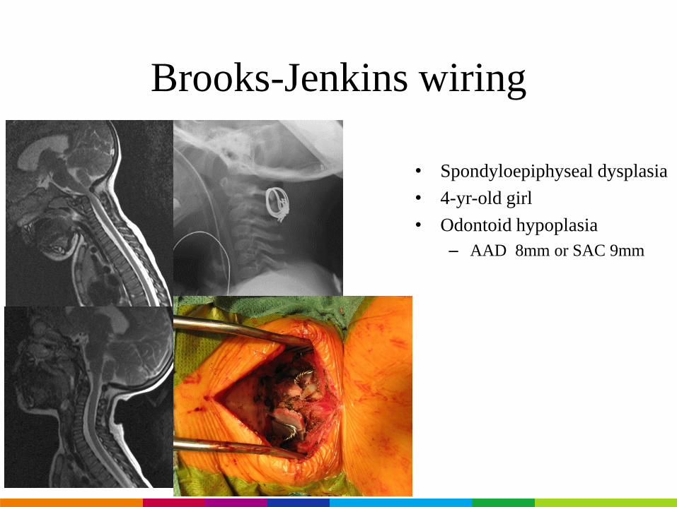
Brooks-Jenkins wiring
• Spondyloepiphyseal dysplasia
• 4-yr-old girl
• Odontoid hypoplasia
– AAD 8mm or SAC 9mm
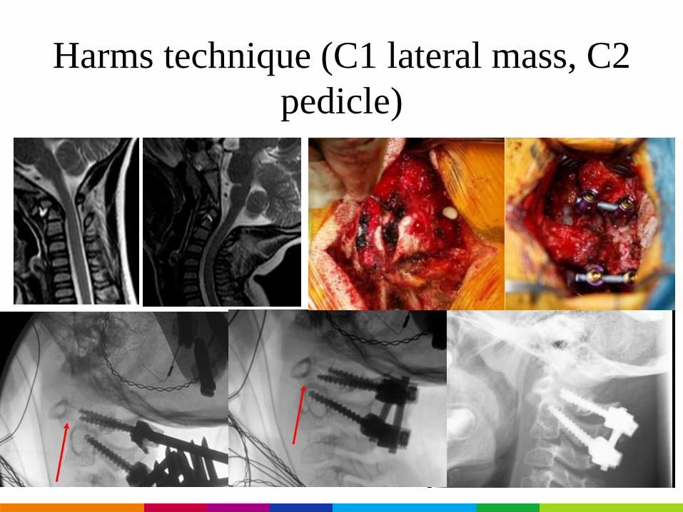
Harms technique (C1 lateral mass, C2
pedicle)
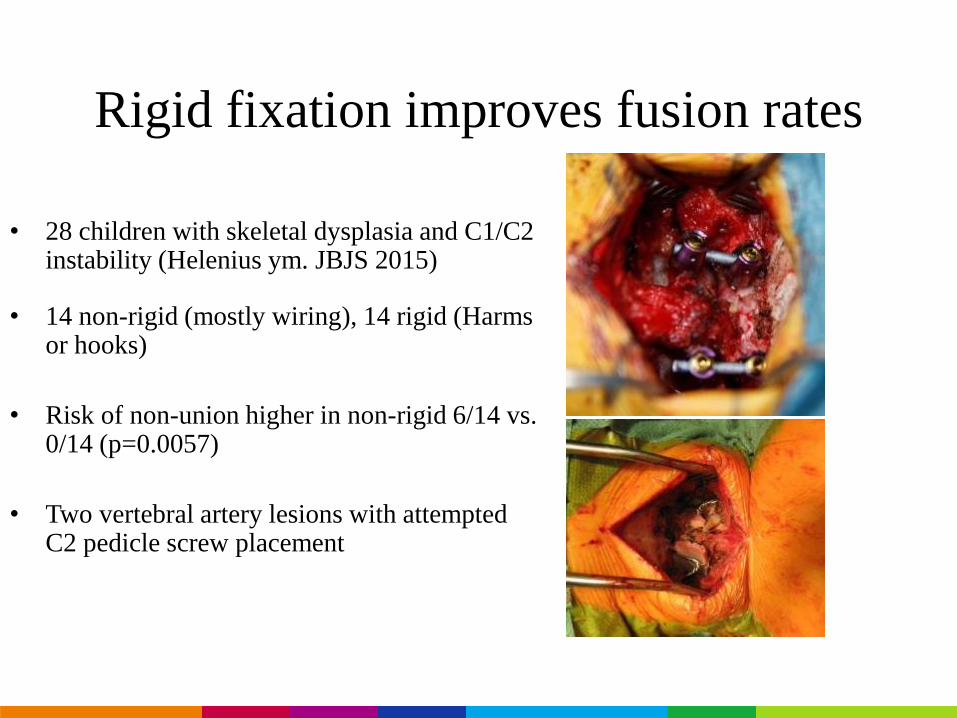
Rigid fixation improves fusion rates
• 28 children with skeletal dysplasia and C1/C2 instability (Helenius ym. JBJS 2015)
• 14 non-rigid (mostly wiring), 14 rigid (Harmsor hooks)
• Risk of non-union higher in non-rigid 6/14 vs. 0/14 (p=0.0057)
• Two vertebral artery lesions with attemptedC2 pedicle screw placement
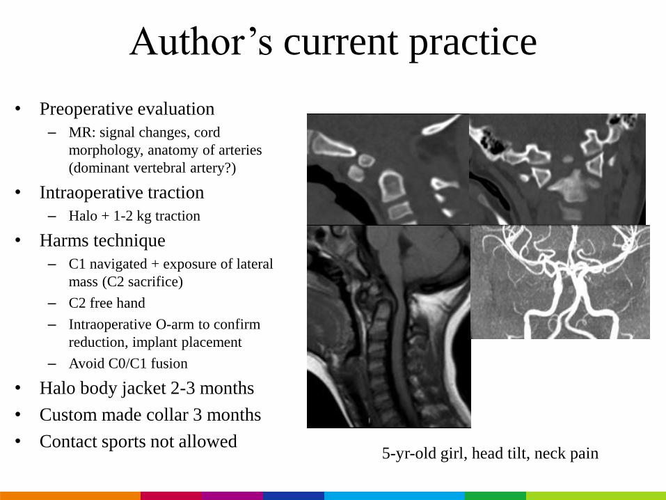
Author’s current practice
• Preoperative evaluation
– MR: signal changes, cord
morphology, anatomy of arteries
(dominant vertebral artery?)
• Intraoperative traction
– Halo + 1-2 kg traction
• Harms technique
– C1 navigated + exposure of lateral
mass (C2 sacrifice)
– C2 free hand
– Intraoperative O-arm to confirm
reduction, implant placement
– Avoid C0/C1 fusion
• Halo body jacket 2-3 months
• Custom made collar 3 months
• Contact sports not allowed5-yr-old girl, head tilt, neck pain

Intraoperative traction
Make sure cervical alignment is acceptable with fluoroscopy
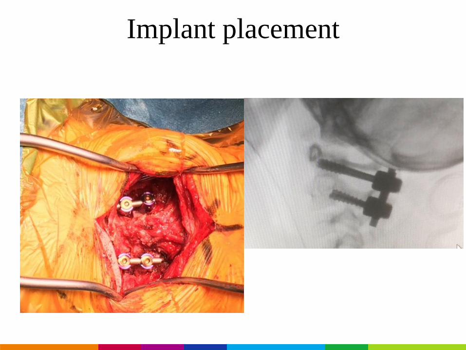
Implant placement
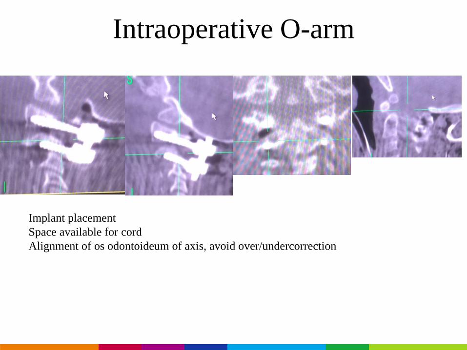
Intraoperative O-arm
Implant placement
Space available for cord
Alignment of os odontoideum of axis, avoid over/undercorrection
Conclusions
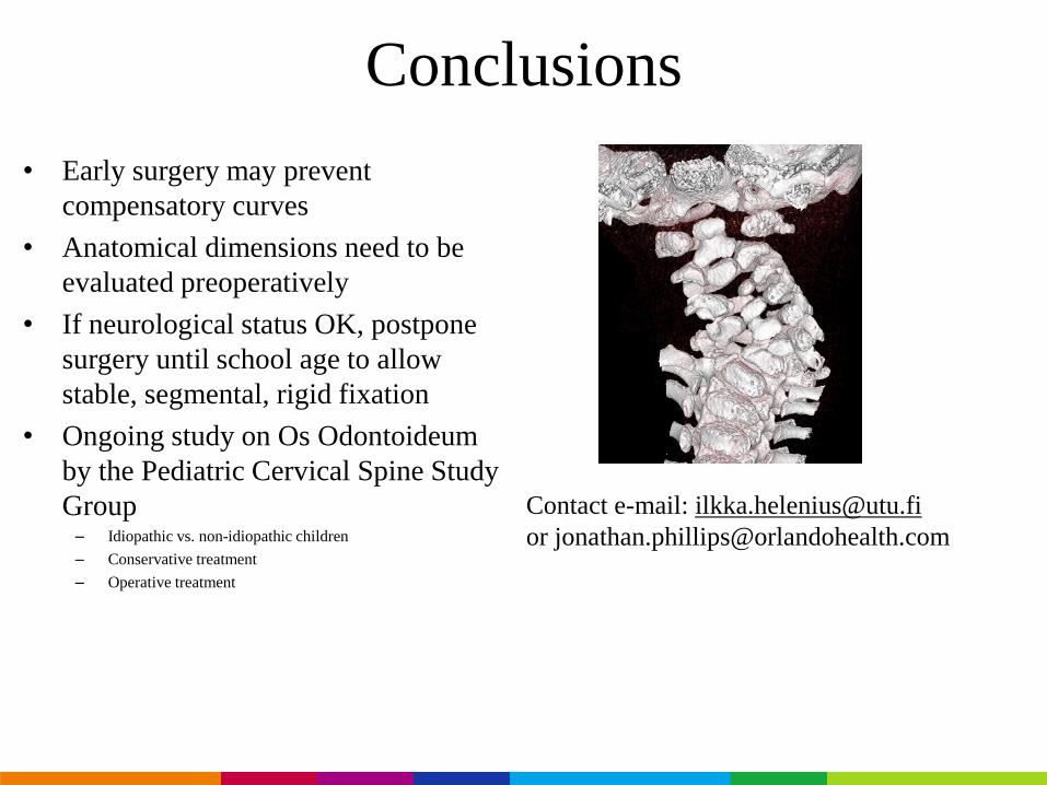
• Early surgery may prevent
compensatory curves
• Anatomical dimensions need to be
evaluated preoperatively
• If neurological status OK, postpone
surgery until school age to allow
stable, segmental, rigid fixation
• Ongoing study on Os Odontoideum
by the Pediatric Cervical Spine Study
Group– Idiopathic vs. non-idiopathic children
– Conservative treatment
– Operative treatment
Contact e-mail: [email protected]