tick importance and disease transmission - afrivip · ticks: tick importance and disease...
TRANSCRIPT
Ticks: Tick importance and disease transmission
Ticks Tick importance and disease transmission
Authors: Prof Maxime Madder, Prof Ivan Horak, Dr Hein Stoltsz
Licensed under a Creative Commons Attribution license.
Ticks: Tick importance and disease transmission
2 | P a g e
TABLE OF CONTENTS
Table of Contents...........................................................................................................2
Introduction ....................................................................................................................4
Importance .....................................................................................................................5
Disease transmission ....................................................................................................6
Transovarial transmission ....................................................................................................... 7
Transstadial transmission ....................................................................................................... 8
Intrastadial transmission ......................................................................................................... 8
Transmission by co-feeding .................................................................................................... 8
Mechanical transmission ........................................................................................................ 9
Transmission by coxal fluid..................................................................................................... 9
Transmission by ingestion ...................................................................................................... 9
Venereal transmission ............................................................................................................ 9
Ticks of veterinary importance ................................................................................... 10
Amblyomma spp. .................................................................................................................. 10
Amblyomma hebraeum – the bont tick ........................................................................................ 10
Amblyomma variegatum – the tropical bont tick ......................................................................... 11
Vector-based epidemiology of heartwater (Norval et al. 1992) .............................................. 12
Reservoirs of infection ................................................................................................................. 12
Acquisition of infection................................................................................................................. 12
Vector status ................................................................................................................................................ 12
Factors leading to infection ......................................................................................................... 13
Control ......................................................................................................................................... 13
Hyalomma spp. .................................................................................................................... 14
Hyalomma dromedarii – the camel tick ....................................................................................... 14
Ticks: Tick importance and disease transmission
3 | P a g e
Hyalomma truncatum – small smooth bont-legged tick .............................................................. 15
Ixodes spp. ........................................................................................................................... 17
Ixodes rubicundus - Karoo paralysis tick..................................................................................... 17
Rhipicephalus spp. ............................................................................................................... 19
Rhipicephalus (Boophilus) microplus - Asian blue tick ............................................................... 19
Rhipicephalus (Boophilus) decoloratus – African blue tick ......................................................... 21
Rhipicephalus appendiculatus - Brown ear tick .......................................................................... 23
Rhipicephalus zambeziensis ....................................................................................................... 25
Rhipicephalus evertsi evertsi - the red-legged tick. .................................................................... 26
Ornithodoros spp. ................................................................................................................. 28
Ornithodoros moubata/porcinus - Eyeless or hut or warthog tampan ........................................ 28
References ................................................................................................................... 30
Ticks: Tick importance and disease transmission
4 | P a g e
INTRODUCTION
Ticks are blood-sucking arthropods found in virtually all terrestrial regions of the planet. Globally, approximately 900 species of ticks are recognized of which about 700 species are ixodid or hard ticks and 200 species are soft ticks. Approximately 200 ixodid and 40 argasid species are present in the Afrotropical region but only a small number are of veterinary and medical importance. Many of the ticks and tick-borne diseases occur usually in specific geographical areas but with globalisation and climate change their range may be expand and even spread intercontinentally.
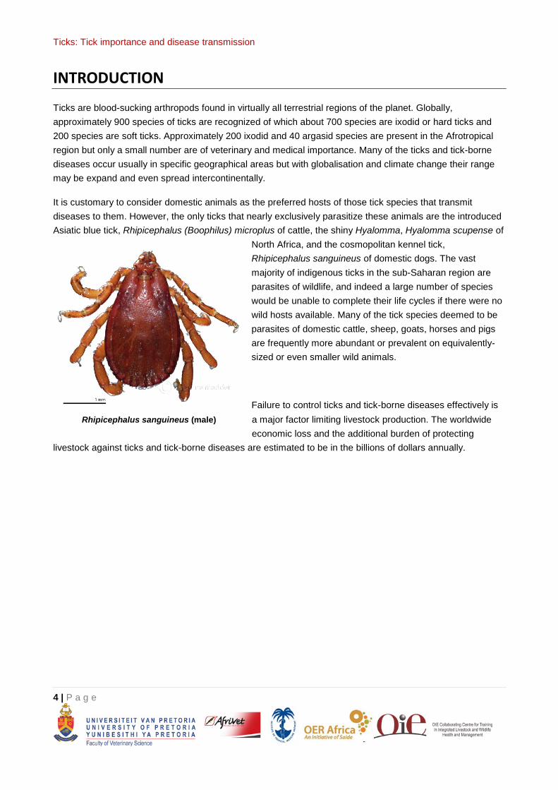
It is customary to consider domestic animals as the preferred hosts of those tick species that transmit diseases to them. However, the only ticks that nearly exclusively parasitize these animals are the introduced Asiatic blue tick, Rhipicephalus (Boophilus) microplus of cattle, the shiny Hyalomma, Hyalomma scupense of
North Africa, and the cosmopolitan kennel tick, Rhipicephalus sanguineus of domestic dogs. The vast majority of indigenous ticks in the sub-Saharan region are parasites of wildlife, and indeed a large number of species would be unable to complete their life cycles if there were no wild hosts available. Many of the tick species deemed to be parasites of domestic cattle, sheep, goats, horses and pigs are frequently more abundant or prevalent on equivalently-sized or even smaller wild animals.
Failure to control ticks and tick-borne diseases effectively is a major factor limiting livestock production. The worldwide economic loss and the additional burden of protecting
livestock against ticks and tick-borne diseases are estimated to be in the billions of dollars annually.
Rhipicephalus sanguineus (male)
Ticks: Tick importance and disease transmission
5 | P a g e
IMPORTANCE
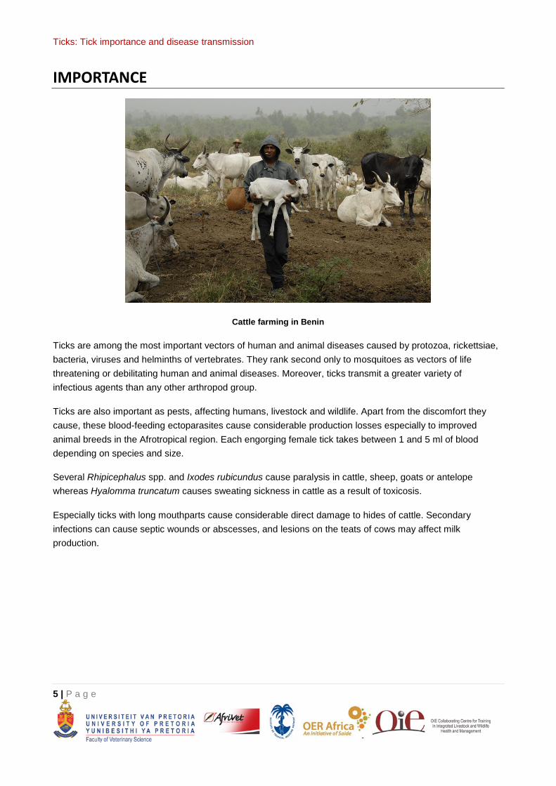
Cattle farming in Benin
Ticks are among the most important vectors of human and animal diseases caused by protozoa, rickettsiae, bacteria, viruses and helminths of vertebrates. They rank second only to mosquitoes as vectors of life threatening or debilitating human and animal diseases. Moreover, ticks transmit a greater variety of infectious agents than any other arthropod group.
Ticks are also important as pests, affecting humans, livestock and wildlife. Apart from the discomfort they cause, these blood-feeding ectoparasites cause considerable production losses especially to improved animal breeds in the Afrotropical region. Each engorging female tick takes between 1 and 5 ml of blood depending on species and size.
Several Rhipicephalus spp. and Ixodes rubicundus cause paralysis in cattle, sheep, goats or antelope whereas Hyalomma truncatum causes sweating sickness in cattle as a result of toxicosis.
Especially ticks with long mouthparts cause considerable direct damage to hides of cattle. Secondary infections can cause septic wounds or abscesses, and lesions on the teats of cows may affect milk production.
Ticks: Tick importance and disease transmission
6 | P a g e
DISEASE TRANSMISSION
Only a small number of tick species are vectors of important economic diseases or toxicosis in sub-Saharan Africa (see Table 1). These are Amblyomma hebraeum, Amblyomma variegatum, Hyalomma dromedarii, Hyalomma truncatum, Ixodes rubicundus, Rhipicephalus (Boophilus) microplus, Rhipicephalus (Boophilus) decoloratus, Rhipicephalus appendiculatus, Rhipicephalus evertsi evertsi, Rhipicephalus zambeziensis and argasid or soft ticks of the Ornithodoros moubata/porcinus complex. Each of these ticks occurs in specific areas suitable for survival and reproduction. In cattle they transmit diseases of economic importance such as heartwater, babesiosis, theileriosis and anaplasmosis but also non-pathogenic or mild theileriosis, spirochaetosis, benign anaplasmosis, benign babesiosis and ehrlichiosis. Diseases affecting sheep and goats are heartwater, anaplasmosis, theileriosis, spirochaetosis and Nairobi sheep disease. Diseases that affect horses, mules and donkeys are piroplasmosis, spirochaetosis and those of pigs are porcine babesiosis and African swine fever.
Several wild ruminant species are susceptible to Ehrlichia ruminantium the causal organism of heartwater or cowdriosis or can act as carriers of the organism. Some are also carriers to certain Theileria spp., while zebra (Equus spp.) are carriers to Babesia caballi and Theileria equi the cause of equine piroplasmosis, and wild suids to Babesia trautmanni the cause of porcine babesiosis and to infection with the virus of African swine fever.
Ixodid ticks are also important vectors of several organisms causing disease in humans in sub-Saharan Africa. These are Rickettsia conori the cause of tick bite fever or tick typhus, Coxiella burneti the cause of Q-fever, and the virus causing Crimean-Congo Haemorrhagic fever. In addition argasid ticks of the Ornithodoros moubata complex can transmit Borrelia duttoni the cause of African relapsing fever to humans.
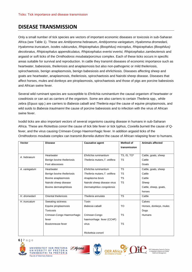
Vector Disease Causative agent Method of transmission
Animals affected
A. hebraeum Heartwater
Benign bovine theileriosis
Foot abscesses
Ehrlichia ruminantium
Theileria mutans,T. velifera
TS, IS, TO*
TS
Cattle, goats, sheep
Cattle
Goats
A. variegatum Heartwater
Benign bovine theileriosis
Bovine anaplasmosis
Nairobi sheep disease
Bovine dermatophilosis
Ehrlichia ruminantium
Theileria mutans,T. velifera
Anaplasma bovis
Nairobi sheep disease virus
Dermatophilus congolensis
TS
TS
TS
TS
Cattle, goats, sheep
Cattle
Cattle
Sheep
Cattle, sheep, goats,
horses
H. dromedarii Oriental theileriosis Theileria annulata TS Cattle
H. truncatum Sweating sickness
Equine piroplasmosis
Toxicosis
Crimean-Congo Haemorrhagic
fever
Boutonneuse fever
Toxin
Babesia caballi
Crimean-Congo
haemorrhage fever (CCHF)
virus
Rickettsia conorii
TO
TS
TS
Calves
Horses, donkeys, mules
Dogs
Humans
Ticks: Tick importance and disease transmission
7 | P a g e
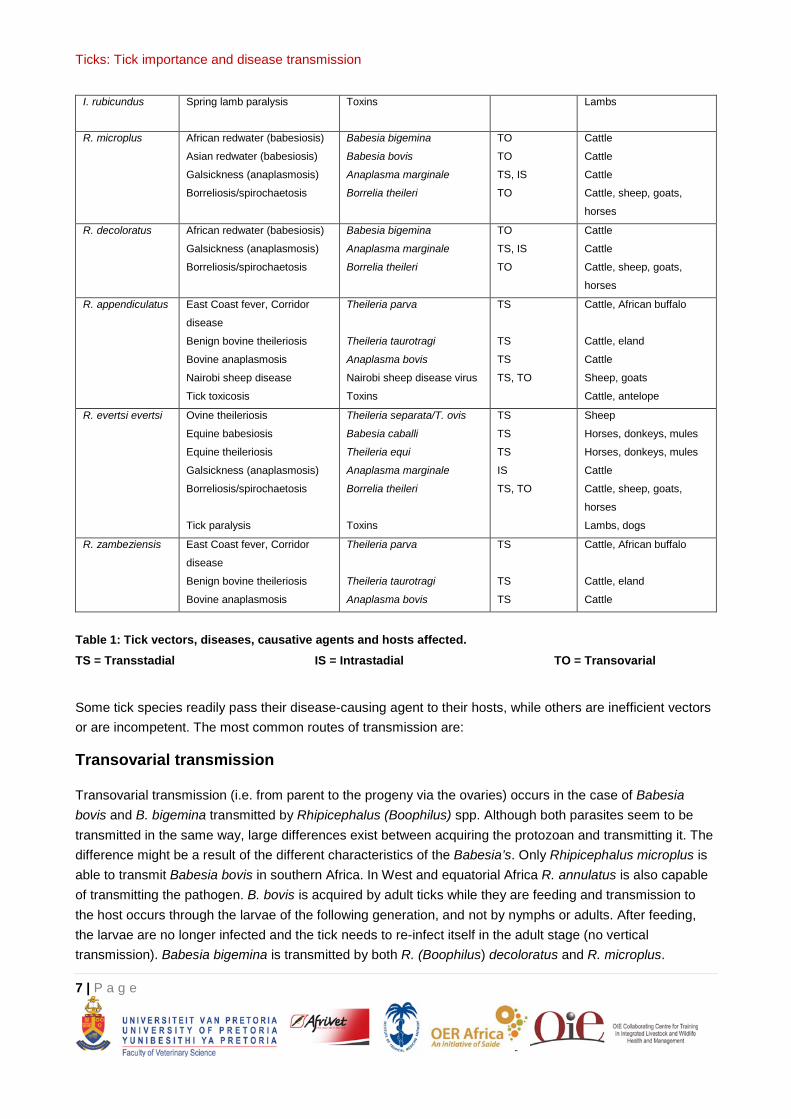
I. rubicundus Spring lamb paralysis Toxins Lambs
R. microplus African redwater (babesiosis)
Asian redwater (babesiosis)
Galsickness (anaplasmosis)
Borreliosis/spirochaetosis
Babesia bigemina
Babesia bovis
Anaplasma marginale
Borrelia theileri
TO
TO
TS, IS
TO
Cattle
Cattle
Cattle
Cattle, sheep, goats,
horses
R. decoloratus African redwater (babesiosis)
Galsickness (anaplasmosis)
Borreliosis/spirochaetosis
Babesia bigemina
Anaplasma marginale
Borrelia theileri
TO
TS, IS
TO
Cattle
Cattle
Cattle, sheep, goats,
horses
R. appendiculatus East Coast fever, Corridor
disease
Benign bovine theileriosis
Bovine anaplasmosis
Nairobi sheep disease
Tick toxicosis
Theileria parva
Theileria taurotragi
Anaplasma bovis
Nairobi sheep disease virus
Toxins
TS
TS
TS
TS, TO
Cattle, African buffalo
Cattle, eland
Cattle
Sheep, goats
Cattle, antelope
R. evertsi evertsi Ovine theileriosis
Equine babesiosis
Equine theileriosis
Galsickness (anaplasmosis)
Borreliosis/spirochaetosis
Tick paralysis
Theileria separata/T. ovis
Babesia caballi
Theileria equi
Anaplasma marginale
Borrelia theileri
Toxins
TS
TS
TS
IS
TS, TO
Sheep
Horses, donkeys, mules
Horses, donkeys, mules
Cattle
Cattle, sheep, goats,
horses
Lambs, dogs
R. zambeziensis East Coast fever, Corridor
disease
Benign bovine theileriosis
Bovine anaplasmosis
Theileria parva
Theileria taurotragi
Anaplasma bovis
TS
TS
TS
Cattle, African buffalo
Cattle, eland
Cattle
Table 1: Tick vectors, diseases, causative agents and hosts affected. TS = Transstadial IS = Intrastadial TO = Transovarial
Some tick species readily pass their disease-causing agent to their hosts, while others are inefficient vectors or are incompetent. The most common routes of transmission are:
Transovarial transmission
Transovarial transmission (i.e. from parent to the progeny via the ovaries) occurs in the case of Babesia bovis and B. bigemina transmitted by Rhipicephalus (Boophilus) spp. Although both parasites seem to be transmitted in the same way, large differences exist between acquiring the protozoan and transmitting it. The difference might be a result of the different characteristics of the Babesia’s. Only Rhipicephalus microplus is able to transmit Babesia bovis in southern Africa. In West and equatorial Africa R. annulatus is also capable of transmitting the pathogen. B. bovis is acquired by adult ticks while they are feeding and transmission to the host occurs through the larvae of the following generation, and not by nymphs or adults. After feeding, the larvae are no longer infected and the tick needs to re-infect itself in the adult stage (no vertical transmission). Babesia bigemina is transmitted by both R. (Boophilus) decoloratus and R. microplus.
Ticks: Tick importance and disease transmission
8 | P a g e
Although the pathogen is also transmitted transovarially and acquired during the engorgement of adult ticks, transmission is via engorging nymphs and/or adults of the next generation. Vertical transmission occurs as well, infection from one generation to another without necessarily reinfection having taken place.
Transstadial transmission
Transstadial (i.e. from one life stage to the next) occurs in East Coast fever (ECF) caused by the protozoan Theileria parva and transmitted mainly by R. appendiculatus but also by R. zambeziensis. In both vectors the parasite is acquired during larval or nymphal feeding and transmitted in the next stage by nymphs (if acquired by larvae) or adults (if acquired by nymphs). After transmission of the parasite to a naïve animal, the ticks are free of infection. The parasite undergoes a complex reproductive cycle in the ticks in contrast to many other pathogens. Anaplasma spp. for instance can also be transmitted transstadially but are already infectious in the same lifecycle stage in which the pathogen was acquired. Intrastadial transmission of Anaplasma spp. is also possible.
Intrastadial transmission
Intrastadial (within the same life stage, by males) in the case of heartwater, caused by the parasite Ehrlichia ruminantium, mainly transmitted by Amblyomma variegatum and A. hebraeum although other Amblyomma spp. like A. pomposum, A. lepidum , A. cohaerens and A. gemma are of lesser importance . Amblyomma male ticks produce a pheromone during feeding that attracts nymphs and adult Amblyomma ticks to the same animal and to area where the male tick is feeding. As a consequence, clustering occurs on some animals, a phenomenon which is often observed. It is important to notice that during the process of clustering mainly male ticks first start a blood meal, during which phase the ticks can acquire infection with E. ruminantium, and after clustering some may detach and continue their blood meal on another animal. Ehrlichia ruminandium can easily be transferred from one host to another during this process and are transmitted by the same tick in the same stage, a process that is known as intrastadial transmission. The same method of transmission is also observed in the case of Anaplasma marginale and A. centrale, by their main vector R. microplus and by Rhipicephalus simus. To be able to transmit pathogens intrastadially, the pathogens needs to be infective for the host in the same lifecycle stage in which it was acquired. If not, the pathogen cannot infect the host (i.e. Theileria parva is acquired as piroplasms in the larval or nymphal stage of the tick and the parasite undergoes a sexual reproduction in the tick and only becomes infective to cattle again in the following tick stage).
Transmission by co-feeding
Some pathogens, like tick-borne encephalitis (TBE) virus, present in some parts of Asia and Eastern and Central Europe, are transmitted in a particular way, which also depends on the vector’s seasonal dynamics. TBE virus is normally short-lived in its rodent hosts and usually does not develop patent systemic infections. This implies that the transmission to ticks during feeding is far from efficient. The feeding mechanism of ticks is different from that of mosquitoes for instance, in that ticks do not probe for superficial blood veins, instead they make a feeding pool with their chelicerae from which they ingest blood and other fluids that flow to the feeding pool. During feeding ticks also produce pheromones (chemical messenger chemicals) that attract other tick stages of the same tick species towards the same feeding pool. Although most commonly found in Amblyomma spp., it has also been observed in other tick genera. When infected nymphs feed next to non-
Ticks: Tick importance and disease transmission
9 | P a g e
infected larvae, the infection can be transmitted from the nymphs, via the feeding pool, to the unfed and non-infected larvae. Only when sufficient larvae and nymphs are active in the same season, TBE foci exist (Randolph, 2005).
Mechanical transmission
Mechanical transmission of Theileria mutans and Anaplasma spp. sometimes occurs by means of other vector (e.g. flies) and even by needles. In this mode of transmission parasites are carried by the mouthparts of vectors from one host to another, just as if parasites are transmitted by infected hypodermic needles.
Transmission by coxal fluid
Blood meal concentration in hard ticks is accomplished by salivary glands and excess fluid is reinjected into the host, a process during which transmission of pathogens occurs. In soft ticks however, coxal glands excrete excess fluid during feeding. During this process pathogens can also be excreted with the coxal fluid and infect the host. This is mainly the case in Borrelia duttonni transmitted by Ornithodoros moubata causing tick-borne relapsing fever and African swine fever transmitted by Ornithodoros porcinus porcinus (Kleiboeker et al., 1998).
Transmission by ingestion
Some pathogens like Hepatozoon spp. are transmitted by ticks, but only when the hosts of the ticks eat them. In the case of Hepatozoon canis and the vector Rhicephalus sanguineus, the ticks are eaten during grooming (Shaw et al., 2001).
Venereal transmission
Trans-ovarial and venereal transmission (infected male ticks transmit the infection during copulation to uninfected female ticks) of Crimean-Congo Haemorrhagic fever virus (CCHF) have been demonstrated amongst some vector species, indicating a mechanism which may contribute to maintaining the circulation of the virus in nature. However, the most important source for acquisition of the CCHF virus by ticks is believed to be infected small vertebrates on which immature Hyalomma ticks feed. Venereal transmission of the virus causing African swine fever can also take place between male and female Ornithodoros porcinus.
Venereal transmission occurs during transfer of the spermatophore produced by male ticks and anchored in the genital aperture of the female tick. The neck of the spermatophore is often seen protruding from the genital aperture of engorged female ticks collected from host animals.
Ticks: Tick importance and disease transmission
10 | P a g e
TICKS OF VETERINARY IMPORTANCE
Below is a selection of tick species of the genera Amblyomma, Hyalomma, Ixodes, Rhipicephalus and Ornithodoros that are of veterinary and human importance. Their distribution, host preference and predilection site, biology and importance are given.
Amblyomma spp.
Amblyomma hebraeum – the bont tick
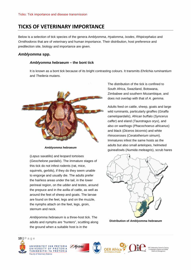
It is known as a bont tick because of its bright contrasting colours. It transmits Ehrlichia ruminantium and Theileria mutans.
The distribution of the tick is confined to South Africa, Swaziland, Botswana, Zimbabwe and southern Mozambique, and does not overlap with that of A. gemma.
Adults feed on cattle, sheep, goats and large wild ruminants, particularly giraffes (Giraffa camelopardalis), African buffalo (Syncerus caffer) and eland (Taurotragus oryx), and also on warthogs (Phacochoerus africanus) and black (Diceros bicornis) and white rhinoceroses (Ceratotherium simum). Immatures infest the same hosts as the adults but also small antelopes, helmeted guineafowls (Numida meleagris), scrub hares
(Lepus saxatilis) and leopard tortoises (Geochelone pardalis). The immature stages of this tick do not infest rodents (rat, mice, squirrels, gerbils), if they do they seem unable to engorge and usually die. The adults prefer the hairless areas under the tail, in the lower perineal region, on the udder and testes, around the prepuce and in the axilla of cattle, as well as around the feet of sheep and goats. The larvae are found on the feet, legs and on the muzzle, the nymphs attach on the feet, legs, groin, sternum and neck.
Amblyomma hebraeum is a three-host tick. The adults and nymphs are “hunters”, scuttling along the ground when a suitable host is in the
Amblyomma hebraeum
Distribution of Amblyomma hebraeum
Ticks: Tick importance and disease transmission
11 | P a g e
vicinity, particularly so if the host is already infested with adult male ticks excreting an attraction pheromone.
This tick requires moisture and warmth, brush and bush and does not survive in open grassland. In South Africa it is found along the coastal belt from Port Elizabeth in the Eastern Cape Province, through KwaZulu-Natal and thence across Mpumalanga, Gauteng, Limpopo and North West Provinces, north of a line running approximately through Pretoria to the Botswana border. It is also present in eastern Swaziland, southern Mozambique, eastern Botswana and in southern and eastern Zimbabwe.
Laboratory studies have demonstrated that A. hebraeum can carry higher infections and also a greater number of strains of E. ruminantium than A. variegatum, the other major vector of this disease. In addition field observations seem to indicate that outbreaks of the disease are more commonly associated with the former than the latter tick and that these outbreaks are also more severe. Besides cattle a number of wild ruminants, as well as other animals such as scrub hares, helmeted guineafowls and leopard tortoises can act as asymptomatic carriers of E. ruminantium. Other organisms transmitted by A. hebraeum are Theileria mutans and Theileria velifera the cause of benign bovine theilerioses. The occurrence of foot abscesses in goats in the Eastern Cape Province, South Africa is significantly related to the seasonal abundance of adult A. hebraeum and Rhipicephalus glabroscutatum. The attachment sites of these ticks around and between the hooves afford entrance to secondary bacterial infections leading to abscessation. These abscesses cause severe lameness in the goats, particularly if more than one foot is affected.
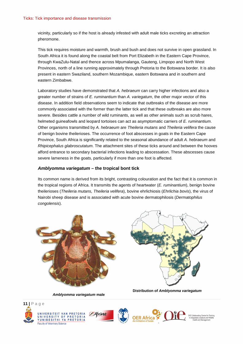
Amblyomma variegatum – the tropical bont tick
Its common name is derived from its bright, contrasting colouration and the fact that it is common in the tropical regions of Africa. It transmits the agents of heartwater (E. ruminantium), benign bovine theilerioses (Theileria mutans, Theileria velifera), bovine ehrlichiosis (Ehrlichia bovis), the virus of Nairobi sheep disease and is associated with acute bovine dermatophilosis (Dermatophilus congolensis).
Amblyomma variegatum male Distribution of Amblyomma variegatum
Ticks: Tick importance and disease transmission
12 | P a g e
It is widely distributed through West, Central, North-East and East Africa and in southern Africa extends into Zambia, north-eastern Botswana, the Caprivi Strip of Namibia, north-western Zimbabwe and central and northern Mozambique. Its spread southwards appears to be limited by a decrease in annual rainfall and interspecific competition with A. hebraeum with which it shares similar habitats, hosts and sites of attachment.
Vector-based epidemiology of heartwater (Norval et al. 1992)
Reservoirs of infection
African buffaloes and possibly other wild ruminants can serve as asymptomatic carriers of E. ruminantium, so can domestic ruminants. Repeated infection of hosts can reinforce the carrier state as the infectivity of immune hosts increases following re-exposure to the organism.
Non-ruminant animals such as scrub hares, guineafowls and tortoises are frequently infested by the immature stages of A. hebraeum, and can also be infected with E. ruminantium and can serve as reservoirs of infection for as yet undetermined lengths of time. Unfed, infected A. hebraeum nymphs and adults can probably retain infection for at least a year. Attached A. hebraeum and A. variegatum males can remain on the same host for 5 months or can transfer to other hosts. Nymphs do not lose the infection if they feed on non-susceptible hosts.
Acquisition of infection
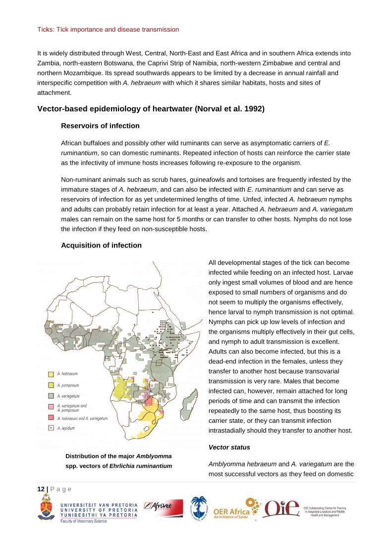
All developmental stages of the tick can become infected while feeding on an infected host. Larvae only ingest small volumes of blood and are hence exposed to small numbers of organisms and do not seem to multiply the organisms effectively, hence larval to nymph transmission is not optimal. Nymphs can pick up low levels of infection and the organisms multiply effectively in their gut cells, and nymph to adult transmission is excellent. Adults can also become infected, but this is a dead-end infection in the females, unless they transfer to another host because transovarial transmission is very rare. Males that become infected can, however, remain attached for long periods of time and can transmit the infection repeatedly to the same host, thus boosting its carrier state, or they can transmit infection intrastadially should they transfer to another host.
Vector status
Amblyomma hebraeum and A. variegatum are the most successful vectors as they feed on domestic
Distribution of the major Amblyomma spp. vectors of Ehrlichia ruminantium
Ticks: Tick importance and disease transmission
13 | P a g e
ruminants in all their stages of development. It would, however, appear that the susceptibility to infection with E. ruminantium can vary considerably within populations of A. hebraeum and of A. variegatum. Field observations seem to indicate that outbreaks of the disease are more commonly associated with A. hebraeum than with A. variegatum, and that these outbreaks are also more severe. The parasite is passed to the host within the first 24 hours of tick attachment, and because the ticks are slow feeders they may still be attached when the animal shows the first signs of illness. The other Amblyomma spp. that can serve as vectors either have limited distribution ranges, or they do not feed on ruminants in all their stages of development, or they have very specific host preferences.
Factors leading to infection
Larvae ascend the surrounding vegetation and infest passing hosts. Nymphs and adults remain on the soil surface and are activated by specific stimuli, namely CO2 exhaled by host animals and by an attraction, aggregation, attachment pheromone produced by attached engorging male ticks. Thus herds of cattle are more likely to trigger tick activity than single animals, large animals more so than small animals, and already infested animals more so than uninfested animals. Most engorged larvae or nymphs detach from their hosts around sunset, and where animals are kraaled at night large accumulations of unfed nymphs or adults that have recently moulted from the detached engorged larvae and nymphs will be present and these will have a readily available source of hosts in the kraaled cattle.
Control
The first effects of intensive tick control in endemic situations will be the reduction in size of the reservoirs of infection that reside in the ticks themselves. This will lead to a reduction in the numbers of animals that become carriers and the re-challenge of existing carriers will be lowered and this will lead to lower infection rates in ticks. The overall effect will be to reduce the numbers of animals that acquire immunity to heartwater through natural infection.
Ticks: Tick importance and disease transmission
14 | P a g e
Hyalomma spp.
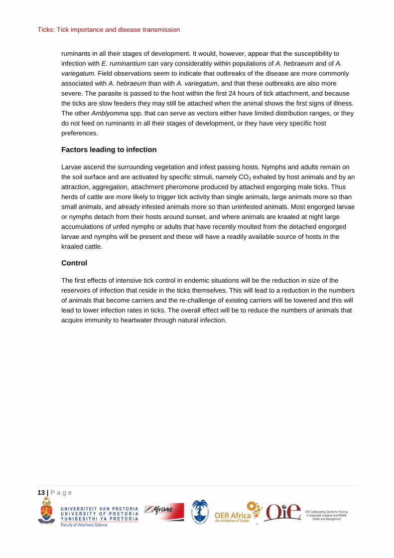
Hyalomma dromedarii – the camel tick
This tick transmits Theileria annulata the cause of tropical theileriosis.
The preferred hosts of this tick are camels (Camelus dromedarius), but cattle, sheep, goats and horses may also be infested. The larvae and the nymphs feed on small burrowing animals and on hares, but the nymphs may also infest camels, cattle and horses. Adults attach on the inner thighs, udder, scrotum and in the nostrils of camels.
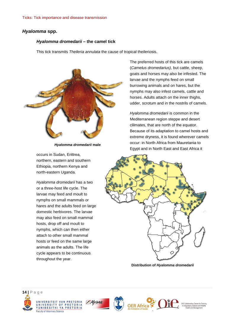
Hyalomma dromedarii is common in the Mediterranean region steppe and desert clilmates, that are north of the equator. Because of its adaptation to camel hosts and extreme dryness, it is found wherever camels occur: in North Africa from Mauretania to Egypt and in North East and East Africa it
occurs in Sudan, Erittrea, northern, eastern and southern Ethiopia, northern Kenya and north-eastern Uganda.
Hyalomma dromedarii has a two or a three-host life cycle. The larvae may feed and moult to nymphs on small mammals or hares and the adults feed on large domestic herbivores. The larvae may also feed on small mammal hosts, drop off and moult to nymphs, which can then either attach to other small mammal hosts or feed on the same large animals as the adults. The life cycle appears to be continuous throughout the year.
Hyalomma dromedarii male
Distribution of Hyalomma dromedarii
Ticks: Tick importance and disease transmission
15 | P a g e
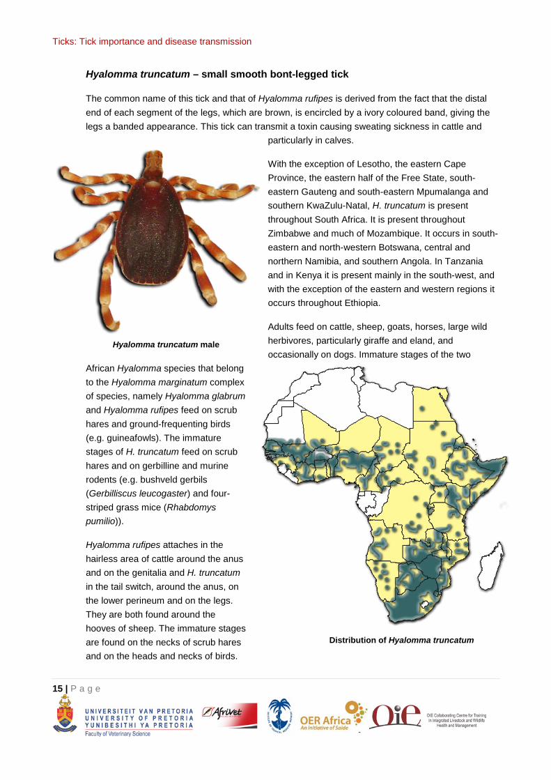
Hyalomma truncatum – small smooth bont-legged tick
The common name of this tick and that of Hyalomma rufipes is derived from the fact that the distal end of each segment of the legs, which are brown, is encircled by a ivory coloured band, giving the legs a banded appearance. This tick can transmit a toxin causing sweating sickness in cattle and
particularly in calves.
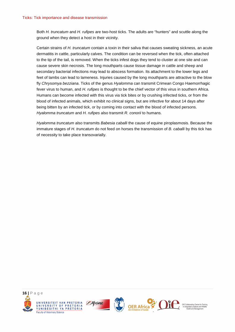
With the exception of Lesotho, the eastern Cape Province, the eastern half of the Free State, south-eastern Gauteng and south-eastern Mpumalanga and southern KwaZulu-Natal, H. truncatum is present throughout South Africa. It is present throughout Zimbabwe and much of Mozambique. It occurs in south-eastern and north-western Botswana, central and northern Namibia, and southern Angola. In Tanzania and in Kenya it is present mainly in the south-west, and with the exception of the eastern and western regions it occurs throughout Ethiopia.
Adults feed on cattle, sheep, goats, horses, large wild herbivores, particularly giraffe and eland, and occasionally on dogs. Immature stages of the two
African Hyalomma species that belong to the Hyalomma marginatum complex of species, namely Hyalomma glabrum and Hyalomma rufipes feed on scrub hares and ground-frequenting birds (e.g. guineafowls). The immature stages of H. truncatum feed on scrub hares and on gerbilline and murine rodents (e.g. bushveld gerbils (Gerbilliscus leucogaster) and four-striped grass mice (Rhabdomys pumilio)).
Hyalomma rufipes attaches in the hairless area of cattle around the anus and on the genitalia and H. truncatum in the tail switch, around the anus, on the lower perineum and on the legs. They are both found around the hooves of sheep. The immature stages are found on the necks of scrub hares and on the heads and necks of birds.
Hyalomma truncatum male
Distribution of Hyalomma truncatum
Ticks: Tick importance and disease transmission
16 | P a g e
Both H. truncatum and H. rufipes are two-host ticks. The adults are “hunters” and scuttle along the ground when they detect a host in their vicinity.
Certain strains of H. truncatum contain a toxin in their saliva that causes sweating sickness, an acute dermatitis in cattle, particularly calves. The condition can be reversed when the tick, often attached to the tip of the tail, is removed. When the ticks infest dogs they tend to cluster at one site and can cause severe skin necrosis. The long mouthparts cause tissue damage in cattle and sheep and secondary bacterial infections may lead to abscess formation. Its attachment to the lower legs and feet of lambs can lead to lameness. Injuries caused by the long mouthparts are attractive to the blow fly Chrysomya bezziana. Ticks of the genus Hyalomma can transmit Crimean Congo Haemorrhagic fever virus to human, and H. rufipes is thought to be the chief vector of this virus in southern Africa. Humans can become infected with this virus via tick bites or by crushing infected ticks, or from the blood of infected animals, which exhibit no clinical signs, but are infective for about 14 days after being bitten by an infected tick, or by coming into contact with the blood of infected persons. Hyalomma truncatum and H. rufipes also transmit R. conorii to humans.
Hyalomma truncatum also transmits Babesia caballi the cause of equine piroplasmosis. Because the immature stages of H. truncatum do not feed on horses the transmission of B. caballi by this tick has of necessity to take place transovarially.
Ticks: Tick importance and disease transmission
17 | P a g e
Ixodes spp.
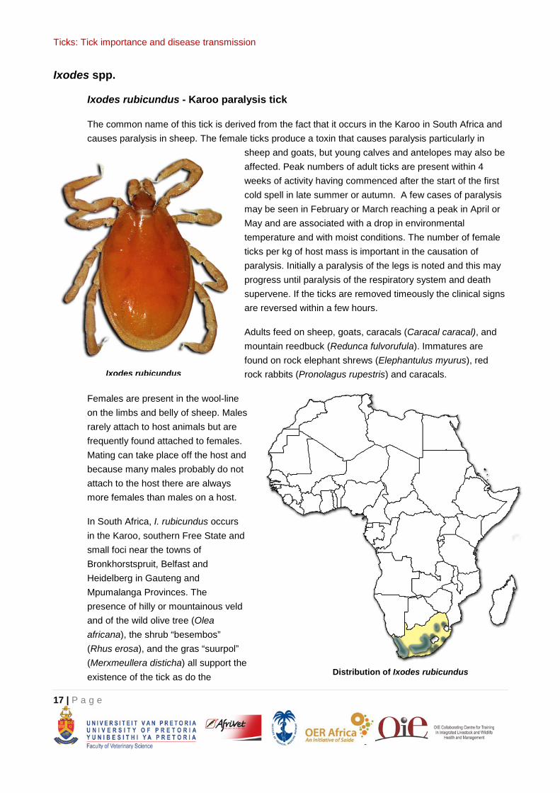
Ixodes rubicundus - Karoo paralysis tick
The common name of this tick is derived from the fact that it occurs in the Karoo in South Africa and causes paralysis in sheep. The female ticks produce a toxin that causes paralysis particularly in
sheep and goats, but young calves and antelopes may also be affected. Peak numbers of adult ticks are present within 4 weeks of activity having commenced after the start of the first cold spell in late summer or autumn. A few cases of paralysis may be seen in February or March reaching a peak in April or May and are associated with a drop in environmental temperature and with moist conditions. The number of female ticks per kg of host mass is important in the causation of paralysis. Initially a paralysis of the legs is noted and this may progress until paralysis of the respiratory system and death supervene. If the ticks are removed timeously the clinical signs are reversed within a few hours.
Adults feed on sheep, goats, caracals (Caracal caracal), and mountain reedbuck (Redunca fulvorufula). Immatures are found on rock elephant shrews (Elephantulus myurus), red rock rabbits (Pronolagus rupestris) and caracals.
Females are present in the wool-line on the limbs and belly of sheep. Males rarely attach to host animals but are frequently found attached to females. Mating can take place off the host and because many males probably do not attach to the host there are always more females than males on a host.
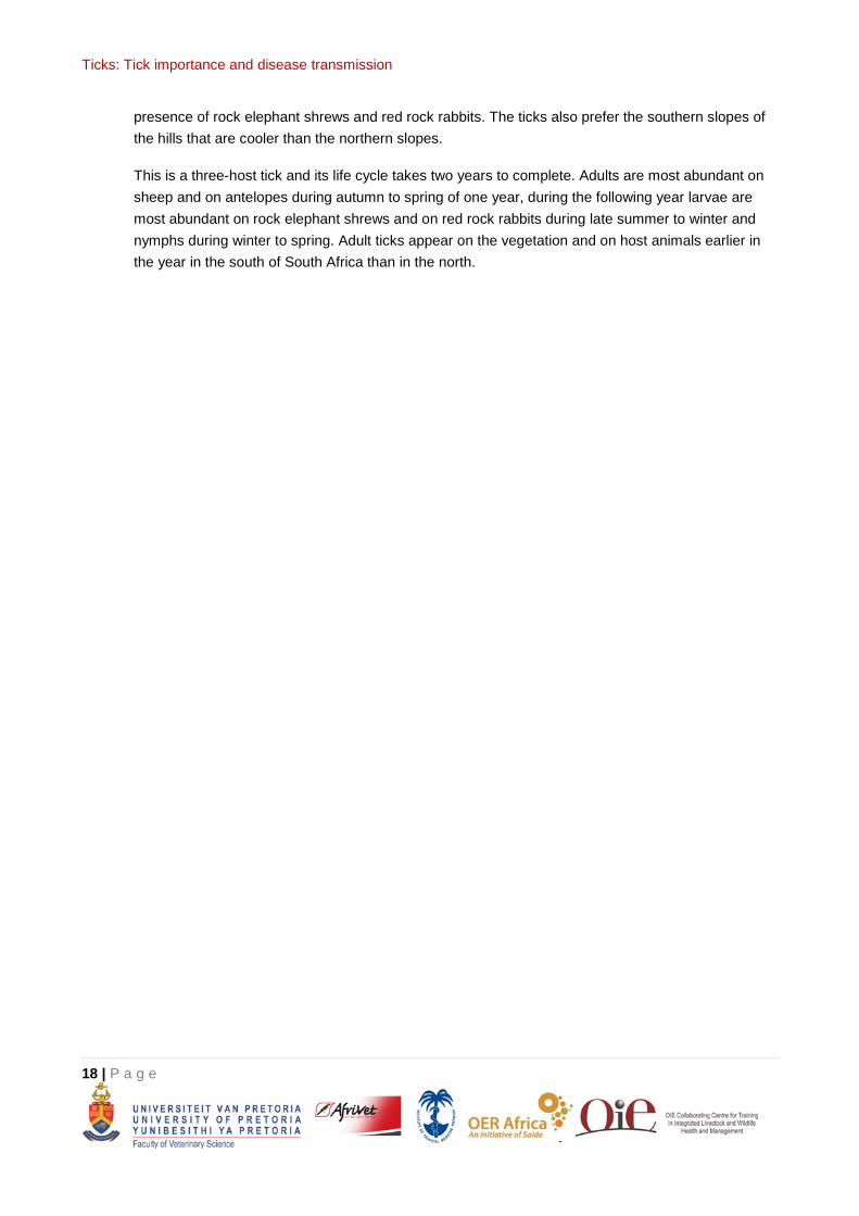
In South Africa, I. rubicundus occurs in the Karoo, southern Free State and small foci near the towns of Bronkhorstspruit, Belfast and Heidelberg in Gauteng and Mpumalanga Provinces. The presence of hilly or mountainous veld and of the wild olive tree (Olea africana), the shrub “besembos” (Rhus erosa), and the gras “suurpol” (Merxmeullera disticha) all support the existence of the tick as do the
Ixodes rubicundus
Distribution of Ixodes rubicundus
Ticks: Tick importance and disease transmission
18 | P a g e
presence of rock elephant shrews and red rock rabbits. The ticks also prefer the southern slopes of the hills that are cooler than the northern slopes.
This is a three-host tick and its life cycle takes two years to complete. Adults are most abundant on sheep and on antelopes during autumn to spring of one year, during the following year larvae are most abundant on rock elephant shrews and on red rock rabbits during late summer to winter and nymphs during winter to spring. Adult ticks appear on the vegetation and on host animals earlier in the year in the south of South Africa than in the north.
Ticks: Tick importance and disease transmission
19 | P a g e
Rhipicephalus spp.
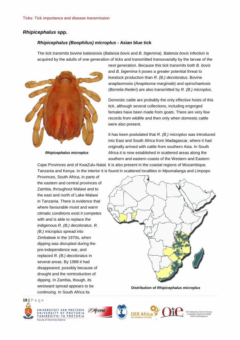
Rhipicephalus (Boophilus) microplus - Asian blue tick
The tick transmits bovine babesiosis (Babesia bovis and B. bigemina). Babesia bovis infection is acquired by the adults of one generation of ticks and transmitted transovarially by the larvae of the
next generation. Because this tick transmits both B. bovis and B. bigemina it poses a greater potential threat to livestock production than R. (B.) decoloratus. Bovine anaplasmosis (Anaplasma marginale) and spirochaetosis (Borrelia theileri) are also transmitted by R. (B.) microplus.
Domestic cattle are probably the only effective hosts of this tick, although several collections, including engorged females have been made from goats. There are very few records from wildlife and then only when domestic cattle were also present.
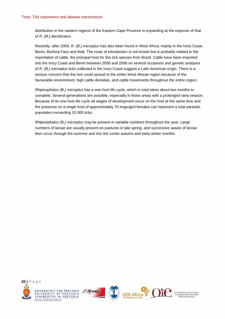
It has been postulated that R. (B.) microplus was introduced into East and South Africa from Madagascar, where it had originally arrived with cattle from southern Asia. In South Africa it is now established in scattered areas along the southern and eastern coasts of the Western and Eastern
Cape Provinces and of KwaZulu-Natal. It is also present in the coastal regions of Mozambique, Tanzania and Kenya. In the interior it is found in scattered localities in Mpumalanga and Limpopo Provinces, South Africa, in parts of the eastern and central provinces of Zambia, throughout Malawi and to the east and north of Lake Malawi in Tanzania. There is evidence that where favourable moist and warm climatic conditions exist it competes with and is able to replace the indigenous R. (B.) decoloratus. R. (B.) microplus spread into Zimbabwe in the 1970s, when dipping was disrupted during the pre-independence war, and replaced R. (B.) decoloratus in several areas. By 1988 it had disappeared, possibly because of drought and the reintroduction of dipping. In Zambia, though, its westward spread appears to be continuing. In South Africa its
Rhipicephalus microplus
Distribution of Rhipicephalus microplus
Ticks: Tick importance and disease transmission
20 | P a g e
distribution in the eastern regions of the Eastern Cape Province is expanding at the expense of that of R. (B.) decoloratus.
Recently, after 2000, R. (B.) microplus has also been found in West Africa, mainly in the Ivory Coast, Benin, Burkina Faso and Mali. The route of introduction is not known but is probably related to the importation of cattle, the principal host for this tick species from Brazil. Cattle have been imported into the Ivory Coast and Benin between 2000 and 2006 on several occasions and genetic analyses of R. (B.) microplus ticks collected in the Ivory Coast suggest a Latin American origin. There is a serious concern that this tick could spread to the entire West African region because of the favourable environment, high cattle densities, and cattle movements throughout the entire region.
Rhipicephalus (B.) microplus has a one-host life cycle, which in total takes about two months to complete. Several generations are possible, especially in those areas with a prolonged rainy season. Because of its one-host life cycle all stages of development occur on the host at the same time and the presence on a single host of approximately 70 engorged females can represent a total parasitic population exceeding 10 000 ticks.
Rhipicephalus (B.) microplus may be present in variable numbers throughout the year. Large numbers of larvae are usually present on pastures in late spring, and successive waves of larvae then occur through the summer and into the cooler autumn and early winter months.
Ticks: Tick importance and disease transmission
21 | P a g e
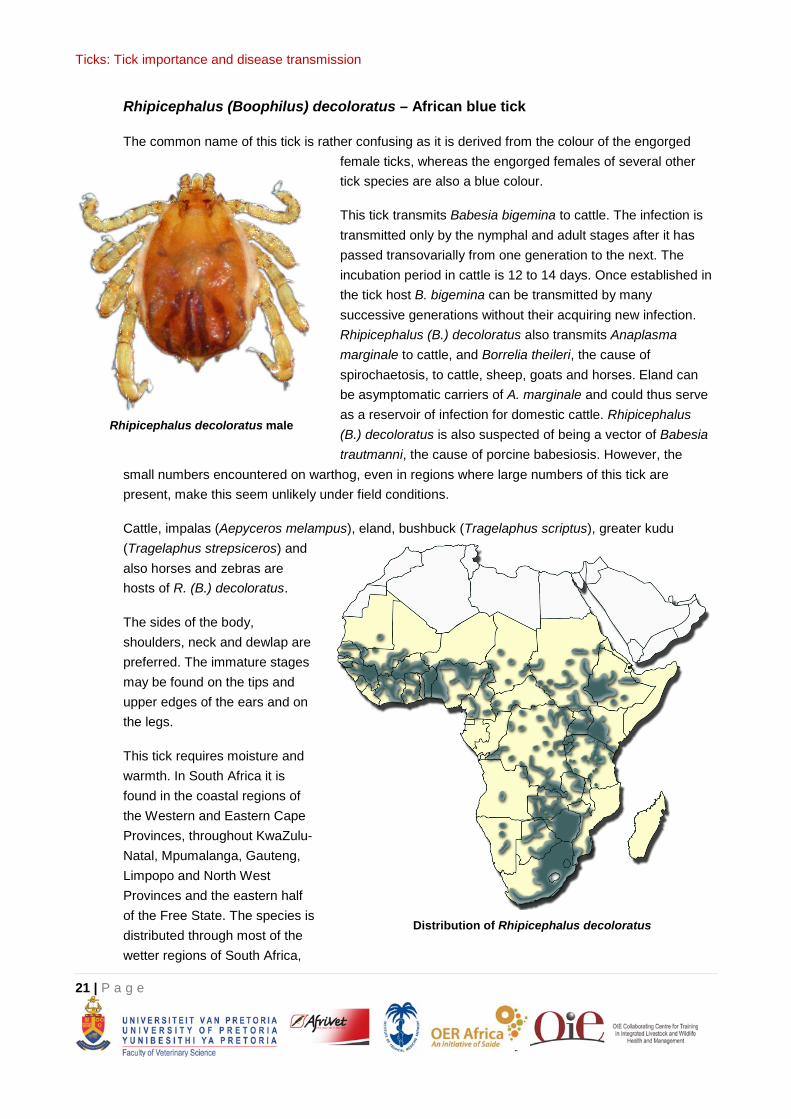
Rhipicephalus (Boophilus) decoloratus – African blue tick
The common name of this tick is rather confusing as it is derived from the colour of the engorged female ticks, whereas the engorged females of several other tick species are also a blue colour.
This tick transmits Babesia bigemina to cattle. The infection is transmitted only by the nymphal and adult stages after it has passed transovarially from one generation to the next. The incubation period in cattle is 12 to 14 days. Once established in the tick host B. bigemina can be transmitted by many successive generations without their acquiring new infection. Rhipicephalus (B.) decoloratus also transmits Anaplasma marginale to cattle, and Borrelia theileri, the cause of spirochaetosis, to cattle, sheep, goats and horses. Eland can be asymptomatic carriers of A. marginale and could thus serve as a reservoir of infection for domestic cattle. Rhipicephalus (B.) decoloratus is also suspected of being a vector of Babesia trautmanni, the cause of porcine babesiosis. However, the
small numbers encountered on warthog, even in regions where large numbers of this tick are present, make this seem unlikely under field conditions.
Cattle, impalas (Aepyceros melampus), eland, bushbuck (Tragelaphus scriptus), greater kudu (Tragelaphus strepsiceros) and also horses and zebras are hosts of R. (B.) decoloratus.
The sides of the body, shoulders, neck and dewlap are preferred. The immature stages may be found on the tips and upper edges of the ears and on the legs.
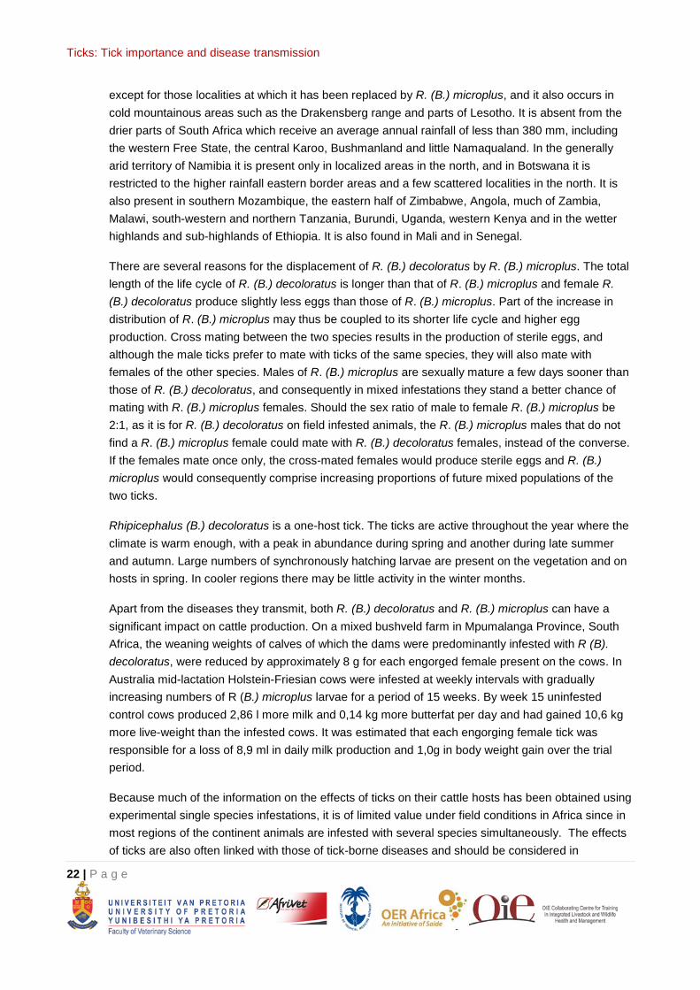
This tick requires moisture and warmth. In South Africa it is found in the coastal regions of the Western and Eastern Cape Provinces, throughout KwaZulu-Natal, Mpumalanga, Gauteng, Limpopo and North West Provinces and the eastern half of the Free State. The species is distributed through most of the wetter regions of South Africa,
Rhipicephalus decoloratus male
Distribution of Rhipicephalus decoloratus
Ticks: Tick importance and disease transmission
22 | P a g e
except for those localities at which it has been replaced by R. (B.) microplus, and it also occurs in cold mountainous areas such as the Drakensberg range and parts of Lesotho. It is absent from the drier parts of South Africa which receive an average annual rainfall of less than 380 mm, including the western Free State, the central Karoo, Bushmanland and little Namaqualand. In the generally arid territory of Namibia it is present only in localized areas in the north, and in Botswana it is restricted to the higher rainfall eastern border areas and a few scattered localities in the north. It is also present in southern Mozambique, the eastern half of Zimbabwe, Angola, much of Zambia, Malawi, south-western and northern Tanzania, Burundi, Uganda, western Kenya and in the wetter highlands and sub-highlands of Ethiopia. It is also found in Mali and in Senegal.
There are several reasons for the displacement of R. (B.) decoloratus by R. (B.) microplus. The total length of the life cycle of R. (B.) decoloratus is longer than that of R. (B.) microplus and female R. (B.) decoloratus produce slightly less eggs than those of R. (B.) microplus. Part of the increase in distribution of R. (B.) microplus may thus be coupled to its shorter life cycle and higher egg production. Cross mating between the two species results in the production of sterile eggs, and although the male ticks prefer to mate with ticks of the same species, they will also mate with females of the other species. Males of R. (B.) microplus are sexually mature a few days sooner than those of R. (B.) decoloratus, and consequently in mixed infestations they stand a better chance of mating with R. (B.) microplus females. Should the sex ratio of male to female R. (B.) microplus be 2:1, as it is for R. (B.) decoloratus on field infested animals, the R. (B.) microplus males that do not find a R. (B.) microplus female could mate with R. (B.) decoloratus females, instead of the converse. If the females mate once only, the cross-mated females would produce sterile eggs and R. (B.) microplus would consequently comprise increasing proportions of future mixed populations of the two ticks.
Rhipicephalus (B.) decoloratus is a one-host tick. The ticks are active throughout the year where the climate is warm enough, with a peak in abundance during spring and another during late summer and autumn. Large numbers of synchronously hatching larvae are present on the vegetation and on hosts in spring. In cooler regions there may be little activity in the winter months.
Apart from the diseases they transmit, both R. (B.) decoloratus and R. (B.) microplus can have a significant impact on cattle production. On a mixed bushveld farm in Mpumalanga Province, South Africa, the weaning weights of calves of which the dams were predominantly infested with R (B). decoloratus, were reduced by approximately 8 g for each engorged female present on the cows. In Australia mid-lactation Holstein-Friesian cows were infested at weekly intervals with gradually increasing numbers of R (B.) microplus larvae for a period of 15 weeks. By week 15 uninfested control cows produced 2,86 l more milk and 0,14 kg more butterfat per day and had gained 10,6 kg more live-weight than the infested cows. It was estimated that each engorging female tick was responsible for a loss of 8,9 ml in daily milk production and 1,0g in body weight gain over the trial period.
Because much of the information on the effects of ticks on their cattle hosts has been obtained using experimental single species infestations, it is of limited value under field conditions in Africa since in most regions of the continent animals are infested with several species simultaneously. The effects of ticks are also often linked with those of tick-borne diseases and should be considered in
Ticks: Tick importance and disease transmission
23 | P a g e
conjunction with the latter. Furthermore cattle herds contain animals of different ages, sexes and types and experiments to determine the effects of ticks should be conducted on entire herds rather than on individual or selected age groups of animals.
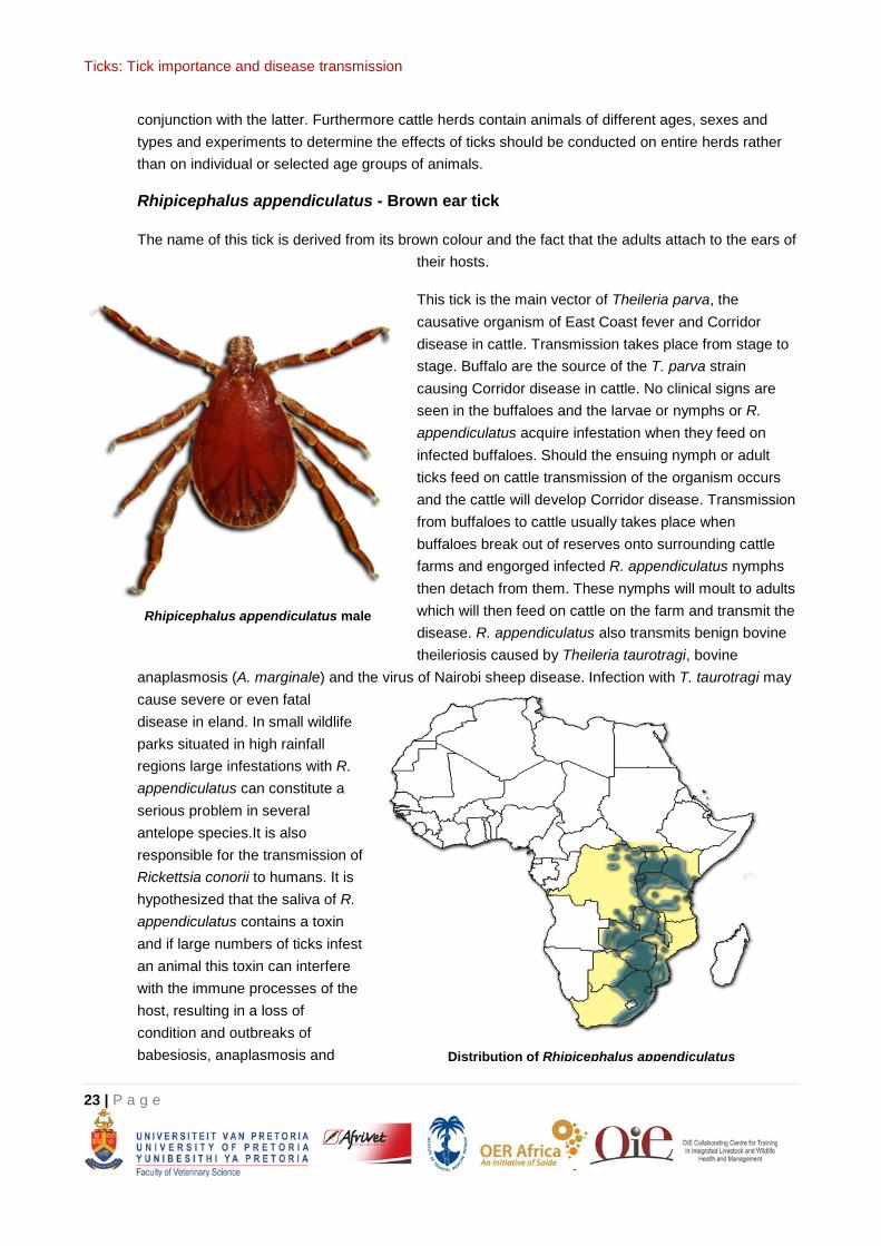
Rhipicephalus appendiculatus - Brown ear tick
The name of this tick is derived from its brown colour and the fact that the adults attach to the ears of their hosts.
This tick is the main vector of Theileria parva, the causative organism of East Coast fever and Corridor disease in cattle. Transmission takes place from stage to stage. Buffalo are the source of the T. parva strain causing Corridor disease in cattle. No clinical signs are seen in the buffaloes and the larvae or nymphs or R. appendiculatus acquire infestation when they feed on infected buffaloes. Should the ensuing nymph or adult ticks feed on cattle transmission of the organism occurs and the cattle will develop Corridor disease. Transmission from buffaloes to cattle usually takes place when buffaloes break out of reserves onto surrounding cattle farms and engorged infected R. appendiculatus nymphs then detach from them. These nymphs will moult to adults which will then feed on cattle on the farm and transmit the disease. R. appendiculatus also transmits benign bovine theileriosis caused by Theileria taurotragi, bovine
anaplasmosis (A. marginale) and the virus of Nairobi sheep disease. Infection with T. taurotragi may cause severe or even fatal disease in eland. In small wildlife parks situated in high rainfall regions large infestations with R. appendiculatus can constitute a serious problem in several antelope species.It is also responsible for the transmission of Rickettsia conorii to humans. It is hypothesized that the saliva of R. appendiculatus contains a toxin and if large numbers of ticks infest an animal this toxin can interfere with the immune processes of the host, resulting in a loss of condition and outbreaks of babesiosis, anaplasmosis and
Rhipicephalus appendiculatus male
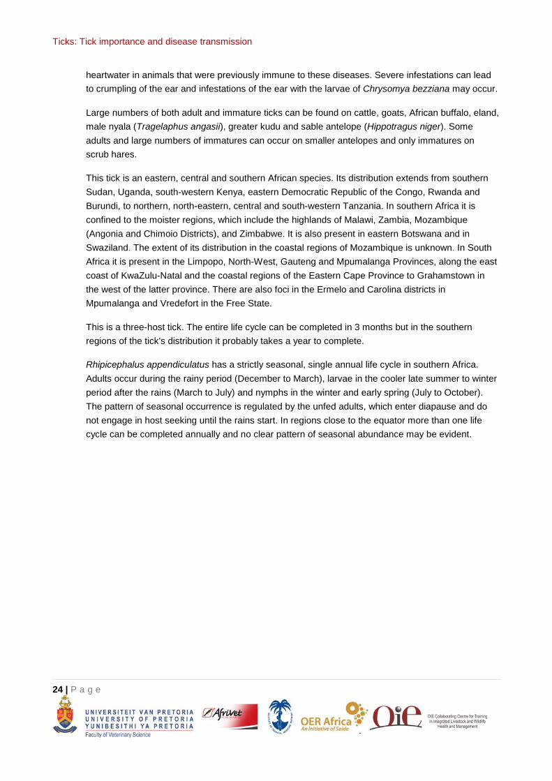
Distribution of Rhipicephalus appendiculatus
Ticks: Tick importance and disease transmission
24 | P a g e
heartwater in animals that were previously immune to these diseases. Severe infestations can lead to crumpling of the ear and infestations of the ear with the larvae of Chrysomya bezziana may occur.
Large numbers of both adult and immature ticks can be found on cattle, goats, African buffalo, eland, male nyala (Tragelaphus angasii), greater kudu and sable antelope (Hippotragus niger). Some adults and large numbers of immatures can occur on smaller antelopes and only immatures on scrub hares.
This tick is an eastern, central and southern African species. Its distribution extends from southern Sudan, Uganda, south-western Kenya, eastern Democratic Republic of the Congo, Rwanda and Burundi, to northern, north-eastern, central and south-western Tanzania. In southern Africa it is confined to the moister regions, which include the highlands of Malawi, Zambia, Mozambique (Angonia and Chimoio Districts), and Zimbabwe. It is also present in eastern Botswana and in Swaziland. The extent of its distribution in the coastal regions of Mozambique is unknown. In South Africa it is present in the Limpopo, North-West, Gauteng and Mpumalanga Provinces, along the east coast of KwaZulu-Natal and the coastal regions of the Eastern Cape Province to Grahamstown in the west of the latter province. There are also foci in the Ermelo and Carolina districts in Mpumalanga and Vredefort in the Free State.
This is a three-host tick. The entire life cycle can be completed in 3 months but in the southern regions of the tick's distribution it probably takes a year to complete.
Rhipicephalus appendiculatus has a strictly seasonal, single annual life cycle in southern Africa. Adults occur during the rainy period (December to March), larvae in the cooler late summer to winter period after the rains (March to July) and nymphs in the winter and early spring (July to October). The pattern of seasonal occurrence is regulated by the unfed adults, which enter diapause and do not engage in host seeking until the rains start. In regions close to the equator more than one life cycle can be completed annually and no clear pattern of seasonal abundance may be evident.
Ticks: Tick importance and disease transmission
25 | P a g e
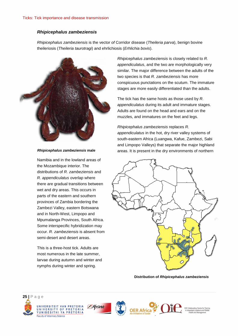
Rhipicephalus zambeziensis
Rhipicephalus zambeziensis is the vector of Corridor disease (Theileria parva), benign bovine theileriosis (Theileria taurotragi) and ehrlichiosis (Erhlichia bovis).
Rhipicephalus zambeziensis is closely related to R. appendiculatus, and the two are morphologically very similar. The major difference between the adults of the two species is that R. zambeziensis has more conspicuous punctations on the scutum. The immature stages are more easily differentiated than the adults.
The tick has the same hosts as those used by R. appendiculatus during its adult and immature stages. Adults are found on the head and ears and on the muzzles, and immatures on the feet and legs.
Rhipicephalus zambeziensis replaces R. appendiculatus in the hot, dry river valley systems of south-eastern Africa (Luangwa, Kafue, Zambezi, Sabi and Limpopo Valleys) that separate the major highland areas. It is present in the dry environments of northern
Namibia and in the lowland areas of the Mozambique interior. The distributions of R. zambeziensis and R. appendiculatus overlap where there are gradual transitions between wet and dry areas. This occurs in parts of the eastern and southern provinces of Zambia bordering the Zambezi Valley, eastern Botswana and in North-West, Limpopo and Mpumalanga Provinces, South Africa. Some interspecific hybridization may occur. R. zambeziensis is absent from semi-desert and desert areas.
This is a three-host tick. Adults are most numerous in the late summer, larvae during autumn and winter and nymphs during winter and spring.
Rhipicephalus zambeziensis male
Distribution of Rhipicephalus zambeziensis
Ticks: Tick importance and disease transmission
26 | P a g e
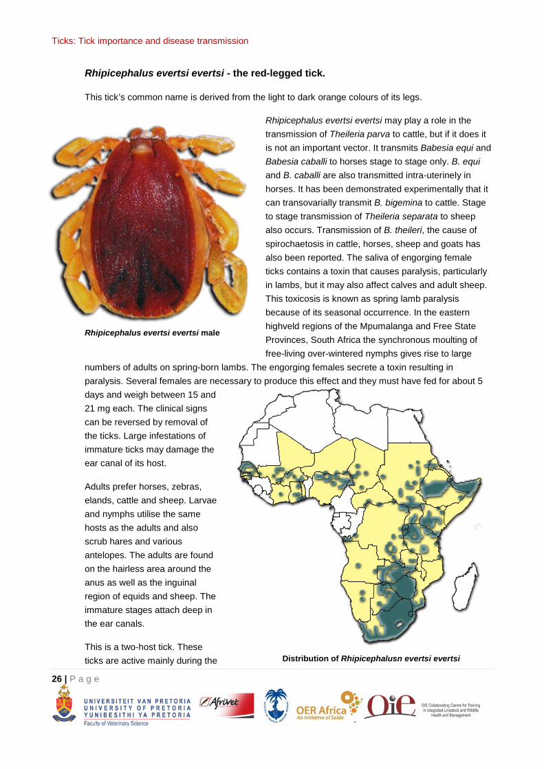
Rhipicephalus evertsi evertsi - the red-legged tick.
This tick’s common name is derived from the light to dark orange colours of its legs.
Rhipicephalus evertsi evertsi may play a role in the transmission of Theileria parva to cattle, but if it does it is not an important vector. It transmits Babesia equi and Babesia caballi to horses stage to stage only. B. equi and B. caballi are also transmitted intra-uterinely in horses. It has been demonstrated experimentally that it can transovarially transmit B. bigemina to cattle. Stage to stage transmission of Theileria separata to sheep also occurs. Transmission of B. theileri, the cause of spirochaetosis in cattle, horses, sheep and goats has also been reported. The saliva of engorging female ticks contains a toxin that causes paralysis, particularly in lambs, but it may also affect calves and adult sheep. This toxicosis is known as spring lamb paralysis because of its seasonal occurrence. In the eastern highveld regions of the Mpumalanga and Free State Provinces, South Africa the synchronous moulting of free-living over-wintered nymphs gives rise to large
numbers of adults on spring-born lambs. The engorging females secrete a toxin resulting in paralysis. Several females are necessary to produce this effect and they must have fed for about 5 days and weigh between 15 and 21 mg each. The clinical signs can be reversed by removal of the ticks. Large infestations of immature ticks may damage the ear canal of its host.
Adults prefer horses, zebras, elands, cattle and sheep. Larvae and nymphs utilise the same hosts as the adults and also scrub hares and various antelopes. The adults are found on the hairless area around the anus as well as the inguinal region of equids and sheep. The immature stages attach deep in the ear canals.
This is a two-host tick. These ticks are active mainly during the
Rhipicephalus evertsi evertsi male
Distribution of Rhipicephalusn evertsi evertsi
Ticks: Tick importance and disease transmission
27 | P a g e
summer but are present throughout the year in warm regions.
A very similar tick Rhipicephalus evertsi mimeticus, known as the Namibian red-legged tick, looks like R. evertsi evertsi, but has red and ivory-coloured banded legs similar to those of certain Hyalomma spp. This tick occurs in western Botswana, central and northern Namibia and southern and western Angola and transmits Theileria equi the cause of equine piroplasmosis and Theileria separata, the cause of ovine theileriosis.
Ticks: Tick importance and disease transmission
28 | P a g e
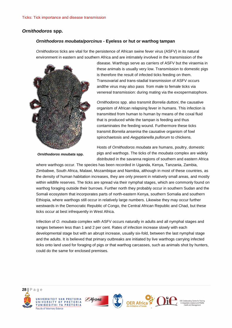
Ornithodoros spp.
Ornithodoros moubata/porcinus - Eyeless or hut or warthog tampan
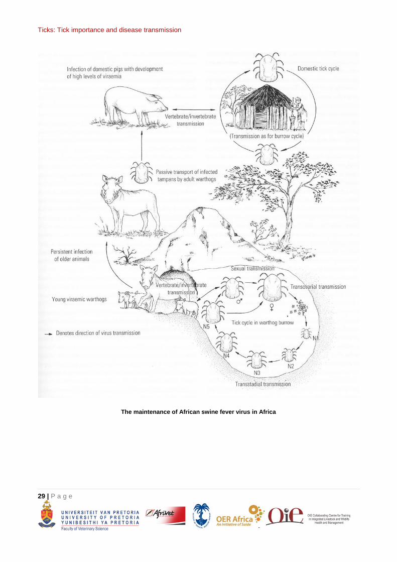
Ornithodoros ticks are vital for the persistence of African swine fever virus (ASFV) in its natural environment in eastern and southern Africa and are intimately involved in the transmission of the
disease. Warthogs serve as carriers of ASFV but the viraemia in these animals is usually very low. Transmission to domestic pigs is therefore the result of infected ticks feeding on them. Transovarial and trans-stadial transmission of ASFV occurs andthe virus may also pass from male to female ticks via venereal transmission: during mating via the exospermatophore.
Ornithodoros spp. also transmit Borrelia duttoni, the causative organism of African relapsing fever in humans. This infection is transmitted from human to human by means of the coxal fluid that is produced while the tampan is feeding and thus contaminates the feeding wound. Furthermore these ticks transmit Borrelia anserina the causative organism of fowl spirochaetosis and Aegyptianella pullorum to chickens.
Hosts of Ornithodoros moubata are humans, poultry, domestic pigs and warthogs. The ticks of the moubata complex are widely distributed in the savanna regions of southern and eastern Africa
where warthogs occur. The species has been recorded in Uganda, Kenya, Tanzania, Zambia, Zimbabwe, South Africa, Malawi, Mozambique and Namibia, although in most of these countries, as the density of human habitation increases, they are only present in relatively small areas, and mostly within wildlife reserves. The ticks are spread via their nymphal stages, which are commonly found on warthog foraging outside their burrows. Further north they probably occur in southern Sudan and the Somali ecosystem that incorporates parts of north-eastern Kenya, southern Somalia and southern Ethiopia, where warthogs still occur in relatively large numbers. Likewise they may occur further westwards in the Democratic Republic of Congo, the Central African Republic and Chad, but these ticks occur at best infrequently in West Africa.
Infection of O. moubata complex with ASFV occurs naturally in adults and all nymphal stages and ranges between less than 1 and 2 per cent. Rates of infection increase slowly with each developmental stage but with an abrupt increase, usually six-fold, between the last nymphal stage and the adults. It is believed that primary outbreaks are initiated by live warthogs carrying infected ticks onto land used for foraging of pigs or that warthog carcasses, such as animals shot by hunters, could do the same for enclosed premises.
Ornithodoros moubata spp.
Ticks: Tick importance and disease transmission
29 | P a g e
The maintenance of African swine fever virus in Africa
Ticks: Tick importance and disease transmission
30 | P a g e
REFERENCES
1. Brown and Hodek (1983). Diapause and Life Cycle Strategies in Insects. Entomologica Vol. 23. Dr. W. Junk Publishers: The Hague.
2. Coetzer, J. A. W. and Tustin, R. C. 2004. Infectious diseases of Livestock (2nd edition). Oxford University Press.
3. Randolph, S. (2005). Tick ecology: processes and patterns behind the epidemiological risk posed by ixodid ticks as vectors. Parasitology 129(S1), S37-S65. (Randolph 2005.pdf) http://www.cbpv.org.br/artigos/CBPV_artigo_021.pdf
4. Kleiboeker et al. (1998). African swine fever virus infection in the argasid host, Ornithodoros porcinus porcinus. J Virol 72(3), 1711-1724. (ASF_Ornithodoros.pdf) http://jvi.asm.org/content/72/3/1711.full.pdf+html
5. Norval, RAI, Andrew, HR, Yunker, CE, & Burridge, MJ 1992 Biological processes in the epidemiology of heartwater. In Fivaz, BH, Petney, TN & Horak, IG (eds). Tick vector biology: medical and veterinary aspects. Springer-Verlag: Berlin, Heidelberg, pp. 71-86.
6. Shaw et al. (2001). Tick-borne infectious diseases of dogs. Trends Parasitol 17(2), 74-80. (Transmission by ingestion.pdf). http://www.sciencedirect.com/science/article/pii/S1471492200018560
7. Walker, AR, Bouattour, A, Camicas, J-L, Estrada-Peña, A, Horak, IG, Latif, AA, Pegram, RG, Preston,PM 2003. Ticks of domestic animals in Africa: a guide to identification of species. Bioscience Reports, 095451730X
8. Tick website of African ticks: http://www.itg.be/photodatabase/African_ticks_files/index.html