thymidylate synthetase - a target enzyme in cancer chemotherapy
TRANSCRIPT

Biochimica et Biophysica Acta, 473 (1977) 73-92 © Elsevier/North-Holland Biomedical Press
BBA 87036
T H Y M I D Y L A T E S Y N T H E T A S E - A T A R G E T E N Z Y M E I N C A N C E R
C H E M O T H E R A P Y
PETER V. DANENBERG
LAC-USC Comprehensive Cancer Center and Department of Biochemistry, University of Southern California, School of Medicine, Los Angeles, Calif. 90031 (U.S.A.}
(Received May 12th, 1977)
CONTENTS
I. Introduction . . . . . . . . . . . . . . . . . . . . . . . . . . . . . . . . . 73
lI. The interaction of thymid~late synthetase with 5-fluoro-2'-deoxyuridylate . . . . . . . 75
III. Interaction of thymidylate synthetase with other dUMP analogs . . . . . . . . . . . 85 A. 5-Trifluoromethyl-2'-deoxyuridylic acid (FadTMP) . . . . . . . . . . . . . . . 85 B. 5-Formyl-2'-deoxyuridylic acid (formyl-dUMP) . . . . . . . . . . . . . . . . 87 C. 5-Mercapto-2'-deoxyuridylic acid I, mercapto-dUMP) . . . . . . . . . . . . . . 87 D. 5-Iodoacetamidomethyl-2'-deoxyuridine-5'-phosphate . . . . . . . . . . . . . . 87
IV. Analogs of 5,10-CH2H4folate . . . . . . . . . . . . . . . . . . . . . . . . . . 88
V. Conclusion . . . . . . . . . . . . . . . . . . . . . . . . . . . . . . . . . . 89
Acknowledgements . . . . . . . . . . . . . . . . . . . . . . . . . . . . . . . . 90
References . . . . . . . . . . . . . . . . . . . . . . . . . . . . . . . . . . . . 90
I. INTRODUCTION
The biosynthesis o f thymid ine nucleot ides is unique in a n u m b e r o f respects.
Thymine first appears in cells as the deoxyr ibonuc leos ide monophospha t e , deoxy-
thymidyl ic acid (dTMP) , whereas the o ther cons t i tuent deoxyr ibonuc leo t ides o f
D N A are all fo rmed at the d iphospha te levels f rom the co r respond ing r ibonucleot ides .
Thus, there are no rma l ly no r ibose-conta in ing thymid ine mononuc leo t ides in cells.
Pe rhaps the mos t s t r iking aspect o f the origin o f d T M P is tha t the reac t ion leading
to its f o rma t ion involves a modif ica t ion o f the pyr imid ine base. This reac t ion is
carr ied out by the enzyme thymidy la te synthetase (EC 2.1.1.45), and consists o f an
unusua l two-s tep process ,in which t ransfer o f a one-ca rbon unit f r o m the cofac tor
NS ,Nl° -me thy lene t e t r ahydro fo l a t e (5,10-CH2H4folate) to the 5-posi t ion o f deoxy-
ur idyla te ( d U M P ) is fo l lowed by reduct ion o f the one -ca rbon unit to a methyl
group. The overal l reac t ion can be wri t ten as :

74
dUMP + 5,10-CH2H4folate -+ dTMP + H2folate
o
H H 2 N ,,,,,,r./N~,/N,,, ' 0 0
HO-- p--O--FO~,] Jr / \ ...N ~ " - L ~ C --Glu ~OH W OH OH 2
oH ,1, O
HN IJJ~cH3 H 0 ~N ~I H 2 N "]//N I~F'N ~ 0 II 0 , N-'~ -~kN-,, ~ -- II
H O - - P - O - ' ~ Jr "]~ " - - N k--'/~r.~C --Gill O~H ~ OH H
OH
S c h e m e I.
The role of the cofactor as both the carrier of the one-carbon fragment and the reducing agent was established by several classical early experiments. Humphreys and Greenberg [1] discovered a direct relationship between the H4folate content of cell-free extracts and dTMP formation. Friedkin [2] demonstrated that tritium- labeled H4folate, prepared by reducing folate with boro[3H]hydride, transferred the label to the methyl group of dTMP. According to the work of McDougall and Blakley [3], this process occurred with negligible isotope dilution, suggesting a direct intramolecular hydride transfer.
Since thymidylate synthetase is the only de novo source of thymidine, it is a pivotal enzyme in the biosynthesis of DNA. Not surprisingly, the activity of this enzyme usually is substantially elevated, compared to normal tissues, in proliferating cellular systems such as regenerating liver [4].
Because thymidylate synthetase is not known to be regulated by any of the naturally occurring nucleotides, except for a weak product inhibition by dTMP, the elevated activity in response to a demand for increased DNA synthesis most likely arises from increased cellular synthesis of the enzyme. Conrad and Ruddle [5] found that puromycin, an inhibitor of protein synthesis, prevented a methotrexate-stimulated increase in thymidylate synthetase activity. Other evidence has led to the conclusion that a masked stable messenger RNA for the enzyme is present in normal liver [6]. Because the thymidylate synthetase content of normal tissues is very low and in many cases undetectable, Blakley has suggested that the enzyme may be the rate-limiting step in DNA synthesis [7]. However, the ribonucleotide reductase system also has an exceedingly low V, and pools of acid-soluble deoxyribosyl nucleotides in most mammalian and bacterial cells are only about 1 ~o of the ribonucleotide pools [8]. Therefore, the question of the rate-limiting step in DNA synthesis has not yet been resolved. It is fikely that either ribonucleotide reduction or thymidylate synthesis can be the controlling factor under different conditions. For example, depletion of H~folate by methotrexate could cause the latter to be rate limiting.

75
From the above considerations, blockade of thymidylate synthetase would clearly exert drastic effects upon tissues that require a high rate of DNA synthesis. Since many tumors fall in this category, thymidylate synthetase is an attractive target for the design of cancer chemotherapeutic agents. The enzyme is apparently not subject to allosteric regulation and in most organisms cannot be circumvented by other pathways (except by thymidine kinase, a salvage enzyme which converts pre- formed thymidine to dTMP). In fact, a number of therapeutically active and widely used antimetabolites have been found to inhibit thymidylate synthetase activity either by a direct interaction, or indirectly by inhibiting dihydrofolate reductase, an enzyme which regenerates the tetrahydrofolate consumed in the thymidylate synthesis re- action. Unfortunately, anti-tumor activity by a drug that inhibits DNA synthesis is usually accompanied by considerable toxicity to normal rapidly proliferating tissues, such as the gastrointestinal tract, mucosa, and hematopoietic tissues.
The mechanism of cell death by inhibition of DNA synthesis has been ex- tensively investigated by Cohen [9]. Compounds that inhibit production of dTMP have been shown to promote unbalanced growth of cells in which protein and RNA are being synthesized, but not DNA, a phenomenon which apparently leads to irreversible DNA damage, and has been termed "thymineless death". It is interesting that a simultaneous inhibition of RNA synthesis markedly retards thymineless death [10].
In this report, we will consider the mechanisms of interaction of thymidylate synthetase with various compounds of established or potential anti-tumor activity.
II. THE INTERACTION OF THYMIDYLATE SYNTHETASE WITH 5-FLUORO-2'-DEOXYURIDYLATE
Since its introduction in 1957 by Heidelberger [11], 5-fluorouracil (5-FUra) and its derivatives have been used extensively in cancer chemotherapy, especially in the treatment of patients with disseminated breast and colon cancers [12]. The conception and design of this drug was based on a very definite and ingenious rationale derived from the observation that tumors utilize uracil to a greater degree than does the surrounding normal tissue [13]. The reasoning was that a derivative of uracil, although not necessarily having more specificity for the enzymes from tumor tissues, would produce a differential cytotoxicity because it would be preferentially taken up by tumors. 5-Fluorouracil was chosen not only because fluorine and hydrogen are very similar in size but also because of the exceptional stability of the carbon-fluorine bond. These considerations led Heidelberger to predict that in vivo 5-FUra would become incorporated into RNA and also inhibit DNA biosynthesis. Indeed, it was discovered early that 5-FUra and its derivatives 5-fluoro-2'-deoxyuridine (FdUrd), 5-fluorouridine, and fluoroorotic acid all inhibited incorporation into DNA of labeled formate, a known precursor of the methyl group of DNA thymidine [14]. These results were confirmed by Cohen and co-workers, who demonstrated that

76
FdUrd induces thymineless death in Escherichia coli [15]. It was further shown that this effect at the enzymatic level was due to a powerful inhibition of thymidylate synthetase by the nucleotide form of the drug, 5-fluoro-2'-deoxyuridylic acid (FdUMP). Cohen's initial observations launched an intensive study of the nature of the complex formed between FdUMP and its target enzyme, a research activity which has only recently reached its peak. The interaction of this nucleotide with thymidylate synthetase has important implications for cancer chemotherapy as well as for an understanding of the puzzling and complex mechanism of this enzyme.
The complexity of the interaction of FdUMP and thymidylate synthetase is illustrated by the confusion surrounding attempts to interpret the inhibition kinetics of FdUMP. Hartmann and Heidelberger [16] performed the first kinetics study using an extract of Ehrlich ascites cells as the enzyme source. They found that the in- hibition by FdUMP, although very potent (Kl/Km = 1 0 - 3 ) , appeared to be a classical competitive one. Ackermann-Potter plots [17], which are diagnostic for tightly binding inhibitors capable of "titrating" an enzyme, were not parallel and appeared to intersect at the origin. These data supported the reversible nature of the inhibition. Mathews and Cohen [18], using the improved spectrophotometric assay for thymi- dylate synthetase activity rather than the discontinuous, single-point assay of the previous study, found that a short pre-incubation of FdUMP and enzyme in the presence of 5,10-CH2H4folate gave Lineweaver-Burk plots that showed non-com- petitive inhibition against dUMP, and Ackermann-Potter plots that indicated stoichiometric inhibition. Without preincubation, the inhibition by FdUMP was competitive with dUMP. The source of the enzyme in this study was phage-infected E. coli. Blakley [19], using thymidylate synthetase from Streptococcus faecalis, also found that inhibition of the enzyme by FdUMP closely approximated the theoretical curves for stoichiometric inhibition. These results and the availability of the spectro- photometric assay prompted Reyes and Heidelberger [20] to reinvestigate the Ehrlich ascites enzyme. Competitive patterns resulted with FdUMP as the inhibitor and dUMP as the variable substrate regardless of whether the components were pre- incubated or not, with the difference that the apparent Ki for FdUMP was lowered 10-fold when pre-incubation was employed. The conclusion that binding of FdUMP to thymidylate synthetase was not stoichiometric, but reversible was supported by dialysis experiments. Enzyme which had been exposed to FdUMP and 5,10-CHzH4 folate regained full activity after a dialysis period of 16 h. The tight binding of the inhibitor was attributed to an ionic interaction resulting from the high acidity of the N-3 proton of FdUMP (pK, = 8.15 compared to 9.45 for uracil). To account for the discrepancies in the kinetics of FdUMP, Reyes and Heidelberger [20] invoked intrinsic species differences among enzymes from mammalian and other sources. Lorenson et al. [21] continued kinetic studies of the FdUMP-thymidylate synthetase interaction with enzyme obtained from chick embryo extracts. The inhibition patterns of FdUMP versus dUMP were again competitive, changing to noncompe- titive upon prior incubation of the inhibitor, cofactor, and enzyme. The apparent inhibition constant of FdUMP also decreased considerably when preincubated. In

77
this investigation, Ackermann-Potter plots clearly showed the change in kinetics resulting from pre-incubation. An identical pattern of behavior has since been observed by Capco et al. [22] with thymidylate synthetase isolated from T4 phage infected E. coli. However, inhibition of enzyme induced by T5 phage in E. coli was not stoichiometric and the inhibitor was readily dissociated by gel filtration [22]. The latter finding supports the opinion of Reyes and Heidelberger [20] that thymidy- late synthetases of different origins could vary in their mechanism of FdUMP binding. How much of a factor enzymatic differences might be in the variability of response of tumors to 5-FUra is not known. The more recent studies of Myers et al. [23] clarified to some extent the discrepancy between the classical competitive inhibition observed with FdUMP and its apparent ability in most cases to form a tight, stoichiometric complex with thymidylate synthetase. Using a highly purified enzyme from methotrexate-resistant Lactobacillus casei [24], these workers confirmed the results obtained previously, that FdUMP did irreversibly inactivate thymidylate synthetase even in the presence of dUMP. However, as dUMP concentrations were raised, the rate at which the enzyme was irreversibly inhibited was progressively slowed. In the absence of dUMP, a condition rarely, if ever, achieved in vivo, the rate of inactivation was too rapid to measure, but at dUMP levels about 1 mM there was essentially no inactivation. Myers et al. [23] proposed that the behavior observed with FdUMP and dUMP in this system is consistent with a two-stage model for tightly binding inhibitors, the initial step being competition for the enzymatic binding site be- tween the substrate and inhibitor. The chemical reaction leading to irreversible (covalent) binding then occurs at a rate proportional to the concentrations of the competing species. This model is similar to the one proposed for the irreversible inhibition of acetyl cholinesterase by diisopropyl fluorophosphate [25].
The ability of dUMP to prevent or retard inhibition of thymidylate synthetase by FdUMP suggests that intracellular dUMP concentrations may have a significant effect on the response of tumors to chemotherapy with 5-FUra or FdUrd. In fact, Myers et al. [26] observed a progressive increase in tumor cell dUMP pools after administration of 5-FUra. Calculations from their data lead to estimates of intra- cellular dUMP concentration as high as 1 mM. Peak values for dUMP levels were attained concomitantly with resumption of DNA and thymidylate synthesis (as measured by [3H]dUrd incorporation). The coincidence of these events suggested that the accumulation of dUMP serves to protect newly synthesized thymidylate synthetase thus removing the block to DNA synthesis caused by FdUMP. Elim- ination of F dUMP was not a significant factor in determining enzyme activity because levels of the inhibitor in P1534 ascites cells, after an initial rapid decrease, declined quite slowly and so could be considered reasonably constant for periods of several days. The specific mechanisms for the dUMP pool expansion following 5-FUra administration were not investigated, but were suggested to involve de- repression of dCMP deaminase and/or decreased feedback inhibition of ribonu- cleotide reductase due to decreased dTTP levels [26]. The conclusions of Myers and coworkers [26] concerning the role of dUMP in the recovery of cells from the effects
78
of 5-FUra supported by the previous finding of Bosch et al. [27] that dUrd could reverse the 5-FUra induced inhibition of DNA synthesis, presumably by increasing the cellular dUMP concentration.
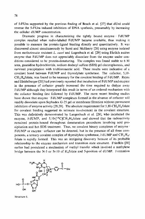
Dramatic progress in characterizing the tightly bound enzyme. FdUMP complex resulted when radio-labeled FdUMP became available, thus making it possible to measure the protein-ligand binding directly and quantitatively. It was discovered almost simultaneously by Santi and McHenry [28] using enzyme isolated from methotrexate resistant L. easel and Lagenbach et al. [29] using Ehrlich ascites enzyme that FdUMP does not appreciably dissociate from the enzyme under con- ditions considered to be protein-denaturing. The complex was found stable to 6 M urea, guanidine hydrochloride, sodium dodecyl sulfate (SDS) gel electrophoresis, and survived precipitation with trichloroacetic acid. These results were indicative of a covalent bond between FdUMP and thymidylate synthetase. The cofactor, 5,10- CH2H4folate, was found to be necessary for the covalent binding of FdUMP. Reyes and Heidelberger [20] had previously reported that incubation of FdUMP and enzyme in the presence of cofactor greatly increased the time required to dialyze away FdUMP although they interpreted this result in terms of an ordered mechanism with the cofactor binding first followed by FdUMP. The more recent binding studies have shown that enzyme • FdUMP complexes formed in the absence of cofactor will readily dissociate upon Sephadex G-25 gel or membrane filtration without permanent inhibition of enzyme activity [28,29]. The absolute requirement for 5,10-CH2H4folate for covalent binding suggested its intimate involvement in the covalent structure. This was definitively demonstrated by Langenbach et al. [29], who incubated the enzyme, FdUMP, and 5,10-[14C]CH2H4folate and showed that the radioactivity remained protein-bound throughout denaturation procedures involving acid pre- cipitation and hot SDS treatment. Thus, no covalent binary complexes of enzyme FdUMP or enzyme • cofactor can be detected, but in the presence of all three com- ponents, a ternary covalent complex of thymidylate synthetase, FdUMP and CH2H4- folate is rapidly formed. This was an intriguing discovery because of its probable relationship to the enzyme mechanism and transition state structure. Friedkin [30] earlier had postulated a mechanism of methyl transfer which involved a methylene bridge between the N-5 or N-10 of H,folate and 5-position of dUMP. Formation
H H 2 N .~//N.,,,/N,., O
£ . . ~ t _ tl
o
0 HN~J~I U 2 II 0 "~kN
HO-- P - - O ' - - ~
IOH ~ = OH
--Glu
Structure 1.

79
of the intermediate I is then followed by intramolecular hydride transfer from the 6-position of H4folate [21,31] to the methylene group to generate thymidylate.
This model would account for the covalent attachment of the cofactor to FdUMP if a similar intermediate did form, but not for the covalent binding of FdUMP to the protein. Clues to the latter had come from model studies carried out by Santi and Brewer [32] and Cushley et al. [33], who showed that the labilization of the C-5 proton of dUMP observed during the course of the enzymatic reaction [34] could most easily be accommodated by postulating a nucleophilic attack at the 6-position of the uracil moiety (Scheme II). This process would render the 5-position more electrophilic and thereby more receptive to electrophilic attack by the electron- deficient methylene group of CH2H~folate.
o o G
HN~'I~ H .... H N~.I/H 020.,,.
dRP dRP
dRP= deoxyribose- 5' -phosphate
5, IO-CH2H4 folate
H
I - - N - - ~ ~)--C oOH H ~
~ N ~ , CH2
0 1~1 Nu-Enz dRP
--glu
Scheme II.
OD
HN M'~H O."~" ? Nu-Enz
dRP
O
dRP
~ elimination
O
HN O u-Enz
dRP
The mechanism now postulated for exchange of the C-5 proton suggests that the structure of the Friedkin intermediate should be modified to one in which an enzymatic nucleophilic species has formed an adduct with the 5,6 double bond of uracil (II). If we assume that FdUMP undergoes an identical reaction in the initial sequence, the analogous structure III is obtained. The presence of a fluorine atom in
H H 2 N ' ~ N iy'N'l O
N ~ L , ~ .~--~ ,=~ II | ' , ' X N - - - ~ \ /) ' -"C--Glu
0 OH I H ";-" ~ L / C H 2
HN" "I~F
O/~'?"L"- Nu_En z
dRP
Structure III.

80
place of hydrogen would prevent the enzymatic reaction from proceeding to com- pletion, and thereby allow stabilization of the whole complex in a facsimile of an enzymatic intermediate. An interesting question is: how far does the reaction proceed when FdUMP is substituted for dUMP? Fluorine could prevent either hydride transfer to the methylene group or, alternately, elimination of the enzymatic nucleo- phile from the 5,6 double bond of thymidylate. Among the dramatic changes that occur in the ultraviolet spectrum upon formation of the complex is a decrease in absorption at 290 nm and an increase at 330 nm [35,36,37]. Sharma and Kisliuk [36] proposed that the absorption increase at 330 nm arose from oxidation of H4folate to H2folate after the enzyme • FdUMP • cofactor complex was formed. However, Santi et al. [38] isolated unchanged H4folate upon dissociation of the complex with no loss of tritium from the 6-position of the cofactor. These results conclusively showed that the reduction step does not occur. In the absence of further data, the ultraviolet absorption changes were attributed by Santi [38] to unspecified per- turbations of the H4folate chromophore. It has also been suggested that the increase at 330 nm is caused by opening of the 5,10-methylene bridge to give a methylene iminium cation without formation of a methylene bridge to FdUMP [37]. The absence of hydride transfer could be rationalized in terms of steric or electrostatic effects resulting from substitution of a fluorine atom. However, there is also an interesting mechanistic interpretation which involves postulating a reactive exocyclic methylene group on the uracil heterocycle as the next intermediate in the reaction sequence (Scheme III). Such a species has been previously considered by Santi and
H H H 2 N ~ N N H 2 N ",~IN ,~,/N,,, ,
"r"1 :" i~u "1 o K i o N ~ , k }~ .,.L-- k ,.=, H N L ~NJ~, =S -
o ; J]. ~CH 2 HN" ~1<~ H
O . ~ ? ..,,I,~ No_E nz H N " ~ CH2 dRP O " ~ ? "J~" N u - E n z
, ~ dRP
:Nu-Enz
Scheme III .
O
H N ~ I CH3 H 2 N "~JN"-./H~ O + + ,
dRP OH H
--Glu
Pogolotti [39,40] in nucleophilic substitution reactions of 5-acetoxymethyl- and 5-(p-nitrophenoxymethyl) uracils. The first step of this mechanism is a proton abstraction from C-5 of uracil followed by elimination of a quaternary ammonium ion. Obviously, due to the stability of the carbon-fluorine bond, the reaction will not occur if there is a fluorine atom in place of hydrogen at the 5-position. The process whereby an enzyme inactivates itself by carrying out a partial catalytic sequence on

81
a compound has been given the descriptive term "suicide inhibition" and the com- pounds have been designated as kcat inhibitors [41 ].
Considerable effort has gone into verifying III as the structure of the covalent thymidylate synthetase • FdUMP • CH2H4folate complex since Langenbach et al. [29] first proposed it. All evidence to date is consistent with structure III. Danenberg et al. [35] found that a reactive methylene group on the cofactor is necessary to form a stable ternary complex. 5-Methyl- and 10-methyl-H4folates, cofactor analogs not chemically capable of forming a methylene bridge, induced binding of FdUMP that was stable to get filtration but not to protein denaturants. A peptide generated by trypsin cleavage of the enzyme • [aH]FdUMP - [14C]cofactor complex contained both isotopes [35]. Sommer and Santi [42] isolated a short pronase peptide (6-8 amino acids) with FdUMP bound to it, and found that it had an ultraviolet spectrum similar to 5-methyl-H4folate. Convincing evidence for a methylene-bridged structure came from an N M R study of the latter pronase generated peptide [43]. The fluorine peak was split into a doublet of triplets, which is consistent with the presence of a methylene group adjacent to the fluorine.
In spite of the apparent stability of the denaturated enzyme • FdUMP • cofactor complex, inhibition of the enzyme by FdUMP is of a "pseudo-irreversible" nature. The aforementioned dialysis experiments of Reyes and Heidelberger showed that enzyme activity can be regenerated after exposure to FdUMP [20]. In a more quantitative study of the reversibility phenomenon, Santi and co-workers [37] used a filter assay to measure dissociation of labeled FdUMP from an L. casei enzyme • [aH]FdUMP complex. The dissociation was found to a first-order process, i.e., the rate was not dependent on the concentration of exogenous FdUMP and, in addition, was highly temperature dependent with a tl/2 at 21°C of about 20 h at a saturating cofactor concentration. The stability of the complex once the enzyme is denatured shows that the bonds involved are not intrinsically labile, but that the enzyme itself catalyzes the breakdown to regenerate FdUMP and free thymidylate synthetase. A model that seems aptly to describe the behavior of the enzyme • FdUMP • cofactor complex is the mechanism proposed for the Mannich reaction in acidic media [44] (Scheme IV).
H* R2NH ÷ H C H O ~ R2NCH2OH~ - '~R2NcH2 (D
H20 3 O (~OH R2N = CH2
I W R'
~OH ..JLCH ,C"2N"2
I R'
Scheme IV.
O
RJLCH/CH2NR2
R'

82
The mechanism involves complex kinetics of equilibria among the various forms of adducts between the amine components and formaldehyde. However, there are two aspects which may be directly relevant to the mechanism of thymidylate synthetase: (1) attack by an electrophilic species, probably an iminium cation, on the enol form of the active hydrogen compound; and (2) a reversible alkylation at the position adjacent to the carbonyl group. The cofactor is present in solution largely as the 5,10-methylene bridged compound [45]. Therefore, if the cationic iminium species, which was first proposed by Kallen and Jencks [46], is indeed an inter- mediate, it is probably generated in the active site by an acid catalyzed ring opening and thus protected from water in a hydrophobic environment until it can react with the 5-position of dUMP. According to the mechanism in Scheme IV, reversal of the alkylation would require an enzyme-mediated protonation on the 4-carbonyl group of the fluorouracil moiety of the complex. Considering the low basicity of the 4-oxo group and the stability of most carbon-carbon bonds, one would expect the equi- librium to lie heavily in favor of the forward reaction, and, in fact, the dissociation constant of FdUMP from the ternary complex (the ratio of koff/kon) obtained from binding studies has been calculated to be 5 • 10 -~ 1 M [37], a factor of 106 lower than the dissociation constant of the binary enzyme • FdUMP complex [47].
A large part of the interest in the structure of the covalent thymidylate syn- thetase • FdUMP • cofactor complex has been derived from its probable resemblance to an enzymatic intermediate or transition state. All the evidence obtained to date has been consistent with this hypothesis, so the probability is good that the group through which FdUMP is attached to the enzyme is actually the nucleophile that initiates the enzymatic reaction by attack on the 6-position of dUMP. With the stable enzyme" FdUMP" cofactor complex available, attempts have been made recently in several laboratories to make a direct and unequivocal identification of this nucleo- philic species. A number of earlier experiments directed at this problem have established the presence of a catalytically essential sulfhydryl group, most likely located within the active site. The evidence is as follows: (1) Treatment of the enzyme with iodoacetamide in the presence of dUMP did not cause inactivation [35], but when the dUMP was removed and the enzyme exposed to iodo[~4C]acetamide, enzymatic activity was lost. Subsequent acid hydrolysis yielded one equivalent of [~4C]carboxymethyl cysteine [35]. The substrate protection against iodoacetamide inactivation indicates, but does not prove, that the essential sulfhydryl residue is in the active site. (2) Showdomycin (3-fl-D-ribofuroanosylmaleimide) is a sulfhydryl reagent and also a uridine analog. Although this compound inactivates thymidylate synthetase, Kalman [48] found that the rate of inactivation is 10-fold faster with the nucleotide, showdomycin 5'-phosphate. The presence of the phosphate presumably confers substrate-like specificity to the compound therefore enabling it to react more quickly with the active site sulfhydryl group. (3) Glutathione and bisulfite have been found to be the most effective nucleophiles at promoting exchange of the C-5 proton of uridine [49,50], which is thought to occur by the previously discussed addition- elimination reaction with the 5,6 double bond. Recently, threonine and histidine

83
emerged as a candidate nucleophiles when Sommer and Santi [24] reported that an acid hydrolysate of a short peptide with FdUMP covalently attached contained Thr, His, Ala, Leu, and Pro2 but no cysteine. Further experiments from the same laboratory showed no significant difference in the uptake of sulfhydryl reagents between the free enzyme and the enzyme" FdUMP complex [51]. However, sub- sequently, Danenberg and Heidelberger [52] demonstrated that the peptide does, in fact, contain cysteine by an unequivocal double-labeling experiment. L. casei cells were grown in the presence of [35S]cysteine, resulting in a [3SS]cysteine-containing thymidylate synthetase. The [3H]FdUMP-peptide isolated from this enzyme con- tained both radiolabels, 3H and 35S, in equivalent amounts. Treatment of the FdUMP-peptide with 2,2'-bis-dithionitrobenzoic acid failed to detect a free sulfhydryl group in the peptide (Danenberg, P. V., unpublished results), indicating that the sulfur was part of a thioether linkage. The presence of a thioether linkage between FdUMP and the protein was strongly confirmed by the behavior of the enzyme - FdUMP • cofactor complex when exposed to Raney nickel [53]. This reagent has the unusual property of excising sulfur atoms from organic molecules and thus causing specific scission of carbon-sulfur bonds. Raney nickel effected cleavage of FdUMP and 5,10-CH2H4folate from the protein at the same rate that it reacted with smaller sulfur-containing molecules, cf., as in the conversion of cysteine to alanine, whereas carbon-oxygen and carbon-nitrogen bonds were unaffected by the reagent. Cleavage of 3H and 35S from the double-labeled [3H]FdUMP. [35S]enzyme occurred at identical rates in the presence of Raney nickel. Maley and coworkers [54] performed an elegant study in which they isolated and sequenced the identical cyanogen bromide fragments of both free and FdUMP-bound thymidylate synthetase from L. casei.
Acid hydrolysis and sequencing of the FdUMP peptide did not reveal cysteine, but analysis of the analogous peptide from enzyme not treated with FdUMP yielded the following sequence: Ala-Leu-Pro-Pro-CMCys-His-Thr-Leu-Tyr. This sequence was identical to that of the FdUMP-containing peptide, except that carboxymethyl- cysteine was found in place of the unidentified amino acid attached to FdUMP. Although these results do not constitute direct evidence of a sulfide linkage between cysteine and FdUMP, nevertheless it is unlikely that the enzyme would contain two fairly long sequences identical except for the cysteine residue. Santi and coworkers [55] were prompted by some of the previously discussed results to re-investigate the composition of their pronase-generated FdUMP-peptide. Cysteine was detected as cysteic acid in the acid hydrolysate, but only when the peptide was first subjected to performic acid oxidation. The following partial sequence was established: Leu-Pro- Pro-(His,Cys)-Thr. Histidine was eliminated as a candidate carrier of bound FdUMP when treatment of the peptide with the Pauly reagent showed a free imidazole moiety, leaving threonine and cysteine as possibilities [55]. Failure to detect cysteine in the peptide hydrolysate may arise from the fact that the bond between the 5-FUra moiety and the amino acid is not easily hydrolyzed. This would not be unexpected, since a thioether bond should be relatively stable under the acidic conditions that will cleave peptide bonds. Methionine, for example, is not appreciably hydrolyzed

84
to homocysteine during amino acid composition analyses. It is also possible that the thioether bond is cleaved in such a way that cysteine is not regenerated. To obtain a completely convincing identification of the amino acid bound to F d U M P it will apparently be necessary to degrade the peptide to that one amino acid residue, and unequivocally determine the structure of the compound so obtained.
The question of whether thymidylate synthetase possesses one or two functional active sites is an interesting one and is presently being actively investigated in several laboratories. The existence of two high affinity binding sites for F d U M P on thy- midylate synthetase isolated from several sources has been demonstrated [37,47,56]. This observation is in accord with results obtained from sodium dodecyl sulfate gel electrophoresis and sedimentation equilibrium studies, which have shown that the enzyme consists of two identical or very similar subunits [22,56,57,58]. In an effort to further characterize the subunits, Loeble and Dunlap [59] showed that the sub- units have the same N-terminal (methionine) and penultimate (valine) amino acids and give rise to identical cyanogen bromide fragments. Based on these data, it would be rea- sonable to suppose that thymidylate synthetase consists of 2 identical subunits, each of which has a functional active site capable of forming a covalent complex with F d U M P and 5,10-CH2H4folate. The actual situation appears to be much more complex.
Strong evidence for non-identity of the F d U M P binding sites was obtained by Aull et al. [60] who followed the breakdown of the ternary enzyme • F d U M P • co- factor complex by native gel electrophoresis (non-denaturing). Three resolvable forms of thymidylate synthetase were observed on the gels: native enzyme, a 1 : 1 : 1 molar complex of enzyme" F d U M P - cofactor, and a 1:2:2 complex of the same com- ponents. The relative amount of each band was dependent on the ratio of F d U M P to enzyme when forming the complex. Upon storage of the complex, the 1 : 2: 2 band disappeared from the gel within 24 h with corresponding appearance of the 1 : 1 : 1
complex, which was much more stable. After 6 days, 60 ~o of the 1 : 1 : 1 complex still remained, with the remainder in the free enzyme from. These experiments provided a dramatic demonstration of the previously discussed reversibility of the non- denatured enzyme ' F d U M P complex. Further evidence that the F d U M P binding sites are not identical comes from the work of Galivan et al. [47] who found that two moles of F d U M P are bound per enzyme in Tris buffer but only one mole in phosphate buffer. Scatchard plots were biphasic, suggesting that the first binding site has a lower dissociation constant than the second. In the presence of urea, one mole of F d U M P dissociated from the T2 phage enzyme but the other remained bound [56]. Danenberg et al. [35] were able to detect only 1 mol of F d U M P bound to the sodium dodecyl sulfate denatured complex. There is a possibility that F d U M P in the pres- ence of 5,10-CH2H4folate, becomes attached covalently to one site but forms a noncovalent complex to the other site. An interesting question is whether the un- bound site of the 1 : 1 : 1 enzyme • F d U M P " cofactor complex has enzymatic activity. In this regard it should be noted that after Leary et al. [60] inactivated the enzyme with one mole of iodoacetate, they were able to regenerate one-half the original activity with 0.1 M fl-mercaptoethanol, suggesting that the unreacted subunit still

85
had catalytic activity. Although 2 mol of FdUMP bind to 1 mol of enzyme, it appears from equilibrium dialysis [47] and circular dichroism [61] studies that dUMP and the product dTMP occupy only one site at a time. This finding supports the presence of only one catalytic site per enzyme molecule in spite of the apparent homogeneity of the two subunits. In accord with the concept of one site, Dunlap and coworkers found that 1 mol of p-chloromercuribenzoate per mol of enzyme causes complete inacti- vation of the enzyme, as does carboxypeptidase cleavage of 1 mol of valine from the carboxy-terminus [57,62]. Kinetics studies with H4folate analogs have also indicated the presence of an alternate noncatalytic binding site. 10-Methyl-folate [63], 5- methyl-HJolate [35,64] and 5,11-CH2H4homofolate [35] all inhibited the enzyme non-competitively with respect to the cofactor. A replot of the slopes from the H4homofolate inhibition pattern furthermore was parabolic, suggestive of the binding of more than one molecule of inhibitor [35]. Thus, the situation with regard to the binding and catalytic sites of thymidylate synthetase is still far from resolved. Several lines of evidence show that the sites are not identical, implying that the sub- units have different primary sequences, or, alternately, identical subunits could be asymmetrically joined in a manner that allows only one site at a time to function, and changes the affinity of the second site for inhibitors and substrates. Assuming that there is only one catalytic site per enzyme molecule, the question is raised as to the role of the other, catalytically non-functional site. A possibility worth considering is that it has a regulatory function.
III. INTERACTION OF THYMIDYLATE SYNTHETASE WITH OTHER dUMP ANALOGS
IliA. 5-Trifluoromethyl-2'-deoxyuridylic acid (F3dTMP) 5-Trifluoromethyl-2'-deoxyuridine (F3dThd) was synthesized by Heidelberger
in 1962 [65]. The drug was conceived on the basis of a rationale similar to the one used in the design of 5-FUra, namely, substitution of fluorine atoms for hydrogen on the pyrimidine base. Since F3dThd is a thymidine analog, Heidelberger predicted that it would become incorporated into DNA, and, in fact, subsequent investigation of phage DNA formed in the presence of FadThd revealed that about 11 ~ of the thymine residues were substituted [66]. On this basis Kaufman and Heidelberger [67] tested F3dThd as an antiviral agent and found remarkable antiviral activity against ocular herpes virus infections, considerably greater than the activities of 5-iododeoxy- uridine, 5-bromodeoxyuridine, or cytosine arabinoside [68]. Heidelberger and Anderson [69] discovered that F3dThd is also active against many transplanted tumors in mice. In clinical trials F3dThd has produced some response in human cancer patients [70,71] but not enough to justify further testing. F3dThd is converted in vivo by thymidine kinase to the nucleotide F3dTMP [71], a powerful inhibitor of thymidy- late synthetase (K~ ---- 10 -8 M) [20]. Binding of this magnitude is probably sufficient to account for most of the tumor inhibitory effect of FadThd, but its incorporation into DNA may also contribute to the anti-tumor activity.

86
Reyes and Heidelberger [20] investigated the kinetics of inhibition of thymidy- late synthetase by F3dTMP and found them to be similar to those of FdUMP. The inhibition changed from competitive with dUMP to noncompetitive after pre- incubation of the inhibitor with the enzyme. However, the results of dialysis ex- periments were quite different for the two inhibitors. Whereas all of the bound FdUMP was eventually dialyzed away, F3dTMP did not dissociate at all upon dialysis following a 2 h preincubation with the enzyme. On this basis, it was postulated that FadTMP reacts chemically with an active site nucleophile to form a carboxamide or ester, via a reaction analogous to that ofglycine with 5-trifluoromethyluracil (FaThy) in a basic medium that generates 5-uracoylglycine [73]. The trifluoromethyl group of FaThy appeared to be quite reactive compared to most trifluoromethyl groups. In order to study this unusual reactivity, Santi and Sakai [74] measured the hydrolysis rates of the trifluoromethyl groups of various F3Thy derivatives, and found that 1-(3-aminopropyl) F3Thy was hydrolyzed to the carboxyuracil derivative 10~-fold faster than 1-methyl FaThy. This extraordinary increase in reactivity was explained on the basis of nucleophilic attack by the amino group on the 6-position of the hetero- cycle with concomitant expulsion of a fluoride ion to generate a reactive exocyclic difluoromethylene group, which could be captured by another proximate nucleophile. Neighboring group participation of the amino group of this compound would be a kinetically favorable process since it would result in the formation of a six-membered ring. Derivatives that could only form smaller or larger rings were relatively inert to hydrolysis. Santi and Sakai [74] suggested that F3dTMP reacts with the enzyme by a similar mechanism, involving rate-determining attack of an enzymatic nucleophile at the 6-position of F3dTMP. However, an irreversible covalent labeling of the enzymatic active site has never been directly demonstrated using radio-labeled F3dTMP. Assuming that it does occur, the rate of alkylation of the enzyme by F3dTMP appears to be quite slow and supports a non-specific bimolecular reaction rather than an active site-directed one. Certainly, if attack by the enzymatic nucleo- phile at the 6-position of F3dTMP is the rate-determining step in alkylation of the enzyme, it must be many orders of magnitude slower than the corresponding reaction with FdUMP. Danenberg et al. [35] found that short incubation periods gave FadTMP • enzyme complexes that were isolable by Sephadex G-25 filtration, but were not bound together covalently. Denaturation of these complexes caused release of
apparently unchanged F3dTMP [35]. The mechanism of F3dTMP binding to thymidylate synthetase differs from that
of FdUMP in another important respect. No dependence of the presence of cofactor for maximal stoichiometric binding of F3dTMP was observed using both L. casei [35] and T4 phage-induced enzyme [22]. Furthermore, a given amount of the T4 phage induced enzyme bound almost 12 times more FadTMP than FdUMP. These results suggested the possibility that F3dTMP binds at numerous non-catalytic sites in addition to the active site. The mode of interaction of FadTMP with the enzyme is still unresolved and its elucidation remains a worthwhile goal.

87
IIIB. 5-Formyl-2'-deoxyuridylic acid (formyl-dUMP)
Santi and Sakai [75] discovered that formyl-dUMP undergoes a strong inter- action with thymidylate synthetase, leading to a Ki of 10 -8 M. The kinetics display the familiar pattern observed with FdUMP and F3dTMP: competitive inhibition with respect to dUMP and non-competitive against 5,10-CH2H4folate, changing to non-competitive with dUMP upon preincubation of the inhibitor and enzyme [76]. The binding of formyl-dUMP to the enzyme resembles that of FdUMP, but not FadTMP, in that the cofactor is required. An attractive mechanism for the strong binding of formyl-dUMP with the enzyme is the formation of a Schiff base between the formyl group and an enzymatic amino group. Several examples of such an inter- action between an enzyme and an inhibitor exist in the literature. In that case, borohydride reduction should lead to irreversible covalent attachment of formyl- dUMP to the protein. However, such experiments have not yet been published and so the mode of binding of formyl-dUMP remains unresolved.
IIIC. 5-Mercapto-2'-deoxyuridylic acid (mercapto-dUMP)
The nucleoside, 5-mercapto-2'-deoxyuridine has been found to possess anti- tumor activity against Ehrlich ascites cells in culture and against L1210 leukemia both in culture and in mice [77]. The discovery that this compound caused inhibition of dTMP synthetase activity in crude extracts of E. coli B cells in the presence of ATP led Kalman and Bardos [77] to synthesize the nucleotide in order to study its inter- action with the enzyme. Mercapto-dUMP was found to be a potent inhibitor (Ki = 4 ' 10-a), competitive with dUMP. Preincubation produced no alteration of the kinetic pattern or change in the inhibition constant, in contrast to all of the previously described inhibitors. It was suggested [77] that the strong binding of mercapto-dUMP is due to ionization of the sulfhydryl group to a sulfide anion, resulting in a favorable charge interaction with a positive group in the active site, possibly the putative amine that forms a Schiff base with formyl-dUMP [76]. This hypothesis was supported by the observation that 5-methylmercapto-2'-deoxyuridine did not show significant inhibition of dTMP synthetase in the cell-free extract even though it was phosphorylated by thymidine kinase [77]. These results are consistent with the hypothesis that the anti-tumor activity or 5-mercapto-2'-deoxyuridine is due to inhibition of thymidylate synthetase.
IIID. 5-Iodoacetamidomethyl-2'-deoxyuridine-5'-phosphate
Barfknecht et al. [78] have recently demonstrated species-specific inhibition of thymidylate synthetase. 5-Iodoacetamidomethyl-dUMP, an analog containing an alkylating group, showed some selectivity toward irreversible inhibition of Ehrlich ascites thymidylate synthetase. About 20~o inactivation of the thymus enzyme was observed over a 30 min period, which was not dependent on the concentration of the inhibitor, whereas over the same period Ehrlich ascites enzyme was inhibited 75 by a 465 ~tM concentration of the inhibitor ( k l n a o t l v a t l o n = 0.057 min-1). The analog

88
was competitive with dUMP in both cases with reversible Kl values of 27 #M for the thymus enzyme and 68 #M for the ascites enzyme.
IV ANALOGS OF 5,10-CH2H4FOLATE
Analogs of 5,10-CH2H4folate as inhibitors of thymidylate synthetase have generally not been as potent as the pyrimidine nucleotides. Some compounds, however, have been found which have binding constants considerably lower than the Km of the substrate. Thus, the possibility that thymidylate synthetase may be an effective target for some folate analogs cannot be excluded.
Baker and coworkers [79,80] synthesized a large number of compounds designed as non-classical inhibitors of thymidylate synthetase, several of which contained alkylating groups. Although this approach yielded very potent inhibitors of dihydro- folate reductase, some of them of the active site-directed, irreversible type, none of Baker's compounds alkylated thymidylate synthetase or bound appreciably better than the cofactor [80].
Goodman et al. [81] discovered that tetrahydrohomofolate, an analog with an extra methylene group between C-6 and N-10, was a very potent inhibitor of thy- midylate synthetase from E. coli (Km/Ki = 100). The compound possessed weak substrate properties for the L. casei enzyme and was a less effective inhibitor (Km/Ki = 2) [35]. The rationale behind the design of homofolate was to utilize dihydrofolate reductase for in situ production of a reduced folate analog that would be a potent inhibitor of thymidylate synthetase [82]. This approach would be expected to be particularly effective in methotrexate resistant tumor cells which had elevated dihydrofolate reductase, and, in fact, levels of this enzyme appear to be related to the anti-tumor activity of the drug. Mead et al. [83] observed anti-tumor activity using H4homofolate against a methotrexate resistant L1210 variant, but the drug had only marginal activity against the antifolate sensitive parent dells. Dihydro- homofolate had activity comparable to H4homofolate [84]. The effectiveness of either reduced form of homofolate is greatly diminished if cells are pre-treated with methotrexate [84]. Nevertheless, it has never been demonstrated that H4homofolate is the active form of the drug, or that inhibition of thymidylate synthetase is the primary mode of action. Recently, in fact, Nahas and Friedkin obtained evidence that H2homofolate is not converted to H4homofolate in leukemia cells in spite of high dihydrofolate reductase levels [85].
It has been known for some time that extracts of E. coli contain substances that have the ability to inhibit markedly thymidylate synthetase activity [86]. Friedkin and coworkers [87] have recently purified and identified these compounds as N 5- formyl-H4pteroyloligoglutamates. Maximum inhibition was achieved by the com- pound containing 6 glutamyl residues, which was found to be 200-fold and 20-fold more inhibitory than the derivatives with 1 and 2 glutamates, respectively. Ki values were not reported, but the Iso for the 6-glutamate homolog was 1.7 ~M in the presence
89
of 0.1 mM 5,10-CH2H4folate. Analogs with a terminal lysine residue were about 20-fold poorer inhibitors than the corresponding glutamyl H4pteroates [87].
In addition to the reduced folate analogs, which suffer from the disadvantage of inherent instability, a number of unreduced folate analogs have been found to be potent inhibitors of thymidylate synthetase. Kisliuk and coworkers [88] synthesized a series of pteroyloligoglutamates containing an unreduced pteridine ring and discovered also that inhibition of thymidylate synthesis increased dramatically with the number of glutamyl residues attached to the p-aminobenzoyl moiety. With the substrate 5,10-CH2H4folate at a concentration of 3" 10 -4 M, 5 0 ~ inhibition was obtained with the pteroylhexaglutamate at 6 • 10 -7 M , the diglutamate at 3 • 10 -5 M, and the monoglutamate at 1.5" 10 -4 M [88]. Interestingly, reduction of the com- pounds to the dihydropteridine forms decreased their binding to the enzyme about 3-fold. (-)H4pteroylglutamates were substrates for thymidylate synthetase, but the ( + ) forms were inhibitors [88]. Kisliuk suggested that the cellular ratio of H 2-
pteroylgtutamate to H4pteroylglutamate could be an effective regulator of thymi- dylate synthetase activity in vivo, due to the strong inhibition exerted by the former.
Bird et al. [89] tested a series of 2-amino-4-hydroxyquinazoline analogs (5,8 deazafolic acid) of folic acid as inhibitors of thymidylate synthetase from S. faecalis in order to study structure-activity correlations for this class of compounds. The most potent inhibitor was the 2-amino-4-hydroxyquinazoline analog with a methyl group on the 10-position (/5o ~- 0.098/zM at a cofactor concentration of 0.28raM). Various other substitutions led to sizeable losses in inbinding. For example, the 2,4 diamino-quinazoline was 20-fold less effective, and substitution of an aspartyl for the glutamyl residue resulted in a 60-fold binding decrease [89]. Identical structure- activity correlations for the quinazolinyl analogs were found with thymidylate synthetases from mouse neuroblastoma, L. casei [90], and D. pneumoniae [91]. The 2-amino-4-hydroxyquinazolines in each case were much more inhibitory than the 2,4-diamino compounds. Ki values were not reported, but the ratio of inhibitor to substrate for 50 ~ inhibition ( ( I /S) 50) ranged from l0 -2 for the Diplococcus pneu. moniae enzyme to about 10 -a for thymidylate synthetase from the other two sources mentioned above.
The 2-amino-4-hydroxyquinazoline analog inhibited the growth of mouse neuroblastoma cells in culture (ECso = 14 b~M) [90]. The cytotoxicity was attributed to inhibition of both thymidylate synthetase and dihydrofolate reductase, based on the observation that both thymidine and leucovorin protected the cells against the drug.
V. CONCLUSION
Drugs that can effectively inhibit or interfere with DNA synthesis have been of great importance in cancer chemotherapy. T h e design of inhibitors of thymidylate synthetase, a k e y enzyme in the biosynthesis of DNA, has yielded clinically useful
90
drugs. Since a number of very potent inhibitors of this enzyme are known it is likely tha t significant improvement s in chemothe rapeu t i c efficacy could be achieved in a
relat ively shor t t ime by concent ra t ing on (1) more effective clinical use o f a l ready
existing drugs, (2) ra t iona l deve lopmen t o f d rug combina t ions a imed toward circum-
vent ing poten t ia l mechanisms o f resistance and innate refractor iness , and (3) a means
o f increas ing the cel lular concen t ra t ion o f the active fo rm of the drug. Nevertheless ,
a search for o ther effective inhib i tors o f the ta rge t enzyme should also be encouraged.
In this regard, an under s t and ing o f the enzymat ic mechan i sm can be o f ines t imable
value. Studies o f the in terac t ions o f the active forms o f var ious drugs, especial ly
F d U M P , have resulted in significant progress t oward unravel ing the complex mech-
anism of thymidy la te synthetase. A n u m b e r o f in t r iguing quest ions remain un-
resolved, which are at the present t ime being actively pursued in several labora tor ies .
We may, therefore, l ook forward to e luc ida t ion o f the comple te mechanism of ac t ion
in the near future.
ACKNOWLEDGEMENTS
The au tho r wishes to thank Dr. Charles Heide lberger and Dr. R icha rd M o r a n
for helpful advice regard ing the p r e p a r a t i o n o f the manuscr ipt .
REFERENCES
1 Humphreys, G. M. and Greenberg, D. M. (1958) Arch. Biochem. Biophys. 78,275-287 2 Friedkin, M. (1959) Fed. Proc. Fed. Am. Soc. Exp. Biol. 18, 230 3 McDougall, B. M. and Blakley, R. L. (1960) Nature 188,944 4 Labow, R., Maley, G. F. and Maley, F. (1969) Cancer Res. 29, 366-372 5 Conrad, A. H. and Ruddle, R. H. (1972) J. Cell. Sci. 19, 471-486 6 Barth, O. and Maass, H. (1970) Z. Krebsforsch. 75, 45-54 7 Blakley, R. L. (1960) The Biochemistry of Folic Acid and Related Pteridines, p. 237, American
Elsevier, New York 8 Beck, W. S. (1964) Medicine 43, 715-726 9 Cohen, S. S. (1971) Ann. N, Y. Acad. Sci. 186, 292-301
10 Barner, H. D. and Cohen: S. S. (1958) Biochim. Biophys. Acta 30, 12-20 11 Heidelberger, C., Chaudhuri, N. K , Danneberg, P., Mooren, D., Griesbach, L., Duschinsky, R.,
Schnitzer, R. J., Pleven, E. and Scheiner, T. (1957) Nature 179, 663-666 12 Heidelberger, C. and Ansfield, F. J. (1963) Cancer Res. 23, 1226-1243 13 Rutman, R. J., Cantarow, A. and Paschkis, K. E. (1954) Cancer Res. 14~ 119-123 14 Danneberg, P. B., Montag, B. J. and Heidelberger, C. (1958) Cancer Res. 18, 329-334 15 Cohen, S. S., Flaks, J. G., Barner, H. D., Loeb, M. R. and Lichtenstein, J. (1958) Proc. Natl.
Acad. Sci. U.S. 44, 1004-1012 16 Hartmann, K. U. and Heidelberger, C. (1961) J. Biol. Chem. 236, 3006-3013 17 Ackermann, W. W. and Potter, V. R, (1949) Proc. Soc. Exp. Biol. Med. 72, 1-9 18 Mathews, C. K. and Cohen, S. S. (1963) J. Biol. Chem. 238, 367-376 19 Blakley, R. L. (1963) J. Biol. Chem. 238, 2113-2118 20 Reyes, P. and Heidelberger, C. (1965) Mol. Pharmacol. 1, 14-30 21 Lorenson, M. Y., Maley, G. F. and Male),, F. (1967) J. Biol. Chem. 242, 3332-3344 22 Capco, G. R., Krupp, J. R. and Mathews, C. K. (1973) Arch. Biochem. Biophys. 158, 726-775

91
23 Myers, C. E., Young, R. C. and Chabner, B. A. (1975) J. Clin. Invest. 56, 1231-1238 24 Dunlap, R. B., Harding, N. G. L. and Huennekens, F. M. (1971) Biochemistry 10, 88-97 25 Aldridge, W. N. and Reiner, R. (1972) Front. Biol. 26, 91-100 26 Myers, C. E., Young, R. C., Johns, D. G. and Chabner, B. A. 0974) Cancer Res. 34, 2682-2688 27 Bosch, L., Harbers, E. and Heidelberger, C. (1958) Cancer Res. 18, 335-343 28 Santi, D. V. and McHenry, C. S. (1972) Proc. Natl. Acad. Sci. U.S. 69, 1855-1857 29 Langenbach, R. J., Danenberg, P. V. and Heidelberger, C. (1972) Biochem. Biophys. Res.
Commun. 48, 1565-1671 30 Friedkin, M. (1959) in The Kinetics of Cellular Proliferation (Stohlmann, Jr., F., ed.), Grune
and Stratton, New York 31 Pastore, E. J. (1967) Abstracts 154th National Meeting, American Clinical Society Division of
Biological Chemistry, 107c 32 Santi, D. V. and Brewer, C. F. (1968) J. Am. Chem. Soc. 90, 6236-6238 33 Cushley, R. J., Lipsky, S. R. and Fox, J. J. (1968) Tetrahedron Lett. 52, 5393-5397 34 Lomax, M. 1. S. and Greenberg, R. G. (1967) J. Biol. Chem. 242, 109-113 35 Danenberg, P. V., Langenbach, R. J. and Heidelberger, C. (1974) Biochemistry 13, 926-933 36 Sharma, R. K. and Kisliuk, R. L. (1973) Fed. Proc. Fed. Amer. Soc. Exp. Biol. 32, 591-592 37 Santi, D. V., McHenry, C. S. and Sommer, H. (1974) Biochemistry 13, 471-480 38 Santi, D. V., Pena, V. A. and Lam, S. S. M. (1976) Biochim. Biophys. Acta 438, 324-331 39 Santi, D. V. and Pogolotti, Sr., A. L. (1971) J. Heterocycl. Chem. 8, 265-273 40 Santi, D. V. and Pogolotti, Jr., A. L. (1974) Biochemistry 13, 456-466 41 Rando, R. R. (1974) Science 185, 320-324 42 Sommer, M. and Santi, D. V. (1974) Biochem. Biophys. Res. Commun. 57, 68%695 43 James, T. L., Pogolotti, A. L., Ivanetich, K. M., Wataya, Y., Lam, S. S. M. and Santi, D. V.
(1976) Biochem. Biophys. Res. Commun. 72, 404-410 44 Cummings, T. F. and Shelton, J. K. (1960) J. Org. Chem. 25, 419-423 45 Blakley, R. L. (1954) Biochem. J. 58, 448-462 46 Kallen, R. G. and Jencks, W. P. (1966) J. Biol. Chem. 241, 5831-5863 47 Galivan, J. H., Maley, G. F. and Maley, F. (1974) Biochemistry 13, 2282-2289 48 Kalman, T. I. (1972) Biochem. Biophys. Res. Commun. 49, 1007-1013 49 Kalman, T. I. (1971) Biochemistry 10, 2567-2573 50 Wataya, Y. and Hayatsu, H. (1972) Biochemistry 11, 3583-3588 51 McHenry, C. S. and Santi, D. V. (1974) Biochem. Biophys. Res. Commun. 57, 539-545 52 Danenberg, P. V. and Heidelberger, C. (1975) Fed. Proc. Fed. Am. Soc. Exp. Biol. 34, 1703 Abstr. 53 Danenberg, P. V. and Heidelbergei, C. (1976) Biochemistry 15, 1331-1337 54 Maley, F., Maley, G. F., Bellisario, R. and Galivan, J. (1976) Proc. Natl. Acad. Sci. U.S. 73,
1848-1851 55 Pogolotti, A. L., Ivanetich, K. M., Sommer, H. and Santi, D. V. (1976) Biochem. Biophys. Res,
Commun. 73, 972-977 56 Galivan, J., Maley, G. F. and Maley, F. (1974) Biochemistry 13, 2282-2289 57 Dunlap, R. B., Harding, N. G. L. and Huennekens, F. M. (1971) Biochemistry 10, 88-97 58 Fridland, A., Langenbach, R. J. and Heidelberger, C. (1971) J. Biol. Chem. 246, 7110-7114 59 Loeble, R. B. and Dunlap, R. B. (1972) Biochem. Biophys. Res. Commun. 49, 1671-1677 60 Aull, J. L., Lyon, J. A. and Dunlap, R. B. (1974) Microchem. J. 19, 210-218 6l Leary, R. P., Beaudette, N. and Kisliuk, R. J. (1975) J. Biol. Chem. 250, 4864-4868 62 Aull, J. L., Loeble, R. B. and Dunlap, R. B. (1974) J. Biol. Chem. 247, 1167-1172 63 Kalman, T. I. and Fung, S.-M. (1972) 104th National Meeting of the American Chemical Society,
1972, New York, N.Y. Sept., Abstract BIOL. 64 Slavik, K. and Zakrzewski, S. F. (1967) Mol. Pharmacol. 3, 370-377 65 Heidelberger, C., Parsons, D. and Remy, D. C. (1962) J. Am. Chem. Soc. 84, 3597-3598 66 Gottschling, H. and Heidelberger, C. (1963) J. Mol. Biol. 7, 541-560 67 Kaufman, H. E. and Heidelberger, C. (1964) Science 145, 585-587 68 Kaufman, H E. (1965) Ann. N.Y. Acad. Sci. 130, 168-180 69 Heidelberger, C. and Anderson, S. W. (1964) Cancer Res. 24, 1979-1985 70 Ansfield, F. J. and Ramirez, G. (1971) Cancer Chemother. Rep. 55, 205-208 71 Helson, L., Yagoda, M., McCarthy, M., Murphy, M. L. and Krakoff, I. H. (1970) Proc. Am.
Assoc. Cancer Res. 11, 53

92
72 Bresnick, E. and Williams, S. S. (1967) Biochem. Pharmacol. 16, 503-507 73 Heidelberger, C., Parsons, D. G. and Remy, D. C. (1964) J. Med. Chem. 7, 1-5 74 Santi, D. V. and Sakai, T. T. (1971) Biochemistry 10, 3598-3607 75 Santi, D. V. and Sakai, T. T. (1971) Biochem. Biophys. Res. Comrnun. 42, 813-817 76 Santi, D. V. and Sakai, T. T. (1972) Biochem. Biophys. Res. Commun. 46, 1320-1325 77 Kalman, T. I. and Bardos, T. J. (1970) Mol. Pharmacol. 6, 621-630 78 Barfknecht, R. L., Huet-Rose, R. A., Kampf, A. and Mertes, M. P. (1976) J. Am. Chem. Soc. 98,
5041-5043 79 Baker, B. R. and Coward, J. K. (1967) J. Heterocycl. Chem. 4, 202-207 80 Baker, B. R. (1967) Design of Active-Site-Directed Enzyme Inhibitor, John Wiley and Sons,
Inc., New York 81 Goodman, L., DeGraw, J.~ Kisliuk, R. L., Friedkin, M., Pastore, E. J., Crawford, E. J., Plante,
L. T., Nahas, A., Morningstar, F. J., Jr., Kwok, G., Wilson, L., Donovan, E. F. and Ratzan, J. (1964) J. Am. Chem. Soc. 86, 308-309
82 Friedkin, M., Crawford, E., Humphreys, S. R. and Goldin, A. (1962) Cancer Res. 22, 600-604 83 Mead, J. A. R., Goldin, A., Kisliuk, R. L., Friedkin, M., Plante, L., Crawford, E. J. and
Kwok, G. (1966) Cancer Res. 26, 2374-2379 84 Mishra, L. C. and Mead, J. A. P. (1972) Chemotherapy 17, 283-292 85 Nahas, A. and Friedkin, M. (1972) Mol. Pharm. 8, 353-361 86 Friedkin, M., Crawford, E. J., Donovan, E. aod Pastore, E. J. (1962) J. Biol. Chem. 237, 3811-
3814 87 Friedkin, M., Plante, L. T., Crawford, E. J. and Crumm, M. (1975) J. Biol. Chem. 250, 5614-5621 88 Kisliuk, R. L., Gaumont, Y. and Baugh, C. M. (1974) J. Biol. Chem. 249, 41(10-4103 89 Bird, O. D., Vaitkus, J. W. and Clarke, J. (1970) Mol. Pharmacol. 6, 573-575 90 Carlin, S. C., Rosenberg, R. N., VandeVenter, L. and Friedkin, M. (1974) Mol. Pharmacol. 10,
194-203 91 McKuen, R. W. and Sirotnak, F. M. (1975) Biochim. Biophys. Acta 384, 369-380