three-dimensional architecture of rat lingual filiform papillae with
TRANSCRIPT

J. Anat. (1989), 165, pp. 177-189 177With 17 figuresPrinted in Great Britain
Three-dimensional architecture of rat lingual filiform papillaewith special reference to the epithelium-connective tissue
interface
TOSHIKAZU NAGATO, MASAMI NAGAKI,MICHITAKA MURAKAMI AND HIROAKI TANIOKA
Department of Oral and Maxillofacial Surgery, Ehime University, School ofMedicine, 454 Shizukawa, Shigenobu, Onsen-gun, Ehime 791-02, Japan
(Accepted 1 December 1988)
INTRODUCTION
Three types of filiform papillae which differ in shape, size, histological features(Fish, Malone & Richter, 1944; Kutuzov & Sicher, 1951) and in surface texture asexamined with the scanning electron microscope (Svejda & Skach, 1971; Yoshioka &Muto, 1976; Iida, Yoshioka & Muto, 1985) are present in the rat tongue. Nevertheless,the functions of the three types of filiform papillae have not been fully described in anyof these reports.
In recent years scanning electron microscopic observations of the epithelium-connective tissue interface have been performed on various tissues including humanoral mucosa (Scaletta & Simmelink, 1973; Squier, Johnson & Hackemann, 1975;Klein-Szanto & Schroeder, 1977; Ooya & Tooya, 1981), rat oral mucosa (Toyoshima& Shimamura, 1982; Hull & Warfel, 1986) and mouse taste buds (Suzuki & Takeda,1987): On the other hand, since Evan, Dail, Dammrose & Palmer (1976) introduceda method for the removal of the connective tissue and the basal lamina usinghydrochloric acid and enzymes before scanning electron microscopy, connectivetissue component-removing techniques suitable for different tissues, includingmyoepithelium (Nagato, 1978; Nagato, Yoshida, Yoshida & Uehara, 1980),neuromuscular junctions (Desaki & Uehara, 1981) and the vascular wall (Fujiwara& Uehara, 1984) have been developed.
In the present study we applied to the rat tongue the connective tissue component-removing technique using hydrochloric acid in an attempt to visualise the three-dimensional architecture of the basal surface of the epithelium and the free surface ofthe connective tissue papillae in various parts of the dorsum of the tongue using ascanning electron microscope.
MATERIALS AND METHODS
Tongues used in the experiment were obtained from Sprague-Dawley rats of bothsexes, aged 3 to 6 months. The animals were perfused with half-strength Karnovsky'sfixative (Karnovsky, 1965) through the heart, after anaesthesia with intraperitonealsodium pentobarbitone (50 mg/kg). The whole tongue was then removed.The epithelium-attached samples, about 2 mm square, were obtained from various
parts of the tongue. They were further fixed by immersing for 3 hours in the samefixative, then washed four times with 0-2 M phosphate buffer. The samples werehydrolysed in 8N hydrochloric acid at 60 °C for 35-45 minutes, and at the end of this

T. NAGATO AND OTHERS
process the sample container was shaken vigorously to detach the epithelium from theconnective tissue at the interface. Immediately thereafter the concentration ofhydrochloric acid was gradually reduced by diluting with water after which thesamples were washed with water four times and postfixed in 1 % osmic acid for 1 hour.After washing with pure water four times, samples were dehydrated with an ethanolseries, critical point dried, coated with platinum and then were observed with a HitachiS-500A scanning electron microscope.
Several samples were processed by conventional methods for scanning electronmicroscopy to observe the free surface of the lingual dorsum.
OBSERVATIONS
Basal surface of the tongue epitheliumAnterior portion of the lingual dorsumThe basal surface of the epithelium in the anterior portion of the lingual dorsum
shows numerous oval depressions about 40-50 ,tm in width and 50-60 ,tm in length.These depressions correspond to the cavities occupied by the connective tissue coresof small conical filiform papillae, and they are separated from one another by bank-like protrusions in a hexagonal pattern, with a distance of 30-40 ,um between adjacentdepressions (Fig. 1). Numerous minute grooves run irregularly on the surface of thehexagonal protrusions. The surface of the depression are relatively smooth on theanterior wall, whereas well-developed protrusions are present on the posterior wall. Asshown in Figure 2, a higher power view of the protrusions reveals the presence ofnumerous minute ridge-like protrusions and furrow-like grooves, indicating a highlycomplex surface morphology. The depressions corresponding to the fungiformpapillae are seen as round depressions about 120 ,m in diameter, scattered amongthose of the small conical filiform papillae. These large depressions are arrangedwithout any specific orientation. The entire wall of these depressions is covered withnumerous wrinkles that tend to be arranged in circles in the upper half and verticallyin the lower half. There is a dome-like protrusion at the bottom of a depression thatcorresponds to the site of a taste bud (Fig. 3).
Intermolar eminenceThe depressions on the basal surface of the epithelium in the intermolar eminence
belong to large conical filiform papillae. Their shapes vary markedly, and includetriangles, squares, pentagons and circles. Also, there are substantial differences in theirdiameter, this ranging from about 70 /sm to about 130 ,um (Fig. 4). The depth of adepression is either equivalent to its diameter or may be deeper; and well-developedwrinkles consisting of 5 to 7 ridge-like protrusions lie vertically on the anterior wall,
Figs. 1-3. Scanning electron micrographs of the basal surface of the epithelium of the anteriorportion in the lingual dorsum. In each photograph the bottom is near the tongue apex.Fig. 1. Numerous oval depressions are seen. Well-developed epithelial protrusions are present onlyon the posterior wall. x 240.Fig. 2. Higher power view of the epithelial protrusions. Numerous fine protrusions and grooves areseen. x 890.Fig. 3. Fungiform papillae. Depressions are arranged without any specific orientation. A dome-likeprotrusion is seen at the bottom of the depression. x 270.Fig. 4. Scanning electron micrograph of the basal surface of the epithelium of the intermolareminence in the lingual dorsum. Numerous depressions are present. Their shapes vary markedly andinclude triangles, squares, pentagons and circles. The bottom of the photograph is near the tongueapex. x 180.
178

SEM of rat lingualfiliform papillae 179

180 T. NAGATO AND OTHERS
making a clear contrast with the relatively flat surface of the posterior wall. Thesurfaces of these wrinkles are covered with numerous minute projecting structures.Minute ridge-like structures are observed both on the lateral and posterior walls, andthey meet at right angles with the ridge-like protrusions on the anterior wall (Fig. 5).The boundary between the depressions is formed by protruded areas about 20-30 ,umin width that are covered with numerous minute furrow-like structures running alongthe edges of the depressions (Fig. 6). At the. boundary between large conical filiformpapillae and thready filiform papillae, the depressions vary in shape, and, frequently,they have wrinkles on the lateral sides. In this area the depressions sometimes appeartransitional, making it difficult to determine whether they belong to large conicalfiliform papillae or thready filiform papillae (Fig. 7).
Figure 8 shows the free surface structure of the boundary between large conical andthready filiform papillae. Large conical filiform papillae in the centre of the intermolareminence are inclined forwards, and the tip faces the apex of the tongue. Those at thesides of the intermolar eminence are inclined so that the tip faces anterolaterally. Thepapillae adjacent to thready filiform papillae are disorganised and differ from otherlarge conical filiform papillae: some are turned laterally, while others have a splitprojecting tip (Fig. 8). A few thready filiform papillae along the boundary betweenthready and large conical filiform papillae are disorganised, frequently facing laterally(Fig. 8).Posterior portion of the lingual dorsumThe basal surface of the epithelium in the posterior portion of the lingual dorsum
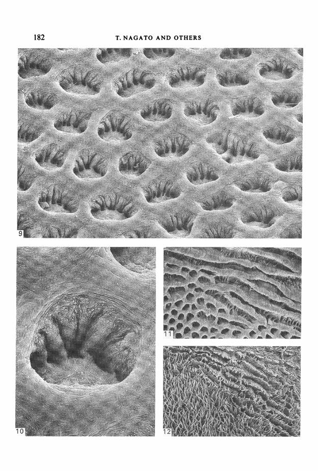
shows a large number of transversely orientated oval depressions about 50-110 ,um inlength and 50-70 ,um in width. An area about 20-30 ,um in width separates adjacentdepressions (Fig. 9). The anterior wall of each depression is relatively smooth, whereasthe posterior wall reveals several well-developed wrinkles lying vertically; the surfaceof each is covered with minute projecting structures (Fig. 10). Several secondarydepressions partitioned by epithelial walls connected to the wrinkles on the posteriorwall are observed at the bottom of a depression, and they correspond to the cavitiesoccupied by the secondary connective tissue papillae of thready filiform papillae(Fig. 10). A substantial change in the structure of the basal surface of the epitheliumoccurs at the posterior edge of the thready filiform papillae distribution area, i.e., nearthe boundary of the root of the tongue. As shown in Figure 11, the depressions in thisarea look like gutters, the bottoms of which show numerous secondary depressionstransversely arranged in rows. Such gutters appear to be caused by the lateral fusionof primary depressions observed on the basal surface of the epithelium in the posteriorportion of the lingual dorsum (Fig. 11).
Figure 12 shows the free surface structure of the posterior edge of the lingual
Figs. 5-7. Scanning electron micrographs of the basal surface of the epithelium of the lingualdorsum. In each photograph the bottom is near the tongue apex.Fig. 5. Large conical filiform papillae. Several epithelial furrow-like protrusions are present only onthe anterior wall. x 360.Fig. 6. Boundary between the depressions of large conical filiform papillae. Numerous fine furrow-like structures running along the edges of the depressions are seen. x 1340.Fig. 7. Boundary between large conical and thready filiform papillae. The depressions vary inappearance. x 150.Fig. 8. Scanning electron micrograph of the free surface of the transitional area between theintermolar eminence and the posterior portion of the lingual dorsum. Filiform papillae, which arepresent at the boundary between large conical and thready filiform papillae, show a transitionalsurface structure. The bottom of the photograph is near the tongue apex. x 110.

SEM of rat lingual filiform papillae 181
182 T. NAGATO AND OTHERS
EC! ;, - o - X < . -MW .x.Ia

SEM of rat lingual filiform papillae 183dorsum. Six to ten rows of thready filiform papillae in the most posterior area arelinked from side to side on the lateral edge of the tongue, and they form laterally-running long, furrow-like primary papillae that have numerous secondary papillae.
Three-dimensional structure of connective tissue papillaeAnterior portion of the lingual dorsum
After the removal of the epithelium, the lamina propria mucosae reveals two typesof connective tissue papillae. One type comprises relatively small projections growingin clusters in the entire area of the anterior portion of the lingual dorsum (Fig. 13), andthe other type consists of large projections scattered among the small projections (Fig.14). The former type represents the connective tissue papillae of small conical filiformpapillae, whereas the latter belongs to those of fungiform papillae, and both types ofpapillae project from the surface of the tongue almost at right angles. The connectivetissue papillae of the small conical filiform papillae are about 70 ,um in height andabout 30 ,um in diameter, and they become larger at their bases to reach about 50 ,umin diameter. These papillae lack one third to a half of the posterior part of theprojection and their lateral view resembles the crown of an incisor tooth (Fig. 15). Onthe other hand, the connective tissue papillae of fungiform papillae are 150-200 #sm inheight and 70-100,m in diameter, and they are easily distinguished from those ofsmall conical filiform papillae because of their size difference. There is a depressionabout 40-50 m in diameter on the tip of the connective tissue papilla of fungiformpapillae and this corresponds to a taste bud (Fig. 14).
Intermolar eminenceThe connective tissue papillae in the intermolar eminence belong to large conical
filiform papillae and they appear in varying sizes of about 100-200 ,um in height, withtheir basal parts about 60-110 um in diameter. The papillae appear as board-likeprojections about 50-100 /um in width and 20,m in thickness. Anteroposteriorlycompressed and slightly protruding toward the root of the tongue, they standvertically on top of a conical base. The surface of their board-like projection isrelatively smooth on the posterior side, but it shows several vertically running parallelgrooves on the anterior side (Fig. 16).
Posterior portion of the lingual dorsumThe connective tissue papillae of thready filiform papillae reflect the three-
dimensional image observed from the free surface, and because of the numerous
Figs. 9-11. Scanning electron micrographs of the basal surface of the epithelium of the lingualdorsum. In each photograph the bottom is near the tongue apex.Fig. 9. Posterior portion of the lingual dorsum. A number of laterally-elongated oval depressions areseen. The posterior wall of each depression reveals several well-developed epithelial wrinkles runningvertically. x 210.Fig. 10. Thready filiform papillae. Several secondary depressions partitioned by the epithelial wallsconnected to the wrinkles on the posterior wall are observed at the bottom of the depression. Thesurface of the area between the depressions shows numerous fine furrow-like structures runningaround the edges of the depressions. x 610.Fig. 11. Near the posterior edge of the posterior portion. Several laterally-running long gutter-likedepressions are seen in the most posterior area. x 75.Fig. 12. Scanning electron micrograph of the surface of the posterior portion of the lingual dorsum.In the most posterior area, several thready filiform papillae are linked from side to side of theirprimary papillae and form lateral long, furrow-like papillae that have numerous secondary threadypapillae. x 50.

184 T. NAGATO AND OTHERS

SEM of rat lingual filiform papillae 185secondary papillae growing in clusters they are easily distinguishable from the othertwo types of filiform papillae (Fig. 17). The primary papillae have a variable width,ranging from 50 to 100 /tm, but they are of almost uniform thickness and height. Thesecondary papillae growing from the tip of the primary papillae differ in number: 6 forthe wide primary papillae, 3 for the narrow ones, with 4-5 being the most commonarrangement. The secondary papillae have a long and narrow cylindrical shape andthey are about 7-i 0 /m in diameter and 30-40 #m in height. The three-dimensionalstructure of a papilla resembles a human hand, with the primary papilla as the palmand the secondary papillae as the fingers.
DISCUSSION
Kutuzov & Sicher (1951) conducted detailed gross and light microscopicobservations of filiform papillae on the rat lingual dorsum. Their study showed thatthere are three types of filiform papillae whose distribution areas are divided by clearboundaries and that the tissue architecture differs between the anterior and posteriorsurfaces of filiform papillae. According to the size and morphological features, theydesignated the filiform papillae in the anterior part of the lingual dorsum as simpleconical papillae, those on the intermolar eminence as giant conical papillae, and thosein the posterior part of the dorsum as (true) filiform papillae. Although these threetypes of papillae have distinct morphological features, including size, direction ofincline and the presence of secondary papillae, all of them have a long and narrowshape with a pointed tip. These common features seem to justify the designation of allthree types of papillae as 'filiform'. In addition, not all of them have taste buds.Taking these common characteristics into account, we have designated all three typesas filiform papillae. At the same time, according to their morphological characteristics,we classified them into small conical filiform papillae, large conical filiform papillae,and thready filiform papillae, which correspond respectively to Kutuzov & Sicher'ssimple conical papillae, giant conical papillae and (true) filiform papillae.
It is well known that the morphology of filiform papillae differs among differentanimal species as well as among different sites on the tongue itself (Sonntag, 1920,1924, 1925). Also, in recent years, observations by scanning electron microscopy haverevealed that many species have several types of filiform papillae. The distributionpatterns of filiform papillae differ among species. For instance, the dog (Iwasaki &Sakata, 1985) and shrew (Kobayashi, Shimoda & Shimamura, 1983) show no clearboundary between different distribution areas, with a gradual transition from onedistribution area to another; the opossum (Krause & Cutts, 1982) has a mixedpopulation of two types of filiform papillae in the same area, whereas the Japanese
Figs. 13-17. Scanning electron micrographs of the connective tissue papillae.Fig. 13. Small conical filiform papillae. The connective tissue papillae project from the surface oflingual dorsum at right angles. The bottom left of the photograph is near the tongue apex. x 250.Fig. 14. Fungiform papillae. A crater-like depression is seen at the tip of a connective tissue papilla.x650.Fig. 15. Small conical filiform papillae. The lateral aspect of a connective tissue papilla resembles thecrown of an incisor tooth. The right side of the photograph is near the tongue apex. x 790.Fig. 16. Large conical filiform papillae. The connective tissue papillae have several vertically-runningparallel grooves on the anterior aspect. The bottom left of the photograph is near the tongue apex.x250.Fig. 17. Thready filiform papillae. Each connective tissue papilla resembles a human hand, withthe primary papilla as the palm and the secondary papillae as the fingers. The bottom of thephotograph is near the tongue apex. x 670.
7 ANA 165

186 T. NAGATO AND OTHERS
long-fingered bat (Kobayashi & Shimamura, 1982), the Mongolian gerbil (Iwasakiet al. 1984) and rodents such as the rat (Kutuzov & Sicher, 1951), mouse (Kutuzov &Sicher, 1953; Iwasaki, Miyata & Kobayashi, 1987) and guinea-pig (Iwasaki & Miyata,1985), show clear boundaries for each type of filiform papillae. The presentobservations on the rat have indicated the presence of filiform papillae exhibiting atransitional shape between large conical and thready filiform papillae. These factssuggest that the shape of the tongue and lingual papillae and the distribution patternof morphologically different filiform papillae have gradually changed as the eatinghabits of different species have evolved.
Several investigators have been interested in the three-dimensional structure of thelamina propria in lingual mucosae, i.e. of the connective tissue papillae (Fish et al.1944; Kunze, 1969; Beckers, 1975a, b). Toyoshima & Shimamura (1982) treated theunfixed rat tongue with EDTA and observed the epithelium-connective tissueinterface by scanning electron microscopy, and they described the three-dimensionalstructure of the basal surface of the epithelium and connective tissue papillae in theanterior portion of the dorsum. Hull & Warfel (1986) also treated unfixed sampleswith 2N sodium bromide and observed the three-dimensional image of the connectivetissue papillae after the removal of the epithelium. The method used in the presentstudy adequately fixes the samples: first by perfusion, then by immersion, therebyminimising potential artefacts, and it allows the samples, the epithelium in particular,to maintain sufficiently their true form for scanning electron microscopy. Comparedwith the epithelium, connective tissue papillae seem to be easily damaged by thehydrochloric acid treatment. This in turn suggests that by adjusting the treatment timefor hydrochloric acid, this method may enable us to elucidate a number of veryinteresting as well as important facts under a scanning electron microscope, includingthe surface structure of the connective tissue papillae, the arrangement of collagenousfibres that form these papillae and the three-dimensional structure of fibroblasts andblood vessels in the papillae.Our present observations offer several new findings. The three types of filiform
papillae not only have clearly different three-dimensional surface images but alsodistinct morphological features in the epithelium-connective tissue interface. Inparticular, the changes in the surface image observed in the vicinity of the boundaryline between large conical and thready filiform papillae and the most posterior part ofthe thready filiform papillae distribution area are in complete agreement with thechanges in the three-dimensional image of the basal surface of the epithelium, even inthe small details. The fungiform papillae indicate no distinct orientation at the basalsurface of the epithelium. On the other hand, in small and large conical filiformpapillae, wrinkles in the depressions of the basal surface of the epithelium are sited onopposite sides. These facts clearly indicate that the morphology of the lingual papillaeare closely related to that of the connective tissue which forms the core of thesepapillae. It is supposed that numerous minute ridge-like protrusions and furrow-likestructures on the basal surface of the tongue epithelium are structures which formstrong connections between the epithelium and the underlying connective tissue.Some workers have already pointed out that, depending on the species and their
eating habits, the shape of the tongue differs, and, similarly, considerable variationexists in the number, distribution and shape of the lingual papillae (Sonntag, 1920,1924, 1925). However, in spite of the fact that the filiform papillae are well known tohave substantial morphological variation among species and among differing sites onthe lingual dorsum, there have been few reports in which their functions are fullydiscussed. One exception is the report by Kutuzov & Sicher (1952), who, after detailed

SEM of rat lingual filiform papillae 187gross and light microscopic observations of the rat palate, discussed the functions ofvarious parts of the palate in relation to the morphological characteristics ofcorresponding parts of the lingual dorsum.Our ideas concerning the functions of the three types of filiform papillae based on
our own results and those of a number of earlier studies are as follows:Small conical filiform papillae, present in the anterior portion of the lingual dorsum,
have a conical shape with a tongue-like projection at the tip. The papillae inclinebackward, and the tips face the pharynx. The tips and posterior surfaces of thepapillae are covered with hard keratin, the keratinisation being similar to thatobserved in the nail. These areas are tough as well as firm (Farbman, 1970). The basalsurface of the epithelium revealed well-developed wrinkles only on the posterior wallsof the depressions. This structure appears to be effective in resisting an external forceand maintaining the backward incline of the filiform papillae. In view of these findings,we postulate that the primary role of small conical filiform papillae is efficiently to takefood into the oral cavity.
Large conical filiform papillae present in the intermolar eminence have a similarshape to the small conical filiform papillae but the size, both in diameter and height,is two or three times that of the small conical filiform papillae. Also, large conicalfiliform papillae are inclined in the opposite direction to the small conical filiformpapillae, being inclined forward with the tips facing the apex of the tongue. The sametrend can be observed in the basal surface of the epithelium, with well-developedepithelial wrinkles present only on the anterior wall of the depressions. In addition, 5well-developed intermolar rugae with numerous sharp secondary papillae have beenshown to exist in the intermolar region of the palate, corresponding to the distributionarea of the large conical filiform papillae (Kutuzov & Sicher, 1952). Therefore, aspointed out by Kutuzov & Sicher, the filiform papillae in this area are believed tofunction further to grind food that has been crushed with the molar teeth, incoordination with the palate. From the direction of the papillae, this grindingmovement seems to be carried out by strongly pressing the intermolar eminence ofthe tongue against the intermolar region of the palate, then moving the tongueforward. Moreover, the wrinkles observed in the anterior wall of the depressionsappear to be effective in preventing the papillae from inclining backward whenperforming this grinding movement.The three-dimensional surface image of thready filiform papillae is morphologically
characterised by the presence of numerous slender and long secondary papillaegrowing in clusters. Also, the thready filiform papillae exhibit a distinct structure ofthe basal surface of the epithelium and the connective tissue papillae that differs fromthose observed in the other two types of filiform papillae. The presence of numeroussecondary papillae suggests the possibility that thready filiform papillae might workas a heat-releasing organ and may thus function in the control of body temperature.
SUMMARY
In an attempt to elucidate the functions of three types of filiform papillae in the rattongue, the epithelium-connective tissue interface of the lingual dorsum was observedby scanning electron microscopy.The basal surface of the epithelium in the anterior portion of the lingual dorsum
revealed numerous anteroposteriorly arranged, slightly elongated, oval depressionswith round depressions scattered among them. The former exhibited distinct epithelialprotrusions or wrinkles on the posterior wall. By contrast, the latter depressions
7-2

188 T. NAGATO AND OTHERS
indicated no such arrangement. The depressions in the intermolar eminence wereeither round or polygonal, and had several vertically-running well-developed wrinkleson the anterior wall. On the other hand, the depressions on the basal surface of theepithelium in the posterior portion of the lingual dorsum were -laterally-elongatedand oval with several vertically-running wrinkles on the posterior wall. At thebottom of the depression there were 3 to 6 secondary depressions, partitioned by thesewrinkles.The three-dimensional structure of the connective tissue papillae observed after the
removal of the epithelium clearly differed among the three types of filiform papillae.The lateral view of the connective tissue papillae of small conical filiform papillaeresembled the crown of an incisor tooth. In large conical filiform papillae, theconnective tissue papillae consisted of a truncated cone-shaped basal part withanteroposteriorly flat board-like projections. The connective tissue papillae of threadyfiliform papillae showed several secondary papillae growing from the tip of theanteroposteriorly orientated flat primary papillae.As a result of these findings, we suggest that the primary role of small conical
filiform papillae is efficiently to take food into the oral cavity. The large conicalfiliform papillae function to grind food that has already been crushed by the molarteeth in coordination with the palate. Thready filiform papillae might work as a heat-releasing organ and function in the control of body temperature.
REFERENCES
BECKERS, H. W. (1975a). Zur Morphologie der Papilla fungiformis einiger Primaten und des Menschen.Advances in Anatomy, Embryology and Cell Biology 50, 7-56.
BECKERS, H. W. (1975b). Zur Morphologie der Papilla fungiformis einiger Nagetiere. Advances in Anatomy,Embryology and Cell Biology 50, 57-116.
DESAKI, J. & UEHARA, Y. (1981). The overall morphology of neuromuscular junctions as revealed by scanningelectron microscopy. Journal of Neurocytology 10, 101-110.
EVAN, A. P., DAIL, W. G., DAMMROSE, D. & PALMER, C. (1976). Scanning electron microscopy of cell surfacefollowing removal of extracellular material. Anatomical Record 185, 433-447.
FARBMAN, A. I. (1970). The dual pattern of keratinization in filiform papillae on rat tongue. Journal ofAnatomy 106, 233-242.
FISH, H. S., MALONE, P. D. & RICHTER, C. P. (1944). The anatomy of the tongue of the domestic Norway rat.I. The skin of the tongue; The various papillae; Their number and distribution. Anatomical Record 89,429-440.
FuJIwARA, T. & UEHARA, Y. (1984). The cytoarchitecture of the wall and the innervation pattern of themicrovessels in the rat mammary gland: A scanning electron microscopic observation. American Journal ofAnatomy 170, 39-54.
HULL, M. T. & WARFEL, K. A. (1986). Basal lamina at the epithelial-connective tissue junction in the rat fore-stomach, esophagus, tongue and palate: Scanning electron microscopic study. Scanning Electron Microscopy1986 IV, 1395-1401.
IIDA, M., YOSHIOKA, I. & MurO, H. (1985). Three-dimensional and surface structures of rat filiform papillae.Acta anatomica 121, 237-244.
IWASAKI, S. & MIYATA, K. (1985). Studies on the lingual dorsal epithelium of the Guinea pig by scanningelectron microscopy. Okajimas folia anatomica japonica 61, 423-436.
IWASAKI, S., MIYATA, K. & KOBAYASHI, K. (1987). The surface structure of the dorsal epithelium of tonguein the mouse. Acta anatomica nipponica 62, 69-76.
IWASAKI, S. & SAKATA, K. (1985). Scanning electron microscopy of the lingual dorsal surface of the Beagle dog.Okajimas folia anatomica japonica 62, 1-14.
IWASAKI, S., SAKATA, K., MIYATA, K., MoRi, H. & KOBAYASHI, K. (1984). Fine structure of the lingual dorsalepithelium of Mongolian gerbil. Japanese Journal of Oral Biology 26, 292-296.
KARNOVSKY, M. J. (1965). A formaldehyde-glutaraldehyde fixative of high osmolality for use in electronmicroscopy. Journal of Cell Biology 27, 137A.
KLEIN-SZANTO, A. J. P. & SCHROEDER, H. E. (1977). Architecture and density of the connective tissue papillaeof the human oral mucosa. Journal ofAnatomy 123, 93-109.
KOBAYASHI, S. & SHIMAMURA, A. (1982). Comparative anatomical observations of the tongue of the Japaneselong-fingered bats, Miniopterus schreibersifuliginosus. Okajimasfolia anatomica japonica 58, 923-932.
KOBAYASHI, S., SHIMODA, T. & SHIMAMURA, A. (1983). Comparative anatomical observations on the tongueof the Inse.'tivora. Okajimasfolia anatomica japonica 60, 211-218.

SEM of rat lingualfiliform papillae 189KRAUSE, W. J. & Cumrs, J. H. (1982). Morphological observations on the papillae of the opossum tongue.Acta anatomica 113, 159-168.
KUNZE, K. (1969). Die Papilla filiformis des Menschen als Tastsinnesorgan. Licht- und elektronen-mikroskopische Untersuchungen. Ergebnisse der Anatomie und Entwicklungsgeschichte 41 (5), 3-64.
KuTuzov, H. & SICHER, H. (1951). The filiform and the conical papillae of the tongue in the white rat.Anatomical Record 110, 275-288.
KuTuzov, H. & SICHER, H. (1952). Anatomy and function of the palate in the white rat. Anatomical Record114, 67-84.
KuTUzov, H. & SICHER, H. (1953). Comparative anatomy of the mucosa of the tongue and the palate of thelaboratory mouse. Anatomical Record 116, 409-425.
NAGATO, T. (1978). Scanning electron microscopical image of myoepithelial cells. Journal of ElectronMicroscopy 27, 235-236.
NAGATO, T., YOSHIDA, H., YOSHIDA, A. & UEHARA, Y. (1980). A scanning electron microscope study ofmyoepithelial cells in exocrine glands. Cell and Tissue Research 209, 1-10.
OOYA, K. & TOOYA, Y. (1981). Scanning electron microscopy of the epithelium-connective tissue interface inhuman gingiva. Journal of Periodontal Research 16, 135-139.
SCALETrA, L. J. & SIMMELINK, J. W. (1973). Scanning electron microscopy of the epithelial-connective tissueinterface. Journal ofDental Research 52, 147. (IADR abstract).
SONNTAG, C. F. (1920). The comparative anatomy of the tongue of the mammalia. I, General description ofthe tongue. Proceedings of the Zoological Society of London, 115-129.
SONNTAG, C. F. (1924). The comparative anatomy of the tongue of the mammalia. X, Rodentia. Proceedingsof the Zoological Society of London, 725-741.
SONNTAG, C. F. (1925). The comparative anatomy of the tongue of the mammalia. XII, Summary,classification and phylogeny. Proceedings of the Zoological Society of London, 701-762.
SQUIER, C. A., JOHNSON, N. W. & HACKEMANN, M. (1975). Structure and function of normal human oralmucosa. In Oral Mucosa in Health and Disease (ed. A. E. Dolby), p. 55, Oxford, London, Edinburgh,Melbourne: Blackwell Scientific Publications.
SUZUKI, Y. & TAKEDA, M. (1987). Scanning electron microscopic observation of the basement membrane inmouse taste buds. Journal of Electron Microscopy 36, 98-106.
SVEJDA, J. & SKACH, M. (1971). Die Zunge der Ratte im Raster-Elektronenmikroskop (Stereoscan). Zeitschriftfir mikroskopisch-anatomische Forschung 84, 101-116.
TOYosHIMA, K. & SHIMAMURA, A. (1982). Scanning electron microscope study of the epithelium-connectivetissue interfaces of the tongue papillae in the rat. Japanese Journal of Oral Biology 24, 1019-1022.
YOSHIOKA, I. & MUTO, H. (1976). Surface structures of the tongue, palate and buccal mucosa of the rat.(Scanning electrpn microscopic studies on the oral mucosa 3.) Okajimas folia anatomica japonica 52,297-312.