three consecutive monthly intravitreal ranibizumab for...
TRANSCRIPT
Open Journal of Ophthalmology, 2012, 2, 93-96 http://dx.doi.org/10.4236/ojoph.2012.23020 Published Online August 2012 (http://www.SciRP.org/journal/ojoph)
93
Three Consecutive Monthly Intravitreal Ranibizumab for Choroidal Neovascularization in Central Serous Choriorethınopathy: A Case Report
Kazim Erol1, Esin Sogutlu Sari2*, Arif Koytak3, A. Karaçor1, D. T. Çoban1, M. Bulut1
1Ophthalmology Department, Antalya Training and Research Hospital, Antalya, Turkey; 2Ophthalmology Department, Kartal Training and Research Hospital, Istanbul, Turkey; 3Ophthalmology Department, Bezmialem Vakif University, Istanbul, Turkey. Email: *[email protected] Received March 19th, 2012; revised April 25th, 2012; accepted May 8th, 2012
ABSTRACT
Purpose: The authors report the result of three consecutive monthly intravitreal ranibizumab injection for choroidal neovascularization (CNV) after bevacizumab injection for chronic central serous rethinopathy (CSR). Methods: A 48- year-old man with chronic CSR was treated with intravitreal single dose 2.5 mg bevacizumab. One year after CNV was occurred, and three consecutive monthly intravitreal ranibizumab injections were performed. Results: Four weeks later the first ranibizumab dose, best corrected visual acuity was improved 20/80 to 20/20 and remained stable within one year. Conclusion: Repeat intravitreal ranibizumab injection in CNV after bevacizumab injection for chronic CSR ap-peared to be an effective treatment option. Keywords: Central Serous Choriorethinopathy; Choroidal Neovascularization; Ranibizumab
1. Introduction
Central serous chorioretinopathy (CSR) is common dis- eases of the posterior segment of the eye characterized by serous detachment of the neurosensory retina in the macula secondary to an idiopathic leakage in the outer blood-retinal barier at the retinal pigment epithelium (RPE). Although visual distortions are usually mild and spontaneous recovery occurs within a few months, some patients with CSR have a poor visual acuity due to retinal pigment epithelium atrophy, persistant or recurrent pig- ment epithelial detachment, subretinal fluid and chor- oidal neovascularization (CNV) [1]. CNV secondary to CSR is an uncommon relation which has been also noted to complicate laser photocoagulation treatment due to the puncture of Bruch’s membrane by laser burns and photo- dynamic theraphy due to the RPE alterations and induces the release of vascular endothelial growth factor (VEGF) [2].
Different treatment options including photodynamic theraphy with vertaporfin, laser photocoagulation, vitro- retinal submacular surgery and intravitreal anti VEGF agents (bevacizumab or ranibizumab) have been reported for the chronic and recurrent CSR with or without CNV. [2-5] We report the results of three consecutive monthly
intravitreal ranibizumab injection for CNV after bevaci- zumab injection for chronic CSR. To our knowledge there have been no previously reported cases of CNV after bevacizumab for the management of CSR.
2. Case Report
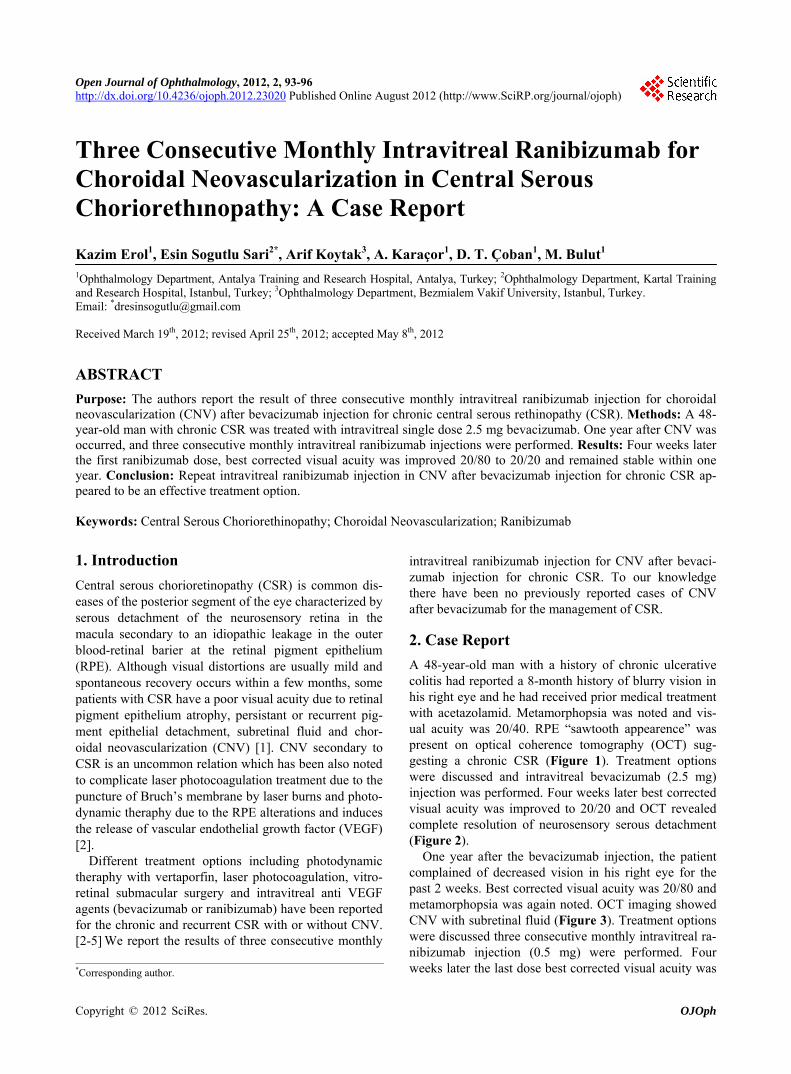
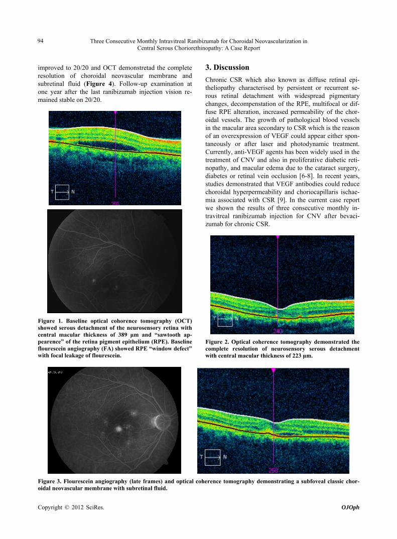
A 48-year-old man with a history of chronic ulcerative colitis had reported a 8-month history of blurry vision in his right eye and he had received prior medical treatment with acetazolamid. Metamorphopsia was noted and vis- ual acuity was 20/40. RPE “sawtooth appearence” was present on optical coherence tomography (OCT) sug- gesting a chronic CSR (Figure 1). Treatment options were discussed and intravitreal bevacizumab (2.5 mg) injection was performed. Four weeks later best corrected visual acuity was improved to 20/20 and OCT revealed complete resolution of neurosensory serous detachment (Figure 2).
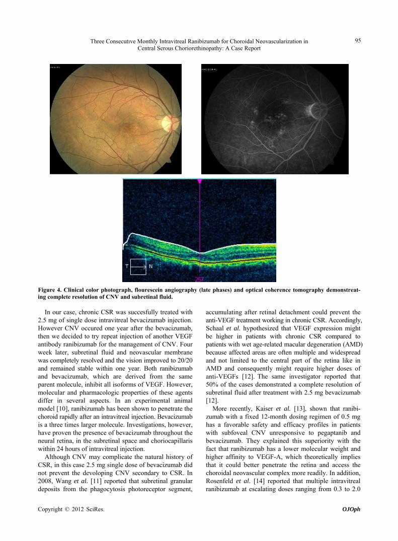
One year after the bevacizumab injection, the patient complained of decreased vision in his right eye for the past 2 weeks. Best corrected visual acuity was 20/80 and metamorphopsia was again noted. OCT imaging showed CNV with subretinal fluid (Figure 3). Treatment options were discussed three consecutive monthly intravitreal ra- nibizumab injection (0.5 mg) were performed. Four weeks later the last dose best corrected visual acuity was *Corresponding author.
Copyright © 2012 SciRes. OJOph
Three Consecutive Monthly Intravitreal Ranibizumab for Choroidal Neovascularization in Central Serous Choriorethinopathy: A Case Report
94
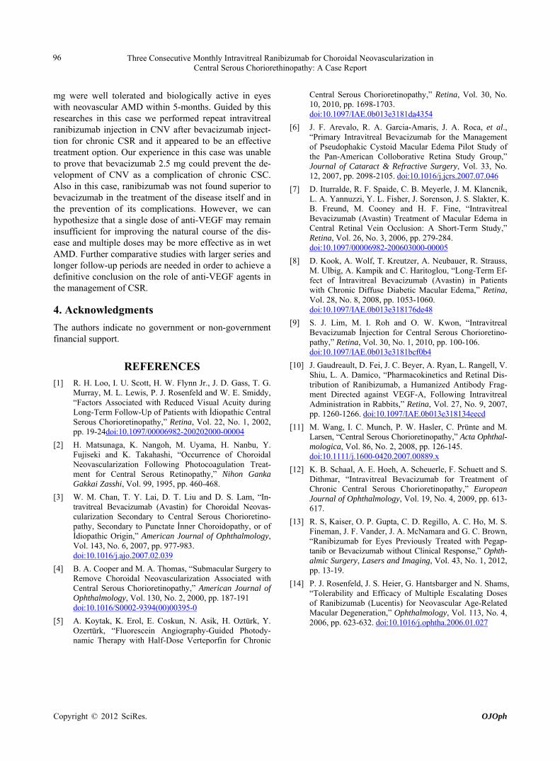
improved to 20/20 and OCT demonstretad the complete resolution of choroidal neovascular membrane and subretinal fluid (Figure 4). Follow-up examination at one year after the last ranibizumab injection vision re- mained stable on 20/20.
Figure 1. Baseline optical cohorence tomography (OCT) showed serous detachment of the neurosensory retina with central macular thickness of 389 μm and “sawtooth ap- pearence” of the retina pigment epithelium (RPE). Baseline flourescein angiography (FA) showed RPE “window defect” with focal leakage of flourescein.
3. Discussion
Chronic CSR which also known as diffuse retinal epi- theliopathy characterised by persistent or recurrent se- rous retinal detachment with widespread pigmentary changes, decompenstation of the RPE, multifocal or dif- fuse RPE alteration, increased permeability of the chor- oidal vessels. The growth of pathological blood vessels in the macular area secondary to CSR which is the reason of an overexpression of VEGF could appear either spon- taneously or after laser and photodynamic treatment. Currently, anti-VEGF agents has been widely used in the treatment of CNV and also in proliferative diabetic reti- nopathy, and macular edema due to the cataract surgery, diabetes or retinal vein occlusion [6-8]. In recent years, studies demonstrated that VEGF antibodies could reduce choroidal hyperpermeability and choriocapillaris ischae- mia associated with CSR [9]. In the current case report we shown the results of three consecutive monthly in- travitreal ranibizumab injection for CNV after bevaci- zumab for chronic CSR.
Figure 2. Optical coherence tomography demonstrated the complete resolution of neurosensory serous detachment with central macular thickness of 223 μm.
Figure 3. Flourescein angiography (late frames) and optical coherence tomography demonstrating a subfoveal classic chor- oidal neovascular membrane with subretinal fluid.
Copyright © 2012 SciRes. OJOph
Three Consecutıve Monthly Intravitreal Ranibizumab for Choroidal Neovascularization in Central Serous Choriorethinopathy: A Case Report
95
Figure 4. Clinical color photograph, flourescein angiography (late phases) and optical coherence tomography demonstreat- ing complete resolution of CNV and subretinal fluid.
In our case, chronic CSR was succesfully treated with 2.5 mg of single dose intravitreal bevacizumab injection. However CNV occured one year after the bevacizumab, then we decided to try repeat injection of another VEGF antibody ranibizumab for the management of CNV. Four week later, subretinal fluid and neovascular membrane was completely resolved and the vision improved to 20/20 and remained stable within one year. Both ranibizumab and bevacizumab, which are derived from the same parent molecule, inhibit all isoforms of VEGF. However, molecular and pharmacologic properties of these agents differ in several aspects. In an experimental animal model [10], ranibizumab has been shown to penetrate the choroid rapidly after an intravitreal injection. Bevacizumab is a three times larger molecule. Investigations, however, have proven the presence of bevacizumab throughout the neural retina, in the subretinal space and choriocapillaris within 24 hours of intravitreal injection.
Although CNV may complicate the natural history of CSR, in this case 2.5 mg single dose of bevacizumab did not prevent the devoloping CNV secondary to CSR. In 2008, Wang et al. [11] reported that subretinal granular deposits from the phagocytosis photoreceptor segment,
accumulating after retinal detachment could prevent the anti-VEGF treatment working in chronic CSR. Accordingly, Schaal et al. hypothesized that VEGF expression might be higher in patients with chronic CSR compared to patients with wet age-related macular degeneration (AMD) because affected areas are often multiple and widespread and not limited to the central part of the retina like in AMD and consequently might require higher doses of anti-VEGFs [12]. The same investigator reported that 50% of the cases demonstrated a complete resolution of subretinal fluid after treatment with 2.5 mg bevacizumab [12].
More recently, Kaiser et al. [13], shown that ranibi- zumab with a fixed 12-month dosing regimen of 0.5 mg has a favorable safety and efficacy profiles in patients with subfoveal CNV unresponsive to pegaptanib and bevacizumab. They explained this superiority with the fact that ranibizumab has a lower molecular weight and higher affinity to VEGF-A, which theoretically implies that it could better penetrate the retina and access the choroidal neovascular complex more readily. In addition, Rosenfeld et al. [14] reported that multiple intravitreal ranibizumab at escalating doses ranging from 0.3 to 2.0
Copyright © 2012 SciRes. OJOph
Three Consecutive Monthly Intravitreal Ranibizumab for Choroidal Neovascularization in Central Serous Choriorethinopathy: A Case Report
96
mg were well tolerated and biologically active in eyes with neovascular AMD within 5-months. Guided by this researches in this case we performed repeat intravitreal ranibizumab injection in CNV after bevacizumab inject- tion for chronic CSR and it appeared to be an effective treatment option. Our experience in this case was unable to prove that bevacizumab 2.5 mg could prevent the de- velopment of CNV as a complication of chronic CSC. Also in this case, ranibizumab was not found superior to bevacizumab in the treatment of the disease itself and in the prevention of its complications. However, we can hypothesize that a single dose of anti-VEGF may remain insufficient for improving the natural course of the dis- ease and multiple doses may be more effective as in wet AMD. Further comparative studies with larger series and longer follow-up periods are needed in order to achieve a definitive conclusion on the role of anti-VEGF agents in the management of CSR.
4. Acknowledgments
The authors indicate no government or non-government financial support.
REFERENCES [1] R. H. Loo, I. U. Scott, H. W. Flynn Jr., J. D. Gass, T. G.
Murray, M. L. Lewis, P. J. Rosenfeld and W. E. Smiddy, “Factors Associated with Reduced Visual Acuity during Long-Term Follow-Up of Patients with İdiopathic Central Serous Chorioretinopathy,” Retina, Vol. 22, No. 1, 2002, pp. 19-24doi:10.1097/00006982-200202000-00004
[2] H. Matsunaga, K. Nangoh, M. Uyama, H. Nanbu, Y. Fujiseki and K. Takahashi, “Occurrence of Choroidal Neovascularization Following Photocoagulation Treat- ment for Central Serous Retinopathy,” Nihon Ganka Gakkai Zasshi, Vol. 99, 1995, pp. 460-468.
[3] W. M. Chan, T. Y. Lai, D. T. Liu and D. S. Lam, “In- travitreal Bevacizumab (Avastin) for Choroidal Neovas- cularization Secondary to Central Serous Chorioretino- pathy, Secondary to Punctate İnner Choroidopathy, or of İdiopathic Origin,” American Journal of Ophthalmology, Vol. 143, No. 6, 2007, pp. 977-983. doi:10.1016/j.ajo.2007.02.039
[4] B. A. Cooper and M. A. Thomas, “Submacular Surgery to Remove Choroidal Neovascularization Associated with Central Serous Chorioretinopathy,” American Journal of Ophthalmology, Vol. 130, No. 2, 2000, pp. 187-191 doi:10.1016/S0002-9394(00)00395-0
[5] A. Koytak, K. Erol, E. Coskun, N. Asik, H. Oztürk, Y. Ozertürk, “Fluorescein Angiography-Guided Photody- namic Therapy with Half-Dose Verteporfin for Chronic
Central Serous Chorioretinopathy,” Retina, Vol. 30, No. 10, 2010, pp. 1698-1703. doi:10.1097/IAE.0b013e3181da4354
[6] J. F. Arevalo, R. A. Garcia-Amaris, J. A. Roca, et al., “Primary Intravitreal Bevacizumab for the Management of Pseudophakic Cystoid Macular Edema Pilot Study of the Pan-American Colloborative Retina Study Group,” Journal of Cataract & Refractive Surgery, Vol. 33, No. 12, 2007, pp. 2098-2105. doi:10.1016/j.jcrs.2007.07.046
[7] D. Iturralde, R. F. Spaide, C. B. Meyerle, J. M. Klancnik, L. A. Yannuzzi, Y. L. Fisher, J. Sorenson, J. S. Slakter, K. B. Freund, M. Cooney and H. F. Fine, “Intravitreal Bevacizumab (Avastin) Treatment of Macular Edema in Central Retinal Vein Occlusion: A Short-Term Study,” Retina, Vol. 26, No. 3, 2006, pp. 279-284. doi:10.1097/00006982-200603000-00005
[8] D. Kook, A. Wolf, T. Kreutzer, A. Neubauer, R. Strauss, M. Ulbig, A. Kampik and C. Haritoglou, “Long-Term Ef- fect of İntravitreal Bevacizumab (Avastin) in Patients with Chronic Diffuse Diabetic Macular Edema,” Retina, Vol. 28, No. 8, 2008, pp. 1053-1060. doi:10.1097/IAE.0b013e318176de48
[9] S. J. Lim, M. I. Roh and O. W. Kwon, “Intravitreal Bevacizumab İnjection for Central Serous Chorioretino- pathy,” Retina, Vol. 30, No. 1, 2010, pp. 100-106. doi:10.1097/IAE.0b013e3181bcf0b4
[10] J. Gaudreault, D. Fei, J. C. Beyer, A. Ryan, L. Rangell, V. Shiu, L. A. Damico, “Pharmacokinetics and Retinal Dis- tribution of Ranibizumab, a Humanized Antibody Frag- ment Directed against VEGF-A, Following Intravitreal Administration in Rabbits,” Retina, Vol. 27, No. 9, 2007, pp. 1260-1266. doi:10.1097/IAE.0b013e318134eecd
[11] M. Wang, I. C. Munch, P. W. Hasler, C. Prünte and M. Larsen, “Central Serous Chorioretinopathy,” Acta Ophthal- mologica, Vol. 86, No. 2, 2008, pp. 126-145. doi:10.1111/j.1600-0420.2007.00889.x
[12] K. B. Schaal, A. E. Hoeh, A. Scheuerle, F. Schuett and S. Dithmar, “Intravitreal Bevacizumab for Treatment of Chronic Central Serous Chorioretinopathy,” European Journal of Ophthalmology, Vol. 19, No. 4, 2009, pp. 613- 617.
[13] R. S, Kaiser, O. P. Gupta, C. D. Regillo, A. C. Ho, M. S. Fineman, J. F. Vander, J. A. McNamara and G. C. Brown, “Ranibizumab for Eyes Previously Treated with Pegap- tanib or Bevacizumab without Clinical Response,” Ophth- almic Surgery, Lasers and Imaging, Vol. 43, No. 1, 2012, pp. 13-19.
[14] P. J. Rosenfeld, J. S. Heier, G. Hantsbarger and N. Shams, “Tolerability and Efficacy of Multiple Escalating Doses of Ranibizumab (Lucentis) for Neovascular Age-Related Macular Degeneration,” Ophthalmology, Vol. 113, No. 4, 2006, pp. 623-632. doi:10.1016/j.ophtha.2006.01.027
Copyright © 2012 SciRes. OJOph