thoracic cavity. right and left pleural cavities parietal pleura visceral (pulmonary) pleura ...
TRANSCRIPT

Thoracic Cavity
Right and Left Pleural Cavities
Parietal Pleura Visceral (Pulmonary) Pleura Parietal
– Costal– Mediastinal– Diaphragmatic– Cupola
Connecting Pleura
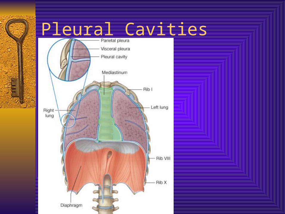
Pleural Cavities

Pleural Cavities
Lungs
Light, soft, spongy Conical in shape, apex, base, costal surface,
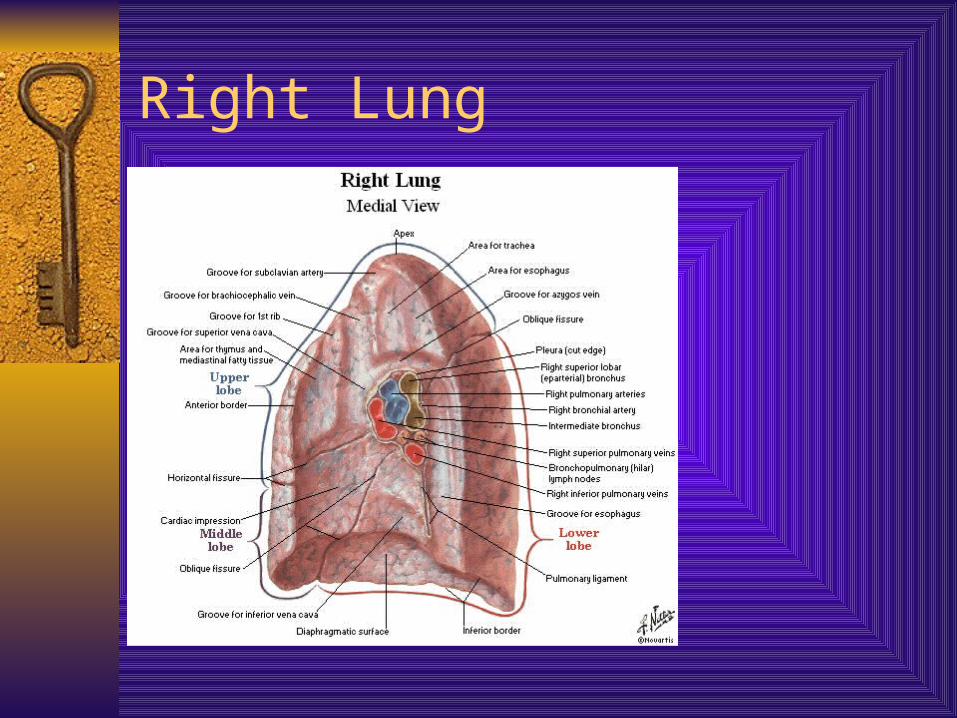
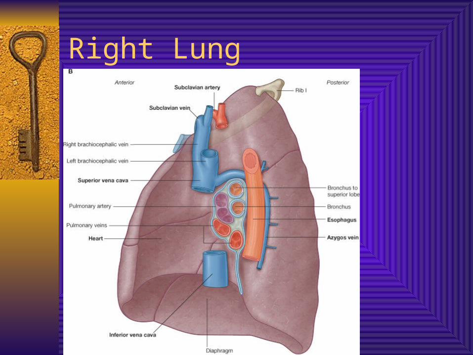
medial surface, hilus. Note various impressions Right lung
– Three lobes; superior, middle and inferior
– Oblique and horizontal fissure
Left Lung– Two lobes; superior and inferior also Lingula and
Cardiac notch, oblique fissure
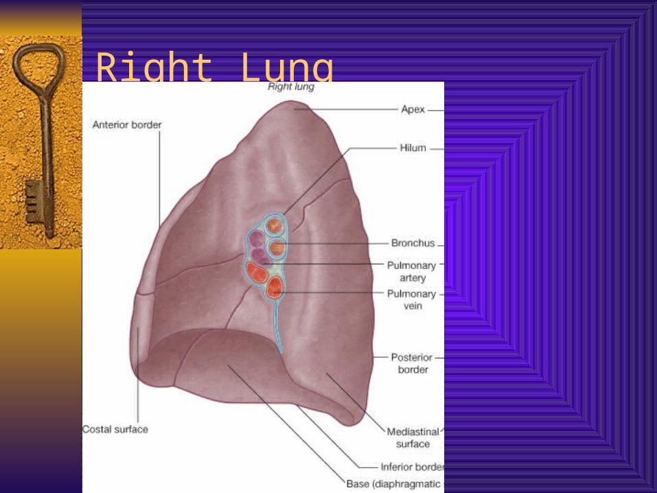
Right Lung
Right Lung
Right Lung

Right Lung
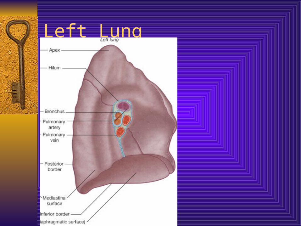
Left Lung
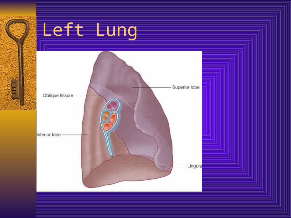
Left Lung
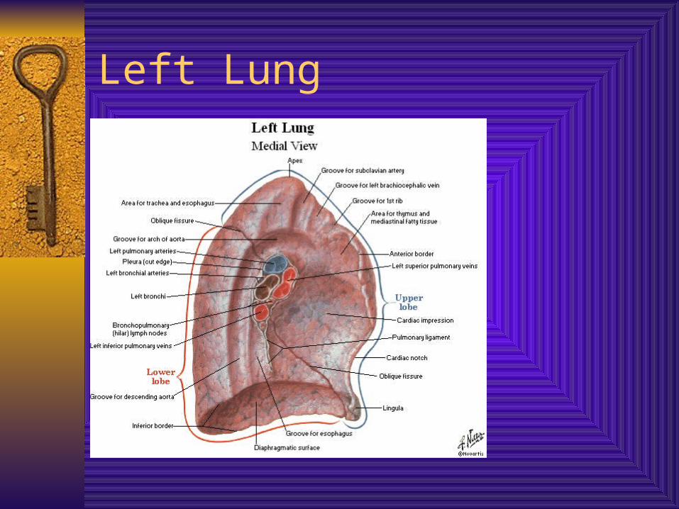
Left Lung
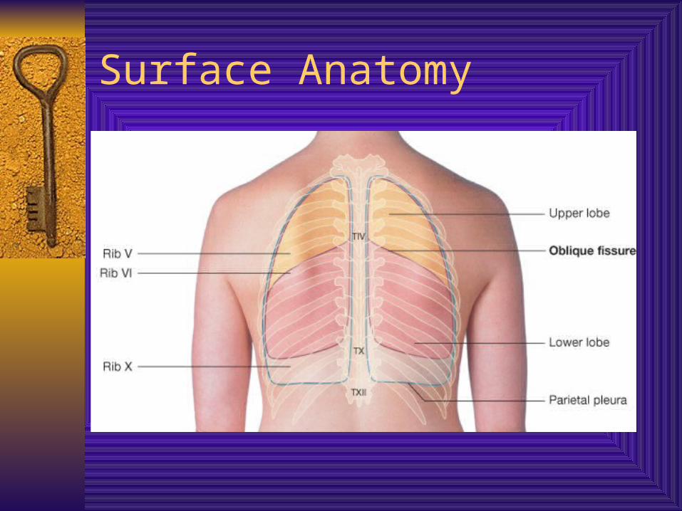
Surface Anatomy
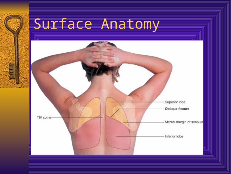
Surface Anatomy

Surface Anatomy
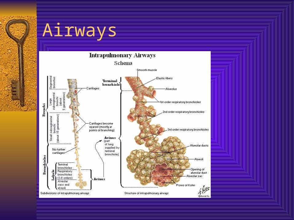
Airways
Trachea, primary bronchi, secondary bronchi, tertiary bronchi out to 25 generations
All comprised of hyaline cartilage Trachea
– Begins where larynx ends (about C6)
– 10 cm long, half in neck, half in mediastinum
– 20 U-Shaped rings of hyaline cartilage – keeps lumen intact but not as brittle as bone
– Lined with epithelium and cilia which work to keep foreign bodies/irritants away from lungs

Airways

Airways

Trachea
Airways
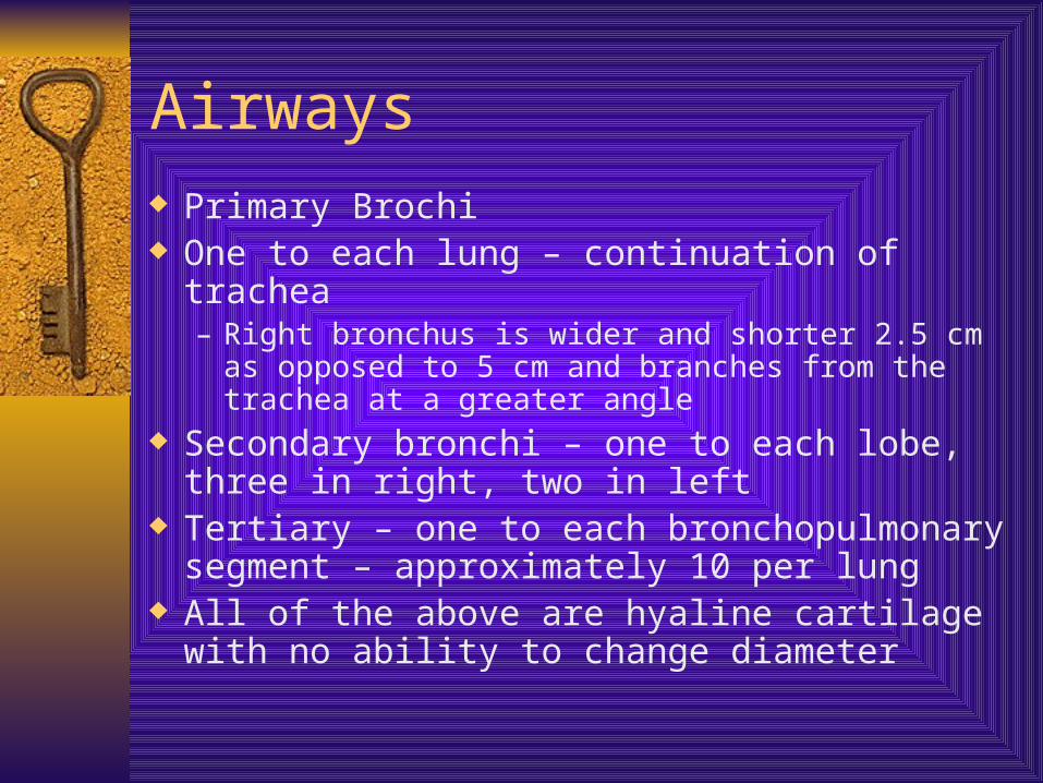
Airways Primary Brochi One to each lung – continuation of trachea
– Right bronchus is wider and shorter 2.5 cm as opposed to 5 cm and branches from the trachea at a greater angle
Secondary bronchi – one to each lobe, three in right, two in left
Tertiary – one to each bronchopulmonary segment – approximately 10 per lung
All of the above are hyaline cartilage with no ability to change diameter

Bronchoscope

Tumor

Tumor

Tumor
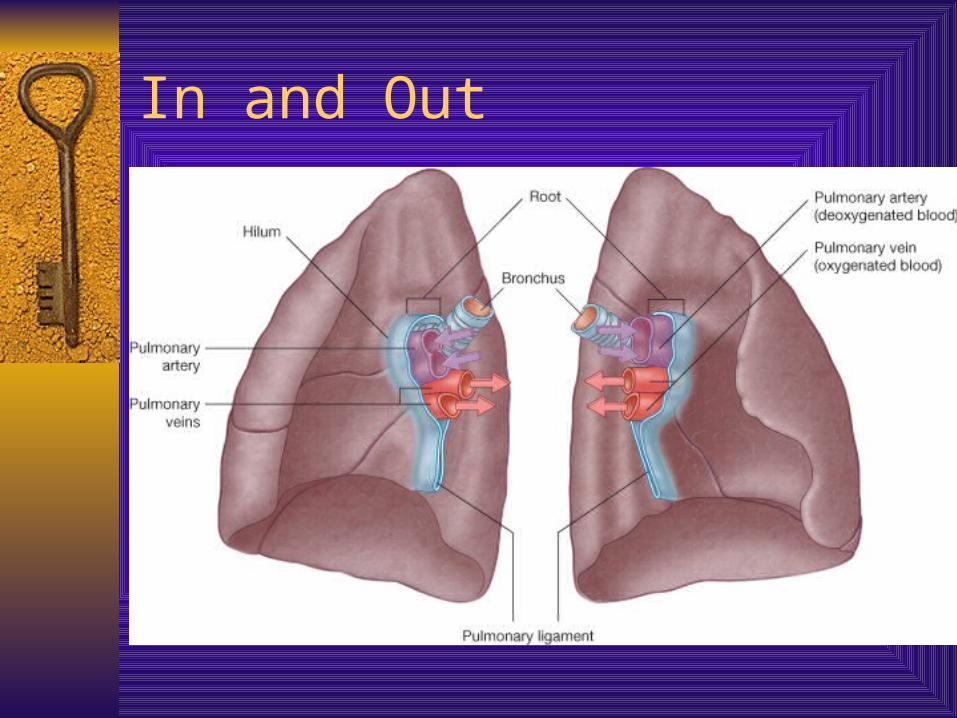
In and Out
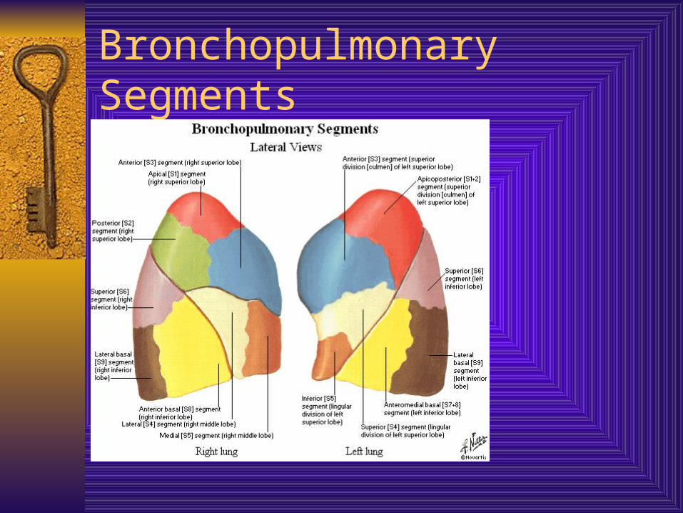
Bronchopulmonary Segments
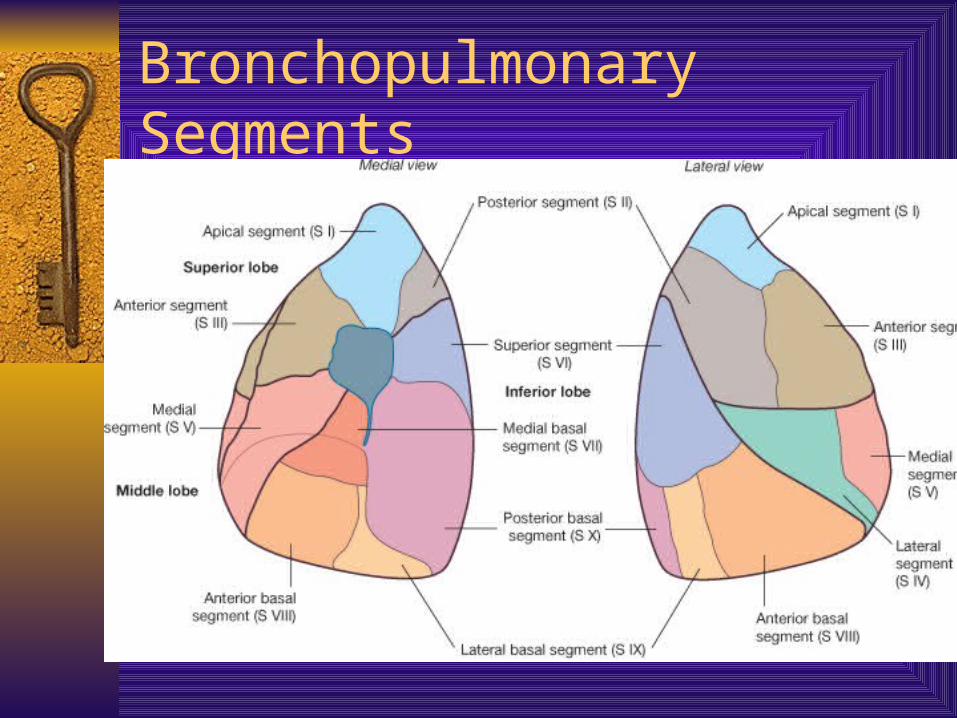
Bronchopulmonary Segments
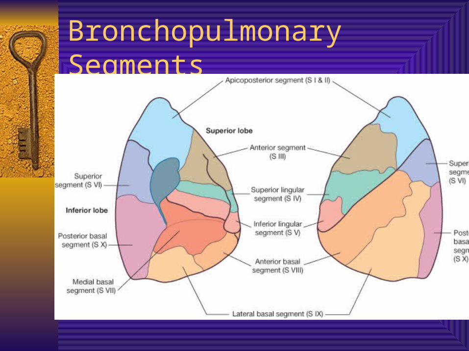
Bronchopulmonary Segments
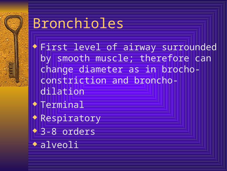
Bronchioles
First level of airway surrounded by smooth muscle; therefore can change diameter as in brocho-constriction and broncho-dilation
Terminal Respiratory 3-8 orders alveoli

Bronchioles
Bronchioles
Gas Exchange Pulmonary arteries carry deoxygenated
blood to aleoli Gas exchange occurs via diffusion through
the capillary beds Returned to heart via pulmonary veins
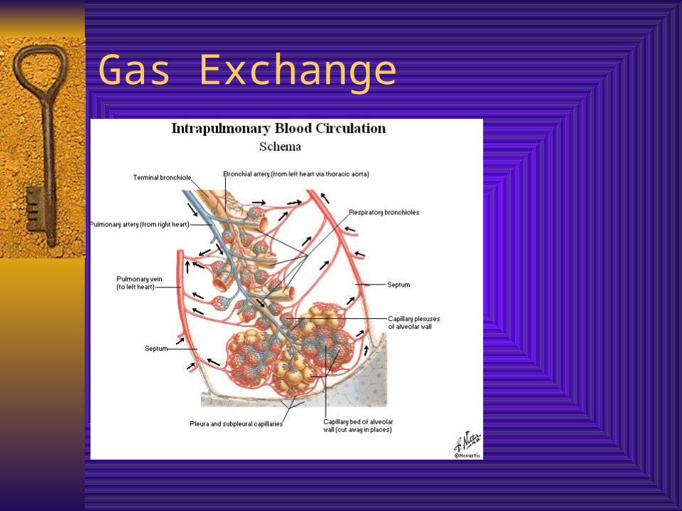
Gas Exchange

Innervation
Pleura via intercostal (thoracic) nerves Tracheobronchial tree Parasympathetic via CN X efferent function
= broncho-constriction via smooth mm., also to epithelial cells in trachea; afferent = responsible for cough reflex
Sympathetic from T1-T5 efferent = brocho-dilation

Intercostal to Pleura
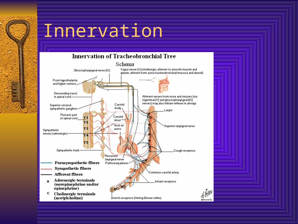
Innervation
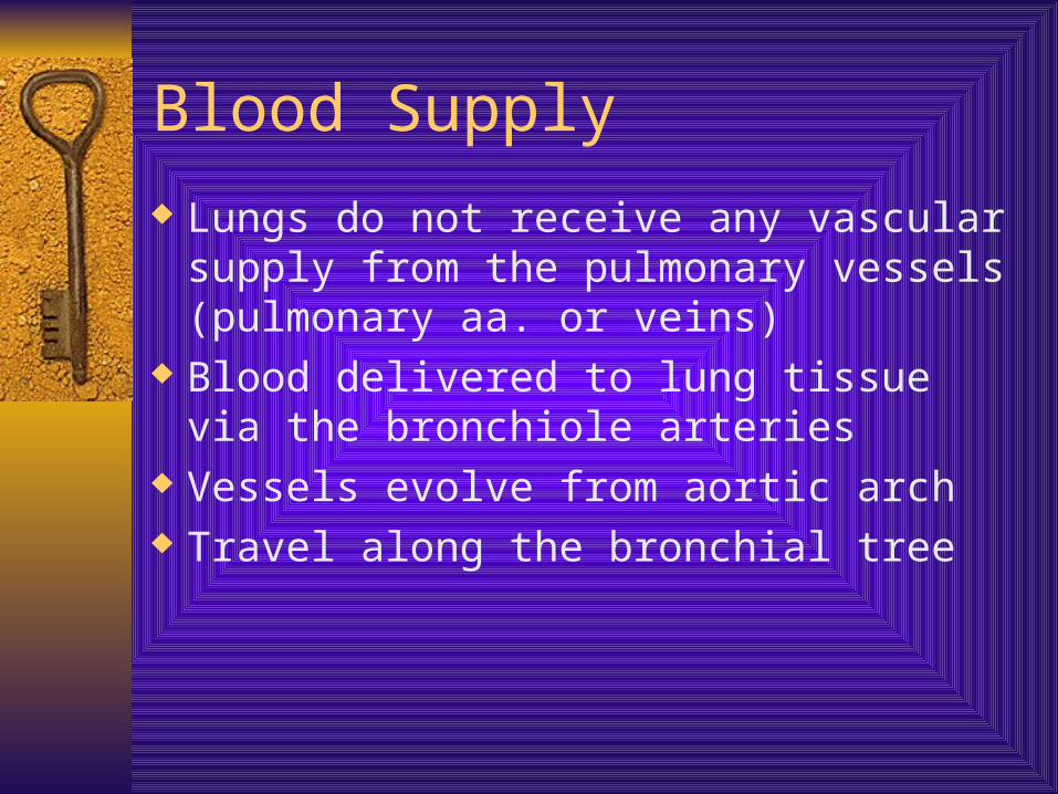
Blood Supply
Lungs do not receive any vascular supply from the pulmonary vessels (pulmonary aa. or veins)
Blood delivered to lung tissue via the bronchiole arteries
Vessels evolve from aortic arch Travel along the bronchial tree
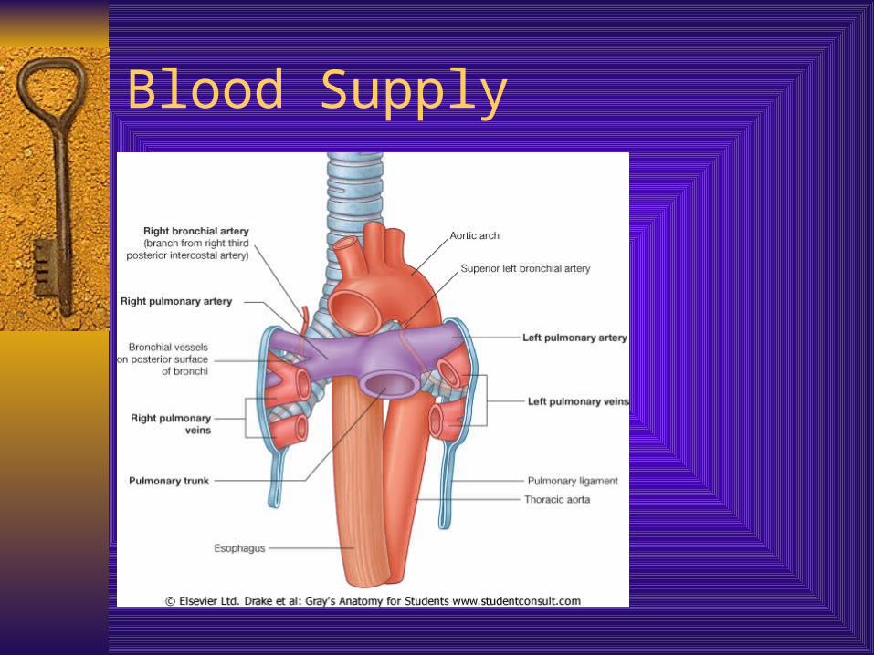
Blood Supply
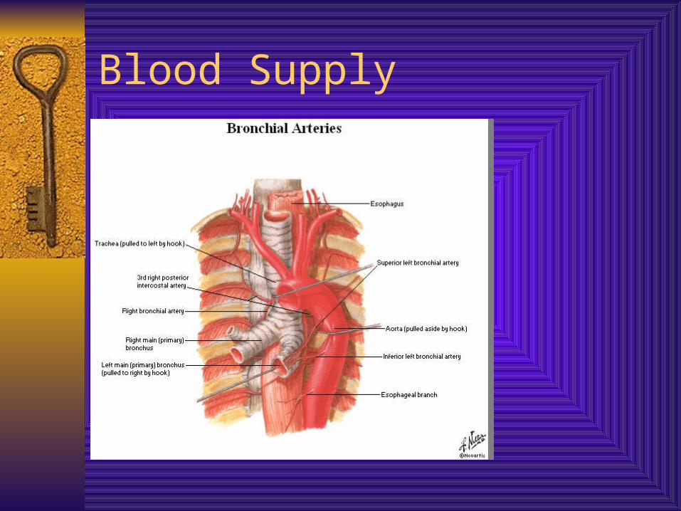
Blood Supply