thioredoxin glutathione reductase-dependent redox networks in platyhelminth parasites
TRANSCRIPT
FORUM REVIEW ARTICLE
Thioredoxin Glutathione Reductase-DependentRedox Networks in Platyhelminth Parasites
David L. Williams,1 Mariana Bonilla,2 Vadim N. Gladyshev,3 and Gustavo Salinas2
Abstract
Significance: Platyhelminth parasites cause chronic infections that are a major cause of disability, mortality, andeconomic losses in developing countries. Maintaining redox homeostasis is a major adaptive problem faced byparasites and its disruption can shift the biochemical balance toward the host. Platyhelminth parasites possess astreamlined thiol-based redox system in which a single enzyme, thioredoxin glutathione reductase (TGR), afusion of a glutaredoxin (Grx) domain to canonical thioredoxin reductase (TR) domains, supplies electrons tooxidized glutathione (GSSG) and thioredoxin (Trx). TGR has been validated as a drug target for schistosomiasis.Recent Advances: In addition to glutathione (GSH) and Trx reduction, TGR supports GSH-independent de-glutathionylation conferring an additional advantage to the TGR redox array. Biochemical and structural studieshave shown that the TR activity does not require the Grx domain, while the glutathione reductase and deglu-tathionylase activities depend on the Grx domain, which receives electrons from the TR domains. The search forTGR inhibitors has identified promising drug leads, notably oxadiazole N-oxides. Critical Issues: A conspicuousfeature of platyhelminth TGRs is that their Grx-dependent activities are temporarily inhibited at high GSSGconcentrations. The mechanism underlying the phenomenon and its biological relevance are not completelyunderstood. Future Directions: The functional diversity of Trxs and Grxs encoded in platyhelminth genomesremains to be further assessed to thoroughly understand the TGR-dependent redox network. Optimization ofTGR inhibitors and identification of compounds targeting other parasite redox enzymes are good options toclinically develop relevant drugs for these neglected, but important diseases. Antioxid. Redox Signal. 00, 000–000.
Introduction
Platyhelminthes is a phylum of metazoan organismsthat includes two major classes of human and livestock
parasites: trematodes (flukes) and cestodes (tapeworms).Platyhelminth parasites have complex life cycles with bothsexual and asexual reproductive phases and undergo meta-morphoses and migrations within their multiple hosts. Pla-tyhelminth infections can result in chronic (lasting for years)infections. The disability and poverty generated by platyhel-minth infections are an enormous health and economic bur-den for low-income countries. Schistosomiasis alone, the mostprevalent and devastating infection caused by trematodes,affects more than 200 million people and causes more than200,000 deaths in tropical and subtropical areas of Africa,South America, and Asia. It is estimated that the globalprevalence of food-borne trematodiases is > 40 million people(25). These infectious agents include Opisthorchis spp. andClonorchis spp. (liver flukes causing cholangitis and bile duct
cancer), Paragonimus spp. (lung flukes), and Fasciolopsis buskiand Fasciola hepatica (intestinal and liver flukes, respectively).Cestode infections are less prevalent, yet some of them, suchas cysticercosis and hydatid disease, constitute significantproblems to human health; for example, neurocysticercosiscaused by Taenia solium is the major cause of acquired epilepsyworldwide (19). Platyhelminthes also result in widespreadinfections in economically important domestic animals.
Parasites causing chronic infections, such as platy-helminthes, are excellent model organisms to study pathwaysinvolved in redox homeostasis. These organisms are exposednot only to the oxidant species endogenously generated as aresult of their own metabolism, but also to those derived fromthe immune response mounted by their hosts. The study ofantioxidant defenses in platyhelminth parasites has revealeda unique biochemical scenario and novel drug targets forplatyhelminth infections. This is highly relevant for the ne-glected diseases caused by these infections: few drugs arecurrently available for the treatment of platyhelminth
1Department of Immunology-Microbiology, Rush University Medical Center, Chicago, Illinois.2Catedra de Inmunologıa, Facultad de Quımica, Instituto de Higiene, Universidad de la Republica, Montevideo, Uruguay.3Harvard Medical School, Brigham and Women’s Hospital, Boston, Massachusetts.
ANTIOXIDANTS & REDOX SIGNALINGVolume 00, Number 00, 2012ª Mary Ann Liebert, Inc.DOI: 10.1089/ars.2012.4670
1
infections. Schistosomiasis treatment relies on the use of onlyone drug, praziquantel, which is also effective against otherplatyhelminthes. However, this drug is massively used totreat millions of people every year (there is little acquiredimmunity to reinfection) and there is justified concern for theemergence of resistance (12).
Antioxidant Defenses in Platyhelminth ParasitesAre More Precarious Than Anticipated
Platyhelminth parasites must cope with the reactive oxy-gen species (ROS) derived from their aerobic metabolism, andshould withstand ROS and reactive nitrogen species (RNS)derived from the host’s activated immune cells. Once acti-vated by parasites, recruited leukocytes and resident macro-phages can release large amounts of nitric oxide radical (�NO)and superoxide radical anion (O2� - ) (Fig. 1). These species arenot intrinsically harmful, but can react with each other givingrise to peroxynitrite, a highly reactive oxidant and nitratingspecies that generates nitrosative stress. Superoxide anion
radical dismutation (spontaneous and catalyzed by superox-ide dismutase [SOD]) leads to hydrogen peroxide (H2O2)production. H2O2 can react with metal ions (Fenton reaction)to generate hydroxyl radical (�OH), a highly oxidizing species(Fig. 1). Recruited and activated neutrophils and eosinophilsalso release the heme-containing enzymes myeloperoxidase(EC 1.11.2.2) and eosinophil peroxidase (EC 1.11.1.7), re-spectively. These enzymes catalyze the conversion of H2O2
and halides into hypohalous acids that are also powerful ox-idants (18, 57). Collectively, ROS and RNS impair proteinfunction, nitrate lipids and initiate lipid peroxidation, whichleads to radical chain reactions further damaging membranes,nucleic acids, and proteins. These processes may ultimatelylead to parasite death. To withstand oxidative stress parasitesrely on antioxidant enzymes. Parasitic platyhelminth antiox-idant enzymes have been characterized in recent years re-vealing both similarities and striking differences with those ofparasitic platyhelminth’s hosts and free-living platyhelminthes.
Similar to most eukaryotic organisms, parasitic platy-helminthes possess Cu/Zn and Mn SOD that decomposeO2� - in the cytosolic and mitochondrial compartments, re-spectively. Surprisingly, early biochemical observations sug-gested that parasitic platyhelminthes lack catalase (36), ahighly efficient enzyme that converts H2O2 to oxygen andwater. The sequencing of platyhelminth genomes from classTrematoda (Schistosoma spp.) and class Cestoda (Echinococcusspp.) confirmed the absence of genes encoding catalase(Fig. 2). It is interesting to note that the catalase gene is presentin free-living platyhelminthes (e.g., Schmidtea mediterranea),and other animal lineages, suggesting that this gene has beenlost in the parasitic neodermata lineage. Detoxification ofperoxides in parasitic platyhelminthes relies on thiol- andselenol-dependent peroxidases: 2-Cys peroxiredoxins (Prxs)and selenocysteine (Sec)-containing glutathione peroxidases(GPxs), which are able to reduce H2O2 to water and lipidhydroperoxides to alcohols. There are three Prx genes inparasitic platyhelminth genomes, two of them encode cyto-solic proteins and a third one encodes a mitochondrial protein(51). These Prxs have been characterized in Schistosoma man-soni and Schistosoma japonicum (30–31, 49, 51), all of them re-duce H2O2 and receive electrons from thioredoxin (Trx), andtwo of them can also receive electrons from glutathione (GSH)(51). One Prx in each schistosome species is found at the wormsurface or in egg secretions, areas of increased oxidativestress. Interestingly, these specific Prx proteins show a bio-chemical transition toward increased activity under higheroxidizing conditions (51). GPxs reduce H2O2 and lipid hy-droperoxides to water and the corresponding alcohols, re-spectively. There are two Sec-containing GPxs present inparasitic platyhelminth genomes corresponding to cytosolicand secreted isoforms, both of them most closely related tomammalian GPx4, which is active mostly against lipid per-oxides (49). Indeed, the biochemically characterized GPxprotein from S. mansoni is more active with lipid peroxidesthan with H2O2 (35).
As already mentioned, metals ions (in particular Fe2 + andCu + 1) are potentially dangerous because they can drive redoxreactions and generate damaging species (Fig. 1). Therefore,sequestration of metal ions under conditions of stress is animportant antioxidant defense strategy. In mammals, amongother proteins, ferritins (an ‘‘iron cage’’) and metallothioneins(Cys-rich proteins that chelate copper and heavy metals) are
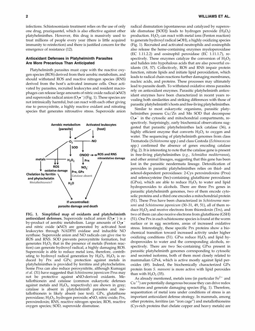
FIG. 1. Simplified map of oxidants and platyhelminthantioxidant defenses. Superoxide radical anion (O2� - ) is aby-product of aerobic metabolism. Large amounts of O2� -
and nitric oxide (�NO) are generated by activated hostleukocytes through NADPH oxidase and inducible NOsynthase. Superoxide anion and NO radicals can give rise toROS and RNS. SOD prevents peroxynitrite formation, butgenerates H2O2 that in the presence of metals (Fenton reac-tion) can generate hydroxyl radical, a highly damaging ROS.Superoxide is able to reduce metal ions, therefore, contrib-uting to hydroxyl radical generation by H2O2. H2O2 is re-duced by Prx and GPx; protection against metals inplatyhelminthes is provided by ferritins and phytochelatins.Some Prxs can also reduce peroxynitrite, although Kumagaiet al. (31) have suggested that Schistosoma japonicum Prxs maynot be protective against �NO-derived oxidants. Me-tallothionein and catalase (common eukaryotic defensesagainst metals and H2O2, respectively) are shown in gray;catalase is absent in platyhelminth parasites and me-tallothionein is likely absent (see text). GPx, glutathioneperoxidase; H2O2, hydrogen peroxide; �NO, nitric oxide; Prx,peroxiredoxin; RNS, reactive nitrogen species; ROS, reactiveoxygen species; SOD, superoxide dismutase.
2 WILLIAMS ET AL.
the key players in maintaining intracellular iron and copperions tightly bound and redox inactive, helping to suppresstheir pro-oxidant effects. Platyhelminth parasites have genesencoding ferritins (16, 28), but metallothioneins have not beendescribed (and their identification by similarity is not trivial).Interestingly, it has recently been proposed that copper andheavy metal sequestration in platyhelminth parasites proba-bly relies on phytochelatins, Cys-rich oligopeptides with thebasic structure of (c-GluCys)nGly, with n = 2–11 (43). Phy-tochelatins are synthesized from GSH by phytochelatin syn-thase; this enzyme recently described in S. mansoni is absent inhumans, and therefore it is a potential new target enzyme fordrug development (43). This finding also highlights the keyrole of thiol-dependent antioxidant defenses in these organ-isms. Figure 2 compares the antioxidant enzymes and pro-teins present in free-living- and parasitic platyhelminthes andin mammalian genomes.
Thioredoxin Glutathione Reductase Is the SoleEnzyme That Supports Both Trx and GSH Pathwaysin Platyhelminth Parasites
In most organisms, including the mammalian hosts ofplatyhelminthes, the Trx and GSH pathways play a key role ina variety of cellular processes, such as DNA synthesis, defenseagainst oxidative stress, detoxification, protein folding, andrepair (Fig. 3A). Both systems operate through redox cascadesthat involve transfer of reducing equivalents from NADPH totargets through reversible dithiol-disulfide reactions.
The Trx system comprises the flavoenzyme thioredoxinreductase (TR) and Trx, a powerful disulfide reductase with acatalytic dithiol/disulfide. Trx provides reducing equivalentsto a variety of enzymes containing redox active cysteine pairsas part of their catalytic cycle, including ribonucleotide re-ductase, an essential catalyst for the synthesis of deox-ynucleotides; antioxidant enzymes such as Prxs; and repairenzymes such as methionine sulfoxide reductases (Msrs, seebelow). Trx is also directly involved in blocking oxidativestress as a generic protein disulfide reductase. In addition, Trx
exerts redox control of regulatory proteins involved in signaltransduction and gene transcription (24). The reduction of thedisulfide of Trx redox-active site by NADPH is catalyzed byTR. Animal TRs belong to class I pyridine-nucleotide disulfideoxidoreductases. They contain an N-terminal CX4C motif atthe active site in the FAD-binding domain and a carboxy-terminal redox active center that mediates electron transferfrom the CX4C redox active site to TR substrates. The carboxy-terminal center usually contains redox active Cys and Secresidues, within the conserved motif GCUG (U denotes Sec).In addition to Trx, TR can reduce other substrates includ-ing low-molecular weight antioxidants such as the oxi-dized forms of lipoamide, lipoic acid, and ascorbic acid(dehydroascorbate). It has been suggested that most of theNADPH-dependent lipoamide and lipoic acid dehydroge-nase activities in mammalian cells should be attributable toTR (7), although the physiological relevance of lipoic acid asantioxidant has been questioned due to the low concentrationof its free lipoic acid.
The GSH system consists of glutathione reductase (GR),GSH, and glutaredoxin (Grx). As with TR, GR is a class Ipyridine-nucleotide disulfide oxidoreductase that contains aCX4C motif at the N-terminal FAD-binding domain, but lacksthe C-terminal redox center present in TR and thioredoxinglutathione reductase (TGR). GR transfers the reducingequivalents of NADPH exclusively to the dimeric oxidizedform of GSH (oxidized glutathione [GSSG]). GSH constitutesthe most abundant thiol-containing compound in cells(ranging from 0.5 to 10 mM), being the major non-proteinthiol-based redox buffer. GSH acts as a general antioxidantmolecule within the cell, provides electrons to GPx, and re-cycles Grx to its reduced state. In addition, GSH serves a de-toxifying role for hydrophobic electrophiles (includingproducts of lipid peroxidation and xenobiotics) and methyl-glyoxal (a side product of glycolysis and other metabolicpathways) in reactions catalyzed by GSH S-transferases andthe glyoxalase system, respectively. Grxs are small thiol-dis-ulfide oxidoreductases that belong to the Trx superfamily,have a similar redox active site, and transfer electrons to their
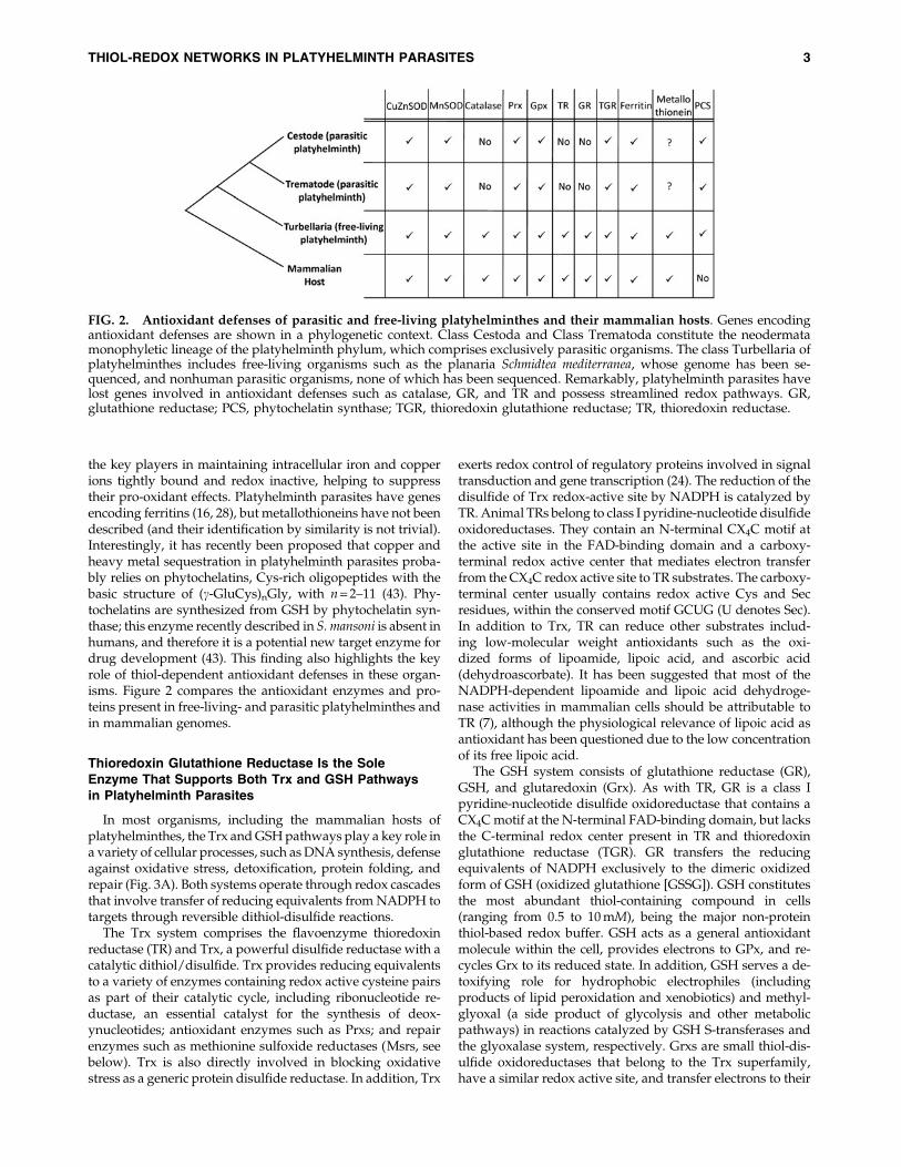
FIG. 2. Antioxidant defenses of parasitic and free-living platyhelminthes and their mammalian hosts. Genes encodingantioxidant defenses are shown in a phylogenetic context. Class Cestoda and Class Trematoda constitute the neodermatamonophyletic lineage of the platyhelminth phylum, which comprises exclusively parasitic organisms. The class Turbellaria ofplatyhelminthes includes free-living organisms such as the planaria Schmidtea mediterranea, whose genome has been se-quenced, and nonhuman parasitic organisms, none of which has been sequenced. Remarkably, platyhelminth parasites havelost genes involved in antioxidant defenses such as catalase, GR, and TR and possess streamlined redox pathways. GR,glutathione reductase; PCS, phytochelatin synthase; TGR, thioredoxin glutathione reductase; TR, thioredoxin reductase.
THIOL-REDOX NETWORKS IN PLATYHELMINTH PARASITES 3
substrates and substrate reductases such as protein-GSHmixed disulfides and ribonucleotide reductase. A particulartype of Grxs, the monothiol group, plays an important role inFe/S cluster assembly and mobilization in the mitochondria.
Typically, Trx and GSH systems are present in both thecytosol and the mitochondria. In mammals three TR isoen-zymes are present. TR1 (also known as TrxR1 and Txnrd1)and TR3 (also known as TrxR2 and Txnrd2), encoded bydifferent genes, function in the cytosol and the mitochondria,respectively. A third gene encodes a TGR (also known as TR2,TrxR3, and Txnrd3) a versatile enzyme whose expression islargely restricted to testis (54). TGR also belongs to class Ipyridine-nucleotide disulfide oxidoreductases and consists ofa natural fusion of conventional TR domains to an N-terminalGrx domain (56). This enzyme contains a CX4C motif proxi-mal to the FAD-binding domain, a carboxy-terminal GCUGredox active center and an additional redox center at the N-terminus extension in a Grx-like domain containing a redox-active CX2C or CX2S motif (56). GR exists as a distinct geneand is alternatively spliced to produce both cytoplasmic andmitochondrial variants (29). GSH and isozymes of Trx andGrx are present in both cytosolic and mitochondrial com-partments. Figure 3B depicts a scheme of primary sequenceand redox centers of TR, GR, TGR, Trx, and Grx.
In contrast to their hosts, parasitic platyhelminthes lackconventional TR and GR (1, 3, 21, 44). Instead, they rely ex-clusively on a linked Trx-GSH system in which TGR providesreducing equivalents to both pathways (Fig. 3A). The earlybiochemical studies were later supported by the genomic
information of platyhelminth parasites. In these organisms,cytosolic and mitochondrial TGR variants are derived from asingle TGR gene and have identical amino acid sequences (1).Nevertheless, the cytosolic isoform of T. crassiceps TGR showeda higher sensitivity to calcium than the mitochondrial variant,suggesting that these isoforms may be conditioned by theirenvironments (e.g., by post-translational modifications) (20). Itis interesting to note that the genome of S. mediterranea encodesTGR and conventional GR and TR, suggesting that GR and TRgenes were lost in the neodermata lineage (39) (Fig. 2).
Structural and Biochemical Studies Have RevealedDetails of TGR, a Complex Selenoenzyme
TGR, like GR and TR, is a homodimer with monomersoriented in an inverted manner forming a twisted ‘‘W’’ shape(5). The current model for the reaction mechanism of TGR,based on biochemical and structural studies, indicates thatelectrons flow from NADPH to FAD, to the CX4C redoxcenter, and then to the C-terminal GCUG redox center ofthe second subunit, as it is the case in mammalian TRs. Thereduced GCUG redox center can shuttle electrons to the CX2Credox center of oxidized Trx (TR activity) or to the CX2C redoxcenter of the Grx domain of the first subunit, which then canreduce GSSG (GR activity; Fig. 4) (4, 5, 8, 26). The proposedelectron pathway implies that the Sec-containing redox centeris within a flexible ‘‘whip’’ at the carboxy terminus of TGR;consistent with this view, the C-terminal tail of TGR has notbeen visualized in the available structures of TGR (4, 6).
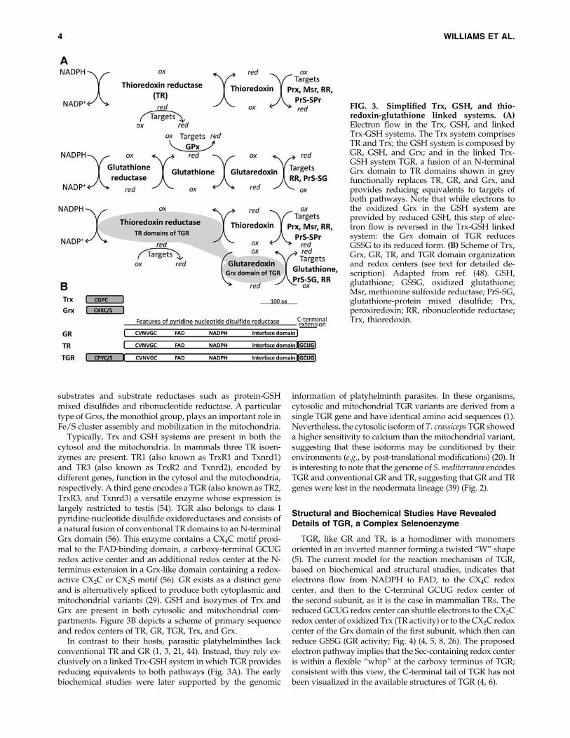
FIG. 3. Simplified Trx, GSH, and thio-redoxin-glutathione linked systems. (A)Electron flow in the Trx, GSH, and linkedTrx-GSH systems. The Trx system comprisesTR and Trx; the GSH system is composed byGR, GSH, and Grx; and in the linked Trx-GSH system TGR, a fusion of an N-terminalGrx domain to TR domains shown in greyfunctionally replaces TR, GR, and Grx, andprovides reducing equivalents to targets ofboth pathways. Note that while electrons tothe oxidized Grx in the GSH system areprovided by reduced GSH, this step of elec-tron flow is reversed in the Trx-GSH linkedsystem: the Grx domain of TGR reducesGSSG to its reduced form. (B) Scheme of Trx,Grx, GR, TR, and TGR domain organizationand redox centers (see text for detailed de-scription). Adapted from ref. (48). GSH,glutathione; GSSG, oxidized glutathione;Msr, methionine sulfoxide reductase; PrS-SG,glutathione-protein mixed disulfide; Prx,peroxiredoxin; RR, ribonucleotide reductase;Trx, thioredoxin.
4 WILLIAMS ET AL.

It has been recently shown that, in addition to having TRand GR activities, TGR can also reduce glutathionylatedpeptides (8). The reversible protein S-thiolation by GSH isemerging as a new mechanism involved in redox control ofprotein function and in the protection of key Cys residuesfrom over-oxidation (15). It is thought that glutathionylationoccurs spontaneously, while deglutathionylation is achievedby thiol-disulfide oxidoreductases, mainly by Grxs. Deglu-tathionylation by Grxs involves a glutathionylated interme-diate generated by the nucleophilic attack of the N-terminalCys of the CX2C active site of the Grx domain, that is thenreduced by GSH (17). Recent studies have shown that inplatyhelminth TGRs, the Grx domain can deglutathionylatesubstrates receiving electrons not only from GSH, but alsofrom the TR domains (1, 8, 26). This dual electron donorspecificity has only been observed for Grxs in a few cases (e.g.,human Grx2) (27). In TGR, the efficiency of this process wouldbe optimized since the electron transfer from TR to Grx occursintramolecularly, avoiding the need for diffusion. Throughthis particular array deglutathionylation may occur under a
broader range of conditions (e.g., when reduction of the Grxdomain by GSH might be thermodynamically unfavorablebecause of a low [GSH]/[GSSG] ratio).
Recently, the role of the CX2C active site of the Grx domainand of the Sec residue of TGR in GR and deglutathionylaseactivities has been examined in recombinant E. granulosus andS. mansoni TGRs (EgTGR and SmTGR, respectively) (8, 26).The N-terminal nucleophilic Cys residue of the Grx domain isessential for both functions; Cys to Ser/Ala mutants havemarginal activity. Therefore, for both activities, the mecha-nism of catalysis involves a glutathionylated intermediatewith the N-terminal Cys of the Grx domain. Cys to Ser mu-tants in the carboxy-terminal Cys residue of EgTGR andSmTGR are fully active in GSSG reduction, while a Cys to Alamutant in SmTGR has significantly lower activity. These re-sults suggest that the C-terminal Cys residue of the Grx do-main may stabilize the thiolate of the N-terminal Cys and thatresolution can be achieved without the formation of the dis-ulfide intermediate within the CX2C motif. These observa-tions, together with the absence of GR activity of the Sec (thepenultimate residue) to stop mutants of EgTGR and SmTGRand the marginal activity of the Sec to Cys mutants, indicatethat the Sec-containing redox center of the TR module canresolve the glutathionylated intermediate (Fig. 5, route A) (8,26). Studies on the deglutathionylase activity have revealedthat alternative routes for the resolution of the glutathiony-lated intermediate may be operative (26). Deglutathionylationhas been studied by different methods for EgTGR andSmTGR. The deglutathionylase activity of EgTGR was mea-sured using a glutathionylated peptide and NADPH as sub-strates (8), while the activity of SmTGR has been evaluatedusing the classical hydroxyethyl disulfide (HED) assay (23),which includes GSH in the reaction mix (Fig. 6 shows ascheme of deglutathionylase activity assays). Similar to theGR activity, the Cys to Ser mutant in the C-terminal Cysresidue of the Grx domain redox center of EgTGR had fulldeglutathionylase activity on a glutathionylated peptidewhile the Sec to Cys mutant is virtually inactive. Using theHED assay, both the Sec to Cys mutant and the Cys to Sermutant in the C-terminal Cys of the Grx domain of SmTGRhad markedly decreased activities, indicating that this Cyswould participate in the resolution of the mechanism (Fig. 5,route B) (26). Taken together, these results indicate that Sec isvital to the GCUG shuttling electrons to the Grx active site; asfor the GR activity, the glutathionylated intermediate can beresolved by the Sec-containing redox center, either directly orindirectly (Fig. 5, routes A and B, respectively). The fact that inSmTGR 40% of the activity was preserved in the Sec to Cysmutant, indicates that the Grx domain can function uncoupledfrom the TR module when GSH is present in the reaction mix(Fig. 5, route C) (26). Thus, resolution of the glutathionylatedintermediate could take place by three different routes, andsome of the differences observed between EgTGR and SmTGRmay be inherent to the enzymes or, more likely, due to theexperimental conditions of the different assays (e.g., presence orabsence of GSH).
Hysteresis Is a Conspicuous Featureof Platyhelminth TGRs
Another piece of the puzzle, to understand how the Grxdomain of platyhelminth TGRs functions and the role of the
FIG. 4. Model of the proposed reaction mechanism ofTGR. The active form of TGR is a dimer of identical sub-units. In the upper part, the flow of reducing equivalents inthe TR module of TGR is shown. In the lower part, reductionof the GCUG redox center produces a structural change inthe flexible carboxy-terminal arm allowing it to be reposi-tioned to either react with the glutaredoxin domain activesite (CX2C) or directly with oxidized Trx (Trx-S2). The re-duced glutaredoxin domain can then interact with its sub-strates, glutathione disulfide (GSSG) or glutathionylatedproteins (PrS-SG). Adapted from ref. (41).
THIOL-REDOX NETWORKS IN PLATYHELMINTH PARASITES 5
C-terminal Cys, arises from the hysteretic behavior of the GRactivity, first described for T. crassiceps TGR (44) and thenobserved in all platyhelminth TGRs characterized so far (9, 21,26). The GR activity of these TGRs, but not their TR activity, istemporarily inhibited at high [GSSG], exhibiting a lag beforeachieving full activity (Fig. 7). The higher the [GSH]/[GSSG]ratio the lower is the lag time. The lag is abolished not only byGSH, but also by Trx and dithiothreitol (DTT) (9, 44); andthere is evidence that GSH and Trx decrease hysteresiswithout the need of preincubation (9). Further, the hystereticbehavior persists after GSSG removal by size exclusionchromatography of a TGR sample treated with GSSG atconditions under which there is hysteresis (9). Taken together,these observations indicate that in all likelihood the phe-nomenon involves thiol-disulfide exchange reactions (9, 44)(Fig. 7). Mutants in the C-terminal Cys of the Grx domain ofEgTGR and SmTGR abolish most of GR hysteresis, implicat-ing this Cys residue in the observed behavior (8, 26). This
suggests that at high GSSG concentrations a disulfide inter-mediate involving C-terminal Cys residue would be rapidlyformed, but slowly resolved, resulting in decreased enzymeactivity. The identity of this alternative disulfide intermediate(i.e., which is the pairing Cys residue of the C-terminal Cysand how its formation would be favored at high GSSG con-centrations remains to be elucidated. If the hysteretic behaviorobserved for this enzyme in vitro occurs under physiologicalconditions, the Grx domain-dependent functions of TGRwould be inhibited under certain circumstances, while Trxreduction is preserved. Further, while the functions of the Grx
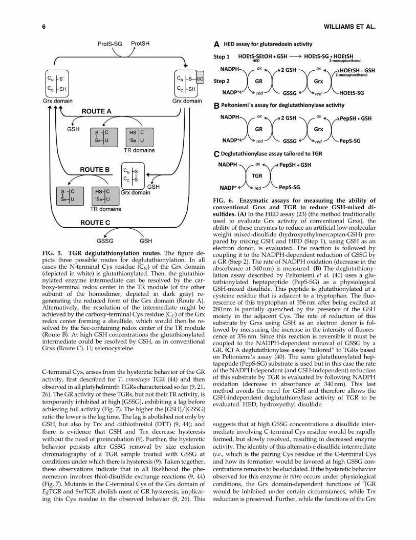
FIG. 5. TGR deglutathionylation routes. The figure de-picts three possible routes for deglutathionylation. In allcases the N-terminal Cys residue (CN) of the Grx domain(depicted in white) is glutathionylated. Then, the glutathio-nylated enzyme intermediate can be resolved by the car-boxy-terminal redox center in the TR module (of the othersubunit of the homodimer, depicted in dark gray) re-generating the reduced form of the Grx domain (Route A).Alternatively, the resolution of the intermediate might beachieved by the carboxy-terminal Cys residue (CC) of the Grxredox center forming a disulfide, which would then be re-solved by the Sec-containing redox center of the TR module(Route B). At high GSH concentrations the glutathionylatedintermediate could be resolved by GSH, as in conventionalGrxs (Route C). U; selenocysteine.
FIG. 6. Enzymatic assays for measuring the ability ofconventional Grxs and TGR to reduce GSH-mixed di-sulfides. (A) In the HED assay (23) (the method traditionallyused to evaluate Grx activity of conventional Grxs), theability of these enzymes to reduce an artificial low-molecularweight mixed-disulfide (hydroxyethylmercaptan-GSH) pre-pared by mixing GSH and HED (Step 1), using GSH as anelectron donor, is evaluated. The reaction is followed bycoupling it to the NADPH-dependent reduction of GSSG bya GR (Step 2). The rate of NADPH oxidation (decrease in theabsorbance at 340 nm) is measured. (B) The deglutathiony-lation assay described by Peltoniemi et al. (40) uses a glu-tathionylated heptapeptide (PepS-SG) as a physiologicalGSH-mixed disulfide. This peptide is glutathionylated at acysteine residue that is adjacent to a tryptophan. The fluo-rescence of this tryptophan at 356 nm after being excited at280 nm is partially quenched by the presence of the GSHmoiety in the adjacent Cys. The rate of reduction of thissubstrate by Grxs using GSH as an electron donor is fol-lowed by measuring the increase in the intensity of fluores-cence at 356 nm. Since this reaction is reversible it must becoupled to the NADPH-dependent removal of GSSG by aGR. (C) A deglutathionylase assay ‘‘tailored’’ to TGRs basedon Peltoniemi’s assay (40). The same glutathionylated hep-tapeptide (PepS-SG) substrate is used but in this case the rateof the NADPH-dependent (and GSH-independent) reductionof this substrate by TGR is evaluated by following NADPHoxidation (decrease in absorbance at 340 nm). This lastmethod avoids the need for GSH and therefore allows theGSH-independent deglutathionylase activity of TGR to beevaluated. HED, hydroxyethyl disulfide.
6 WILLIAMS ET AL.
domain are inhibited TGR can support Trx-based GSSG re-duction and deglutathionylation through alternative pathwaysthat involve its TR module (8). Therefore, if hysteresis occursin vivo, this inhibition would be transient: Trxs can reduceGSSG, protein disulfides, and protein–GSH mixed disulfides,and consequently relieve hysteresis. If hysteresis would have aphysiological function, it might serve a role in enzyme regula-tion or signaling, or it might have a protective role for the activeCys residue from irreversible oxidation.
Expanding the Thiol-Dependent Redox Network:More Players Than Anticipated
As already mentioned, a first line of antioxidant defenses inplatyhelminth parasites relies on the thiol- and selenol-dependent peroxidases: Prxs and GPxs. Msrs are a second lineof defense against oxidative stress. Msrs are selenol- and thiol-dependent repair enzymes that reduce methionine sulfoxide(Met-SO) back to methionine and receive electrons from Trxs.Oxidation produces the S and R sulfoxide diastereomers ofmethionine, which are reduced by two distinct stereospecificMsrs: MsrA and MsrB that reduce Met-(S)-SO and Met-(R)-SO, respectively (33). In S. mansoni, two forms of MsrB wererecently cloned and characterized (38). They are able to reduceMet-SO to Met, and were found to be expressed in all stages ofthe parasite’s life cycle. The search for MsrA gene in theS. mansoni genome and transcriptomic data did not reveal anyhomolog. In contrast, Echinococcus spp. genomes contain onecopy of each MsrA and MsrB genes (our unpublished obser-vations). The absence of MsrA gene in Schistosoma is intrigu-ing, and it suggests an incomplete genome sequence or anovel mechanism to repair Met-(S)-SO. In contrast to mam-
malian MsrB proteins, none of the platyhelminth MsrB pro-teins contain Sec.
The sequencing of parasitic platyhelminth genomes hasprovided a view of the complexity of the redox network de-pendent on TGR. So far, cytosolic and mitochondrial Trxshave been characterized in platyhelminth parasites (2, 10, 13,34). Data mining of the Echinococcus spp. genome revealed theexistence of five additional cytosolic monodomain Trxs,which are actively expressed (our unpublished observations).One of them possesses an atypical CPHS active site, found inhuman Trx3, which participates in disulfide isomerization inmammalian testis and can accept electrons from GSH or fromTR. Interestingly, the Grx domain of mammalian TGR, whichis also expressed in testis and participates in disulfide isom-erization contains a CPHS active site too. The data mining ofE. granulosus genome also revealed the existence of Grx di-versity. We identified two Grxs with CGFS active site andtwo monodomain cytosolic Grxs with CPYC redox active site,and all four show evidence of expression (our unpublishedinformation). Mitochondrial Grxs with CGFS active sites havebeen shown to participate in Fe/S cluster biogenesis andmobilization; they are dimeric and ligate a [2Fe-2S] cluster bythe active site Cys of two Grx monomers and two GSH mol-ecules (46). Grxs with CPYC active site have been shown tocatalyze the GSH-dependent reduction of protein disulfidesand GSH-protein mixed disulfides (17), and some of theseGrxs have also been recently shown to bind Fe/S clusters (11).Despite these general considerations, the specific functionsand targets of these newly identified Trxs and Grxs remain tobe explored.
Finally, platyhelminth parasite genomes encode a Sec-containing selenoprotein W (SelW) (41, 47). The members ofthis class of selenoproteins have a redoxin domain related toTrx domain and have been shown to be GSH-dependent an-tioxidant proteins in vivo, although their precise function isnot known. Transcriptomic surveys indicate that this gene ishighly expressed.
TGR Inhibition Has Led to the Identificationof Promising Drug Leads for Platyhelminth Infections
Given that both TGR (32) and Prx (49) had been shown tobe essential S. mansoni proteins, the redox pathway,NADPH/TGR/GSH/Prx2/H2O2, was reconstitutedand miniaturized and used in a quantitative high throughputscreen of 70,000 compounds (52). The screen identified severalpotent TGR inhibitors (but no Prx inhibitors), including ox-adiazole 2-oxides (also known as furoxans), phosphinic am-ides, and isoxazolones (Fig. 8). It was subsequently shownthat the furoxan 4-phenyl-1,2,5-oxadiazole-3-carbonitrile-2-oxide (Fx) was (i) active against adult ex vivo S. mansoni,S. japonicum, and S. haematobium; (ii) active against all devel-opmental stages of ex vivo S. mansoni (skin, lung, liver, andadult); (iii) able to cure mice infected with S. mansoni at theskin, liver, and adult stages; and (iv) significantly reducepathology (50). Fx proved to have a unique mechanism ofaction: �NO donation through TGR activity was followed byTGR inhibition and worm death (50). Although �NO donationwas not essential for Fx activity, it significantly increased itspotency. The reaction of TGR with Fx was more efficient atgenerating �NO and Fx killed worms more rapidly and atlower concentrations than other oxadiazole 2-oxides tested
0 2 4 6 8 10
0.0
0.1
0.2
0.3
0.4
0.5
0.6
0.7A
340
time (min)
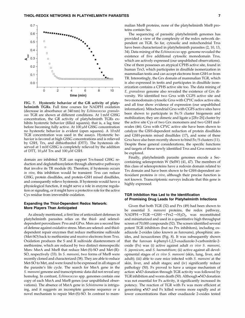
FIG. 7. Hysteretic behavior of the GR activity of platy-helminth TGRs. Full time courses for NADPH oxidation(decrease in absorbance at 340 nm) by Echinococcus granulo-sus TGR are shown at different conditions. At 1 mM GSSGconcentration, the GR activity of platyhelminth TGRs ex-hibits hysteretic behavior (filled squares); that is, a lag timebefore becoming fully active. At 100 lM GSSG concentrationno hysteretic behavior is evident (open squares). A 10 nMTGR concentration was used in the assays. Hysteretic be-havior is favored at high GSSG concentrations and is relievedby GSH, Trx, and dithiothreitol (DTT). The hysteresis ob-served at 1 mM GSSG is completely relieved by the additionof DTT, 10 lM Trx and 100 lM GSH.
THIOL-REDOX NETWORKS IN PLATYHELMINTH PARASITES 7
(50). Further, pretreating worms with an �NO scavenger re-duced the worm-killing activity of Fx to that seen with lessreactive oxadiazole 2-oxides (50). Recent evidence indicatesthat TGR can also be targeted in S. japonicum (22, 53). Theseresults underscore the importance of TGR in trematode redoxbiology and its usefulness as a drug target. Structure-activityrelationship studies on a wide range of Fx-related moleculesdetermined the importance of the 2-oxide, a strong electron-withdrawing group at position 3 and less importance on the
nature of the substitution at position 4 (42). Fx and its analogsshow differential activity between SmTGR and mammalianTR and GR enzymes (42). As indicated, the mechanism ofaction of Fx involves release of �NO; this is followed by the S-nitrosylation and inactivation of TGR (42). Importantly, it hasbeen recently shown that Fx inhibits other trematode andcestode TGRs and leads to parasite death (45), indicating thatFx can be used to target both flukes and tapeworms. Thesefindings reinforce the concept that interference with parasite
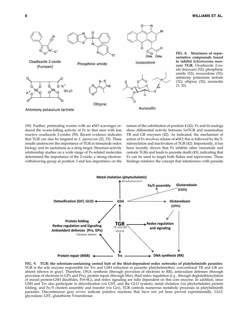
FIG. 8. Structures of repre-sentative compounds foundto inhibit Schistosoma man-soni TGR. Oxadiazole 2-ox-ide (furoxan) (52); phosphinicamide (52); isoxazolone (52);antimony potassium tartrate(32); oltipraz (32); auranofin(3, 32).
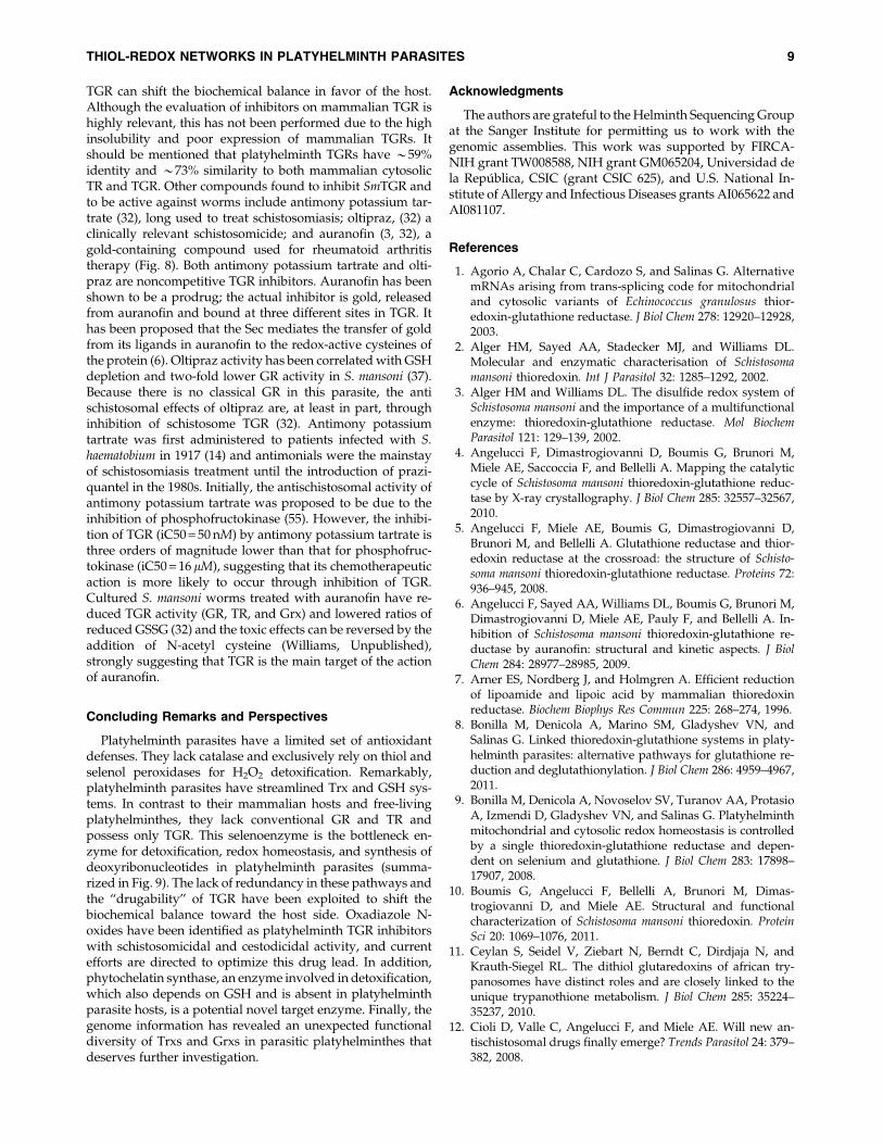
FIG. 9. TGR: the selenium-containing central hub of the thiol-dependent redox networks of platyhelminth parasites.TGR is the sole enzyme responsible for Trx and GSH reduction in parasitic platyhelminthes: conventional TR and GR areabsent (shown in gray). Therefore, DNA synthesis (through provision of electrons to RR), antioxidant defenses (throughprovision of electrons to GPx and Prx), protein repair (through Msr), thiol redox regulation (e.g., through deglutathionylationof mixed protein-GSH disulfides, PrS-SG), and redox signaling are fully dependent on this core enzyme. In addition, sinceGSH and Trx also participate in detoxification (via GST, and the GLO system), metal chelation (via phytochelatin) proteinfolding, and Fe/S clusters assembly and transfer (via Grx), TGR controls numerous metabolic processes in platyhelminthparasites. Discontinuous gray arrows indicate putative reactions that have not yet been proved experimentally. GLO,glyoxalase; GST, glutathione S-transferase.
8 WILLIAMS ET AL.
TGR can shift the biochemical balance in favor of the host.Although the evaluation of inhibitors on mammalian TGR ishighly relevant, this has not been performed due to the highinsolubility and poor expression of mammalian TGRs. Itshould be mentioned that platyhelminth TGRs have *59%identity and *73% similarity to both mammalian cytosolicTR and TGR. Other compounds found to inhibit SmTGR andto be active against worms include antimony potassium tar-trate (32), long used to treat schistosomiasis; oltipraz, (32) aclinically relevant schistosomicide; and auranofin (3, 32), agold-containing compound used for rheumatoid arthritistherapy (Fig. 8). Both antimony potassium tartrate and olti-praz are noncompetitive TGR inhibitors. Auranofin has beenshown to be a prodrug; the actual inhibitor is gold, releasedfrom auranofin and bound at three different sites in TGR. Ithas been proposed that the Sec mediates the transfer of goldfrom its ligands in auranofin to the redox-active cysteines ofthe protein (6). Oltipraz activity has been correlated with GSHdepletion and two-fold lower GR activity in S. mansoni (37).Because there is no classical GR in this parasite, the antischistosomal effects of oltipraz are, at least in part, throughinhibition of schistosome TGR (32). Antimony potassiumtartrate was first administered to patients infected with S.haematobium in 1917 (14) and antimonials were the mainstayof schistosomiasis treatment until the introduction of prazi-quantel in the 1980s. Initially, the antischistosomal activity ofantimony potassium tartrate was proposed to be due to theinhibition of phosphofructokinase (55). However, the inhibi-tion of TGR (iC50 = 50 nM) by antimony potassium tartrate isthree orders of magnitude lower than that for phosphofruc-tokinase (iC50 = 16 lM), suggesting that its chemotherapeuticaction is more likely to occur through inhibition of TGR.Cultured S. mansoni worms treated with auranofin have re-duced TGR activity (GR, TR, and Grx) and lowered ratios ofreduced GSSG (32) and the toxic effects can be reversed by theaddition of N-acetyl cysteine (Williams, Unpublished),strongly suggesting that TGR is the main target of the actionof auranofin.
Concluding Remarks and Perspectives
Platyhelminth parasites have a limited set of antioxidantdefenses. They lack catalase and exclusively rely on thiol andselenol peroxidases for H2O2 detoxification. Remarkably,platyhelminth parasites have streamlined Trx and GSH sys-tems. In contrast to their mammalian hosts and free-livingplatyhelminthes, they lack conventional GR and TR andpossess only TGR. This selenoenzyme is the bottleneck en-zyme for detoxification, redox homeostasis, and synthesis ofdeoxyribonucleotides in platyhelminth parasites (summa-rized in Fig. 9). The lack of redundancy in these pathways andthe ‘‘drugability’’ of TGR have been exploited to shift thebiochemical balance toward the host side. Oxadiazole N-oxides have been identified as platyhelminth TGR inhibitorswith schistosomicidal and cestodicidal activity, and currentefforts are directed to optimize this drug lead. In addition,phytochelatin synthase, an enzyme involved in detoxification,which also depends on GSH and is absent in platyhelminthparasite hosts, is a potential novel target enzyme. Finally, thegenome information has revealed an unexpected functionaldiversity of Trxs and Grxs in parasitic platyhelminthes thatdeserves further investigation.
Acknowledgments
The authors are grateful to the Helminth Sequencing Groupat the Sanger Institute for permitting us to work with thegenomic assemblies. This work was supported by FIRCA-NIH grant TW008588, NIH grant GM065204, Universidad dela Republica, CSIC (grant CSIC 625), and U.S. National In-stitute of Allergy and Infectious Diseases grants AI065622 andAI081107.
References
1. Agorio A, Chalar C, Cardozo S, and Salinas G. AlternativemRNAs arising from trans-splicing code for mitochondrialand cytosolic variants of Echinococcus granulosus thior-edoxin-glutathione reductase. J Biol Chem 278: 12920–12928,2003.
2. Alger HM, Sayed AA, Stadecker MJ, and Williams DL.Molecular and enzymatic characterisation of Schistosomamansoni thioredoxin. Int J Parasitol 32: 1285–1292, 2002.
3. Alger HM and Williams DL. The disulfide redox system ofSchistosoma mansoni and the importance of a multifunctionalenzyme: thioredoxin-glutathione reductase. Mol BiochemParasitol 121: 129–139, 2002.
4. Angelucci F, Dimastrogiovanni D, Boumis G, Brunori M,Miele AE, Saccoccia F, and Bellelli A. Mapping the catalyticcycle of Schistosoma mansoni thioredoxin-glutathione reduc-tase by X-ray crystallography. J Biol Chem 285: 32557–32567,2010.
5. Angelucci F, Miele AE, Boumis G, Dimastrogiovanni D,Brunori M, and Bellelli A. Glutathione reductase and thior-edoxin reductase at the crossroad: the structure of Schisto-soma mansoni thioredoxin-glutathione reductase. Proteins 72:936–945, 2008.
6. Angelucci F, Sayed AA, Williams DL, Boumis G, Brunori M,Dimastrogiovanni D, Miele AE, Pauly F, and Bellelli A. In-hibition of Schistosoma mansoni thioredoxin-glutathione re-ductase by auranofin: structural and kinetic aspects. J BiolChem 284: 28977–28985, 2009.
7. Arner ES, Nordberg J, and Holmgren A. Efficient reductionof lipoamide and lipoic acid by mammalian thioredoxinreductase. Biochem Biophys Res Commun 225: 268–274, 1996.
8. Bonilla M, Denicola A, Marino SM, Gladyshev VN, andSalinas G. Linked thioredoxin-glutathione systems in platy-helminth parasites: alternative pathways for glutathione re-duction and deglutathionylation. J Biol Chem 286: 4959–4967,2011.
9. Bonilla M, Denicola A, Novoselov SV, Turanov AA, ProtasioA, Izmendi D, Gladyshev VN, and Salinas G. Platyhelminthmitochondrial and cytosolic redox homeostasis is controlledby a single thioredoxin-glutathione reductase and depen-dent on selenium and glutathione. J Biol Chem 283: 17898–17907, 2008.
10. Boumis G, Angelucci F, Bellelli A, Brunori M, Dimas-trogiovanni D, and Miele AE. Structural and functionalcharacterization of Schistosoma mansoni thioredoxin. ProteinSci 20: 1069–1076, 2011.
11. Ceylan S, Seidel V, Ziebart N, Berndt C, Dirdjaja N, andKrauth-Siegel RL. The dithiol glutaredoxins of african try-panosomes have distinct roles and are closely linked to theunique trypanothione metabolism. J Biol Chem 285: 35224–35237, 2010.
12. Cioli D, Valle C, Angelucci F, and Miele AE. Will new an-tischistosomal drugs finally emerge? Trends Parasitol 24: 379–382, 2008.
THIOL-REDOX NETWORKS IN PLATYHELMINTH PARASITES 9
13. Chalar C, Martinez C, Agorio A, Salinas G, Soto J, andEhrlich R. Molecular cloning and characterization of athioredoxin gene from Echinococcus granulosus. Biochem Bio-phys Res Commun 262: 302–307, 1999.
14. Christopherson JB. The successful use of antimony in bil-harziosis administered as intravenous injections of anti-monium tartrate, tartar emetic. Lancet ii:325–327, 1918.
15. Dalle-Donne I, Rossi R, Colombo G, Giustarini D, and Mil-zani A. Protein S-glutathionylation: a regulatory device frombacteria to humans. Trends Biochem Sci 34: 85–96, 2009.
16. Dietzel J, Hirzmann J, Preis D, Symmons P, and Kunz W.Ferritins of Schistosoma mansoni: sequence comparison andexpression in female and male worms. Mol Biochem Parasitol50: 245–254, 1992.
17. Fernandes AP and Holmgren A. Glutaredoxins: glutathione-dependent redox enzymes with functions far beyond asimple thioredoxin backup system. Antioxid Redox Signal 6:63–74, 2004.
18. Furtmuller PG, Obinger C, Hsuanyu Y, and Dunford HB.Mechanism of reaction of myeloperoxidase with hydrogenperoxide and chloride ion. Eur J Biochem 267: 5858–5864,2000.
19. Garcıa HH, Gonzalez AE, Evans CAW, and Gilman RH.Taenia solium cysticercosis. Lancet 362: 547–556, 2003.
20. Guevara-Flores A, Del Arenal IP, Mendoza-Hernandez G,Pardo JP, Flores-Herrera O, and Rendon JL. Mitochondrialthioredoxin-glutathione reductase from larval Taenia crassi-ceps (Cysticerci). J Parasitol Res 2010 [Epub ahead of print];DOI:10.1155/2010/719856.
21. Guevara-Flores A, Pardo JP, and Rendon JL. Hysteresis inthioredoxin-glutathione reductase (TGR) from the adultstage of the liver fluke Fasciola hepatica. Parasitol Int 60: 156–160, 2011.
22. Han Y, Zhang M, Hong Y, Zhu Z, Li D, Li X, Fu Z, and Lin J.Characterization of thioredoxin glutathione reductase inSchiotosoma japonicum. Parasitol Int 61: 475–480, 2012.
23. Holmgren A and Aslund F. Glutaredoxin. Methods Enzymol252: 283–292, 1995.
24. Holmgren A, Johansson C, Berndt C, Lonn ME, HudemannC, and Lillig CH. Thiol redox control via thioredoxin andglutaredoxin systems. Biochem Soc Trans 33: 1375–1377,2005.
25. Hotez PJ, Brindley PJ, Bethony JM, King CH, Pearce EJ, andJacobson J. Helminth infections: the great neglected tropicaldiseases. J Clin Invest 118: 1311–1321, 2008.
26. Huang H-H, Day L, Cass CL, Ballou DP, Williams CH, andWilliams DL. Investigations of the catalytic mechanism ofthioredoxin glutathione reductase from Schistosoma mansoni.Biochemistry 50: 5870–5882, 2011.
27. Johansson C, Lillig CH, and Holmgren A. Human mito-chondrial glutaredoxin reduces S-glutathionylated proteinswith high affinity accepting electrons from either glutathi-one or thioredoxin reductase. J Biol Chem 279: 7537–7543,2004.
28. Jones MK, McManus DP, Sivadorai P, Glanfield A, MoertelL, Belli SI, and Gobert GN. Tracking the fate of iron in earlydevelopment of human blood flukes. Int J Biochem Cell Biol39: 1646–1658, 2007.
29. Kelner MJ and Montoya MA. Structural organization of thehuman glutathione reductase gene: determination of correctcDNA sequence and identification of a mitochondrial leadersequence. Biochem Biophys Res Commun 269: 366–368, 2000.
30. Kumagai T, Osada Y, and Kanazawa T. 2-Cys peroxir-edoxins from Schistosoma japonicum: the expression profile
and localization in the life cycle. Mol Biochem Parasitol 149:135–143, 2006.
31. Kumagai T, Osada Y, Ohta N, and Kanazawa T. Peroxir-edoxin-1 from Schistosoma japonicum functions as a scaven-ger against hydrogen peroxide but not nitric oxide. MolBiochem Parasitol 164: 26–31, 2009.
32. Kuntz AN, Davioud-Charvet E, Sayed AA, Califf LL,Dessolin J, Arner ESJ, and Williams DL. Thioredoxin-glutathione reductase from Schistosoma mansoni: an essen-tial parasite enzyme and a key drug target. PLoS Med 4:e206, 2007.
33. Lee BC, Dikiy A, Kim HY, and Gladyshev VN. Functionsand evolution of selenoprotein methionine sulfoxide reduc-tases. Biochim Biophys Acta 1790: 1471–1477, 2009.
34. Line K, Isupov MN, Garcia-Rodriguez E, Maggioli G, ParraF, and Littlechild JA. The Fasciola hepatica thioredoxin: highresolution structure reveals two oxidation states. Mol Bio-chem Parasitol 161: 44–48, 2008.
35. Maiorino M, Roche C, Kiess M, Koenig K, Gawlik D,Matthes M, Naldini E, Pierce R, and Flohe L. A selenium-containing phospholipid-hydroperoxide glutathione peroxi-dase in Schistosoma mansoni. Eur J Biochem 238: 838–844,1996.
36. Mkoji GM, Smith JM, and Prichard RK. Antioxidant systemsin Schistosoma mansoni: evidence for their role in protectionof the adult worms against oxidant killing. Int J Parasitol 18:667–673, 1988.
37. Moreau N, Martens T, Fleury MB, and Leroy JP. Metabolismof oltipraz and glutathione reductase inhibition. BiochemPharmacol 40: 1299–1305, 1990.
38. Oke TT, Moskovitz J, and Williams DL. Characterization ofthe methionine sulfoxide reductases of Schistosoma mansoni. JParasitol 95: 1421–1428, 2009.
39. Otero L, Bonilla M, Protasio A, Fernandez C, Gladyshev V,and Salinas G. Thioredoxin and glutathione systems differ inparasitic and free-living platyhelminths. BMC Genomics 11:237, 2010.
40. Peltoniemi MJ, Karala A-R, Jurvansuu JK, Kinnula VL, andRuddock LW. Insights into deglutathionylation reactions. JBiol Chem 281: 33107–33114, 2006.
41. Prast-Nielsen S, Huang H-H, and Williams DL. Thioredoxin-glutathione reductase: its role in redox biology and potentialas a target for drugs against neglected diseases. BiochimBiophys Acta 1810: 1262–1271, 2011.
42. Rai G, Sayed AA, Lea WA, Luecke HF, Chakrapani H, Prast-Nielsen S, Jadhav A, Leister W, Shen M, Inglese J, Austin CP,Keefer L, Arner ES, Simeonov A, Maloney DJ, Williams DL,and Thomas CJ. Structure mechanism insights and the role ofnitric oxide donation guide the development of oxadiazole-2-oxides as therapeutic agents against schistosomiasis. J MedChem 52: 6474–6483, 2009.
43. Ray D and Williams DL. Characterization of the phytoche-latin synthase of Schistosoma mansoni. PLoS Negl Trop Dis 5:e1168, 2011.
44. Rendon JL, del Arenal IP, Guevara-Flores A, Uribe A,Plancarte A, and Mendoza-Hernandez G. Purification,characterization and kinetic properties of the multifunctionalthioredoxin-glutathione reductase from Taenia crassicepsmetacestode (cysticerci). Mol Biochem Parasitol 133: 61–69,2004.
45. Ross F, Hernandez P, Porcal W, Lopez GV, Cerecetto H,Gonzalez M, Basika T, Carmona C, Flo M, Maggioli G,Bonilla M, Gladyshev VN, Boiani M, and Salinas G. Iden-tification of thioredoxin glutathione reductase inhibitors
10 WILLIAMS ET AL.
that kill cestode and trematode parasites. PLoS One 7:e35033, 2012.
46. Rouhier N, Couturier J, Johnson MK, and Jacquot J-P. Glu-taredoxins: roles in iron homeostasis. Trends Biochem Sci 35:43–52, 2010.
47. Salinas G, Bonilla M, Otero L, Lobanov AV, and GladyshevVN. selenoproteins in parasites. In: Selenium: its MolecularBiology and Role in Human Health, edited by Hatfield DL,Berry MJ, and Gladyshev VN. New York: Springer-VerlagInc., 2011.
48. Salinas G, Selkirk ME, Chalar C, Maizels RM, and FernandezC. Linked thioredoxin-glutathione systems in platyhel-minths. Trends Parasitol 20: 340–346, 2004.
49. Sayed AA, Cook SK, and Williams DL. Redox balancemechanisms in Schistosoma mansoni rely on peroxiredoxinsand albumin and implicate peroxiredoxins as novel drugtargets. J Biol Chem 281: 17001–17010, 2006.
50. Sayed AA, Simeonov A, Thomas CJ, Inglese J, Austin CP,and Williams DL. Identification of oxadiazoles as new drugleads for the control of schistosomiasis. Nat Med 14: 407–412,2008.
51. Sayed AA and Williams DL. Biochemical characterization of2-Cys peroxiredoxins from Schistosoma mansoni. J Biol Chem279: 26159–26166, 2004.
52. Simeonov A, Jadhav A, Sayed AA, Wang Y, Nelson ME,Thomas CJ, Inglese J, Williams DL, and Austin CP. Quan-titative high-throughput screen identifies inhibitors of theSchistosoma mansoni redox cascade. PLoS Negl Trop Dis 2:e127, 2008.
53. Song L, Li J, Xie S, Qian C, Wang J, Zhang W, Yin X, Hua Z,and Yu C. Thioredoxin glutathione reductase as a noveldrug target: evidence from Schistosoma japonicum. PLoS One7: e31456, 2012.
54. Su D, Novoselov SV, Sun Q-A, Moustafa ME, Zhou Y, OkoR, Hatfield DL, and Gladyshev VN. Mammalian seleno-protein thioredoxin-glutathione reductase: roles in disulfidebond formation and sperm maturation. J Biol Chem 280:26491–26498, 2005.
55. Su JG, Mansour JM, and Mansour TE. Purification, kineticsand inhibition by antimonials of recombinant phosphofruc-tokinase from Schistosoma mansoni. Mol Biochem Parasitol 81:171–178, 1996.
56. Sun Q-A, Kirnarsky L, Sherman S, and Gladyshev VN. Se-lenoprotein oxidoreductase with specificity for thioredoxinand glutathione systems. Proc Natl Acad Sci U S A 98: 3673–3678, 2001.
57. van Dalen CJ and Kettle AJ. Substrates and products of eo-sinophil peroxidase. Biochem J 358: 233–239, 2001.
Address correspondence to:Prof. Gustavo Salinas
Catedra de Inmunologıa, Facultad de QuımicaInstituto de Higiene
Universidad de la RepublicaAvda. A. Navarro 3051, Piso 2
Montevideo 11600Uruguay
E-mail: [email protected]
Dr. David L. WilliamsDepartment of Immunology-Microbiology
Rush University Medical CenterChicago, IL 60612
E-mail: [email protected]
Date of first submission to ARS Central, April 23, 2012; date offinal revised submission, August 16, 2012; date of acceptance,August 19, 2012.
Abbreviations Used
DTT¼dithiothreitolGLO¼ glyoxalaseGPx¼ glutathione peroxidaseGR¼ glutathione reductase
GSH¼ glutathioneGSSG¼ oxidized glutathione
GST¼ glutathione S-transferaseH2O2¼hydrogen peroxideHED¼hydroxyethyl disulfideiC50¼half maximal inhibitory concentration
Met-SO¼methionine sulfoxideMsr¼methionine sulfoxide reductaseNO¼nitric oxidePCS¼phytochelatin synthase
PrS-SG¼ glutathione-protein mixed disulfidePrx¼peroxiredoxin
RNS¼ reactive nitrogen speciesROS¼ reactive oxygen species
RR¼ ribonucleotidereductaseSec¼ selenocysteine
SOD¼ superoxide dismutaseTGR¼ thioredoxin glutathione reductase
TR¼ thioredoxin reductaseTrx¼ thioredoxin
THIOL-REDOX NETWORKS IN PLATYHELMINTH PARASITES 11