thermo scientific pierce cell lysis technical handbook · pdf filethermo scientific pierce...
TRANSCRIPT
To order, call 800-874-3723 or 815-968-0747. Outside the United States, contact your local branch office or distributor.
Thermo Scientific Pierce Cell Lysis Technical HandbookFeaturing Cell Lysis Reagents and Detergents
Version 2
Table of Contents
Thermo Scientific Pierce Cell Lysis Reagents Selection Guide 1
Introduction to Protein Extraction 2-5
Cell Lysis Methods 2-5
Introduction to Thermo Scientific Cell Lysis Solutions 6-15
B-PER® Bacterial Protein Extraction Reagents 7-9
I-PER® Insect Cell Protein Extraction Reagent 10
M-PER® Mammalian Protein Extraction Reagent 11
P-PER® Plant Protein Extraction Reagent 12-13
T-PER® Tissue Protein Extraction Reagents 14
Y-PER® Yeast Protein Extraction Reagents 15
Buffers 16
RIPA Buffer 16
Fusion Protein Purification 17-22
Fusion Protein Purification Kits 17-22
GST Orientation Kit 22
Mammalian and Yeast β-Gal Kits 23-24
Thermo Scientific Cell Fractionation Kits 25-37
Subcellular Fractionation Kit 25-26
Mem-PER® Eukaryotic Membrane 27-28 Protein Extraction Kit
Mitochondria Isolation Kits 29-31
NE-PER® Nuclear and Cytoplasmic Extraction Kit 32-34
Cell Surface Protein Isolation Kit 35
Organelle Enrichment Kits 36-37
DNA Extraction 38
Yeast DNA Extraction Kit 38
Thermo Scientific Lyse and Go™ PCR Reagent 38
Detergents 39-43
Introduction to Detergents 39-40
Properties of Common Detergents 40
Thermo Scientific Surfact-Amps and Surfact-Pak Detergents 41
Specialized Detergents 41-43
Detergent Removal 43
Protease Inhibitors 44-47
Halt™ Protease Inhibitor Single-Use Cocktails 44-45
Halt Phosphatase Inhibitor Cocktails 45
Halt Combined Cocktails 46
Protein Stabilizing Cocktail 47
PMSF 47
Soybean Trypsin Inhibitor 47
Protein Refolding 48-49
Pierce Protein Refolding Kit 48
Inclusion Body Solubilization Reagent 49
1 For more information, or to download product instructions, visit www.thermo.com/pierce
1. The detergent can be removed by dialysis 2. Immunoprecipitation 3. See Thermo Scientific Halt Protease and Phosphatase
Inhibitors on pages 44–46
4. Samples prepared in Mem-PER Reagent can be dialyzed if the buffer contains detergent (e.g., CHAPS), otherwise use Pierce SDS-PAGE Sample Prep Kit (Product # 89888)
5. Thermo Scientific Slide-A-Lyzer MINI Dialysis Units
6. Thermo Scientific 2-D Sample Prep for Nuclear Proteins (Product # 89863) and 2-D Sample Prep for Membrane Proteins (Product # 89864) were designed using our NE-PER and Mem-PER Reagents.
7. Need to lyse mitochondria first.
To order, call 800-874-3723 or 815-968-0747. Outside the United States, contact your local branch office or distributor.
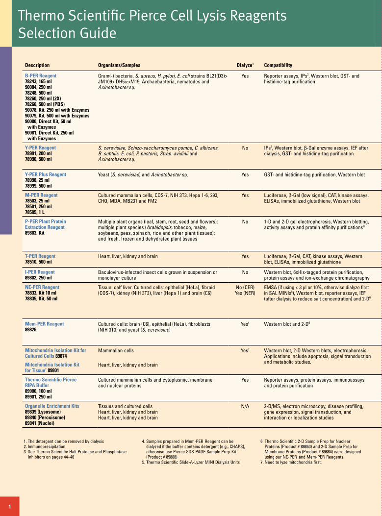
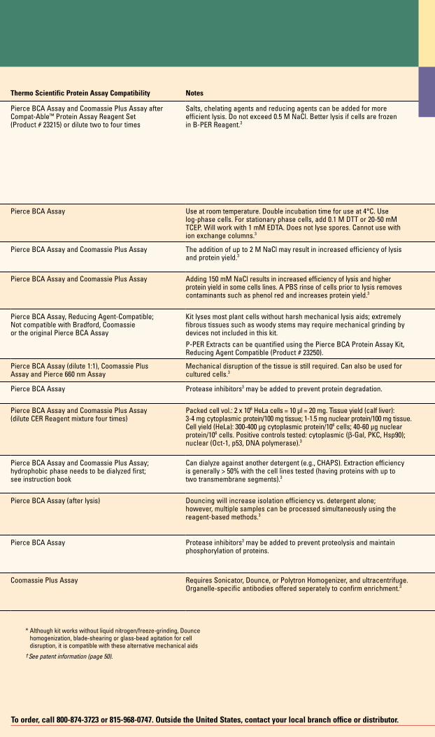
Description Organisms/Samples Dialyze1 Compatibility Thermo Scientific Protein Assay Compatibility Notes
B-PER Reagent78243, 165 ml90084, 250 ml78248, 500 ml78260, 250 ml (2X)78266, 500 ml (PBS)90078, Kit, 250 ml with Enzymes90079, Kit, 500 ml with Enzymes90080, Direct Kit, 50 ml with Enzymes90081, Direct Kit, 250 ml with Enzymes
Gram(-) bacteria, S. aureus, H. pylori, E. coli strains BL21(D3)> JM109> DH5α>M15, Archaebacteria, nematodes and Acinetobacter sp.
Yes Reporter assays, IPs2, Western blot, GST- and histidine-tag purification
Pierce BCA Assay and Coomassie Plus Assay after Compat-Able™ Protein Assay Reagent Set (Product # 23215) or dilute two to four times
Salts, chelating agents and reducing agents can be added for more efficient lysis. Do not exceed 0.5 M NaCl. Better lysis if cells are frozen in B-PER Reagent.3
Y-PER Reagent78991, 200 ml78990, 500 ml
S. cerevisiae, Schizo-saccharomyces pombe, C. albicans, B. subtilis, E. coli, P. pastoris, Strep. avidinii and Acinetobacter sp.
No IPs2, Western blot, β-Gal enzyme assays, IEF after dialysis, GST- and histidine-tag purification
Pierce BCA Assay Use at room temperature. Double incubation time for use at 4°C. Use log-phase cells. For stationary phase cells, add 0.1 M DTT or 20-50 mM TCEP. Will work with 1 mM EDTA. Does not lyse spores. Cannot use with ion exchange columns.3
Y-PER Plus Reagent78998, 25 ml78999, 500 ml
Yeast (S. cerevisiae) and Acinetobacter sp. Yes GST- and histidine-tag purification, Western blot Pierce BCA Assay and Coomassie Plus Assay The addition of up to 2 M NaCl may result in increased efficiency of lysis and protein yield.3
M-PER Reagent78503, 25 ml78501, 250 ml78505, 1 L
Cultured mammalian cells, COS-7, NIH 3T3, Hepa 1-6, 293, CHO, MDA, MB231 and FM2
Yes Luciferase, β-Gal (low signal), CAT, kinase assays, ELISAs, immobilized glutathione, Western blot
Pierce BCA Assay and Coomassie Plus Assay Adding 150 mM NaCl results in increased efficiency of lysis and higher protein yield in some cells lines. A PBS rinse of cells prior to lysis removes contaminants such as phenol red and increases protein yield.3
P-PER Plant Protein Extraction Reagent89803, Kit
Multiple plant organs (leaf, stem, root, seed and flowers); multiple plant species (Arabidopsis, tobacco, maize, soybeans, peas, spinach, rice and other plant tissues); and fresh, frozen and dehydrated plant tissues
No 1-D and 2-D gel electrophoresis, Western blotting, activity assays and protein affinity purifications*
Pierce BCA Assay, Reducing Agent-Compatible;Not compatible with Bradford, Coomassie or the original Pierce BCA Assay
Kit lyses most plant cells without harsh mechanical lysis aids; extremely fibrous tissues such as woody stems may require mechanical grinding by devices not included in this kit.
P-PER Extracts can be quantified using the Pierce BCA Protein Assay Kit, Reducing Agent Compatible (Product # 23250).
T-PER Reagent78510, 500 ml
Heart, liver, kidney and brain Yes Luciferase, β-Gal, CAT, kinase assays, Western blot, ELISAs, immobilized glutathione
Pierce BCA Assay (dilute 1:1), Coomassie Plus Assay and Pierce 660 nm Assay
Mechanical disruption of the tissue is still required. Can also be used for cultured cells.3
I-PER Reagent89802, 250 ml
Baculovirus-infected insect cells grown in suspension or monolayer culture
No Western blot, 6xHis-tagged protein purification, protein assays and ion-exchange chromatography
Pierce BCA Assay Protease inhibitors3 may be added to prevent protein degradation.
NE-PER Reagent78833, Kit 10 ml78835, Kit, 50 ml
Tissue: calf liver. Cultured cells: epithelial (HeLa), fibroid (COS-7), kidney (NIH 3T3), liver (Hepa 1) and brain (C6)
No (CER)Yes (NER)
EMSA (if using < 3 µl or 10%, otherwise dialyze first in SAL MINIs5), Western blot, reporter assays, IEF (after dialysis to reduce salt concentration) and 2-D6
Pierce BCA Assay and Coomassie Plus Assay (dilute CER Reagent mixture four times)
Packed cell vol.: 2 x 10⁶ HeLa cells = 10 µl = 20 mg. Tissue yield (calf liver): 3-4 mg cytoplasmic protein/100 mg tissue; 1-1.5 mg nuclear protein/100 mg tissue. Cell yield (HeLa): 300-400 µg cytoplasmic protein/106 cells; 40-60 µg nuclear protein/106 cells. Positive controls tested: cytoplasmic (β-Gal, PKC, Hsp90); nuclear (Oct-1, p53, DNA polymerase).3
Mem-PER Reagent89826
Cultured cells: brain (C6), epithelial (HeLa), fibroblasts (NIH 3T3) and yeast (S. cerevisiae)
Yes4 Western blot and 2-D6 Pierce BCA Assay and Coomassie Plus Assay; hydrophobic phase needs to be dialyzed first; see instruction book
Can dialyze against another detergent (e.g., CHAPS). Extraction efficiency is generally > 50% with the cell lines tested (having proteins with up to two transmembrane segments).3
Mitochondria Isolation Kit for Cultured Cells 89874
Mitochondria Isolation Kit for Tissue† 89801
Mammalian cells
Heart, liver, kidney and brain
Yes7 Western blot, 2-D Western blots, electrophoresis. Applications include apoptosis, signal transduction and metabolic studies.
Pierce BCA Assay (after lysis) Douncing will increase isolation efficiency vs. detergent alone; however, multiple samples can be processed simultaneously using the reagent-based methods.3
Thermo Scientific Pierce RIPA Buffer89900, 100 ml 89901, 250 ml
Cultured mammalian cells and cytoplasmic, membrane and nuclear proteins
Yes Reporter assays, protein assays, immunoassays and protein purification
Pierce BCA Assay Protease inhibitors3 may be added to prevent proteolysis and maintain phosphorylation of proteins.
Organelle Enrichment Kits89839 (Lysosome)89840 (Peroxisome)89841 (Nuclei)
Tissues and cultured cells Heart, liver, kidney and brain Heart, liver, kidney and brain
N/A 2-D/MS, electron microscopy, disease profiling, gene expression, signal transduction, and interaction or localization studies
Coomassie Plus Assay Requires Sonicator, Dounce, or Polytron Homogenizer, and ultracentrifuge. Organelle-specific antibodies offered seperately to confirm enrichment.3
Thermo Scientific Pierce Cell Lysis Reagents Selection Guide
1
* Although kit works without liquid nitrogen/freeze-grinding, Dounce homogenization, blade-shearing or glass-bead agitation for cell disruption, it is compatible with these alternative mechanical aids
† See patent information (page 50).
To order, call 800-874-3723 or 815-968-0747. Outside the United States, contact your local branch office or distributor.
Description Organisms/Samples Dialyze1 Compatibility Thermo Scientific Protein Assay Compatibility Notes
B-PER Reagent78243, 165 ml90084, 250 ml78248, 500 ml78260, 250 ml (2X)78266, 500 ml (PBS)90078, Kit, 250 ml with Enzymes90079, Kit, 500 ml with Enzymes90080, Direct Kit, 50 ml with Enzymes90081, Direct Kit, 250 ml with Enzymes
Gram(-) bacteria, S. aureus, H. pylori, E. coli strains BL21(D3)> JM109> DH5α>M15, Archaebacteria, nematodes and Acinetobacter sp.
Yes Reporter assays, IPs2, Western blot, GST- and histidine-tag purification
Pierce BCA Assay and Coomassie Plus Assay after Compat-Able™ Protein Assay Reagent Set (Product # 23215) or dilute two to four times
Salts, chelating agents and reducing agents can be added for more efficient lysis. Do not exceed 0.5 M NaCl. Better lysis if cells are frozen in B-PER Reagent.3
Y-PER Reagent78991, 200 ml78990, 500 ml
S. cerevisiae, Schizo-saccharomyces pombe, C. albicans, B. subtilis, E. coli, P. pastoris, Strep. avidinii and Acinetobacter sp.
No IPs2, Western blot, β-Gal enzyme assays, IEF after dialysis, GST- and histidine-tag purification
Pierce BCA Assay Use at room temperature. Double incubation time for use at 4°C. Use log-phase cells. For stationary phase cells, add 0.1 M DTT or 20-50 mM TCEP. Will work with 1 mM EDTA. Does not lyse spores. Cannot use with ion exchange columns.3
Y-PER Plus Reagent78998, 25 ml78999, 500 ml
Yeast (S. cerevisiae) and Acinetobacter sp. Yes GST- and histidine-tag purification, Western blot Pierce BCA Assay and Coomassie Plus Assay The addition of up to 2 M NaCl may result in increased efficiency of lysis and protein yield.3
M-PER Reagent78503, 25 ml78501, 250 ml78505, 1 L
Cultured mammalian cells, COS-7, NIH 3T3, Hepa 1-6, 293, CHO, MDA, MB231 and FM2
Yes Luciferase, β-Gal (low signal), CAT, kinase assays, ELISAs, immobilized glutathione, Western blot
Pierce BCA Assay and Coomassie Plus Assay Adding 150 mM NaCl results in increased efficiency of lysis and higher protein yield in some cells lines. A PBS rinse of cells prior to lysis removes contaminants such as phenol red and increases protein yield.3
P-PER Plant Protein Extraction Reagent89803, Kit
Multiple plant organs (leaf, stem, root, seed and flowers); multiple plant species (Arabidopsis, tobacco, maize, soybeans, peas, spinach, rice and other plant tissues); and fresh, frozen and dehydrated plant tissues
No 1-D and 2-D gel electrophoresis, Western blotting, activity assays and protein affinity purifications*
Pierce BCA Assay, Reducing Agent-Compatible;Not compatible with Bradford, Coomassie or the original Pierce BCA Assay
Kit lyses most plant cells without harsh mechanical lysis aids; extremely fibrous tissues such as woody stems may require mechanical grinding by devices not included in this kit.
P-PER Extracts can be quantified using the Pierce BCA Protein Assay Kit, Reducing Agent Compatible (Product # 23250).
T-PER Reagent78510, 500 ml
Heart, liver, kidney and brain Yes Luciferase, β-Gal, CAT, kinase assays, Western blot, ELISAs, immobilized glutathione
Pierce BCA Assay (dilute 1:1), Coomassie Plus Assay and Pierce 660 nm Assay
Mechanical disruption of the tissue is still required. Can also be used for cultured cells.3
I-PER Reagent89802, 250 ml
Baculovirus-infected insect cells grown in suspension or monolayer culture
No Western blot, 6xHis-tagged protein purification, protein assays and ion-exchange chromatography
Pierce BCA Assay Protease inhibitors3 may be added to prevent protein degradation.
NE-PER Reagent78833, Kit 10 ml78835, Kit, 50 ml
Tissue: calf liver. Cultured cells: epithelial (HeLa), fibroid (COS-7), kidney (NIH 3T3), liver (Hepa 1) and brain (C6)
No (CER)Yes (NER)
EMSA (if using < 3 µl or 10%, otherwise dialyze first in SAL MINIs5), Western blot, reporter assays, IEF (after dialysis to reduce salt concentration) and 2-D6
Pierce BCA Assay and Coomassie Plus Assay (dilute CER Reagent mixture four times)
Packed cell vol.: 2 x 10⁶ HeLa cells = 10 µl = 20 mg. Tissue yield (calf liver): 3-4 mg cytoplasmic protein/100 mg tissue; 1-1.5 mg nuclear protein/100 mg tissue. Cell yield (HeLa): 300-400 µg cytoplasmic protein/106 cells; 40-60 µg nuclear protein/106 cells. Positive controls tested: cytoplasmic (β-Gal, PKC, Hsp90); nuclear (Oct-1, p53, DNA polymerase).3
Mem-PER Reagent89826
Cultured cells: brain (C6), epithelial (HeLa), fibroblasts (NIH 3T3) and yeast (S. cerevisiae)
Yes4 Western blot and 2-D6 Pierce BCA Assay and Coomassie Plus Assay; hydrophobic phase needs to be dialyzed first; see instruction book
Can dialyze against another detergent (e.g., CHAPS). Extraction efficiency is generally > 50% with the cell lines tested (having proteins with up to two transmembrane segments).3
Mitochondria Isolation Kit for Cultured Cells 89874
Mitochondria Isolation Kit for Tissue† 89801
Mammalian cells
Heart, liver, kidney and brain
Yes7 Western blot, 2-D Western blots, electrophoresis. Applications include apoptosis, signal transduction and metabolic studies.
Pierce BCA Assay (after lysis) Douncing will increase isolation efficiency vs. detergent alone; however, multiple samples can be processed simultaneously using the reagent-based methods.3
Thermo Scientific Pierce RIPA Buffer89900, 100 ml 89901, 250 ml
Cultured mammalian cells and cytoplasmic, membrane and nuclear proteins
Yes Reporter assays, protein assays, immunoassays and protein purification
Pierce BCA Assay Protease inhibitors3 may be added to prevent proteolysis and maintain phosphorylation of proteins.
Organelle Enrichment Kits89839 (Lysosome)89840 (Peroxisome)89841 (Nuclei)
Tissues and cultured cells Heart, liver, kidney and brain Heart, liver, kidney and brain
N/A 2-D/MS, electron microscopy, disease profiling, gene expression, signal transduction, and interaction or localization studies
Coomassie Plus Assay Requires Sonicator, Dounce, or Polytron Homogenizer, and ultracentrifuge. Organelle-specific antibodies offered seperately to confirm enrichment.3
trim-fpo-don’t print solid red line
Introduction to Protein ExtractionProtein purification encompasses total protein extraction from
a sample (lysis), specific enrichment and/or isolation of a particular
protein of interest (affinity purification), and removal of interfering
or contaminating substances (sample preparation or clean-up).
Cell lysis is the first step in cell fractionation and protein purification
and, as such, opens the door to a myriad of biological studies. Many
techniques are available for the disruption of cells, including physical
and detergent-based methods. Historically, physical lysis has
been the method of choice for cell disruption; however, physical
methods often require expensive, cumbersome equipment and
involve protocols that can be difficult to repeat due to variability
in the apparatus (such as loose-fitting compared with tight-fitting
homogenization pestles). In recent years, detergent-based lysis
has become very popular due to ease of use, low cost and efficient
protocols. We offer several detergent-based lysis reagents for
preparing whole and fractionated cell lysates that are faster and
more convenient than traditional methods.
For more information, or to download product instructions, visit www.thermo.com/pierce2
fold-fpo-don’t print dashed red linetrim-fpo-don’t print solid red line
For more information, or to download product instructions, visit www.thermo.com/pierce
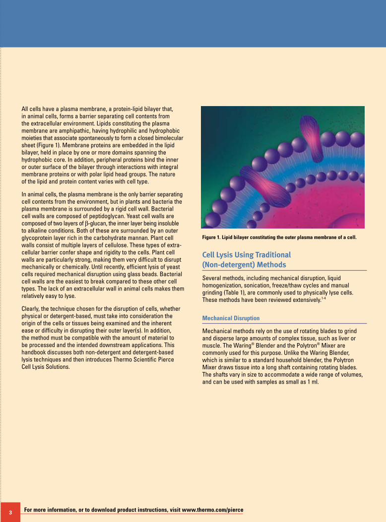
All cells have a plasma membrane, a protein-lipid bilayer that, in animal cells, forms a barrier separating cell contents from the extracellular environment. Lipids constituting the plasma membrane are amphipathic, having hydrophilic and hydrophobic moieties that associate spontaneously to form a closed bimolecular sheet (Figure 1). Membrane proteins are embedded in the lipid bilayer, held in place by one or more domains spanning the hydrophobic core. In addition, peripheral proteins bind the inner or outer surface of the bilayer through interactions with integral membrane proteins or with polar lipid head groups. The nature of the lipid and protein content varies with cell type.
In animal cells, the plasma membrane is the only barrier separating cell contents from the environment, but in plants and bacteria the plasma membrane is surrounded by a rigid cell wall. Bacterial cell walls are composed of peptidoglycan. Yeast cell walls are composed of two layers of β-glucan, the inner layer being insoluble to alkaline conditions. Both of these are surrounded by an outer glycoprotein layer rich in the carbohydrate mannan. Plant cell walls consist of multiple layers of cellulose. These types of extra-cellular barrier confer shape and rigidity to the cells. Plant cell walls are particularly strong, making them very difficult to disrupt mechanically or chemically. Until recently, efficient lysis of yeast cells required mechanical disruption using glass beads. Bacterial cell walls are the easiest to break compared to these other cell types. The lack of an extracellular wall in animal cells makes them relatively easy to lyse.
Clearly, the technique chosen for the disruption of cells, whether physical or detergent-based, must take into consideration the origin of the cells or tissues being examined and the inherent ease or difficulty in disrupting their outer layer(s). In addition, the method must be compatible with the amount of material to be processed and the intended downstream applications. This handbook discusses both non-detergent and detergent-based lysis techniques and then introduces Thermo Scientific Pierce Cell Lysis Solutions.
3
Figure 1. Lipid bilayer constituting the outer plasma membrane of a cell.
Cell Lysis Using Traditional (Non-detergent) Methods
Several methods, including mechanical disruption, liquid homogenization, sonication, freeze/thaw cycles and manual grinding (Table 1), are commonly used to physically lyse cells. These methods have been reviewed extensively.1-4
Mechanical Disruption
Mechanical methods rely on the use of rotating blades to grind and disperse large amounts of complex tissue, such as liver or muscle. The Waring® Blender and the Polytron® Mixer are commonly used for this purpose. Unlike the Waring Blender, which is similar to a standard household blender, the Polytron Mixer draws tissue into a long shaft containing rotating blades. The shafts vary in size to accommodate a wide range of volumes, and can be used with samples as small as 1 ml.
fold-fpo-don’t print dashed red line
To order, call 800-874-3723 or 815-968-0747. Outside the United States, contact your local branch office or distributor. 4
Liquid Homogenization
Liquid-based homogenization is the most widely used cell-disruption technique for small volumes and cultured cells. Cells are lysed by forcing the cell or tissue suspension through a narrow space, thereby shearing the cell membranes. Three different types of homogenizers are in common use. A Dounce homogenizer consists of a round glass pestle that is manually driven into a glass tube. A Potter-Elvehjem homogenizer consists of a manually or mechanically driven pestle coated with PTFE Material and shaped to fit a rounded or conical vessel. The number of strokes and the speed at which the strokes are administered influences the effectiveness of Dounce and Potter-Elvehjem homogenization methods. Both homogenizers can be obtained in a variety of sizes to accommodate a range of volumes. A French press consists of a piston that is used to apply high pressure to a sample volume of 40 to 250 ml, forcing it through a tiny hole in the press. Only two passes are required for efficient lysis due to the high pressures used with this process. The equipment is expensive, but the French press is often the method of choice for breaking bacterial cells mechanically.
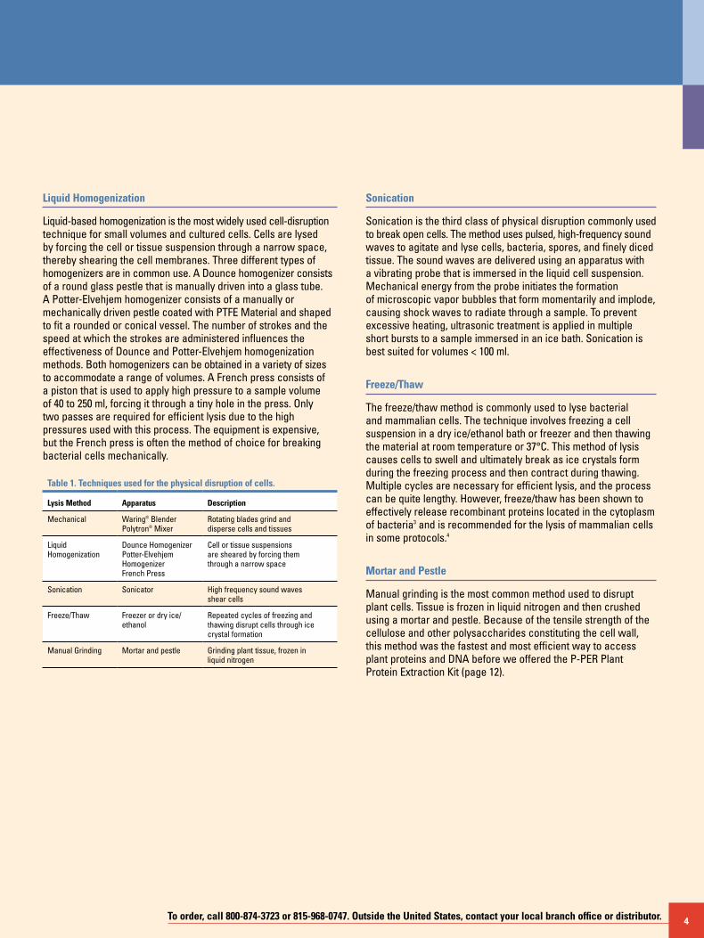
Table 1. Techniques used for the physical disruption of cells.
Lysis Method Apparatus Description
Mechanical Waring® Blender Polytron® Mixer
Rotating blades grind and disperse cells and tissues
Liquid Homogenization
Dounce Homogenizer Potter-Elvehjem Homogenizer French Press
Cell or tissue suspensions are sheared by forcing them through a narrow space
Sonication Sonicator High frequency sound waves shear cells
Freeze/Thaw Freezer or dry ice/ ethanol
Repeated cycles of freezing and thawing disrupt cells through ice crystal formation
Manual Grinding Mortar and pestle Grinding plant tissue, frozen in liquid nitrogen
Sonication
Sonication is the third class of physical disruption commonly used to break open cells. The method uses pulsed, high-frequency sound waves to agitate and lyse cells, bacteria, spores, and finely diced tissue. The sound waves are delivered using an apparatus with a vibrating probe that is immersed in the liquid cell suspension. Mechanical energy from the probe initiates the formation of microscopic vapor bubbles that form momentarily and implode, causing shock waves to radiate through a sample. To prevent excessive heating, ultrasonic treatment is applied in multiple short bursts to a sample immersed in an ice bath. Sonication is best suited for volumes < 100 ml.
Freeze/Thaw
The freeze/thaw method is commonly used to lyse bacterial and mammalian cells. The technique involves freezing a cell suspension in a dry ice/ethanol bath or freezer and then thawing the material at room temperature or 37°C. This method of lysis causes cells to swell and ultimately break as ice crystals form during the freezing process and then contract during thawing. Multiple cycles are necessary for efficient lysis, and the process can be quite lengthy. However, freeze/thaw has been shown to effectively release recombinant proteins located in the cytoplasm of bacteria3 and is recommended for the lysis of mammalian cells in some protocols.4
Mortar and Pestle
Manual grinding is the most common method used to disrupt plant cells. Tissue is frozen in liquid nitrogen and then crushed using a mortar and pestle. Because of the tensile strength of the cellulose and other polysaccharides constituting the cell wall, this method was the fastest and most efficient way to access plant proteins and DNA before we offered the P-PER Plant Protein Extraction Kit (page 12).
5 For more information, or to download product instructions, visit www.thermo.com/pierce
5 For more information, or to download product instructions, visit www.thermo.com/pierce
Additives/Facilitators
Various agents can aid the cell disruption process. Cells sus-pended in a hypotonic buffer swell and burst readily by physical shearing. Adding lysozyme (200 µg/ml) (Product # 89833; page 9) digests the polysaccharide component of yeast and bacterial cell walls. Alternatively, processing can be expedited by treating cells with glass beads to facilitate the crushing of cell walls, which is commonly used with yeast cells. Viscosity of a sample typically increases during lysis due to the release of nucleic acid material. Nucleases such as Thermo Scientific Pierce DNase (25-50 µg/ml) (Product # 89835; page 9) can be added to lysate along with RNase (50 µg/ml) to reduce this problem. Nuclease treatment is not required for sonicated material because sonica-tion shears chromosomes. Finally, proteolysis can be a problem whenever cells are manipulated; therefore, protease inhibitors (Halt Protease Inhibitors, Product #s 78425 and 78430; page 45) should be added to all samples undergoing lysis.
Cell Lysis Using Detergents
Detergent cell lysis is a milder and easier alternative to physical disruption of cell membranes, although it is often used in conjunction with homogenization and mechanical grinding with a Polytron Mixer.
Detergents break the lipid barrier surrounding cells by solubilizing proteins and disrupting lipid:lipid, protein:protein and protein:lipid interactions. Detergents, like lipids, self-associate and bind to hydrophobic surfaces. They are composed of a polar hydrophilic head group and a nonpolar hydrophobic tail and are categorized by the nature of the head group as either ionic (cationic or anionic), nonionic or zwitterionic. Their behavior depends on the properties of the head group and tail.
Unfortunately, there is no standard protocol available for selecting a detergent to use for membrane lysis. The ideal detergent will depend on the intended application. In general, nonionic and zwitterionic detergents are milder and less denaturing than ionic detergents and are used to solubilize membrane proteins when it is critical to maintain protein function and/or retain native protein:protein interactions for enzyme assays or immunoassays. CHAPS, a zwitterionic detergent, and the Triton® X Brand series of nonionic detergents are commonly used for these purposes. In contrast, ionic detergents are strong solubilizing agents and tend to denature proteins, thereby destroying protein activity and function. Studies assessing protein levels strictly through gel electrophoresis and Western blotting typically use SDS to fully denature protein samples by boiling. There are a few commonly used ionic detergents that are only mildly denaturing, including sodium cholate and sodium deoxycholate.
The choice of detergent for cell lysis also depends on sample type. Animal cells, bacteria and yeast all have differing requirements for optimal lysis due to the presence or absence of a cell wall. Because of the dense and complex nature of animal tissues, they require both detergent and mechanical lysis. In addition to the choice of detergent, other important considerations for optimal cell lysis include the buffer, pH, salt concentration and temperature. Consideration should be given to the compatibility of the chosen detergent with downstream applications. If the detergent used for lysis must be removed, then a dialyzable detergent should be selected.
References1. Evans, W.H. (1987). In J.B.C. Findlay and W.H. Evans (Eds.), Biological Membranes:
A Practical Approach. IRL Press, Oxford, England, p. 1-25.2. McNamee, M.G. (1989). Biotechniques 7, 466-475.3. Johnson, B.H. and Hecht, M.H. (1994). Bio/Technology 12, 1357-1360.4. Current Protocols in Molecular Biology (1995). John Wiley and Sons, Inc.
(supplement 29) pp. 9.7.1-9.7.2.
To order, call 800-874-3723 or 815-968-0747. Outside the United States, contact your local branch office or distributor. 6
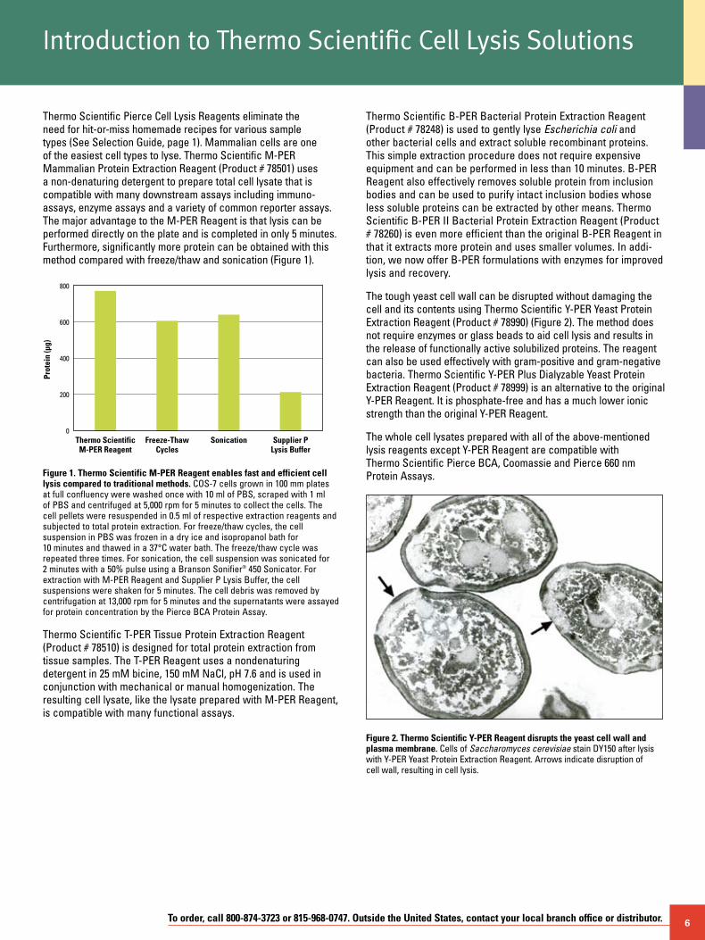
Thermo Scientific Pierce Cell Lysis Reagents eliminate the need for hit-or-miss homemade recipes for various sample types (See Selection Guide, page 1). Mammalian cells are one of the easiest cell types to lyse. Thermo Scientific M-PER Mammalian Protein Extraction Reagent (Product # 78501) uses a non-denaturing detergent to prepare total cell lysate that is compatible with many downstream assays including immuno- assays, enzyme assays and a variety of common reporter assays. The major advantage to the M-PER Reagent is that lysis can be performed directly on the plate and is completed in only 5 minutes. Furthermore, significantly more protein can be obtained with this method compared with freeze/thaw and sonication (Figure 1).
Introduction to Thermo Scientific Cell Lysis Solutions
Thermo Scientific B-PER Bacterial Protein Extraction Reagent (Product # 78248) is used to gently lyse Escherichia coli and other bacterial cells and extract soluble recombinant proteins. This simple extraction procedure does not require expensive equipment and can be performed in less than 10 minutes. B-PER Reagent also effectively removes soluble protein from inclusion bodies and can be used to purify intact inclusion bodies whose less soluble proteins can be extracted by other means. Thermo Scientific B-PER II Bacterial Protein Extraction Reagent (Product # 78260) is even more efficient than the original B-PER Reagent in that it extracts more protein and uses smaller volumes. In addi-tion, we now offer B-PER formulations with enzymes for improved lysis and recovery.
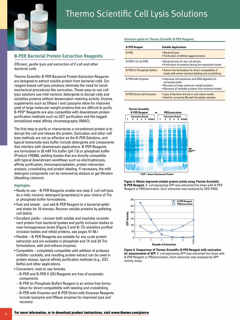
The tough yeast cell wall can be disrupted without damaging the cell and its contents using Thermo Scientific Y-PER Yeast Protein Extraction Reagent (Product # 78990) (Figure 2). The method does not require enzymes or glass beads to aid cell lysis and results in the release of functionally active solubilized proteins. The reagent can also be used effectively with gram-positive and gram-negative bacteria. Thermo Scientific Y-PER Plus Dialyzable Yeast Protein Extraction Reagent (Product # 78999) is an alternative to the original Y-PER Reagent. It is phosphate-free and has a much lower ionic strength than the original Y-PER Reagent.
The whole cell lysates prepared with all of the above-mentioned lysis reagents except Y-PER Reagent are compatible with Thermo Scientific Pierce BCA, Coomassie and Pierce 660 nm Protein Assays.Figure 1. Thermo Scientific M-PER Reagent enables fast and efficient cell
lysis compared to traditional methods. COS-7 cells grown in 100 mm plates at full confluency were washed once with 10 ml of PBS, scraped with 1 ml of PBS and centrifuged at 5,000 rpm for 5 minutes to collect the cells. The cell pellets were resuspended in 0.5 ml of respective extraction reagents and subjected to total protein extraction. For freeze/thaw cycles, the cell suspension in PBS was frozen in a dry ice and isopropanol bath for 10 minutes and thawed in a 37°C water bath. The freeze/thaw cycle was repeated three times. For sonication, the cell suspension was sonicated for 2 minutes with a 50% pulse using a Branson Sonifier® 450 Sonicator. For extraction with M-PER Reagent and Supplier P Lysis Buffer, the cell suspensions were shaken for 5 minutes. The cell debris was removed by centrifugation at 13,000 rpm for 5 minutes and the supernatants were assayed for protein concentration by the Pierce BCA Protein Assay.
Thermo Scientific T-PER Tissue Protein Extraction Reagent (Product # 78510) is designed for total protein extraction from tissue samples. The T-PER Reagent uses a nondenaturing detergent in 25 mM bicine, 150 mM NaCl, pH 7.6 and is used in conjunction with mechanical or manual homogenization. The resulting cell lysate, like the lysate prepared with M-PER Reagent, is compatible with many functional assays.
Figure 2. Thermo Scientific Y-PER Reagent disrupts the yeast cell wall and plasma membrane. Cells of Saccharomyces cerevisiae stain DY150 after lysis with Y-PER Yeast Protein Extraction Reagent. Arrows indicate disruption of cell wall, resulting in cell lysis.
Thermo ScientificM-PER Reagent
Freeze-ThawCycles
Prot
ein
(µg)
Sonication Supplier PLysis Buffer
800
600
400
200
0
7 For more information, or to download product instructions, visit www.thermo.com/pierceFor more information, or to download product instructions, visit www.thermo.com/pierce
B-PER Bacterial Protein Extraction Reagents
Efficient, gentle lysis and extraction of E.coli and other bacterial cells.
Thermo Scientific B-PER Bacterial Protein Extraction Reagents are designed to extract soluble protein from bacterial cells. Our reagent-based cell lysis solutions eliminate the need for harsh mechanical procedures like sonication. These easy-to-use cell lysis solutions use mild nonionic detergents to disrupt cells and solubilize proteins without denaturation retaining activity. Enzyme supplements such as DNase I and Lysozyme allow for improved yield of large molecular weight proteins that are difficult to purify. B-PER* Reagents are also compatible with downstream protein purification methods such as GST purification and His-tagged immobilized metal affinity chromatography (IMAC).
The first step to purify or characterize a recombinant protein is to disrupt the cell and release the protein. Sonication and other cell lysis methods are not as effective as the B-PER Solutions, and typical homemade lysis buffer include detergents and components that interfere with downstream applications. B-PER Reagents are formulated in 20 mM Tris buffer (pH 7.5) or phosphate buffer (Product #78266), yielding lysates that are directly compatible with typical downstream workflows such as electrophoresis, affinity purification, immunoprecipitation, protein interaction analysis, crosslinking and protein labeling. If necessary, the mild detergent components can be removed by dialysis or gel filtration (desalting columns).
Highlights:• Ready to use – B-PER Reagents enable one-step E. coli cell lysis
by a mild, nonionic detergent (proprietary) in your choice of Tris or phosphate buffer formulations.
• Fast and simple – just add B-PER Reagent to a bacterial pellet and shake for 10 minutes. Recover soluble proteins by pelleting cell debris.
• Excellent yields – recover both soluble and insoluble recombi-nant protein from bacterial lysates and purify inclusion bodies to near-homogeneous levels (Figure 3 and 4). (To solubilize purified inclusion bodies and refold proteins, see pages 47-48.)
• Flexible – B-PER Reagents are suitable for any scale protein extraction and are available in phosphate and 1X and 2X Tris formulations, with and without enzymes.
• Compatible – completely compatible with addition of protease inhibitor cocktails, and resulting protein extract can be used in protein assays, typical affinity purification methods (e.g., GST, 6xHis) and other applications.
• Convenient, read-to-use formats: – B-PER and B-PER II (2X) Reagents are free of enzymatic
components – B-PER (in Phosphate Buffer) Reagent is an amine-free formu-
lation for direct compatibility with labeling and crosslinking – B-PER with Enzymes and B-PER Direct with Enzymes Reagents
include lysozyme and DNase enzymes for improved lysis and recovery
Selection guide for Thermo Scientific B-PER Reagents.
B-PER Reagent Suitable Applications
B-PER • Bacterial lysis• Purification of affinity tagged proteins
B-PER II (2 x B-PER) • Bacterial lysis for low cell density• Purification of proteins having low expression levels
B-PER (in Phosphate Buffer) • Amine-free formulation for direct compatibility of lysate with amine-reactive labeling and crosslinking
B-PER with Enzymes • Improved cell membrane and DNA digestion for increased yields
• Recovery of large molecular weight proteins• Recovery of insoluble proteins from inclusion bodies
B-PER Direct with Enzymes • Lysis of bacteria directly in cell culture media• Ideal for screening 96-well microplate samples
Thermo Scientific B-PER ReagentExtraction Round
**GFP = Green Fluorescent Protein
Extraction Round
vs.
1 2 3 4 5 Pellet
PBS/sonication
1 2 3 4 5 Pellet
GFP**
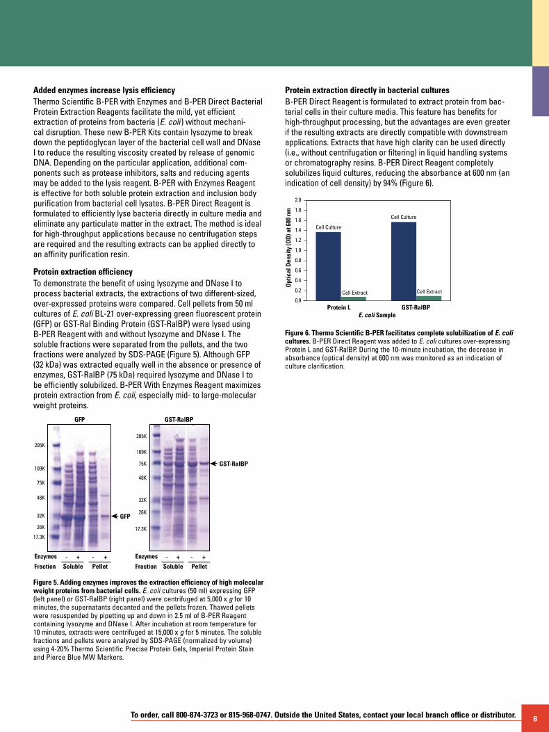
Figure 3. Obtain improved soluble protein yields using Thermo Scientific B-PER Reagent. E. coli expressing GFP was extracted five times with B-PER Reagent or PBS/sonication. Each extraction was analyzed by SDS-PAGE.
Rounds of Extraction
GFP
Act
ivity
31 2 5
Pelle
t4
300
250
200
150
100
50
0
B-PER ReagentPBS/sonication
Figure 4. Comparison of Thermo Scientific B-PER Reagent with sonication for measurement of GFP. E. coli expressing GFP was extracted five times with B-PER Reagent or PBS/sonication. Each extraction was analyzed by GFP activity assay.
Thermo Scientific Cell Lysis Solutions
8To order, call 800-874-3723 or 815-968-0747. Outside the United States, contact your local branch office or distributor.
Added enzymes increase lysis efficiencyThermo Scientific B-PER with Enzymes and B-PER Direct Bacterial Protein Extraction Reagents facilitate the mild, yet efficient extraction of proteins from bacteria (E. coli ) without mechani-cal disruption. These new B-PER Kits contain lysozyme to break down the peptidoglycan layer of the bacterial cell wall and DNase I to reduce the resulting viscosity created by release of genomic DNA. Depending on the particular application, additional com-ponents such as protease inhibitors, salts and reducing agents may be added to the lysis reagent. B-PER with Enzymes Reagent is effective for both soluble protein extraction and inclusion body purification from bacterial cell lysates. B-PER Direct Reagent is formulated to efficiently lyse bacteria directly in culture media and eliminate any particulate matter in the extract. The method is ideal for high-throughput applications because no centrifugation steps are required and the resulting extracts can be applied directly to an affinity purification resin.
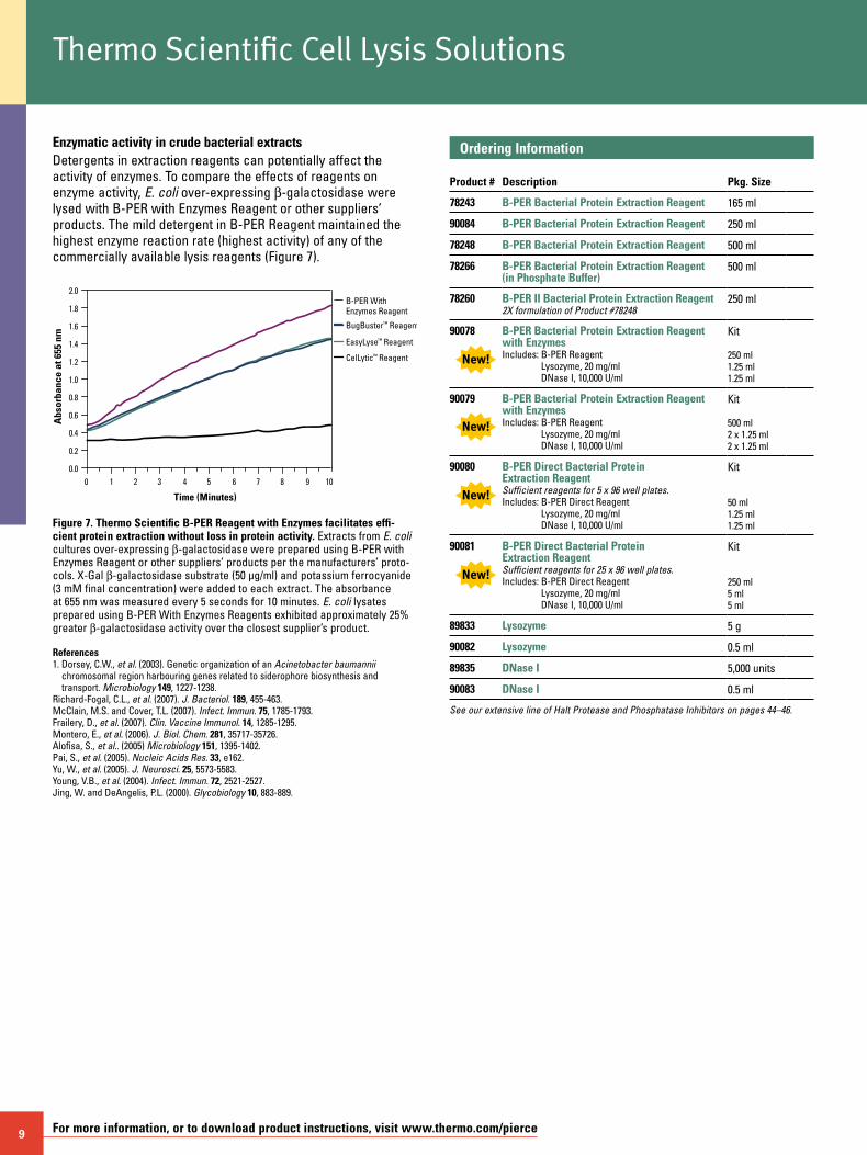
Protein extraction efficiencyTo demonstrate the benefit of using lysozyme and DNase I to process bacterial extracts, the extractions of two different-sized, over-expressed proteins were compared. Cell pellets from 50 ml cultures of E. coli BL-21 over-expressing green fluorescent protein (GFP) or GST-Ral Binding Protein (GST-RalBP) were lysed using B-PER Reagent with and without lysozyme and DNase I. The soluble fractions were separated from the pellets, and the two fractions were analyzed by SDS-PAGE (Figure 5). Although GFP (32 kDa) was extracted equally well in the absence or presence of enzymes, GST-RalBP (75 kDa) required lysozyme and DNase I to be efficiently solubilized. B-PER With Enzymes Reagent maximizes protein extraction from E. coli, especially mid- to large-molecular weight proteins.
GFP GST-RalBP
205K
109K
75K
48K
32K
26K
17.3K
205K
109K
75K
48K
32K
26K
17.3K
Enzymes - + - +Fraction Soluble Pellet
Enzymes - + - +Fraction
GFP
GST-RalBP
Soluble Pellet
Figure 5. Adding enzymes improves the extraction efficiency of high molecular weight proteins from bacterial cells. E. coli cultures (50 ml) expressing GFP (left panel) or GST-RalBP (right panel) were centrifuged at 5,000 x g for 10 minutes, the supernatants decanted and the pellets frozen. Thawed pellets were resuspended by pipetting up and down in 2.5 ml of B-PER Reagent containing lysozyme and DNase I. After incubation at room temperature for 10 minutes, extracts were centrifuged at 15,000 x g for 5 minutes. The soluble fractions and pellets were analyzed by SDS-PAGE (normalized by volume) using 4-20% Thermo Scientific Precise Protein Gels, Imperial Protein Stain and Pierce Blue MW Markers.
Protein extraction directly in bacterial culturesB-PER Direct Reagent is formulated to extract protein from bac-terial cells in their culture media. This feature has benefits for high-throughput processing, but the advantages are even greater if the resulting extracts are directly compatible with downstream applications. Extracts that have high clarity can be used directly (i.e., without centrifugation or filtering) in liquid handling systems or chromatography resins. B-PER Direct Reagent completely solubilizes liquid cultures, reducing the absorbance at 600 nm (an indication of cell density) by 94% (Figure 6).
Protein L GST-RalBP0.0
0.2
0.4
0.6
0.8
1.0
1.2
1.4
1.6
1.8
2.0
Cell Culture
Cell Extract Cell Extract
Cell Culture
E. coli Sample
Opt
ical
Den
sity
(OD
) at 6
00 n
m
Figure 6. Thermo Scientific B-PER facilitates complete solubilization of E. coli cultures. B-PER Direct Reagent was added to E. coli cultures over-expressing Protein L and GST-RalBP. During the 10-minute incubation, the decrease in absorbance (optical density) at 600 nm was monitored as an indication of culture clarification.
9 For more information, or to download product instructions, visit www.thermo.com/pierce
Enzymatic activity in crude bacterial extractsDetergents in extraction reagents can potentially affect the activity of enzymes. To compare the effects of reagents on enzyme activity, E. coli over-expressing b-galactosidase were lysed with B-PER with Enzymes Reagent or other suppliers’ products. The mild detergent in B-PER Reagent maintained the highest enzyme reaction rate (highest activity) of any of the commercially available lysis reagents (Figure 7).
0 1 2 3 4 5 6 7 8 9 100.0
0.2
0.4
0.6
0.8
1.0
1.2
1.4
1.6
1.8
2.0
BugBuster™ Reagent
EasyLyse™ Reagent
B-PER With Enzymes Reagent
CelLytic™ Reagent
Time (Minutes)
Abs
orba
nce
at 6
55 n
m
Figure 7. Thermo Scientific B-PER Reagent with Enzymes facilitates effi-cient protein extraction without loss in protein activity. Extracts from E. coli cultures over-expressing b-galactosidase were prepared using B-PER with Enzymes Reagent or other suppliers’ products per the manufacturers’ proto-cols. X-Gal b-galactosidase substrate (50 µg/ml) and potassium ferrocyanide (3 mM final concentration) were added to each extract. The absorbance at 655 nm was measured every 5 seconds for 10 minutes. E. coli lysates prepared using B-PER With Enzymes Reagents exhibited approximately 25% greater b-galactosidase activity over the closest supplier’s product.
References1. Dorsey, C.W., et al. (2003). Genetic organization of an Acinetobacter baumannii
chromosomal region harbouring genes related to siderophore biosynthesis and transport. Microbiology 149, 1227-1238.
Richard-Fogal, C.L., et al. (2007). J. Bacteriol. 189, 455-463.McClain, M.S. and Cover, T.L. (2007). Infect. Immun. 75, 1785-1793.Frailery, D., et al. (2007). Clin. Vaccine Immunol. 14, 1285-1295.Montero, E., et al. (2006). J. Biol. Chem. 281, 35717-35726.Alofisa, S., et al.. (2005) Microbiology 151, 1395-1402.Pai, S., et al. (2005). Nucleic Acids Res. 33, e162.Yu, W., et al. (2005). J. Neurosci. 25, 5573-5583.Young, V.B., et al. (2004). Infect. Immun. 72, 2521-2527.Jing, W. and DeAngelis, P.L. (2000). Glycobiology 10, 883-889.
Thermo Scientific Cell Lysis Solutions
Ordering Information Product #
Description
Pkg. Size
78243 B-PER Bacterial Protein Extraction Reagent 165 ml
90084 B-PER Bacterial Protein Extraction Reagent 250 ml
78248 B-PER Bacterial Protein Extraction Reagent 500 ml
78266 B-PER Bacterial Protein Extraction Reagent (in Phosphate Buffer)
500 ml
78260 B-PER II Bacterial Protein Extraction Reagent2X formulation of Product #78248
250 ml
90078 B-PER Bacterial Protein Extraction Reagent with EnzymesIncludes: B-PER Reagent
Lysozyme, 20 mg/ml DNase I, 10,000 U/ml
Kit 250 ml 1.25 ml 1.25 ml
90079 B-PER Bacterial Protein Extraction Reagent with EnzymesIncludes: B-PER Reagent
Lysozyme, 20 mg/ml DNase I, 10,000 U/ml
Kit 500 ml 2 x 1.25 ml 2 x 1.25 ml
90080 B-PER Direct Bacterial Protein Extraction ReagentSufficient reagents for 5 x 96 well plates.Includes: B-PER Direct Reagent
Lysozyme, 20 mg/ml DNase I, 10,000 U/ml
Kit 50 ml 1.25 ml 1.25 ml
90081 B-PER Direct Bacterial Protein Extraction ReagentSufficient reagents for 25 x 96 well plates.Includes: B-PER Direct Reagent
Lysozyme, 20 mg/ml DNase I, 10,000 U/ml
Kit 250 ml 5 ml 5 ml
89833 Lysozyme 5 g
90082 Lysozyme 0.5 ml
89835 DNase I 5,000 units
90083 DNase I 0.5 ml
See our extensive line of Halt Protease and Phosphatase Inhibitors on pages 44–46.
To order, call 800-874-3723 or 815-968-0747. Outside the United States, contact your local branch office or distributor. 10
I-PER Insect Cell Protein Extraction Reagent
An efficient, gentle reagent that provides maximum extraction of soluble proteins.
Highlights:• Gentle extraction – optimized, mild nonionic detergent provides maximum extraction of soluble proteins from insect cells • Effective – provides better protein extraction than sonication• Compatible – downstream compatibility with Western blotting, 6xHis-tagged protein purification, protein assays and ion-exchange chromatography • Flexible – useful for protein extraction from suspended or
adherent cultured insect cells
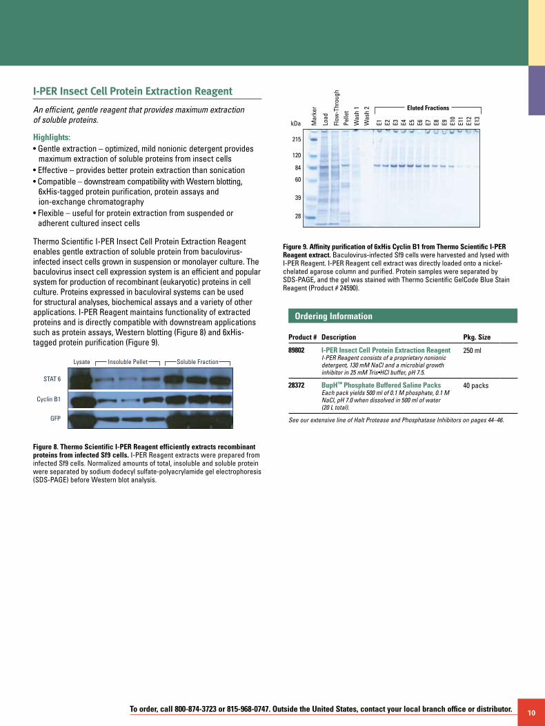
Thermo Scientific I-PER Insect Cell Protein Extraction Reagent enables gentle extraction of soluble protein from baculovirus-infected insect cells grown in suspension or monolayer culture. The baculovirus insect cell expression system is an efficient and popular system for production of recombinant (eukaryotic) proteins in cell culture. Proteins expressed in baculoviral systems can be used for structural analyses, biochemical assays and a variety of other applications. I-PER Reagent maintains functionality of extracted proteins and is directly compatible with downstream applications such as protein assays, Western blotting (Figure 8) and 6xHis-tagged protein purification (Figure 9).
STAT 6
Cyclin B1
GFP
Lysate Insoluble Pellet Soluble Fraction
Figure 8. Thermo Scientific I-PER Reagent efficiently extracts recombinant proteins from infected Sf9 cells. I-PER Reagent extracts were prepared from infected Sf9 cells. Normalized amounts of total, insoluble and soluble protein were separated by sodium dodecyl sulfate-polyacrylamide gel electrophoresis (SDS-PAGE) before Western blot analysis.
kDa
Eluted Fractions
E1 E2 E3 E4 E5 E13
E6 E7 E8 E9 E10
E11
E12
Mar
ker
Load
Flow
-Thr
ough
Pelle
t
Was
h 1
Was
h 2
215
120
84
60
39
28
Figure 9. Affinity purification of 6xHis Cyclin B1 from Thermo Scientific I-PER Reagent extract. Baculovirus-infected Sf9 cells were harvested and lysed with I-PER Reagent. I-PER Reagent cell extract was directly loaded onto a nickel-chelated agarose column and purified. Protein samples were separated by SDS-PAGE, and the gel was stained with Thermo Scientific GelCode Blue Stain Reagent (Product # 24590).
Ordering Information Product #
Description
Pkg. Size
89802 I-PER Insect Cell Protein Extraction ReagentI-PER Reagent consists of a proprietary nonionic detergent, 130 mM NaCl and a microbial growth inhibitor in 25 mM Tris•HCl buffer, pH 7.5.
250 ml
28372 BupH™ Phosphate Buffered Saline PacksEach pack yields 500 ml of 0.1 M phosphate, 0.1 M NaCl, pH 7.0 when dissolved in 500 ml of water (20 L total).
40 packs
See our extensive line of Halt Protease and Phosphatase Inhibitors on pages 44–46.
11 For more information, or to download product instructions, visit www.thermo.com/pierce
M-PER Mammalian Protein Extraction Reagent
Provides highly efficient total protein extraction from cultured mammalian cells.
Highlights:• Quantifiable – mild detergent lysis, yielding extracts that are
immediately compatible with Coomassie (Bradford), the Pierce BCA and Pierce 660 nm Protein Assays or SDS-PAGE1
• Compatible – extracts soluble proteins in nondenatured state, enabling direct use in immunoprecipitation and other affinity purification procedures
• Easy to use – amine-free and fully dialyzable formulation enables compatibility with subsequent assay systems
• Convenient – lyse adherent cells directly in plate or after scraping and washing in suspension
• Recover active protein – maintain luciferase, b-galactosidase, chloramphenicol acetyltransferase (CAT) and other reporter gene activities as well or better than competitor products and freeze/thaw methods
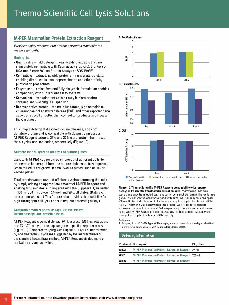
This unique detergent dissolves cell membranes, does not denature protein and is compatible with downstream assays. M-PER Reagent extracts 25% and 20% more protein than freeze/thaw cycles and sonication, respectively (Figure 10).
Suitable for cell lysis on all sizes of culture plates
Lysis with M-PER Reagent is so efficient that adherent cells do not need to be scraped from the culture dish, especially important when the cells are grown in small-welled plates, such as 96- or 24-well plates.
Total protein was recovered efficiently without scraping the cells by simply adding an appropriate amount of M-PER Reagent and shaking for 5 minutes as compared with the Supplier P lysis buffer in 100 mm, 60 mm, 6-well, 24-well and 96-well plates. (Data avail-able on our website.) This feature also provides the feasibility for high-throughput cell lysis and subsequent screening assays.
Compatible with reporter assays, kinase assays, immunoassays and protein assays
M-PER Reagent is compatible with (A) luciferase, (B) b-galactosidase and (C) CAT assays, three popular gene regulation reporter assays (Figure 10). Compared to lysing with Supplier P’s lysis buffer followed by one freeze/thaw cycle (as suggested by the manufacturer) or the standard freeze/thaw method, M-PER Reagent yielded more or equivalent enzyme activities.
Thermo Scientific M-PER Reagent
Supplier P + Freeze/Thaw Cycles Freeze/Thaw Cycles
Exp. 1
A. Renilla Luciferase
Exp. 2
RLU
6
5
4
3
2
1
0
Exp. 1
B. b-galactosidase
Exp. 2 Exp. 3A
bsor
banc
e at
420
nm
0.16
0.12
0.06
0.02
0.00
Exp. 1
C. CAT
Exp. 2
CAT
Rela
tive
Act
ivity
100
10
1
0
Figure 10. Thermo Scientific M-PER Reagent compatibility with reporter assays in transiently transfected mammalian cells. Mammalian FM2 cells were transiently transfected with a reporter construct containing the luciferase gene. The transfected cells were lysed with either M-PER Reagent or Supplier P Lysis Buffer and subjected to luciferase assay. For b-galactosidase and CAT assays, MDA-MB-231 cells were cotransfected with reporter constructs expressing b-galactosidase and CAT, respectively. The transfected cells were lysed with M-PER Reagent or the freeze/thaw method, and the lysates were assayed for b-galactosidase and CAT activity.
Reference1. Banyard, J., et al. (2003). Type XXIII collagen, a new transmembrane collagen identified
in metastatic tumor cells. J. Biol. Chem. 278(23), 20989-20994.
Ordering Information Product #
Description
Pkg. Size
78503 M-PER Mammalian Protein Extraction Reagent 25 ml
78501 M-PER Mammalian Protein Extraction Reagent 250 ml
78505 M-PER Mammalian Protein Extraction Reagent 1 L
Thermo Scientific Cell Lysis Solutions
To order, call 800-874-3723 or 815-968-0747. Outside the United States, contact your local branch office or distributor. 12
P-PER Plant Protein Extraction Kit
Lyses plant leaves, stem, root, seed and flower cells without liquid nitrogen.
The P-PER Plant Protein Extraction Kit offers a new method for performing plant cell lysis and subsequent protein extraction. The P-PER Kit includes an organic lysing reagent and two aqueous reagents which, in conjunction with mild mechanical agitation, effectively extract plant protein.
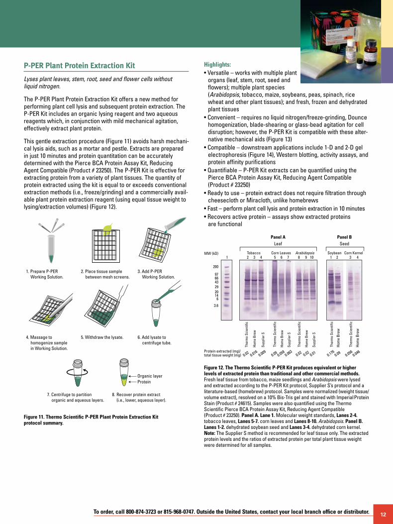
This gentle extraction procedure (Figure 11) avoids harsh mechani-cal lysis aids, such as a mortar and pestle. Extracts are prepared in just 10 minutes and protein quantitation can be accurately determined with the Pierce BCA Protein Assay Kit, Reducing Agent Compatible (Product # 23250). The P-PER Kit is effective for extracting protein from a variety of plant tissues. The quantity of protein extracted using the kit is equal to or exceeds conventional extraction methods (i.e., freeze/grinding) and a commercially avail-able plant protein extraction reagent (using equal tissue weight to lysing/extraction volumes) (Figure 12).
8. Recover protein extract (i.e., lower, aqueous layer).
1. Prepare P-PER Working Solution.
2. Place tissue sample between mesh screens.
3. Add P-PER Working Solution.
4. Massage to homogenize sample in Working Solution.
5. Withdraw the lysate. 6. Add lysate to centrifuge tube.
7. Centrifuge to partition organic and aqueous layers.
ProteinOrganic layer
Figure 11. Thermo Scientific P-PER Plant Protein Extraction Kit protocol summary.
Highlights:• Versatile – works with multiple plant
organs (leaf, stem, root, seed and flowers); multiple plant species (Arabidopsis, tobacco, maize, soybeans, peas, spinach, rice wheat and other plant tissues); and fresh, frozen and dehydrated plant tissues
• Convenient – requires no liquid nitrogen/freeze-grinding, Dounce homogenization, blade-shearing or glass-bead agitation for cell disruption; however, the P-PER Kit is compatible with these alter-native mechanical aids (Figure 13)
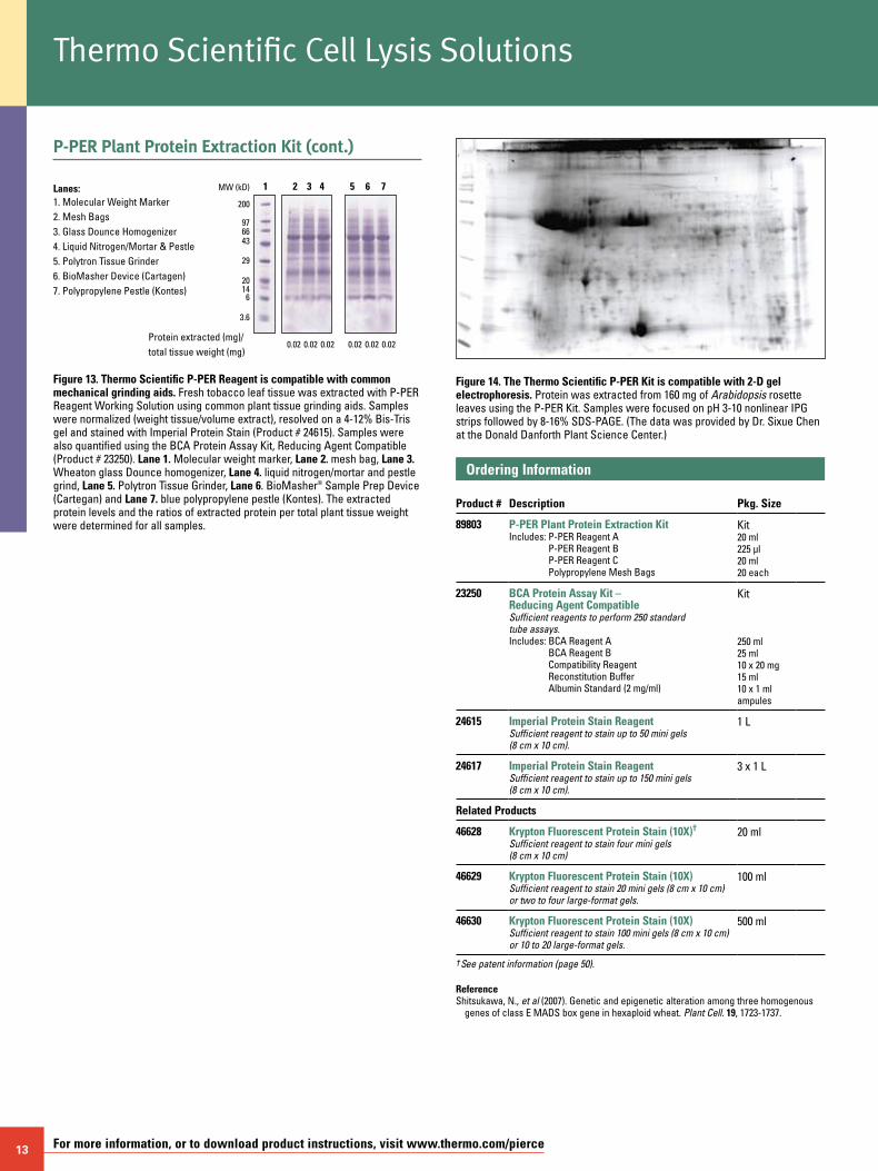
• Compatible – downstream applications include 1-D and 2-D gel electrophoresis (Figure 14), Western blotting, activity assays, and protein affinity purifications
• Quantifiable – P-PER Kit extracts can be quantified using the Pierce BCA Protein Assay Kit, Reducing Agent Compatible (Product # 23250)
• Ready to use – protein extract does not require filtration through cheesecloth or Miracloth, unlike homebrews
• Fast – perform plant cell lysis and protein extraction in 10 minutes • Recovers active protein – assays show extracted proteins
are functional
200
976643292014
6
3.6
1 2 3 4 5 6 7 8 9 10 1 2 3 4
0.020.016
0.009
LeafPanel A Panel B
MW (kD)
Protein extracted (mg)/total tissue weight (mg)
TobaccoH
ome
Bre
w
Supp
lier S
0.020.02
0.01
Hom
e B
rew
Supp
lier S
0.1760.09
Hom
e B
rew
0.0560.046
Hom
e B
rew
0.090.058
0.063
Hom
e B
rew
Supp
lier S
Corn Leaves Arabidopsis Soybean Corn Kernel
SeedTh
erm
o Sc
ient
ific
Ther
mo
Scie
ntifi
c
Ther
mo
Scie
ntifi
c
Ther
mo
Scie
ntifi
c
Ther
mo
Scie
ntifi
c
Figure 12. The Thermo Scientific P-PER Kit produces equivalent or higher levels of extracted protein than traditional and other commercial methods. Fresh leaf tissue from tobacco, maize seedlings and Arabidopsis were lysed and extracted according to the P-PER Kit protocol, Supplier S’s protocol and a literature-based (homebrew) protocol. Samples were normalized (weight tissue/volume extract), resolved on a 10% Bis-Tris gel and stained with Imperial Protein Stain (Product # 24615). Samples were also quantified using the Thermo Scientific Pierce BCA Protein Assay Kit, Reducing Agent Compatible (Product # 23250). Panel A. Lane 1. Molecular weight standards, Lanes 2-4. tobacco leaves, Lanes 5-7. corn leaves and Lanes 8-10. Arabidopsis. Panel B. Lanes 1-2. dehydrated soybean seed and Lanes 3-4. dehydrated corn kernel. Note: The Supplier S method is recommended for leaf tissue only. The extracted protein levels and the ratios of extracted protein per total plant tissue weight were determined for all samples.
13 For more information, or to download product instructions, visit www.thermo.com/pierce
P-PER Plant Protein Extraction Kit (cont.)
200
976643
29
2014
6
3.6
0.02
1 2 3 4 5 6 7
0.02 0.02 0.02 0.02 0.02
MW (kD)
Protein extracted (mg)/total tissue weight (mg)
1. Molecular Weight Marker2. Mesh Bags3. Glass Dounce Homogenizer4. Liquid Nitrogen/Mortar & Pestle5. Polytron Tissue Grinder6. BioMasher Device (Cartagen)7. Polypropylene Pestle (Kontes)
Lanes:
Figure 13. Thermo Scientific P-PER Reagent is compatible with common mechanical grinding aids. Fresh tobacco leaf tissue was extracted with P-PER Reagent Working Solution using common plant tissue grinding aids. Samples were normalized (weight tissue/volume extract), resolved on a 4-12% Bis-Tris gel and stained with Imperial Protein Stain (Product # 24615). Samples were also quantified using the BCA Protein Assay Kit, Reducing Agent Compatible (Product # 23250). Lane 1. Molecular weight marker, Lane 2. mesh bag, Lane 3. Wheaton glass Dounce homogenizer, Lane 4. liquid nitrogen/mortar and pestle grind, Lane 5. Polytron Tissue Grinder, Lane 6. BioMasher® Sample Prep Device (Cartegan) and Lane 7. blue polypropylene pestle (Kontes). The extracted protein levels and the ratios of extracted protein per total plant tissue weight were determined for all samples.
Figure 14. The Thermo Scientific P-PER Kit is compatible with 2-D gel electrophoresis. Protein was extracted from 160 mg of Arabidopsis rosette leaves using the P-PER Kit. Samples were focused on pH 3-10 nonlinear IPG strips followed by 8-16% SDS-PAGE. (The data was provided by Dr. Sixue Chen at the Donald Danforth Plant Science Center.)
Ordering Information Product #
Description
Pkg. Size
89803 P-PER Plant Protein Extraction KitIncludes: P-PER Reagent A
P-PER Reagent B P-PER Reagent C Polypropylene Mesh Bags
Kit 20 ml 225 µl 20 ml 20 each
23250 BCA Protein Assay Kit – Reducing Agent CompatibleSufficient reagents to perform 250 standard tube assays.Includes: BCA Reagent A
BCA Reagent B Compatibility Reagent Reconstitution Buffer Albumin Standard (2 mg/ml)
Kit
250 ml 25 ml 10 x 20 mg 15 ml 10 x 1 ml ampules
24615 Imperial Protein Stain Reagent Sufficient reagent to stain up to 50 mini gels (8 cm x 10 cm).
1 L
24617 Imperial Protein Stain Reagent Sufficient reagent to stain up to 150 mini gels (8 cm x 10 cm).
3 x 1 L
Related Products
46628 Krypton Fluorescent Protein Stain (10X)†
Sufficient reagent to stain four mini gels (8 cm x 10 cm)
20 ml
46629 Krypton Fluorescent Protein Stain (10X)Sufficient reagent to stain 20 mini gels (8 cm x 10 cm) or two to four large-format gels.
100 ml
46630 Krypton Fluorescent Protein Stain (10X)Sufficient reagent to stain 100 mini gels (8 cm x 10 cm) or 10 to 20 large-format gels.
500 ml
† See patent information (page 50).
ReferenceShitsukawa, N., et al (2007). Genetic and epigenetic alteration among three homogenous
genes of class E MADS box gene in hexaploid wheat. Plant Cell. 19, 1723-1737.
Thermo Scientific Cell Lysis Solutions
To order, call 800-874-3723 or 815-968-0747. Outside the United States, contact your local branch office or distributor. 14
T-PER Tissue Protein Extraction Reagent
Extracts total protein from tissue samples.
Highlights:• Simple – just homogenize tissue sample in 1:20 (w/v) of tissue
to T-PER Reagent, then centrifuge to pellet cell/tissue debris• Easy to use – mild detergent is dialyzable for quick and
easy removal• Versatile – can be used with additional components (e.g., pro-
tease inhibitors, salts, reducing agents, chelating agents, etc.)1-5
• Compatible – lysate may be used for reporter assays, protein kinase assays, immunoassays, ELISAs, Western blots and/or protein purifications1-5
• Quantifiable – the lysate is compatible with standard protein assays such as Thermo Scientific Pierce Coomassie Plus (Bradford) Protein Assay (Product # 23236) or Pierce 600 nm Protein Assay (Product # 22660)1-5
This reagent uses a proprietary detergent in 25 mM bicine, 150 mM sodium chloride (pH 7.6) for tissue cell lysis. The simple composition of this reagent provides versatility for many different applications. Depending on the application, it may be advanta-geous to add other components, such as protease inhibitors, salts, reducing agents, chelating agents, etc., to the reagent before proceeding with the cell lysis. The cell lysate prepared with this reagent may be used for reporter assays (e.g., luciferase, b-galactosidase, chloramphenicol acetyl transferase), protein kinase assays (e.g., PKA, PKC, tyrosine kinase), immunoassays (e.g., Western blots, ELISAs, RIAs) and/or protein purifications.
PROTOCOL:1. Weigh tissue samples. A 1:20 (w/v) ratio of tissue to T-PER Reagent is optimal for this procedure.
Notes: a. Protease inhibitors may be added to the T-PER Reagent (if necessary).
b. Smaller volumes of T-PER Reagent may be used if a more concentrated protein extract is required.
2. Add the appropriate amount of T-PER Reagent to the tissue sample and homogenize.
3. Centrifuge the sample for 5 minutes to pellet cell/tissue debris.
4. Collect supernatant and continue with downstream analysis or further purification.
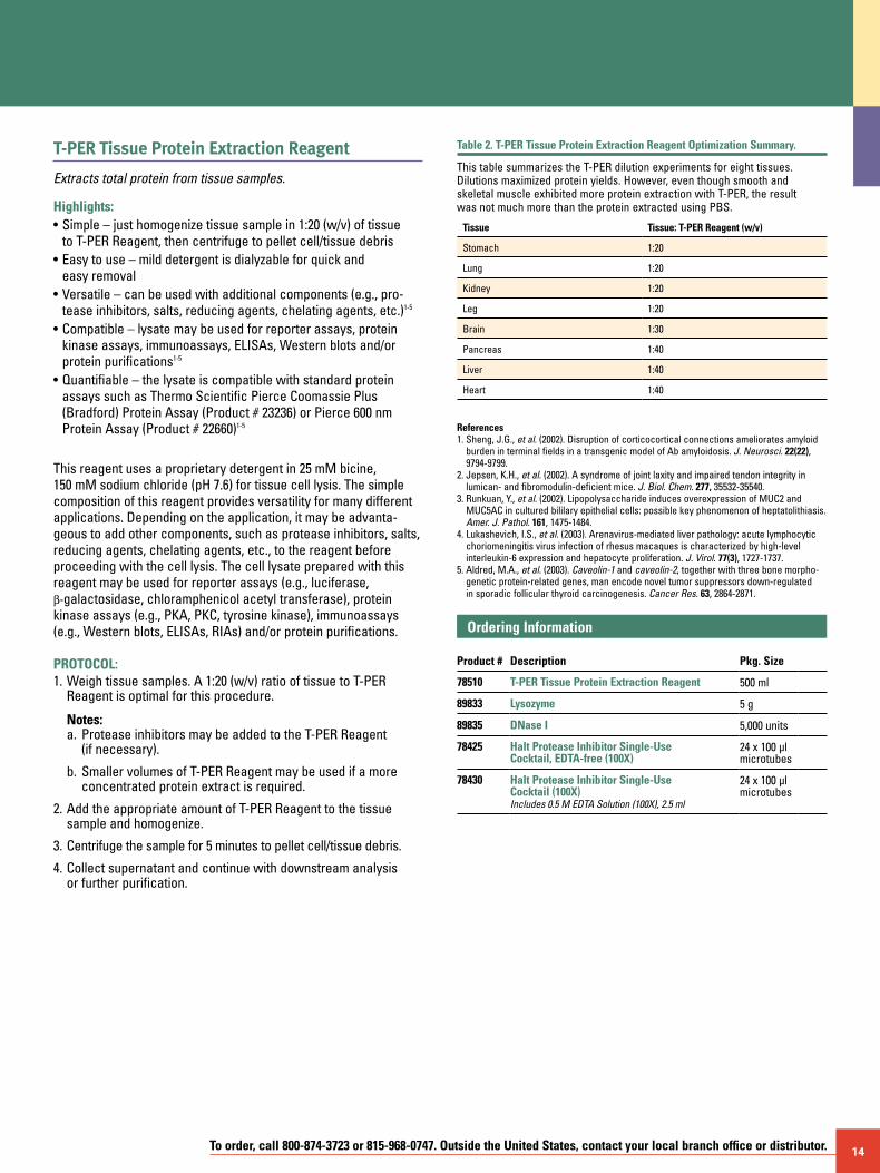
Table 2. T-PER Tissue Protein Extraction Reagent Optimization Summary.
This table summarizes the T-PER dilution experiments for eight tissues. Dilutions maximized protein yields. However, even though smooth and skeletal muscle exhibited more protein extraction with T-PER, the result was not much more than the protein extracted using PBS.
Tissue Tissue: T-PER Reagent (w/v)
Stomach 1:20
Lung 1:20
Kidney 1:20
Leg 1:20
Brain 1:30
Pancreas 1:40
Liver 1:40
Heart 1:40
References1. Sheng, J.G., et al. (2002). Disruption of corticocortical connections ameliorates amyloid
burden in terminal fields in a transgenic model of Ab amyloidosis. J. Neurosci. 22(22), 9794-9799.
2. Jepsen, K.H., et al. (2002). A syndrome of joint laxity and impaired tendon integrity in lumican- and fibromodulin-deficient mice. J. Biol. Chem. 277, 35532-35540.
3. Runkuan, Y., et al. (2002). Lipopolysaccharide induces overexpression of MUC2 and MUC5AC in cultured bililary epithelial cells: possible key phenomenon of heptatolithiasis. Amer. J. Pathol. 161, 1475-1484.
4. Lukashevich, I.S., et al. (2003). Arenavirus-mediated liver pathology: acute lymphocytic choriomeningitis virus infection of rhesus macaques is characterized by high-level interleukin-6 expression and hepatocyte proliferation. J. Virol. 77(3), 1727-1737.
5. Aldred, M.A., et al. (2003). Caveolin-1 and caveolin-2, together with three bone morpho-genetic protein-related genes, man encode novel tumor suppressors down-regulated in sporadic follicular thyroid carcinogenesis. Cancer Res. 63, 2864-2871.
Ordering Information Product #
Description
Pkg. Size
78510 T-PER Tissue Protein Extraction Reagent 500 ml
89833 Lysozyme 5 g
89835 DNase I 5,000 units
78425 Halt Protease Inhibitor Single-Use Cocktail, EDTA-free (100X)
24 x 100 µl microtubes
78430 Halt Protease Inhibitor Single-Use Cocktail (100X) Includes 0.5 M EDTA Solution (100X), 2.5 ml
24 x 100 µl microtubes
15 For more information, or to download product instructions, visit www.thermo.com/pierce
Y-PER Yeast Protein Extraction Reagent
Easy-to-use solution gently disrupts the tough yeast cell wall in less than 20 minutes at room temperature.
Traditionally, protein extraction from yeast required physical dis-ruption to break through the thick proteinaceous cell envelope; less disruptive lysis methods were possible only with other organ-isms like E. coli. Y-PER Yeast Protein Extraction Reagent was the first commercially available yeast lysis reagent to use a mild detergent lysis procedure to efficiently release functionally active solubilized proteins. Several uses for the Y-PER Reagent have been optimized that encompass a broad array of applications ranging from fusion-tagged protein purification to microplate-compatible enzyme assays, and genomic and plasmid DNA extraction from yeast. Y-PER Reagent has even been used to isolate yeast killer virus double-stranded RNA from killer strains of S. cerevisiae.1
The standard protocol for protein extraction is easy: simply add an appropriate volume of Y-PER Reagent to pelleted yeast cells, incubate at room temperature for approximately 20 minutes and spin down the debris. The resulting supernatant is a concentrated protein solution, surpassing typical yields obtained with traditional glass bead disruption.
Thermo ScientificY-PER ReagentGlass Beads
S. cerevisiae S. pombe
Prot
ein
(µg)
B. subtilis
700
600
500
400
300
200
100
0
Figure 15. Thermo Scientific Y-PER Reagent extraction yields greater amounts of usable protein. In all three organisms tested, Y-PER Reagent extracts contain more usable protein than traditional glass bead lysis.
Highlights:• Excellent yields – extracts more than twice as much protein as
glass bead methods (Figure 15)• Easy to use – eliminates the physical problems associated with
traditional glass bead lysis (e.g., clinging static-charged beads, protein/bead clumps and runaway beads)
• Compatible – works with Saccharomyces cerevisiae, Schizosaccharomyces pombe, Pichia pastoris and Bacillus subtilus
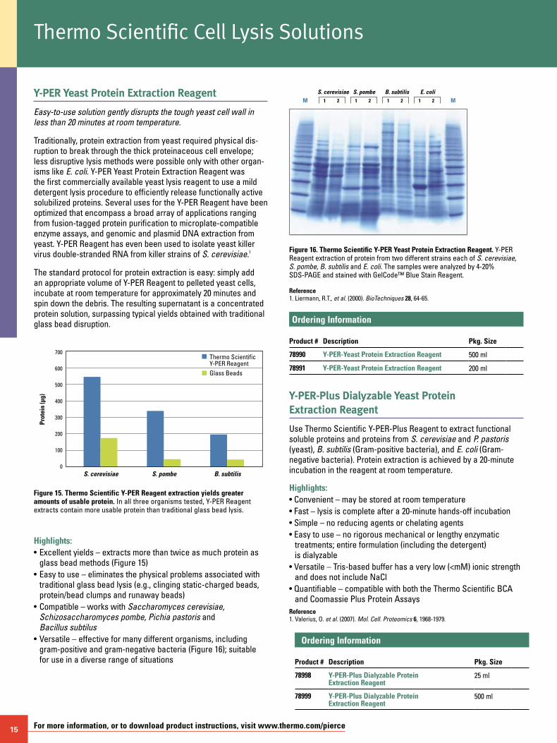
• Versatile – effective for many different organisms, including gram-positive and gram-negative bacteria (Figure 16); suitable for use in a diverse range of situations
M M1
S. cerevisiae S. pombe B. subtilis E. coli2 1 2 1 2 1 2
Figure 16. Thermo Scientific Y-PER Yeast Protein Extraction Reagent. Y-PER Reagent extraction of protein from two different strains each of S. cerevisiae, S. pombe, B. subtilis and E. coli. The samples were analyzed by 4-20% SDS-PAGE and stained with GelCode™ Blue Stain Reagent.
Reference1. Liermann, R.T., et al. (2000). BioTechniques 28, 64-65.
Ordering Information Product #
Description
Pkg. Size
78990 Y-PER-Yeast Protein Extraction Reagent 500 ml
78991 Y-PER-Yeast Protein Extraction Reagent 200 ml
Y-PER-Plus Dialyzable Yeast Protein Extraction Reagent
Use Thermo Scientific Y-PER-Plus Reagent to extract functional soluble proteins and proteins from S. cerevisiae and P. pastoris (yeast), B. subtilis (Gram-positive bacteria), and E. coli (Gram-negative bacteria). Protein extraction is achieved by a 20-minute incubation in the reagent at room temperature.
Highlights:• Convenient – may be stored at room temperature• Fast – lysis is complete after a 20-minute hands-off incubation• Simple – no reducing agents or chelating agents• Easy to use – no rigorous mechanical or lengthy enzymatic
treatments; entire formulation (including the detergent) is dialyzable
• Versatile – Tris-based buffer has a very low (<mM) ionic strength and does not include NaCl
• Quantifiable – compatible with both the Thermo Scientific BCA and Coomassie Plus Protein Assays
Reference1. Valerius, O. et al. (2007). Mol. Cell. Proteomics 6, 1968-1979.
Ordering Information Product #
Description
Pkg. Size
78998 Y-PER-Plus Dialyzable Protein Extraction Reagent
25 ml
78999 Y-PER-Plus Dialyzable Protein Extraction Reagent
500 ml
Thermo Scientific Cell Lysis Solutions
To order, call 800-874-3723 or 815-968-0747. Outside the United States, contact your local branch office or distributor. 16
Thermo Scientific Pierce RIPA Buffer
Compatibility with protease inhibitors prevents proteolysis.
The Thermo Scientific Pierce RIPA Buffer is a reliable buffer used to lyse cultured mammalian cells from both plated cells and cells pelleted from suspension cultures. It enables the extraction of membrane, nuclear and cytoplasmic proteins and is compatible with many applications, including reporter assays, the Pierce BCA Protein Assay, immunoassays and protein purification. Inhibitors such as Halt Protease Inhibitor Cocktail (Product # 78425) and Halt Phosphatase Inhibitor Cocktail (Product # 78420) are also compat-ible with the Pierce RIPA Buffer and can be added before use to prevent proteolysis and maintain protein phosphorylation.
Highlights:• Convenient – ready-to-use solution• Flexible – compatible with many applications, including reporter
assays, protein assays, immunoassays and protein purification• Versatile – enables extraction of cytoplasmic, membrane and
nuclear proteins
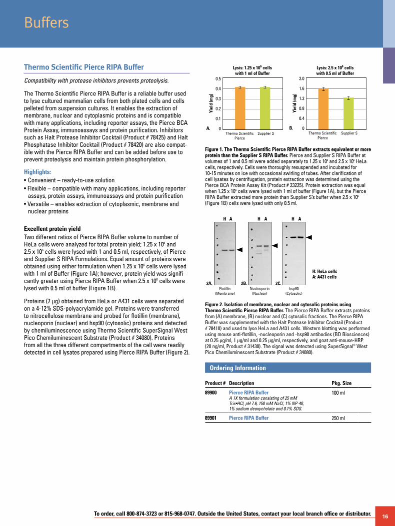
Excellent protein yieldTwo different ratios of Pierce RIPA Buffer volume to number of HeLa cells were analyzed for total protein yield; 1.25 x 10⁶ and 2.5 x 10⁶ cells were lysed with 1 and 0.5 ml, respectively, of Pierce and Supplier S RIPA Formulations. Equal amount of proteins were obtained using either formulation when 1.25 x 10⁶ cells were lysed with 1 ml of Buffer (Figure 1A); however, protein yield was signifi-cantly greater using Pierce RIPA Buffer when 2.5 x 10⁶ cells were lysed with 0.5 ml of buffer (Figure 1B).
Proteins (7 µg) obtained from HeLa or A431 cells were separated on a 4-12% SDS-polyacrylamide gel. Proteins were transferred to nitrocellulose membrane and probed for flotillin (membrane), nucleoporin (nuclear) and hsp90 (cytosolic) proteins and detected by chemiluminescence using Thermo Scientific SuperSignal West Pico Chemiluminescent Substrate (Product # 34080). Proteins from all the three different compartments of the cell were readily detected in cell lysates prepared using Pierce RIPA Buffer (Figure 2).
2.0
1.6
1.2
0.8
0.4
0
Yiel
d (m
g)
B.Thermo Scientific
PierceSupplier S
0.5
0.4
0.3
0.2
0.1
0
Yiel
d (m
g)
Lysis: 1.25 x 10⁶ cellswith 1 ml of Buffer
Lysis: 2.5 x 10⁶ cellswith 0.5 ml of Buffer
A.Thermo Scientific
PierceSupplier S
Figure 1. The Thermo Scientific Pierce RIPA Buffer extracts equivalent or more protein than the Supplier S RIPA Buffer. Pierce and Supplier S RIPA Buffer at volumes of 1 and 0.5 ml were added separately to 1.25 x 10⁶ and 2.5 x 10⁶ HeLa cells, respectively. Cells were thoroughly resuspended and incubated for 10-15 minutes on ice with occasional swirling of tubes. After clarification of cell lysates by centrifugation, protein extraction was determined using the Pierce BCA Protein Assay Kit (Product # 23225). Protein extraction was equal when 1.25 x 10⁶ cells were lysed with 1 ml of buffer (Figure 1A), but the Pierce RIPA Buffer extracted more protein than Supplier S’s buffer when 2.5 x 10⁶ (Figure 1B) cells were lysed with only 0.5 ml.
hsp90(Cytosolic)
Nucleoporin(Nuclear)
H: HeLa cellsA: A431 cells
Flotillin(Membrane)
H A H AH
2A. 2B. 2C.
A
Figure 2. Isolation of membrane, nuclear and cytosolic proteins using Thermo Scientific Pierce RIPA Buffer. The Pierce RIPA Buffer extracts proteins from (A) membrane, (B) nuclear and (C) cytosolic fractions. The Pierce RIPA Buffer was supplemented with the Halt Protease Inhibitor Cocktail (Product # 78410) and used to lyse HeLa and A431 cells. Western blotting was performed using mouse anti-flotillin, -nucleoporin and -hsp90 antibodies (BD Biosciences) at 0.25 µg/ml, 1 µg/ml and 0.25 µg/ml, respectively, and goat anti-mouse-HRP (20 ng/ml, Product # 31430). The signal was detected using SuperSignal® West Pico Chemiluminescent Substrate (Product # 34080).
Ordering Information Product #
Description
Pkg. Size
89900 Pierce RIPA BufferA 1X formulation consisting of 25 mM Tris•HCl, pH 7.6, 150 mM NaCl, 1% NP-40, 1% sodium deoxycholate and 0.1% SDS.
100 ml
89901 Pierce RIPA Buffer 250 ml
Buffers
17 For more information, or to download product instructions, visit www.thermo.com/pierce
B-PER 6xHis Fusion Protein Column and Spin Purification Kits
Optimized high-capacity purifications.
Highlights of both kits:• Greater convenience – no sonication required; complete cell
lysis achieved with B-PER Reagent• Ready-to-use components
Highlights of the B-PER 6xHis Fusion Protein Column Purification Kit:• Fast and efficient – optimized system provides the best purity in
the least amount of time (2.5-3 hours)• High capacity – kit makes it possible to purify more than 10 mg of
over-expressed protein per column
Highlights of the B-PER 6xHis Fusion Protein Spin Purification Kit:• Fast – purify in less than 30 minutes• Excellent yields – achieve yields of approximately 1 mg of pure
6xHis-tagged fusion protein• Complete – includes Spin Columns and Collection Tubes
The B-PER 6xHis Column and Spin Purification Kits rapidly and efficiently purify 6xHis-tagged fusion proteins from bacteria and from baculovirus-infected insect cells. The fusion protein is extracted using B-PER Bacterial Protein Extraction Reagent and then purified using a Nickel Chelated Column (Ni-Chelated Columns) or Spin Column included. The patented detergent in B-PER Reagent, combined with a small amount of imidazole, effi-ciently removes nonspecifically and/or weakly bound proteins (e.g., proteins rich in histidine residues). The 6xHis-tagged proteins are then eluted with excess imidazole (Elution Buffer).
The B-PER 6xHis Fusion Protein Column Kit
The kit protocol produces a high yield of pure 6xHis fusion protein. The column has been tested for loading up to 20 ml lysates from 500 ml cultures. However, for optimal results, a 10 ml lysate from 250 ml bacterial culture (OD600 ~1.5-3) is suggested as the starting material. The yield and purity greatly depend on the expression level and the nature of the recombinant protein. As an example, we routinely obtain 10-12 mg of 6xHis-tagged green fluorescent protein (GFP) from 250 ml overnight bacterial culture with more than 90% purity.
Obtain purified 6xHis-tagged protein using B-PER Column Kit (Product # 78100) in under 3 hours
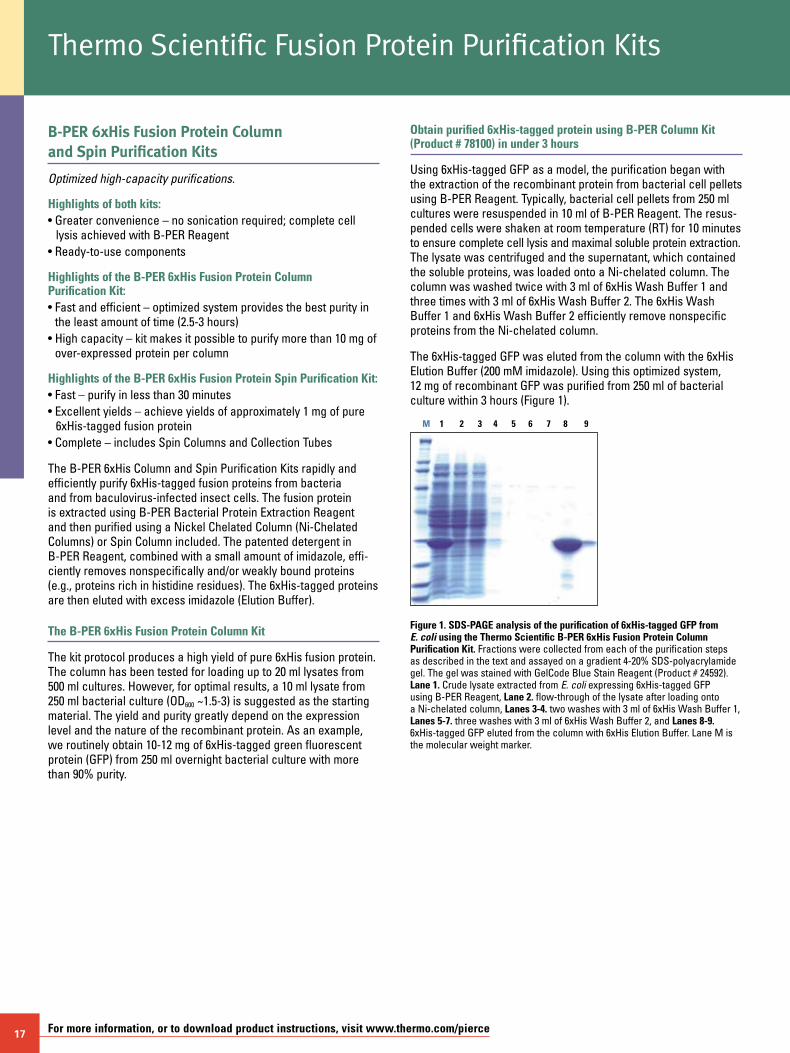
Using 6xHis-tagged GFP as a model, the purification began with the extraction of the recombinant protein from bacterial cell pellets using B-PER Reagent. Typically, bacterial cell pellets from 250 ml cultures were resuspended in 10 ml of B-PER Reagent. The resus-pended cells were shaken at room temperature (RT) for 10 minutes to ensure complete cell lysis and maximal soluble protein extraction. The lysate was centrifuged and the supernatant, which contained the soluble proteins, was loaded onto a Ni-chelated column. The column was washed twice with 3 ml of 6xHis Wash Buffer 1 and three times with 3 ml of 6xHis Wash Buffer 2. The 6xHis Wash Buffer 1 and 6xHis Wash Buffer 2 efficiently remove nonspecific proteins from the Ni-chelated column.
The 6xHis-tagged GFP was eluted from the column with the 6xHis Elution Buffer (200 mM imidazole). Using this optimized system, 12 mg of recombinant GFP was purified from 250 ml of bacterial culture within 3 hours (Figure 1).
Figure 1. SDS-PAGE analysis of the purification of 6xHis-tagged GFP from E. coli using the Thermo Scientific B-PER 6xHis Fusion Protein Column Purification Kit. Fractions were collected from each of the purification steps as described in the text and assayed on a gradient 4-20% SDS-polyacrylamide gel. The gel was stained with GelCode Blue Stain Reagent (Product # 24592). Lane 1. Crude lysate extracted from E. coli expressing 6xHis-tagged GFP using B-PER Reagent, Lane 2. flow-through of the lysate after loading onto a Ni-chelated column, Lanes 3-4. two washes with 3 ml of 6xHis Wash Buffer 1, Lanes 5-7. three washes with 3 ml of 6xHis Wash Buffer 2, and Lanes 8-9. 6xHis-tagged GFP eluted from the column with 6xHis Elution Buffer. Lane M is the molecular weight marker.
M 1 2 3 4 5 6 7 8 9
Thermo Scientific Fusion Protein Purification Kits
To order, call 800-874-3723 or 815-968-0747. Outside the United States, contact your local branch office or distributor. 18
B-PER 6xHis Fusion Protein Column and Spin Purification Kits (cont.)
B-PER Spin Kit (Product # 78300) enables quick purification of 6xHis-tagged protein
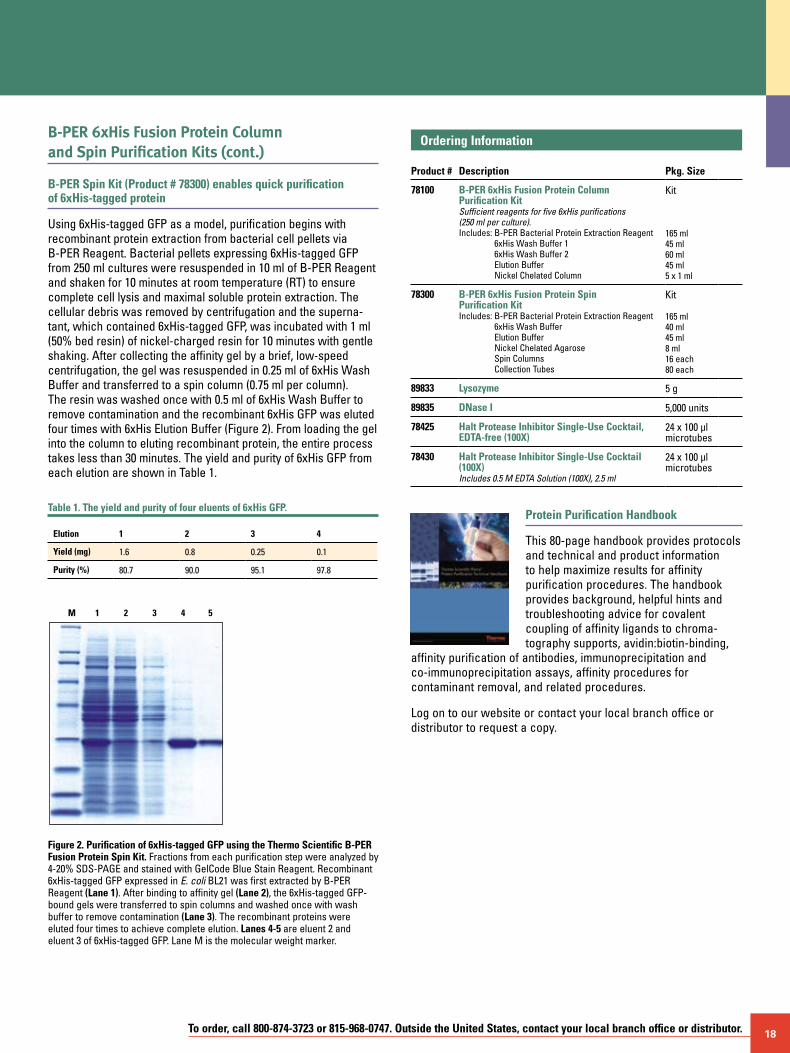
Using 6xHis-tagged GFP as a model, purification begins with recombinant protein extraction from bacterial cell pellets via B-PER Reagent. Bacterial pellets expressing 6xHis-tagged GFP from 250 ml cultures were resuspended in 10 ml of B-PER Reagent and shaken for 10 minutes at room temperature (RT) to ensure complete cell lysis and maximal soluble protein extraction. The cellular debris was removed by centrifugation and the superna-tant, which contained 6xHis-tagged GFP, was incubated with 1 ml (50% bed resin) of nickel-charged resin for 10 minutes with gentle shaking. After collecting the affinity gel by a brief, low-speed centrifugation, the gel was resuspended in 0.25 ml of 6xHis Wash Buffer and transferred to a spin column (0.75 ml per column). The resin was washed once with 0.5 ml of 6xHis Wash Buffer to remove contamination and the recombinant 6xHis GFP was eluted four times with 6xHis Elution Buffer (Figure 2). From loading the gel into the column to eluting recombinant protein, the entire process takes less than 30 minutes. The yield and purity of 6xHis GFP from each elution are shown in Table 1.
Table 1. The yield and purity of four eluents of 6xHis GFP.
Elution 1 2 3 4
Yield (mg) 1.6 0.8 0.25 0.1
Purity (%) 80.7 90.0 95.1 97.8
Figure 2. Purification of 6xHis-tagged GFP using the Thermo Scientific B-PER Fusion Protein Spin Kit. Fractions from each purification step were analyzed by 4-20% SDS-PAGE and stained with GelCode Blue Stain Reagent. Recombinant 6xHis-tagged GFP expressed in E. coli BL21 was first extracted by B-PER Reagent (Lane 1). After binding to affinity gel (Lane 2), the 6xHis-tagged GFP-bound gels were transferred to spin columns and washed once with wash buffer to remove contamination (Lane 3). The recombinant proteins were eluted four times to achieve complete elution. Lanes 4-5 are eluent 2 and eluent 3 of 6xHis-tagged GFP. Lane M is the molecular weight marker.
1 2 3 4 5M
Ordering Information Product #
Description
Pkg. Size
78100 B-PER 6xHis Fusion Protein Column Purification KitSufficient reagents for five 6xHis purifications (250 ml per culture).Includes: B-PER Bacterial Protein Extraction Reagent
6xHis Wash Buffer 1 6xHis Wash Buffer 2 Elution Buffer Nickel Chelated Column
Kit
165 ml 45 ml 60 ml 45 ml 5 x 1 ml
78300 B-PER 6xHis Fusion Protein Spin Purification KitIncludes: B-PER Bacterial Protein Extraction Reagent
6xHis Wash Buffer Elution Buffer Nickel Chelated Agarose Spin Columns Collection Tubes
Kit
165 ml 40 ml 45 ml 8 ml 16 each 80 each
89833 Lysozyme 5 g
89835 DNase I 5,000 units
78425 Halt Protease Inhibitor Single-Use Cocktail, EDTA-free (100X)
24 x 100 µl microtubes
78430 Halt Protease Inhibitor Single-Use Cocktail (100X)Includes 0.5 M EDTA Solution (100X), 2.5 ml
24 x 100 µl microtubes
Protein Purification Handbook
This 80-page handbook provides protocols and technical and product information to help maximize results for affinity purification procedures. The handbook provides background, helpful hints and troubleshooting advice for covalent coupling of affinity ligands to chroma-tography supports, avidin:biotin-binding,
affinity purification of antibodies, immunoprecipitation and co-immunoprecipitation assays, affinity procedures for contaminant removal, and related procedures.
Log on to our website or contact your local branch office or distributor to request a copy.
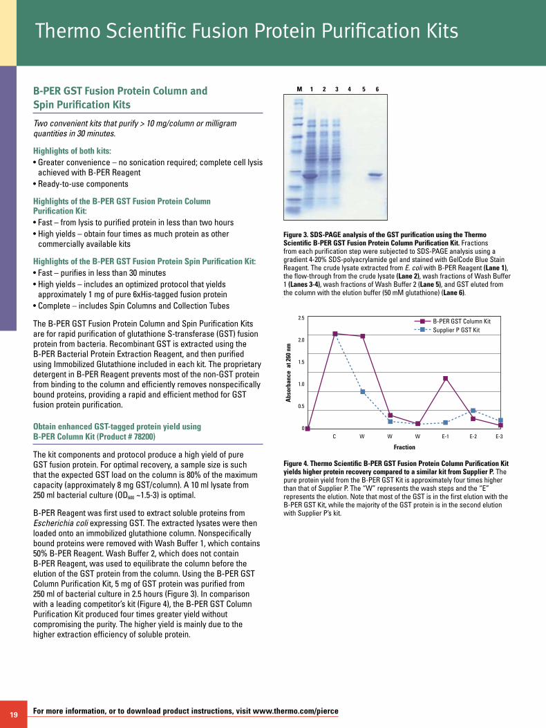
19 For more information, or to download product instructions, visit www.thermo.com/pierce
B-PER GST Fusion Protein Column and Spin Purification Kits
Two convenient kits that purify > 10 mg/column or milligram quantities in 30 minutes.
Highlights of both kits:• Greater convenience – no sonication required; complete cell lysis
achieved with B-PER Reagent• Ready-to-use components
Highlights of the B-PER GST Fusion Protein Column Purification Kit:• Fast – from lysis to purified protein in less than two hours• High yields – obtain four times as much protein as other
commercially available kits
Highlights of the B-PER GST Fusion Protein Spin Purification Kit:• Fast – purifies in less than 30 minutes• High yields – includes an optimized protocol that yields
approximately 1 mg of pure 6xHis-tagged fusion protein• Complete – includes Spin Columns and Collection Tubes
The B-PER GST Fusion Protein Column and Spin Purification Kits are for rapid purification of glutathione S-transferase (GST) fusion protein from bacteria. Recombinant GST is extracted using the B-PER Bacterial Protein Extraction Reagent, and then purified using Immobilized Glutathione included in each kit. The proprietary detergent in B-PER Reagent prevents most of the non-GST protein from binding to the column and efficiently removes nonspecifically bound proteins, providing a rapid and efficient method for GST fusion protein purification.
Obtain enhanced GST-tagged protein yield using B-PER Column Kit (Product # 78200)
The kit components and protocol produce a high yield of pure GST fusion protein. For optimal recovery, a sample size is such that the expected GST load on the column is 80% of the maximum capacity (approximately 8 mg GST/column). A 10 ml lysate from 250 ml bacterial culture (OD600 ~1.5-3) is optimal.
B-PER Reagent was first used to extract soluble proteins from Escherichia coli expressing GST. The extracted lysates were then loaded onto an immobilized glutathione column. Nonspecifically bound proteins were removed with Wash Buffer 1, which contains 50% B-PER Reagent. Wash Buffer 2, which does not contain B-PER Reagent, was used to equilibrate the column before the elution of the GST protein from the column. Using the B-PER GST Column Purification Kit, 5 mg of GST protein was purified from 250 ml of bacterial culture in 2.5 hours (Figure 3). In comparison with a leading competitor’s kit (Figure 4), the B-PER GST Column Purification Kit produced four times greater yield without compromising the purity. The higher yield is mainly due to the higher extraction efficiency of soluble protein.
M 1 2 3 4 5 6
Thermo Scientific Fusion Protein Purification Kits
Figure 3. SDS-PAGE analysis of the GST purification using the Thermo Scientific B-PER GST Fusion Protein Column Purification Kit. Fractions from each purification step were subjected to SDS-PAGE analysis using a gradient 4-20% SDS-polyacrylamide gel and stained with GelCode Blue Stain Reagent. The crude lysate extracted from E. coli with B-PER Reagent (Lane 1), the flow-through from the crude lysate (Lane 2), wash fractions of Wash Buffer 1 (Lanes 3-4), wash fractions of Wash Buffer 2 (Lane 5), and GST eluted from the column with the elution buffer (50 mM glutathione) (Lane 6).
Supplier P GST KitB-PER GST Column Kit
C
Fraction
W
Abs
orba
nce
at 2
60 n
m
E-3E-2W W E-1
2.5
2.0
1.5
1.0
0.5
0
Figure 4. Thermo Scientific B-PER GST Fusion Protein Column Purification Kit yields higher protein recovery compared to a similar kit from Supplier P. The pure protein yield from the B-PER GST Kit is approximately four times higher than that of Supplier P. The “W” represents the wash steps and the “E” represents the elution. Note that most of the GST is in the first elution with the B-PER GST Kit, while the majority of the GST protein is in the second elution with Supplier P’s kit.
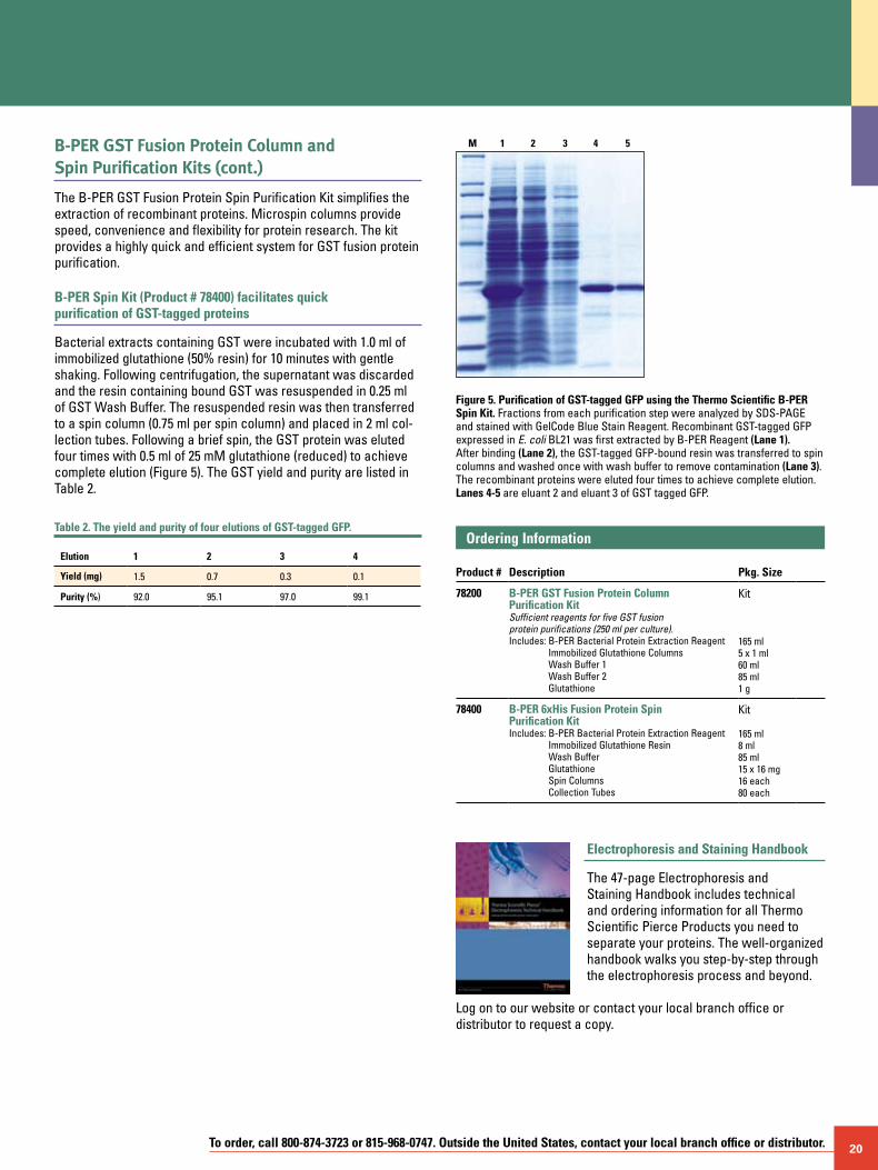
To order, call 800-874-3723 or 815-968-0747. Outside the United States, contact your local branch office or distributor. 20
B-PER GST Fusion Protein Column and Spin Purification Kits (cont.)
The B-PER GST Fusion Protein Spin Purification Kit simplifies the extraction of recombinant proteins. Microspin columns provide speed, convenience and flexibility for protein research. The kit provides a highly quick and efficient system for GST fusion protein purification.
B-PER Spin Kit (Product # 78400) facilitates quick purification of GST-tagged proteins
Bacterial extracts containing GST were incubated with 1.0 ml of immobilized glutathione (50% resin) for 10 minutes with gentle shaking. Following centrifugation, the supernatant was discarded and the resin containing bound GST was resuspended in 0.25 ml of GST Wash Buffer. The resuspended resin was then transferred to a spin column (0.75 ml per spin column) and placed in 2 ml col-lection tubes. Following a brief spin, the GST protein was eluted four times with 0.5 ml of 25 mM glutathione (reduced) to achieve complete elution (Figure 5). The GST yield and purity are listed in Table 2.
Table 2. The yield and purity of four elutions of GST-tagged GFP.
Elution 1 2 3 4
Yield (mg) 1.5 0.7 0.3 0.1
Purity (%) 92.0 95.1 97.0 99.1
M 1 2 3 4 5
Figure 5. Purification of GST-tagged GFP using the Thermo Scientific B-PER Spin Kit. Fractions from each purification step were analyzed by SDS-PAGE and stained with GelCode Blue Stain Reagent. Recombinant GST-tagged GFP expressed in E. coli BL21 was first extracted by B-PER Reagent (Lane 1). After binding (Lane 2), the GST-tagged GFP-bound resin was transferred to spin columns and washed once with wash buffer to remove contamination (Lane 3). The recombinant proteins were eluted four times to achieve complete elution. Lanes 4-5 are eluant 2 and eluant 3 of GST tagged GFP.
Ordering Information Product #
Description
Pkg. Size
78200 B-PER GST Fusion Protein Column Purification KitSufficient reagents for five GST fusion protein purifications (250 ml per culture).Includes: B-PER Bacterial Protein Extraction Reagent
Immobilized Glutathione Columns Wash Buffer 1 Wash Buffer 2 Glutathione
Kit
165 ml 5 x 1 ml 60 ml 85 ml 1 g
78400 B-PER 6xHis Fusion Protein Spin Purification KitIncludes: B-PER Bacterial Protein Extraction Reagent
Immobilized Glutathione Resin Wash Buffer Glutathione Spin Columns Collection Tubes
Kit
165 ml 8 ml 85 ml 15 x 16 mg 16 each 80 each
Electrophoresis and Staining Handbook
The 47-page Electrophoresis and Staining Handbook includes technical and ordering information for all Thermo Scientific Pierce Products you need to separate your proteins. The well-organized handbook walks you step-by-step through the electrophoresis process and beyond.
Log on to our website or contact your local branch office or distributor to request a copy.
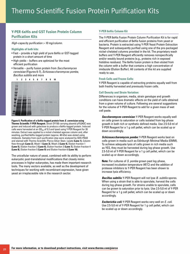
21 For more information, or to download product instructions, visit www.thermo.com/pierce
Y-PER 6xHis and GST Fusion Protein Column Purification Kits
High-capacity purification > 10 mg/column.
Highlights of both kits:• Fast – provide a high yield of pure 6xHis or GST-tagged
protein in a short amount of time• High yields – buffers are optimized for the most
efficient purification• Versatile – purify fusion protein from Saccharomyces
cerevisiae (Figures 6-7), Schizosaccharomyces pombe, Bacillus subtilis and more
Thermo Scientific Fusion Protein Purification Kits
Figure 6. Purification of a 6xHis-tagged protein from S. cerevisiae using Thermo Scientific Y-PER Reagent. Strain DY150 carrying plasmid pYIL042C was grown and induced with galactose to produce a 6xHis-tagged protein. Induced cells were harvested at an OD600 of 5.3 and lysed using Y-PER Reagent for 20 minutes. Extract was applied to a nickel-chelated agarose column and, after washing, purified 6xHis-tagged protein (approx. 48 kDa) was eluted using imidazole. Samples from each purification step were analyzed by SDS-PAGE and stained with Thermo Scientific Pierce Silver Stain. Lysate (Lane 1), Column flow-through (Lane 2), Wash 1 (Lane 3), Wash 2 (Lane 4), Elution fraction 1 (Lane 5), Elution fraction 2 (Lane 6), Elution fraction 3 (Lane 7), Elution fraction 4 (Lane 8), Elution fraction 5 (Lane 9) and Elution fraction 6 (Lane 10).
The unicellular nature of yeast, combined with its ability to perform eukaryotic post-translational modifications that closely mimic processes in higher eukaryotes, has made them important research tools. The many vectors available, as well as the development of techniques for working with recombinant expression, have given yeast an irreplaceable role in the research sector.
1 2 3 4 5 6 7 8 9 10
Y-PER 6xHis Column Kit
The Y-PER 6xHis Fusion Protein Column Purification Kit is for rapid and efficient purification of 6xHis fusion proteins from yeast or bacteria. Protein is extracted using Y-PER Yeast Protein Extraction Reagent and subsequently purified using one of the pre-packaged nickel-chelated columns provided in the kit. The proprietary wash buffers and Y-PER Reagent efficiently removes nonspecifically and/or weakly bound proteins (e.g., proteins rich in exposed histidine residues). The 6xHis fusion protein is then eluted from the column with a buffer that contains a high concentration of imidazole (Elution Buffer). All contents of this kit are supplied ready to use.
Fresh Cells and Frozen Cells: Y-PER Reagent is capable of extracting proteins equally well from both freshly harvested and previously frozen cells.
Cell Density and Strain Variation: Differences in organism, media, strain genotype and growth conditions can have dramatic effects on the yield of cells obtained from a given volume of culture. Following are several suggestions for the volume of Y-PER Reagent to add for a given mass of wet cell paste.
Saccharomyces cerevisiae: Y-PER Reagent works equally well on cells grown to saturation or cells isolated from log-phase growth in both rich or synthetic defined media. Use 2.5-5.0 ml of Y-PER Reagent for a 1 g cell pellet, which can be scaled up or down accordingly.
Schizosaccharomyces pombe: Y-PER Reagent works best on cells grown in media such as Edinburgh Minimal Media (EMM). To achieve adequate lysis of cells grown in rich media such as YES, they must be harvested during log-phase growth. Use 2.5-5.0 ml of Y-PER Reagent for a 1 g cell pellet, which can be scaled up or down accordingly.
Note: For cultures of S. pombe grown past log-phase, increased incubation temperature (45˚C) and the addition of protease inhibitors to Y-PER Reagent has been shown to increase lysis efficiency.
Bacillus subtilis: Y-PER Reagent will not lyse B. subtilis spores. When using a strain that is able to sporulate, harvest the cells during log-phase growth. For strains unable to sporulate, cells can be grown to saturation prior to lysis. Use 2.5-5.0 ml of Y-PER Reagent for a 1 g cell pellet, which can be scaled up or down accordingly.
Escherichia coli: Y-PER Reagent works very well on E. coli. Use 2.5-5.0 ml of Y-PER Reagent for 1 g cell pellet, which can be scaled up or down accordingly.
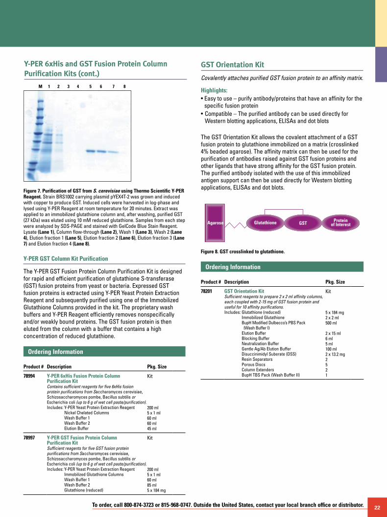
To order, call 800-874-3723 or 815-968-0747. Outside the United States, contact your local branch office or distributor. 22
M 1 2 3 4 5 6 7 8
Figure 7. Purification of GST from S. cerevisiae using Thermo Scientific Y-PER Reagent. Strain BRS1002 carrying plasmid pYEX4T-2 was grown and induced with copper to produce GST. Induced cells were harvested in log-phase and lysed using Y-PER Reagent at room temperature for 20 minutes. Extract was applied to an immobilized glutathione column and, after washing, purified GST (27 kDa) was eluted using 10 mM reduced glutathione. Samples from each step were analyzed by SDS-PAGE and stained with GelCode Blue Stain Reagent. Lysate (Lane 1), Column flow-through (Lane 2), Wash 1 (Lane 3), Wash 2 (Lane 4), Elution fraction 1 (Lane 5), Elution fraction 2 (Lane 6), Elution fraction 3 (Lane 7) and Elution fraction 4 (Lane 8).
Y-PER GST Column Kit Purification
The Y-PER GST Fusion Protein Column Purification Kit is designed for rapid and efficient purification of glutathione S-transferase (GST) fusion proteins from yeast or bacteria. Expressed GST fusion proteins is extracted using Y-PER Yeast Protein Extraction Reagent and subsequently purified using one of the Immobilized Glutathione Columns provided in the kit. The proprietary wash buffers and Y-PER Reagent efficiently removes nonspecifically and/or weakly bound proteins. The GST fusion protein is then eluted from the column with a buffer that contains a high concentration of reduced glutathione.
Ordering Information Product #
Description
Pkg. Size
78994 Y-PER 6xHis Fusion Protein Column Purification KitContains sufficient reagents for five 6xHis fusion protein purifications from Saccharomyces cerevisiae, Schizosaccharomyces pombe, Bacillus subtilis or Escherichia coli (up to 6 g of wet cell paste/purification).Includes: Y-PER Yeast Protein Extraction Reagent
Nickel Chelated Columns Wash Buffer 1 Wash Buffer 2 Elution Buffer
Kit
200 ml 5 x 1 ml 60 ml 60 ml 45 ml
78997 Y-PER GST Fusion Protein Column Purification KitSufficient reagents for five GST fusion protein purifications from Saccharomyces cerevisiae, Schizosaccharomyces pombe, Bacillus subtilis or Escherichia coli (up to 6 g of wet cell paste/purification).Includes: Y-PER Yeast Protein Extraction Reagent
Immobilized Glutathione Columns Wash Buffer 1 Wash Buffer 2 Glutathione (reduced)
Kit
200 ml 5 x 1 ml 60 ml 85 ml 5 x 184 mg
GST Orientation Kit
Covalently attaches purified GST fusion protein to an affinity matrix.
Highlights:• Easy to use – purify antibody/proteins that have an affinity for the
specific fusion protein• Compatible – The purified antibody can be used directly for
Western blotting applications, ELISAs and dot blots
The GST Orientation Kit allows the covalent attachment of a GST fusion protein to glutathione immobilized on a matrix (crosslinked 4% beaded agarose). The affinity matrix can then be used for the purification of antibodies raised against GST fusion proteins and other ligands that have strong affinity for the GST fusion protein. The purified antibody isolated with the use of this immobilized antigen support can then be used directly for Western blotting applications, ELISAs and dot blots.
GlutathioneAgarose GST Proteinof Interest
Figure 8. GST crosslinked to glutathione.
Ordering Information Product #
Description
Pkg. Size
78201 GST Orientation KitSufficient reagents to prepare 2 x 2 ml affinity columns, each coupled with 2-15 mg of GST fusion protein and useful for 10 affinity purifications.Includes: Glutathione (reduced)
Immobilized Glutathione BupH Modified Dulbecco’s PBS Pack (Wash Buffer I) Elution Buffer Blocking Buffer Neutralization Buffer Gentle Ag/Ab Elution Buffer Disuccinimidyl Suberate (DSS) Resin Separators Porous Discs Column Extenders BupH TBS Pack (Wash Buffer II)
Kit 5 x 184 mg 2 x 2 ml 500 ml
2 x 15 ml 6 ml 5 ml 100 ml 2 x 13.2 mg 2 5 2 1
Y-PER 6xHis and GST Fusion Protein Column Purification Kits (cont.)
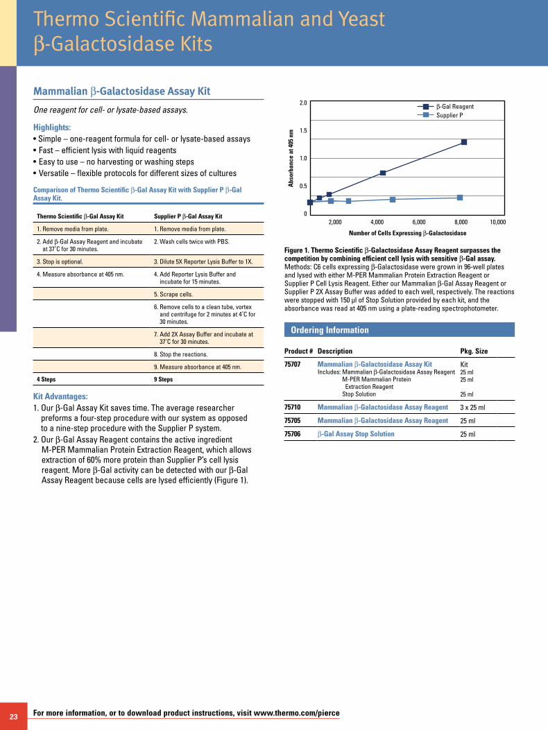
23 For more information, or to download product instructions, visit www.thermo.com/pierce
Thermo Scientific Mammalian and Yeast b-Galactosidase Kits
Mammalian b-Galactosidase Assay Kit
One reagent for cell- or lysate-based assays.
Highlights:• Simple – one-reagent formula for cell- or lysate-based assays• Fast – efficient lysis with liquid reagents• Easy to use – no harvesting or washing steps• Versatile – flexible protocols for different sizes of cultures
Comparison of Thermo Scientific b-Gal Assay Kit with Supplier P b-Gal Assay Kit.
Thermo Scientific b-Gal Assay Kit Supplier P b-Gal Assay Kit
1. Remove media from plate. 1. Remove media from plate.
2. Add b-Gal Assay Reagent and incubate at 37˚C for 30 minutes.
2. Wash cells twice with PBS.
3. Stop is optional. 3. Dilute 5X Reporter Lysis Buffer to 1X.
4. Measure absorbance at 405 nm. 4. Add Reporter Lysis Buffer and incubate for 15 minutes.
5. Scrape cells.
6. Remove cells to a clean tube, vortex and centrifuge for 2 minutes at 4˚C for 30 minutes.
7. Add 2X Assay Buffer and incubate at 37˚C for 30 minutes.
8. Stop the reactions.
9. Measure absorbance at 405 nm.
4 Steps 9 Steps
Kit Advantages:1. Our b-Gal Assay Kit saves time. The average researcher
preforms a four-step procedure with our system as opposed to a nine-step procedure with the Supplier P system.
2. Our b-Gal Assay Reagent contains the active ingredient M-PER Mammalian Protein Extraction Reagent, which allows extraction of 60% more protein than Supplier P’s cell lysis reagent. More b-Gal activity can be detected with our b-Gal Assay Reagent because cells are lysed efficiently (Figure 1).
Supplier Pb-Gal Reagent
2,000
Number of Cells Expressing b-Galactosidase
4,000
Abs
orba
nce
at 4
05 n
m
10,0008,0006,000
2.0
1.5
1.0
0.5
0
Figure 1. Thermo Scientific b-Galactosidase Assay Reagent surpasses the competition by combining efficient cell lysis with sensitive b-Gal assay. Methods: C6 cells expressing b-Galactosidase were grown in 96-well plates and lysed with either M-PER Mammalian Protein Extraction Reagent or Supplier P Cell Lysis Reagent. Either our Mammalian b-Gal Assay Reagent or Supplier P 2X Assay Buffer was added to each well, respectively. The reactions were stopped with 150 µl of Stop Solution provided by each kit, and the absorbance was read at 405 nm using a plate-reading spectrophotometer.
Ordering Information Product #
Description
Pkg. Size
75707 Mammalian b-Galactosidase Assay KitIncludes: Mammalian b-Galactosidase Assay Reagent
M-PER Mammalian Protein Extraction Reagent Stop Solution
Kit25 ml25 ml
25 ml
75710 Mammalian b-Galactosidase Assay Reagent 3 x 25 ml
75705 Mammalian b-Galactosidase Assay Reagent 25 ml
75706 b-Gal Assay Stop Solution 25 ml
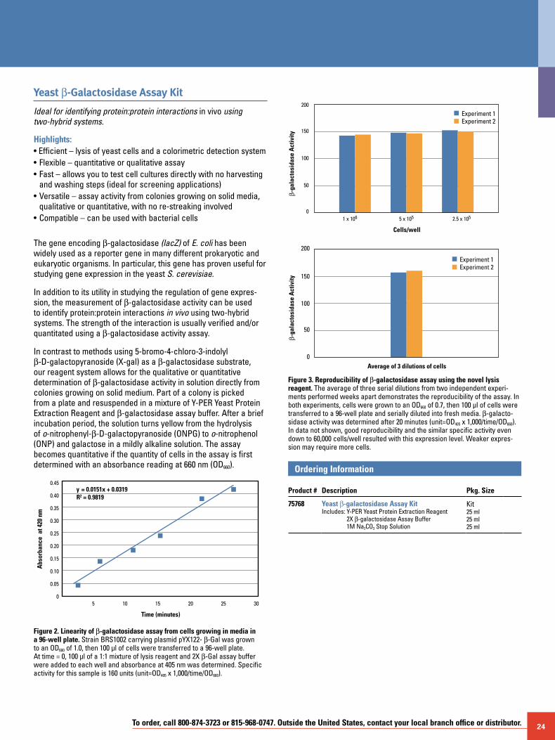
To order, call 800-874-3723 or 815-968-0747. Outside the United States, contact your local branch office or distributor. 24
Yeast b-Galactosidase Assay Kit
Ideal for identifying protein:protein interactions in vivo using two-hybrid systems.
Highlights:• Efficient – lysis of yeast cells and a colorimetric detection system • Flexible – quantitative or qualitative assay• Fast – allows you to test cell cultures directly with no harvesting
and washing steps (ideal for screening applications)• Versatile – assay activity from colonies growing on solid media,
qualitative or quantitative, with no re-streaking involved• Compatible – can be used with bacterial cells
The gene encoding b-galactosidase (lacZ) of E. coli has been widely used as a reporter gene in many different prokaryotic and eukaryotic organisms. In particular, this gene has proven useful for studying gene expression in the yeast S. cerevisiae.
In addition to its utility in studying the regulation of gene expres-sion, the measurement of b-galactosidase activity can be used to identify protein:protein interactions in vivo using two-hybrid systems. The strength of the interaction is usually verified and/or quantitated using a b-galactosidase activity assay.
In contrast to methods using 5-bromo-4-chloro-3-indolyl b-D-galactopyranoside (X-gal) as a b-galactosidase substrate, our reagent system allows for the qualitative or quantitative determination of b-galactosidase activity in solution directly from colonies growing on solid medium. Part of a colony is picked from a plate and resuspended in a mixture of Y-PER Yeast Protein Extraction Reagent and b-galactosidase assay buffer. After a brief incubation period, the solution turns yellow from the hydrolysis of o-nitrophenyl-b-D-galactopyranoside (ONPG) to o-nitrophenol (ONP) and galactose in a mildly alkaline solution. The assay becomes quantitative if the quantity of cells in the assay is first determined with an absorbance reading at 660 nm (OD660).
Figure 2. Linearity of b-galactosidase assay from cells growing in media in a 96-well plate. Strain BRS1002 carrying plasmid pYX122- b-Gal was grown to an OD600 of 1.0, then 100 µl of cells were transferred to a 96-well plate. At time = 0, 100 µl of a 1:1 mixture of lysis reagent and 2X b-Gal assay buffer were added to each well and absorbance at 405 nm was determined. Specific activity for this sample is 160 units (unit=OD405 x 1,000/time/OD660).
5
Time (minutes)
10
Abs
orba
nce
at 4
20 n
m
3020 2515
0.45
0.40
0.35
0.30
0.25
0.20
0.15
0.10
0.05
0
y = 0.0151x + 0.0319R2 = 0.9819
Figure 3. Reproducibility of b-galactosidase assay using the novel lysis reagent. The average of three serial dilutions from two independent experi-ments performed weeks apart demonstrates the reproducibility of the assay. In both experiments, cells were grown to an OD660 of 0.7, then 100 µl of cells were transferred to a 96-well plate and serially diluted into fresh media. b-galacto-sidase activity was determined after 20 minutes (unit=OD405 x 1,000/time/OD660). In data not shown, good reproducibility and the similar specific activity even down to 60,000 cells/well resulted with this expression level. Weaker expres-sion may require more cells.
Ordering Information Product #
Description
Pkg. Size
75768 Yeast b-galactosidase Assay KitIncludes: Y-PER Yeast Protein Extraction Reagent
2X b-galactosidase Assay Buffer 1M Na2CO3 Stop Solution
Kit25 ml25 ml25 ml
b-ga
lact
osid
ase
Act
ivity
b-ga
lact
osid
ase
Act
ivity
200
150
100
50
0
200
150
100
50
0
Cells/well
1 x 10⁶ 5 x 10⁵ 2.5 x 10⁵
Average of 3 dilutions of cells
Experiment 1Experiment 2
Experiment 1Experiment 2
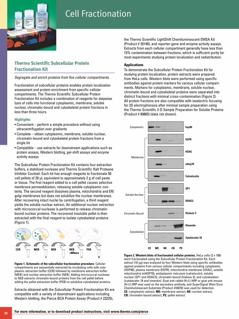
25 For more information, or to download product instructions, visit www.thermo.com/pierce
Thermo Scientific Subcellular Protein Fractionation Kit
Segregate and enrich proteins from five cellular compartments.
Fractionation of subcellular proteins enables protein localization assessment and protein enrichment from specific cellular compartments. The Thermo Scientific Subcellular Protein Fractionation Kit includes a combination of reagents for stepwise lysis of cells into functional cytoplasmic, membrane, soluble nuclear, chromatin-bound and cytoskeletal protein fractions in less than three hours.
Highlights:• Convenient – perform a simple procedure without using
ultracentrifugation over gradients• Complete – obtain cytoplasmic, membrane, soluble nuclear,
chromatin-bound and cytoskeletal protein fractions from a single kit
• Compatible – use extracts for downstream applications such as protein assays, Western blotting, gel-shift assays and enzyme activity assays
The Subcellular Protein Fractionation Kit contains four extraction buffers, a stabilized nuclease and Thermo Scientific Halt Protease Inhibitor Cocktail. Each kit has enough reagents to fractionate 50 cell pellets of 20 µl, equivalent to approximately 2 g of cell paste or tissue. The first reagent added to a cell pellet causes selective membrane permeablization, releasing soluble cytoplasmic con-tents. The second reagent dissolves plasma, mitochondria and ER/golgi membranes but does not solubilize the nuclear membranes. After recovering intact nuclei by centrifugation, a third reagent yields the soluble nuclear extract. An additional nuclear extraction with micrococcal nuclease is performed to release chromatin-bound nuclear proteins. The recovered insoluble pellet is then extracted with the final reagent to isolate cytoskeletal proteins (Figure 1).
CEB MEB NEB PEBNEB+MNase
Figure 1. Schematic of the subcellular fractionation procedure. Cellular compartments are sequentially extracted by incubating cells with cyto-plasmic extraction buffer (CEB) followed by membrane extraction buffer (MEB) and nuclear extraction buffer (NEB). Adding micrococcal nuclease to NEB extracts chromatin-bound proteins from the cell pellet before adding the pellet extraction buffer (PEB) to solubilize cytoskeletal proteins.
Extracts obtained with the Subcellular Protein Fractionation Kit are compatible with a variety of downstream applications including Western blotting, the Pierce BCA Protein Assay (Product # 23225),
the Thermo Scientific LightShift Chemiluminescent EMSA Kit (Product # 20148), and reporter-gene and enzyme-activity assays. Extracts from each cellular compartment generally have less than 15% contamination between fractions, which is sufficient purity for most experiments studying protein localization and redistribution.
ApplicationsTo demonstrate the Subcellular Protein Fractionation Kit for studying protein localization, protein extracts were prepared from HeLa cells. Western blots were performed using specific antibodies against protein markers for various cellular compart-ments. Markers for cytoplasmic, membrane, soluble nuclear, chromatin-bound and cytoskeletal proteins were separated into distinct fractions with minimal cross-contamination (Figure 2). All protein fractions are also compatible with isoelectric focusing for 2D electrophoresis after minimal sample preparation using the Thermo Scientific 2-D Sample Preparation for Soluble Proteins (Product # 89865) (data not shown).
Cytoplasmic hsp90
CE ME NE CB PE
EGFR
VDAC
mhsp70
Calreticulin
SP1
HDAC2
Histone 3
Vimentin
Cytokeratin 18
Membrane
Soluble Nuclear
Chromatin Bound
Cytoskeletal
Figure 2. Western blots of fractionated cellular proteins. HeLa cells (2 x 10⁶) were fractionated using the Subcellular Protein Fractionation Kit. Each extract (10 µg) was analyzed by four Western blots using specific antibodies against proteins from various cellular compartments including cytoplasmic (HSP90), plasma membrane (EGFR), mitochondria membrane (VDAC), soluble mitochondria (mHSP70), endoplasmic reticulum (calreticulin), soluble nuclear (SP1 and HDAC2); chromatin-bound (histone 3); and cytoskeleton (cytokeratin 18 and vimentin). Goat anti-rabbit (H+L) HRP or goat anti-mouse (H+L) HRP was used as the secondary antibody, and SuperSignal West Dura Chemiluminescent Substrate (Product #34076) was used for detection. CE: cytoplasmic extract, ME: membrane extract, NE: nuclear extract, CB: chromatin-bound extract, PE: pellet extract.
Cell Fractionation
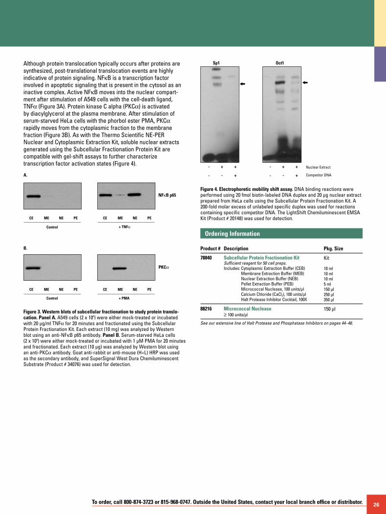
To order, call 800-874-3723 or 815-968-0747. Outside the United States, contact your local branch office or distributor. 26
Although protein translocation typically occurs after proteins are synthesized, post-translational translocation events are highly indicative of protein signaling. NFkB is a transcription factor involved in apoptotic signaling that is present in the cytosol as an inactive complex. Active NFkB moves into the nuclear compart-ment after stimulation of A549 cells with the cell-death ligand, TNFa (Figure 3A). Protein kinase C alpha (PKCa) is activated by diacylglycerol at the plasma membrane. After stimulation of serum-starved HeLa cells with the phorbol ester PMA, PKCa rapidly moves from the cytoplasmic fraction to the membrane fraction (Figure 3B). As with the Thermo Scientific NE-PER Nuclear and Cytoplasmic Extraction Kit, soluble nuclear extracts generated using the Subcellular Fractionation Protein Kit are compatible with gel-shift assays to further characterize transcription factor activation states (Figure 4).
NFκB p65
CE PENEME
PKCα
_
B.
+ TNFαControl
Control
CE PENEME
CE PENEME
+ PMA
CE PENEME
A.
Figure 3. Western blots of subcellular fractionation to study protein translo- cation. Panel A. A549 cells (2 x 10⁶) were either mock-treated or incubated with 20 µg/ml TNFa for 20 minutes and fractionated using the Subcellular Protein Fractionation Kit. Each extract (10 mg) was analyzed by Western blot using an anti-NFkB p65 antibody. Panel B. Serum-starved HeLa cells (2 x 10⁶) were either mock-treated or incubated with 1 µM PMA for 20 minutes and fractionated. Each extract (10 µg) was analyzed by Western blot using an anti-PKCa antibody. Goat anti-rabbit or anti-mouse (H+L) HRP was used as the secondary antibody, and SuperSignal West Dura Chemiluminescent Substrate (Product # 34076) was used for detection.
Sp1
-
-
+
- +
+ -
-
+
- +
+
Oct1
Nuclear Extract
Competitor DNA
Figure 4. Electrophoretic mobility shift assay. DNA binding reactions were performed using 20 fmol biotin-labeled DNA duplex and 20 µg nuclear extract prepared from HeLa cells using the Subcellular Protein Fractionation Kit. A 200-fold molar excess of unlabeled specific duplex was used for reactions containing specific competitor DNA. The LightShift Chemiluminescent EMSA Kit (Product # 20148) was used for detection.
Ordering Information Product #
Description
Pkg. Size
78840 Subcellular Protein Fractionation KitSufficient reagent for 50 cell preps.Includes: Cytoplasmic Extraction Buffer (CEB)
Membrane Extraction Buffer (MEB) Nuclear Extraction Buffer (NEB) Pellet Extraction Buffer (PEB) Micrococcal Nuclease, 100 units/µl Calcium Chloride (CaCl2), 100 units/µl Halt Protease Inhibitor Cocktail, 100X
Kit 10 ml 10 ml 10 ml 5 ml 150 µl 250 µl 350 µl
88216 Micrococcal Nuclease ≥ 100 units/µl
150 µl
See our extensive line of Halt Protease and Phosphatase Inhibitors on pages 44–46.
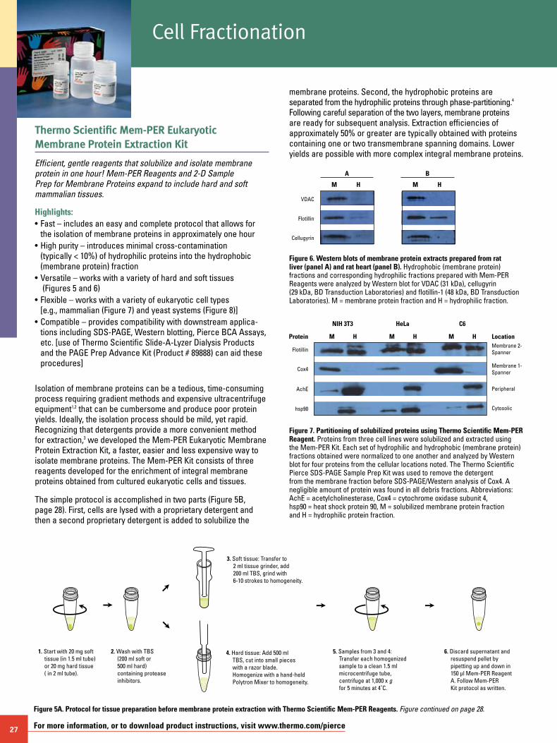
27 For more information, or to download product instructions, visit www.thermo.com/pierce
Thermo Scientific Mem-PER Eukaryotic Membrane Protein Extraction Kit
Efficient, gentle reagents that solubilize and isolate membrane protein in one hour! Mem-PER Reagents and 2-D Sample Prep for Membrane Proteins expand to include hard and soft mammalian tissues.
Highlights:• Fast – includes an easy and complete protocol that allows for
the isolation of membrane proteins in approximately one hour • High purity – introduces minimal cross-contamination
(typically < 10%) of hydrophilic proteins into the hydrophobic (membrane protein) fraction
• Versatile – works with a variety of hard and soft tissues (Figures 5 and 6)
• Flexible – works with a variety of eukaryotic cell types [e.g., mammalian (Figure 7) and yeast systems (Figure 8)]
• Compatible – provides compatibility with downstream applica-tions including SDS-PAGE, Western blotting, Pierce BCA Assays, etc. [use of Thermo Scientific Slide-A-Lyzer Dialysis Products and the PAGE Prep Advance Kit (Product # 89888) can aid these procedures]
Isolation of membrane proteins can be a tedious, time-consuming process requiring gradient methods and expensive ultracentrifuge equipment1,2 that can be cumbersome and produce poor protein yields. Ideally, the isolation process should be mild, yet rapid. Recognizing that detergents provide a more convenient method for extraction,3 we developed the Mem-PER Eukaryotic Membrane Protein Extraction Kit, a faster, easier and less expensive way to isolate membrane proteins. The Mem-PER Kit consists of three reagents developed for the enrichment of integral membrane proteins obtained from cultured eukaryotic cells and tissues.
The simple protocol is accomplished in two parts (Figure 5B, page 28). First, cells are lysed with a proprietary detergent and then a second proprietary detergent is added to solubilize the
membrane proteins. Second, the hydrophobic proteins are separated from the hydrophilic proteins through phase-partitioning.4 Following careful separation of the two layers, membrane proteins are ready for subsequent analysis. Extraction efficiencies of approximately 50% or greater are typically obtained with proteins containing one or two transmembrane spanning domains. Lower yields are possible with more complex integral membrane proteins.
A
VDAC
M H
Flotillin
B
M H
Cellugyrin
Figure 6. Western blots of membrane protein extracts prepared from rat liver (panel A) and rat heart (panel B). Hydrophobic (membrane protein) fractions and corresponding hydrophilic fractions prepared with Mem-PER Reagents were analyzed by Western blot for VDAC (31 kDa), cellugyrin (29 kDa, BD Transduction Laboratories) and flotillin-1 (48 kDa, BD Transduction Laboratories). M = membrane protein fraction and H = hydrophilic fraction.
Figure 7. Partitioning of solubilized proteins using Thermo Scientific Mem-PER Reagent. Proteins from three cell lines were solubilized and extracted using the Mem-PER Kit. Each set of hydrophilic and hydrophobic (membrane protein) fractions obtained were normalized to one another and analyzed by Western blot for four proteins from the cellular locations noted. The Thermo Scientific Pierce SDS-PAGE Sample Prep Kit was used to remove the detergent from the membrane fraction before SDS-PAGE/Western analysis of Cox4. A negligible amount of protein was found in all debris fractions. Abbreviations: AchE = acetylcholinesterase, Cox4 = cytochrome oxidase subunit 4, hsp90 = heat shock protein 90, M = solubilized membrane protein fraction and H = hydrophilic protein fraction.
1. Start with 20 mg soft tissue (in 1.5 ml tube) or 20 mg hard tissue ( in 2 ml tube).
3. Soft tissue: Transfer to 2 ml tissue grinder, add 200 ml TBS, grind with 6-10 strokes to homogeneity.
4. Hard tissue: Add 500 ml TBS, cut into small pieces with a razor blade. Homogenize with a hand-held Polytron Mixer to homogeneity.
6. Discard supernatant and resuspend pellet by pipetting up and down in 150 µl Mem-PER Reagent A. Follow Mem-PER Kit protocol as written.
2. Wash with TBS (200 ml soft or 500 ml hard) containing protease inhibitors.
5. Samples from 3 and 4: Transfer each homogenized sample to a clean 1.5 ml microcentrifuge tube, centrifuge at 1,000 x g for 5 minutes at 4˚C.
Figure 5A. Protocol for tissue preparation before membrane protein extraction with Thermo Scientific Mem-PER Reagents. Figure continued on page 28.
M
NIH 3T3 HeLa C6
Protein LocationMembrane 2-Spanner
Membrane 1-Spanner
Peripheral
Cytosolic
Flotillin
Cox4
AchE
hsp90
H M H M H
Cell Fractionation
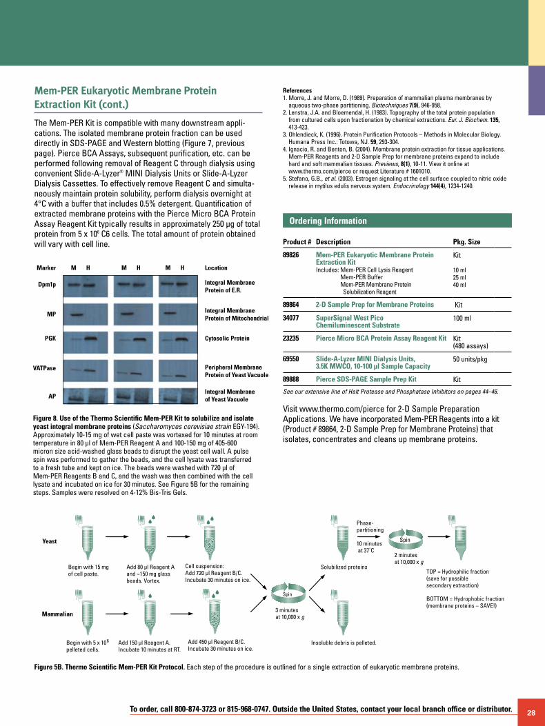
To order, call 800-874-3723 or 815-968-0747. Outside the United States, contact your local branch office or distributor. 28
Mem-PER Eukaryotic Membrane Protein Extraction Kit (cont.)
The Mem-PER Kit is compatible with many downstream appli-cations. The isolated membrane protein fraction can be used directly in SDS-PAGE and Western blotting (Figure 7, previous page). Pierce BCA Assays, subsequent purification, etc. can be performed following removal of Reagent C through dialysis using convenient Slide-A-Lyzer® MINI Dialysis Units or Slide-A-Lyzer Dialysis Cassettes. To effectively remove Reagent C and simulta-neously maintain protein solubility, perform dialysis overnight at 4°C with a buffer that includes 0.5% detergent. Quantification of extracted membrane proteins with the Pierce Micro BCA Protein Assay Reagent Kit typically results in approximately 250 µg of total protein from 5 x 10⁶ C6 cells. The total amount of protein obtained will vary with cell line.
References1. Morre, J. and Morre, D. (1989). Preparation of mammalian plasma membranes by
aqueous two-phase partitioning. Biotechniques 7(9), 946-958.2. Lenstra, J.A. and Bloemendal, H. (1983). Topography of the total protein population
from cultured cells upon fractionation by chemical extractions. Eur. J. Biochem. 135, 413-423.
3. Ohlendieck, K. (1996). Protein Purification Protocols – Methods in Molecular Biology. Humana Press Inc.: Totowa, NJ. 59, 293-304.
4. Ignacio, R. and Benton, B. (2004). Membrane protein extraction for tissue applications. Mem-PER Reagents and 2-D Sample Prep for membrane proteins expand to include hard and soft mammalian tissues. Previews, 8(1), 10-11. View it online at www.thermo.com/pierce or request Literature # 1601010.
5. Stefano, G.B., et al. (2003). Estrogen signaling at the cell surface coupled to nitric oxide release in mytilus edulis nervous system. Endocrinology 144(4), 1234-1240.
Ordering Information Product #
Description
Pkg. Size
89826 Mem-PER Eukaryotic Membrane Protein Extraction KitIncludes: Mem-PER Cell Lysis Reagent
Mem-PER Buffer Mem-PER Membrane Protein Solubilization Reagent
Kit 10 ml 25 ml 40 ml
89864 2-D Sample Prep for Membrane Proteins Kit
34077 SuperSignal West Pico Chemiluminescent Substrate
100 ml
23235 Pierce Micro BCA Protein Assay Reagent Kit Kit (480 assays)
69550 Slide-A-Lyzer MINI Dialysis Units, 3.5K MWCO, 10-100 µl Sample Capacity
50 units/pkg
89888 Pierce SDS-PAGE Sample Prep Kit Kit
See our extensive line of Halt Protease and Phosphatase Inhibitors on pages 44–46.
Visit www.thermo.com/pierce for 2-D Sample Preparation Applications. We have incorporated Mem-PER Reagents into a kit (Product # 89864, 2-D Sample Prep for Membrane Proteins) that isolates, concentrates and cleans up membrane proteins.
MMarker
Dpm1p
MP
PGK
VATPase
AP
Location
Integral MembraneProtein of E.R.
Integral MembraneProtein of Mitochondrial
Cytosolic Protein
Peripheral MembraneProtein of Yeast Vacuole
Integral Membraneof Yeast Vacuole
H M H M H
Figure 8. Use of the Thermo Scientific Mem-PER Kit to solubilize and isolate yeast integral membrane proteins (Saccharomyces cerevisiae strain EGY-194). Approximately 10-15 mg of wet cell paste was vortexed for 10 minutes at room temperature in 80 µl of Mem-PER Reagent A and 100-150 mg of 405-600 micron size acid-washed glass beads to disrupt the yeast cell wall. A pulse spin was performed to gather the beads, and the cell lysate was transferred to a fresh tube and kept on ice. The beads were washed with 720 µl of Mem-PER Reagents B and C, and the wash was then combined with the cell lysate and incubated on ice for 30 minutes. See Figure 5B for the remaining steps. Samples were resolved on 4-12% Bis-Tris Gels.
Begin with 15 mg of cell paste.
Phase- partitioning
10 minutes at 37˚C
TOP = Hydrophilic fraction (save for possible secondary extraction)
BOTTOM = Hydrophobic fraction (membrane proteins – SAVE!)
Solubilized proteins
Insoluble debris is pelleted.
3 minutes at 10,000 x g
Spin
2 minutes at 10,000 x g
Spin
Cell suspension: Add 720 µl Reagent B/C. Incubate 30 minutes on ice.
Add 80 µl Reagent Aand ~150 mg glassbeads. Vortex.
Yeast
Mammalian
Begin with 5 x 10⁶ pelleted cells.
Add 450 µl Reagent B/C.Incubate 30 minutes on ice.
Add 150 µl Reagent A. Incubate 10 minutes at RT.
Figure 5B. Thermo Scientific Mem-PER Kit Protocol. Each step of the procedure is outlined for a single extraction of eukaryotic membrane proteins.
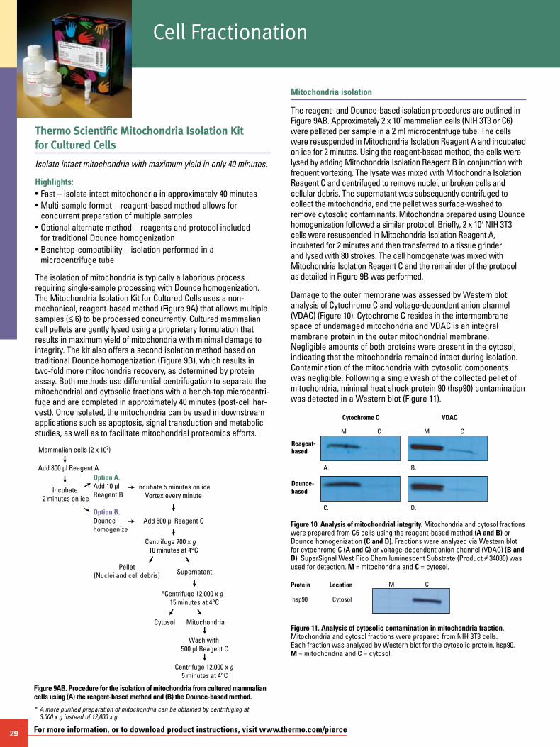
29 For more information, or to download product instructions, visit www.thermo.com/pierce
Thermo Scientific Mitochondria Isolation Kit for Cultured Cells
Isolate intact mitochondria with maximum yield in only 40 minutes.
Highlights:• Fast – isolate intact mitochondria in approximately 40 minutes• Multi-sample format – reagent-based method allows for concurrent preparation of multiple samples• Optional alternate method – reagents and protocol included for traditional Dounce homogenization• Benchtop-compatibility – isolation performed in a
microcentrifuge tube
The isolation of mitochondria is typically a laborious process requiring single-sample processing with Dounce homogenization. The Mitochondria Isolation Kit for Cultured Cells uses a non-mechanical, reagent-based method (Figure 9A) that allows multiple samples (≤ 6) to be processed concurrently. Cultured mammalian cell pellets are gently lysed using a proprietary formulation that results in maximum yield of mitochondria with minimal damage to integrity. The kit also offers a second isolation method based on traditional Dounce homogenization (Figure 9B), which results in two-fold more mitochondria recovery, as determined by protein assay. Both methods use differential centrifugation to separate the mitochondrial and cytosolic fractions with a bench-top microcentri-fuge and are completed in approximately 40 minutes (post-cell har-vest). Once isolated, the mitochondria can be used in downstream applications such as apoptosis, signal transduction and metabolic studies, as well as to facilitate mitochondrial proteomics efforts.
Mitochondria isolation
The reagent- and Dounce-based isolation procedures are outlined in Figure 9AB. Approximately 2 x 10⁷ mammalian cells (NIH 3T3 or C6) were pelleted per sample in a 2 ml microcentrifuge tube. The cells were resuspended in Mitochondria Isolation Reagent A and incubated on ice for 2 minutes. Using the reagent-based method, the cells were lysed by adding Mitochondria Isolation Reagent B in conjunction with frequent vortexing. The lysate was mixed with Mitochondria Isolation Reagent C and centrifuged to remove nuclei, unbroken cells and cellular debris. The supernatant was subsequently centrifuged to collect the mitochondria, and the pellet was surface-washed to remove cytosolic contaminants. Mitochondria prepared using Dounce homogenization followed a similar protocol. Briefly, 2 x 10⁷ NIH 3T3 cells were resuspended in Mitochondria Isolation Reagent A, incubated for 2 minutes and then transferred to a tissue grinder and lysed with 80 strokes. The cell homogenate was mixed with Mitochondria Isolation Reagent C and the remainder of the protocol as detailed in Figure 9B was performed.
Damage to the outer membrane was assessed by Western blot analysis of Cytochrome C and voltage-dependent anion channel (VDAC) (Figure 10). Cytochrome C resides in the intermembrane space of undamaged mitochondria and VDAC is an integral membrane protein in the outer mitochondrial membrane. Negligible amounts of both proteins were present in the cytosol, indicating that the mitochondria remained intact during isolation. Contamination of the mitochondria with cytosolic components was negligible. Following a single wash of the collected pellet of mitochondria, minimal heat shock protein 90 (hsp90) contamination was detected in a Western blot (Figure 11).
Figure 9AB. Procedure for the isolation of mitochondria from cultured mammalian cells using (A) the reagent-based method and (B) the Dounce-based method.
* A more purified preparation of mitochondria can be obtained by centrifuging at 3,000 x g instead of 12,000 x g.
Mammalian cells (2 x 10⁷)
Add 800 µl Reagent A
Incubate 2 minutes on ice
Option A. Add 10 µl Reagent B
Option B. Dounce homogenize
Incubate 5 minutes on iceVortex every minute
Add 800 µl Reagent C
Pellet(Nuclei and cell debris) Supernatant
*Centrifuge 12,000 x g 15 minutes at 4°C
Cytosol Mitochondria
Wash with 500 µl Reagent C
Centrifuge 700 x g 10 minutes at 4°C
Centrifuge 12,000 x g 5 minutes at 4°C
Figure 10. Analysis of mitochondrial integrity. Mitochondria and cytosol fractions were prepared from C6 cells using the reagent-based method (A and B) or Dounce homogenization (C and D). Fractions were analyzed via Western blot for cytochrome C (A and C) or voltage-dependent anion channel (VDAC) (B and D). SuperSignal West Pico Chemiluminescent Substrate (Product # 34080) was used for detection. M = mitochondria and C = cytosol.
Cytochrome C
Reagent-based
A.
C.
B.
D.
M C
Dounce-based
VDAC
M C
Cytosolhsp90
Protein Location M C
Figure 11. Analysis of cytosolic contamination in mitochondria fraction. Mitochondria and cytosol fractions were prepared from NIH 3T3 cells. Each fraction was analyzed by Western blot for the cytosolic protein, hsp90. M = mitochondria and C = cytosol.
Cell Fractionation
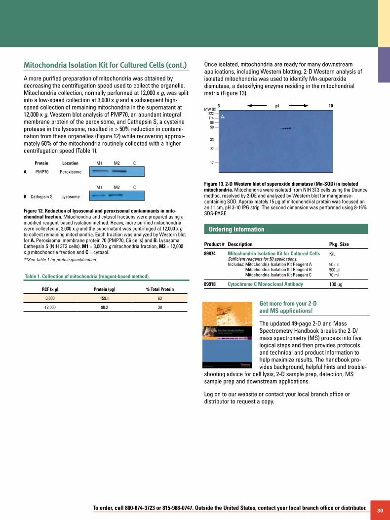
To order, call 800-874-3723 or 815-968-0747. Outside the United States, contact your local branch office or distributor. 30
Mitochondria Isolation Kit for Cultured Cells (cont.)
A more purified preparation of mitochondria was obtained by decreasing the centrifugation speed used to collect the organelle. Mitochondria collection, normally performed at 12,000 x g, was split into a low-speed collection at 3,000 x g and a subsequent high-speed collection of remaining mitochondria in the supernatant at 12,000 x g. Western blot analysis of PMP70, an abundant integral membrane protein of the peroxisome, and Cathepsin S, a cysteine protease in the lysosome, resulted in > 50% reduction in contami-nation from these organelles (Figure 12) while recovering approxi-mately 60% of the mitochondria routinely collected with a higher centrifugation speed (Table 1).
PeroxisomePMP70
Protein Location M1 M2 C
M1 M2 C
LysosomeCathepsin S
A.
B.
Figure 12. Reduction of lysosomal and peroxisomal contaminants in mito- chondrial fraction. Mitochondria and cytosol fractions were prepared using a modified reagent-based isolation method. Heavy, more purified mitochondria were collected at 3,000 x g and the supernatant was centrifuged at 12,000 x g to collect remaining mitochondria. Each fraction was analyzed by Western blot for A. Peroxisomal membrane protein 70 (PMP70, C6 cells) and B. Lysosomal Cathepsin S (NIH 3T3 cells). M1 = 3,000 x g mitochondria fraction, M2 = 12,000 x g mitochondria fraction and C = cytosol. **See Table 1 for protein quantification.
Table 1. Collection of mitochondria (reagent-based method).
RCF (x g) Protein (µg) % Total Protein
3,000 159.1 62
12,000 98.2 38
Once isolated, mitochondria are ready for many downstream applications, including Western blotting. 2-D Western analysis of isolated mitochondria was used to identify Mn-superoxide dismutase, a detoxifying enzyme residing in the mitochondrial matrix (Figure 13).
B.
MW (K)222 —114 —
88 —50 —
33 —
27 —
17 —
222 —114 —
88 —50 —
33 —
27 —
17 —
222 —114 —
88 —50 —33 —
27 —
17 —
3 10pl
C.
Figure 13. 2-D Western blot of superoxide dismutase (Mn-SOD) in isolated mitochondria. Mitochondria were isolated from NIH 3T3 cells using the Dounce method, resolved by 2-DE and analyzed by Western blot for manganese-containing SOD. Approximately 15 µg of mitochondrial protein was focused on an 11 cm, pH 3-10 IPG strip. The second dimension was performed using 8-16% SDS-PAGE.
Ordering Information Product #
Description
Pkg. Size
89874 Mitochondria Isolation Kit for Cultured CellsSufficient reagents for 50 applications.Includes: Mitochondria Isolation Kit Reagent A
Mitochondria Isolation Kit Reagent B Mitochondria Isolation Kit Reagent C
Kit
50 ml 500 µl 70 ml
89918 Cytochrome C Monoclonal Antibody 100 µg
Get more from your 2-D and MS applications!
The updated 49-page 2-D and Mass Spectrometry Handbook breaks the 2-D/mass spectrometry (MS) process into five logical steps and then provides protocols and technical and product information to help maximize results. The handbook pro-vides background, helpful hints and trouble-
shooting advice for cell lysis, 2-D sample prep, detection, MS sample prep and downstream applications.
Log on to our website or contact your local branch office or distributor to request a copy.
A.
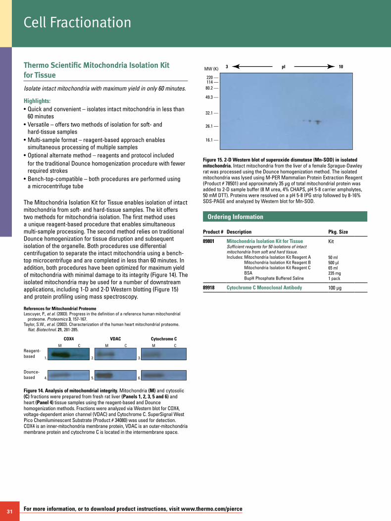
31 For more information, or to download product instructions, visit www.thermo.com/pierce
Thermo Scientific Mitochondria Isolation Kit for Tissue
Isolate intact mitochondria with maximum yield in only 60 minutes.
Highlights:• Quick and convenient – isolates intact mitochondria in less than
60 minutes• Versatile – offers two methods of isolation for soft- and
hard-tissue samples• Multi-sample format – reagent-based approach enables
simultaneous processing of multiple samples• Optional alternate method – reagents and protocol included for the traditional Dounce homogenization procedure with fewer
required strokes• Bench-top-compatible – both procedures are performed using
a microcentrifuge tube
The Mitochondria Isolation Kit for Tissue enables isolation of intact mitochondria from soft- and hard-tissue samples. The kit offers two methods for mitochondria isolation. The first method uses a unique reagent-based procedure that enables simultaneous multi-sample processing. The second method relies on traditional Dounce homogenization for tissue disruption and subsequent isolation of the organelle. Both procedures use differential centrifugation to separate the intact mitochondria using a bench-top microcentrifuge and are completed in less than 60 minutes. In addition, both procedures have been optimized for maximum yield of mitochondria with minimal damage to its integrity (Figure 14). The isolated mitochondria may be used for a number of downstream applications, including 1-D and 2-D Western blotting (Figure 15) and protein profiling using mass spectroscopy.
References for Mitochondrial ProteomeLescuyer, P., et al. (2003). Progress in the definition of a reference human mitochondrial
proteome. Proteomics 3, 157-167.Taylor, S.W., et al. (2003). Characterization of the human heart mitochondrial proteome.
Nat. Biotechnol. 21, 281-285.
Reagent-based
Dounce-based
VDACCOX4M C M C M C
Cytochrome C
1. 2. 3.
4. 5. 6.
Figure 14. Analysis of mitochondrial integrity. Mitochondria (M) and cytosolic (C) fractions were prepared from fresh rat liver (Panels 1, 2, 3, 5 and 6) and heart (Panel 4) tissue samples using the reagent-based and Dounce homogenization methods. Fractions were analyzed via Western blot for COX4, voltage-dependent anion channel (VDAC) and Cytochrome C. SuperSignal West Pico Chemiluminescent Substrate (Product # 34080) was used for detection. COX4 is an inner-mitochondria membrane protein, VDAC is an outer-mitochondria membrane protein and cytochrome C is located in the intermembrane space.
MW (K)
220 —114 —80.2 —
49.3 —
32.1 —
26.1 —
16.1 —
3 10pl
Figure 15. 2-D Western blot of superoxide dismutase (Mn-SOD) in isolated mitochondria. Intact mitochondria from the liver of a female Sprague-Dawley rat was processed using the Dounce homogenization method. The isolated mitochondria was lysed using M-PER Mammalian Protein Extraction Reagent (Product # 78501) and approximately 35 µg of total mitochondrial protein was added to 2-D sample buffer (8 M urea, 4% CHAPS, pH 5-8 carrier ampholytes, 50 mM DTT). Proteins were resolved on a pH 5-8 IPG strip followed by 8-16% SDS-PAGE and analyzed by Western blot for Mn-SOD.
Ordering Information Product #
Description
Pkg. Size
89801 Mitochondria Isolation Kit for TissueSufficient reagents for 50 isolations of intact mitochondria from soft and hard tissue.Includes: Mitochondria Isolation Kit Reagent A
Mitochondria Isolation Kit Reagent B Mitochondria Isolation Kit Reagent C BSA BupH Phosphate Buffered Saline
Kit 50 ml 500 µl 65 ml 235 mg 1 pack
89918 Cytochrome C Monoclonal Antibody 100 µg
Cell Fractionation
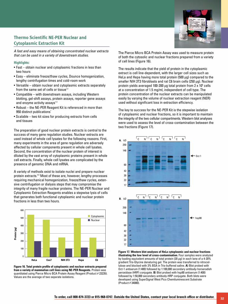
To order, call 800-874-3723 or 815-968-0747. Outside the United States, contact your local branch office or distributor. 32
Thermo Scientific NE-PER Nuclear and Cytoplasmic Extraction Kit
A fast and easy means of obtaining concentrated nuclear extracts that can be used in a variety of downstream studies.
Highlights:• Fast – obtain nuclear and cytoplasmic fractions in less than
two hours• Easy – eliminate freeze/thaw cycles, Dounce homogenization,
lengthy centrifugation times and cold-room work• Versatile – obtain nuclear and cytoplasmic extracts separately
from the same set of cells or tissue1,2
• Compatible – with downstream assays, including Western blotting, gel-shift assays, protein assays, reporter gene assays and enzyme activity assays1-3
• Robust – the NE-PER Reagent Kit is referenced in more than 950 distinct publications*
• Scalable – two kit sizes for producing extracts from cells and tissues
The preparation of good nuclear protein extracts is central to the success of many gene regulation studies. Nuclear extracts are used instead of whole cell lysates for the following reasons. First, many experiments in the area of gene regulation are adversely affected by cellular components present in whole cell lysates. Second, the concentration of the nuclear protein of interest is diluted by the vast array of cytoplasmic proteins present in whole cell extracts. Finally, whole cell lysates are complicated by the presence of genomic DNA and mRNA.
A variety of methods exist to isolate nuclei and prepare nuclear protein extracts.4-6 Most of these are, however, lengthy processes requiring mechanical homogenization, freeze/thaw cycles, exten-sive centrifugation or dialysis steps that may compromise the integrity of many fragile nuclear proteins. The NE-PER Nuclear and Cytoplasmic Extraction Reagents enables a stepwise lysis of cells that generates both functional cytoplasmic and nuclear protein fractions in less than two hours.
The Pierce Micro BCA Protein Assay was used to measure protein of both the cytosolic and nuclear fractions prepared from a variety of cell lines (Figure 16).
The results indicate that the yield of protein in the cytoplasmic extract is cell line-dependent, with the larger cell sizes such as HeLa and Hepa having more total protein (500 µg) compared to the smaller NIH 3T3 fibroblasts and rat C6 brain cells (250 µg). Nuclear protein yields averaged 100-200 µg total protein from 2 x 10⁶ cells at a concentration of 1.5 mg/ml, independent of cell type. The protein concentration of the nuclear extracts can be manipulated easily by varying the volume of nuclear extraction reagent (NER) used without significant loss in extraction efficiency.
The key to success for the NE-PER Kit is the stepwise isolation of cytoplasmic and nuclear fractions, so it is important to maintain the integrity of the two cellular compartments. Western blot analyses were used to assess the level of cross-contamination between the two fractions (Figure 17).
Oct 1
hsp90
A. C N C N C N C NkD
250
148
60
42
30
22
17
C N C N C N C NB. kD
250
148
60
42
30
22
17
CytoplasmicNuclear
HeLa Cos7
Prot
ein
(µg)
C6NIH 3T3 Hepa
600
500
400
300
200
100
0
Figure 16. Total protein profile of cytoplasmic and nuclear extracts prepared from a variety of mammalian cell lines using NE-PER Reagents. Protein was quantitated using Pierce Micro BCA Protein Assay Reagent (Product # 23235). Values are the average of two separate isolations.
Figure 17. Western blot analyses of HeLa cytoplasmic and nuclear fractions illustrating the low level of cross-contamination. Four samples were analyzed by loading equivalent amounts of total protein (20 µg) in each lane of a 4-20% gradient Tris-Glycine denaturing gel. The protein was transferred to nitrocel- lulose and blocked with 3% BSA in Tris-buffered saline. A) Blot probed with Oct-1 antiserum (1:400) followed by 1:100,000 secondary antibody-horseradish peroxidase (HRP) conjugate. B) Blot probed with hsp90 antiserum (1:400) followed by 1:50,000 secondary antibody-HRP conjugate. Both blots were developed using SuperSignal West Pico Chemiluminescent Substrate (Product # 34080).
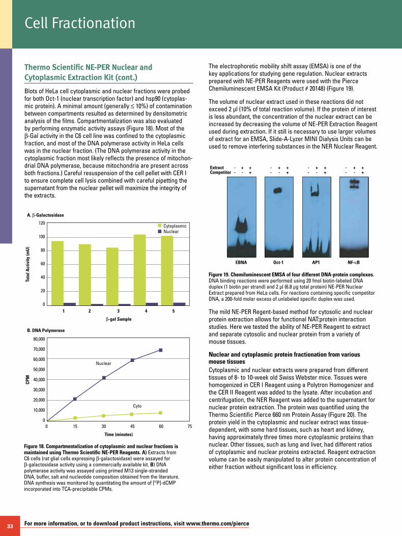
33 For more information, or to download product instructions, visit www.thermo.com/pierce
Thermo Scientific NE-PER Nuclear and Cytoplasmic Extraction Kit (cont.)
Blots of HeLa cell cytoplasmic and nuclear fractions were probed for both Oct-1 (nuclear transcription factor) and hsp90 (cytoplas-mic protein). A minimal amount (generally ≤ 10%) of contamination between compartments resulted as determined by densitometric analysis of the films. Compartmentalization was also evaluated by performing enzymatic activity assays (Figure 18). Most of the b-Gal activity in the C6 cell line was confined to the cytoplasmic fraction, and most of the DNA polymerase activity in HeLa cells was in the nuclear fraction. (The DNA polymerase activity in the cytoplasmic fraction most likely reflects the presence of mitochon-drial DNA polymerase, because mitochondria are present across both fractions.) Careful resuspension of the cell pellet with CER I to ensure complete cell lysis combined with careful pipetting the supernatant from the nuclear pellet will maximize the integrity of the extracts.
CytoplasmicNuclear
1
A. b-Galactosidase
b-gal Sample
2
Tota
l Act
ivity
(mU
)
53 4
120
100
80
60
40
20
0
Cyto
Nuclear
0
B. DNA Polymerase
Time (minutes)
15
CPM
7530 45 60
80,000
70,000
60,000
50,000
40,000
30,000
20,000
10,000
0
Figure 18. Compartmentalization of cytoplasmic and nuclear fractions is maintained using Thermo Scientific NE-PER Reagents. A) Extracts from C6 cells (rat glial cells expressing b-galactosidase) were assayed for b-galactosidase activity using a commercially available kit. B) DNA polymerase activity was assayed using primed M13 single-stranded DNA, buffer, salt and nucleotide composition obtained from the literature. DNA synthesis was monitored by quantitating the amount of [32P]-dCMP incorporated into TCA-precipitable CPMs.
The electrophoretic mobility shift assay (EMSA) is one of the key applications for studying gene regulation. Nuclear extracts prepared with NE-PER Reagents were used with the Pierce Chemiluminescent EMSA Kit (Product # 20148) (Figure 19).
The volume of nuclear extract used in these reactions did not exceed 2 µl (10% of total reaction volume). If the protein of interest is less abundant, the concentration of the nuclear extract can be increased by decreasing the volume of NE-PER Extraction Reagent used during extraction. If it still is necessary to use larger volumes of extract for an EMSA, Slide-A-Lyzer MINI Dialysis Units can be used to remove interfering substances in the NER Nuclear Reagent.
Extract - + +Competitor - - +
- + +- - +
- + +- - +
- + +- - +
EBNA Oct-1 AP1 NF- B
Figure 19. Chemiluminescent EMSA of four different DNA-protein complexes. DNA binding reactions were performed using 20 fmol biotin-labeled DNA duplex (1 biotin per strand) and 2 µl (6.8 µg total protein) NE-PER Nuclear Extract prepared from HeLa cells. For reactions containing specific competitor DNA, a 200-fold molar excess of unlabeled specific duplex was used.
The mild NE-PER Regent-based method for cytosolic and nuclear protein extraction allows for functional NAT:protein interaction studies. Here we tested the ability of NE-PER Reagent to extract and separate cytosolic and nuclear protein from a variety of mouse tissues.
Nuclear and cytoplasmic protein fractionation from various mouse tissuesCytoplasmic and nuclear extracts were prepared from different tissues of 8- to 10-week old Swiss Webster mice. Tissues were homogenized in CER I Reagent using a Polytron Homogenizer and the CER II Reagent was added to the lysate. After incubation and centrifugation, the NER Reagent was added to the supernatant for nuclear protein extraction. The protein was quantified using the Thermo Scientific Pierce 660 nm Protein Assay (Figure 20). The protein yield in the cytoplasmic and nuclear extract was tissue-dependent, with some hard tissues, such as heart and kidney, having approximately three times more cytoplasmic proteins than nuclear. Other tissues, such as lung and liver, had different ratios of cytoplasmic and nuclear proteins extracted. Reagent extraction volume can be easily manipulated to alter protein concentration of either fraction without significant loss in efficiency.
Cell Fractionation
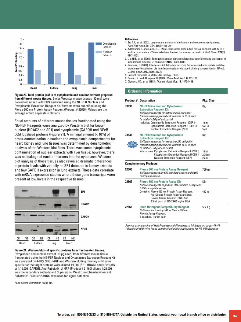
To order, call 800-874-3723 or 815-968-0747. Outside the United States, contact your local branch office or distributor. 34
0
0.2
Tota
l Pro
tein
(mg)
0.4
0.6
0.8
1
1.2
1.4
1.6
1.8
2
Heart Kidney Lung Liver
CytoplasmicExtract
NuclearExtract
Figure 20. Total protein profile of cytoplasmic and nuclear extracts prepared from different mouse tissues. Swiss Webster mouse tissues (40 mg) were harvested, rinsed with PBS and lysed using the NE-PER Nuclear and Cytoplasmic Extraction Reagent Kit. Extracts were quantified using the Pierce 660 nm Protein Assay Reagent (Product # 22660). Values are the average of two separate isolations.
Equal amounts of different mouse tissues fractionated using the NE-PER Reagents were analyzed by Western blot for known nuclear (HDAC2 and SP1) and cytoplasmic (GAPDH and NFkB p65) localized proteins (Figure 21). A minimal amount (< 10%) of cross-contamination in nuclear and cytoplasmic compartments for heart, kidney and lung tissues was determined by densitometric analysis of the Western blot films. There was some cytoplasmic contamination of nuclear extracts with liver tissue; however, there was no leakage of nuclear markers into the cytoplasm. Western blot analysis of these tissues also revealed dramatic differences in protein levels with virtually no SP1 detected in kidney extracts and low GAPDH expression in lung extracts. These data correlate with mRNA expression studies where these gene transcripts were present at low levels in the respective tissues.1
CE NE
Heart
HDAC2
SP1
NFκB
GAPDH
CE NE CE NE CE NE
Kidney Lung Liver
Figure 21. Western blots of specific proteins from fractionated tissues. Cytoplasmic and nuclear extract (10 µg each) from different mouse tissue fractionated using the NE-PER Nuclear and Cytoplasmic Extraction Reagent Kit was analyzed by 4-20% SDS-PAGE and Western blotting. Primary antibodies specific for the target proteins were diluted 1:1,000 (SP1, HDAC2 and NFkB p65, or 1:10,000 (GAPDH). Anti-Rabbit (H+L) HRP (Product # 31460) diluted 1:25,000 was the secondary antibody and SuperSignal West Dura Chemiluminescent Substrate† (Product # 34076) was used for signal detection.
† See patent information (page 50).
References1. Su, A.I., et al. (2002). Large-scale analysis of the human and mouse transcriptomes.
Proc Natl Acad Sci USA. 99(7): 4465-70.2. Adilakshmi, T. and Laine, R.O. (2002). Ribosomal protein S25 mRNA partners with MTF-1
and La to provide a p53-mediated mechanism for survival or death. J. Biol. Chem. 277(6), 4147-4151.
3. Liu, H.B., et al. (2003). Estrogen receptor alpha mediates estrogen’s immune protection in autoimmune disease. J. Immunol. 171(12), 6936-6940.
4. Sanceau, J. (2002). Interferons inhibit tumor necrosis factor-a-mediated matrix metallo-proteinase-9 activation via interferon regulatory factor-1 binding competition for NF-kb. J. Biol. Chem. 277, 35766-35775.
5. Current Protocols in Molecular Biology (1993).6. Zerivitz, K. and Akusjarvi, G. (1989). Gene Anal. Tech. 6, 101-109.7. Dignam, J.D., et al. (1983). Nucleic Acids Res. 11, 1475-1489.
Ordering Information Product #
Description
Pkg. Size
78833 NE-PER Nuclear and Cytoplasmic Extraction Reagent KitSufficient reagents for extracting 50 cell pellet fractions having packed cell volumes of 20 µl each (a total of ~2.0 g of cell paste).Includes: Cytoplasmic Extraction Reagent I (CER I)
Cytoplasmic Extraction Reagent II (CER II) Nuclear Extraction Reagent (NER)
Kit 10 ml 550 µl 5 ml
78835
NEWLarger Kit!
NE-PER Nuclear and Cytoplasmic Extraction Reagent KitSufficient reagents for extracting 250 cell pelletfractions having packed cell volumes of 20 µl each (a total of ~ 10 g of cell paste).Kit contains: Cytoplasmic Extraction Reagent I (CER I)
Cytoplasmic Extraction Reagent II CER II Nuclear Extraction Reagent (NER)
Kit 10 ml 2.75 ml25 ml
Complementary Products
22660 Pierce 660 nm Protein Assay ReagentSufficient reagent for 500 standard assays and 5,000 microplate assays.
750 ml
22662 Pierce 660 nm Protein Assay KitSufficient reagents to perform 300 standard assays and 3,000 microplate assays.Contains: Pierce 660 nm Protein Assay Reagent
Pre-Diluted Protein Assay Standards, Bovine Serum Albumin (BSA) Set 3.5 ml each of 125-2,000 mg/ml BSA
Kit 450 ml
22663 Ionic Detergent Compatibility ReagentSufficient for treating 100 ml Pierce 660 nm Protein Assay Reagent.5 pouches, 1 gram each
5 x 1 g
See our extensive line of Halt Protease and Phosphatase Inhibitors on pages 44–46. * Results of HighWire Press search of scientific publications for NE-PER Reagent.
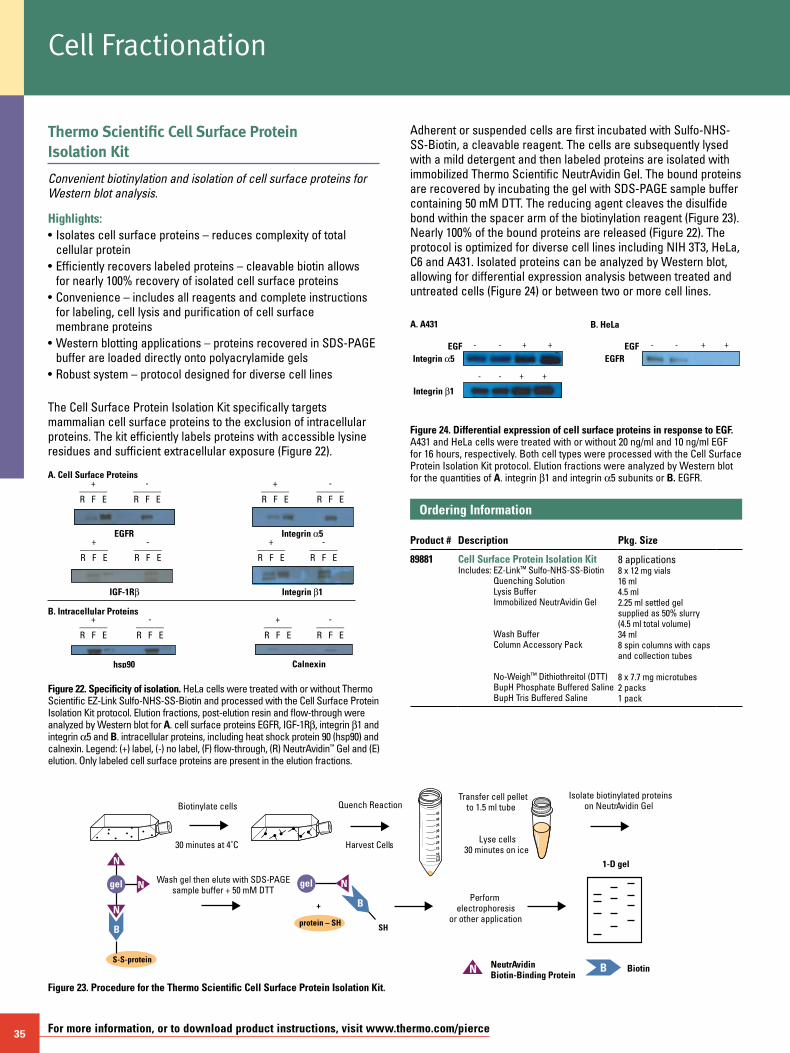
35 For more information, or to download product instructions, visit www.thermo.com/pierce
gel
B+
protein – SH
N
N
gel
N
B
S-S-protein
N
N B
45
40
35
30
25
20
15
107.55.0
Biotinylate cells
30 minutes at 4˚C
ransfer cell pelleto 1.5 ml tube
yse cell30 minutes on ice
Isolate biotinylated proteins on Neutr
Performelectrophoresis
or other application
Wash gel then elute with SDS-PAGE sample buffer + 50 mM DTT
Harvest Cells
Quench Reaction
SH
BiotinNeutrBiotin-Binding Protein
1-D gel
Avidin GelT t
L s
Avidin
Thermo Scientific Cell Surface Protein Isolation Kit
Convenient biotinylation and isolation of cell surface proteins for Western blot analysis.
Highlights:• Isolates cell surface proteins – reduces complexity of total cellular protein• Efficiently recovers labeled proteins – cleavable biotin allows for nearly 100% recovery of isolated cell surface proteins• Convenience – includes all reagents and complete instructions for labeling, cell lysis and purification of cell surface membrane proteins• Western blotting applications – proteins recovered in SDS-PAGE buffer are loaded directly onto polyacrylamide gels• Robust system – protocol designed for diverse cell lines
The Cell Surface Protein Isolation Kit specifically targets mammalian cell surface proteins to the exclusion of intracellular proteins. The kit efficiently labels proteins with accessible lysine residues and sufficient extracellular exposure (Figure 22).
A. Cell Surface Proteins
EGFR
IGF-1Rβ
hsp90
Integrin α5
Integrin β1
Calnexin
+———R F E
-———R F E
B. Intracellular Proteins
+———R F E
-———R F E
+———R F E
-———R F E
+———R F E
-———R F E
+———R F E
-———R F E
+———R F E
-———R F E
Figure 22. Specificity of isolation. HeLa cells were treated with or without Thermo Scientific EZ-Link Sulfo-NHS-SS-Biotin and processed with the Cell Surface Protein Isolation Kit protocol. Elution fractions, post-elution resin and flow-through were analyzed by Western blot for A. cell surface proteins EGFR, IGF-1Rb, integrin b1 and integrin a5 and B. intracellular proteins, including heat shock protein 90 (hsp90) and calnexin. Legend: (+) label, (-) no label, (F) flow-through, (R) NeutrAvidin™ Gel and (E) elution. Only labeled cell surface proteins are present in the elution fractions.
Adherent or suspended cells are first incubated with Sulfo-NHS-SS-Biotin, a cleavable reagent. The cells are subsequently lysed with a mild detergent and then labeled proteins are isolated with immobilized Thermo Scientific NeutrAvidin Gel. The bound proteins are recovered by incubating the gel with SDS-PAGE sample buffer containing 50 mM DTT. The reducing agent cleaves the disulfide bond within the spacer arm of the biotinylation reagent (Figure 23). Nearly 100% of the bound proteins are released (Figure 22). The protocol is optimized for diverse cell lines including NIH 3T3, HeLa, C6 and A431. Isolated proteins can be analyzed by Western blot, allowing for differential expression analysis between treated and untreated cells (Figure 24) or between two or more cell lines.
A. A431
EGFIntegrin a5
Integrin b1
++- - EGFEGFR
++- -
++- -
B. HeLa
Figure 24. Differential expression of cell surface proteins in response to EGF. A431 and HeLa cells were treated with or without 20 ng/ml and 10 ng/ml EGF for 16 hours, respectively. Both cell types were processed with the Cell Surface Protein Isolation Kit protocol. Elution fractions were analyzed by Western blot for the quantities of A. integrin b1 and integrin a5 subunits or B. EGFR.
Ordering Information Product #
Description
Pkg. Size
89881 Cell Surface Protein Isolation KitIncludes: EZ-Link™ Sulfo-NHS-SS-Biotin
Quenching Solution Lysis Buffer Immobilized NeutrAvidin Gel Wash Buffer Column Accessory Pack No-Weigh™ Dithiothreitol (DTT) BupH Phosphate Buffered Saline BupH Tris Buffered Saline
8 applications 8 x 12 mg vials 16 ml 4.5 ml 2.25 ml settled gel supplied as 50% slurry (4.5 ml total volume) 34 ml 8 spin columns with caps and collection tubes
8 x 7.7 mg microtubes 2 packs 1 pack
Figure 23. Procedure for the Thermo Scientific Cell Surface Protein Isolation Kit.
Cell Fractionation
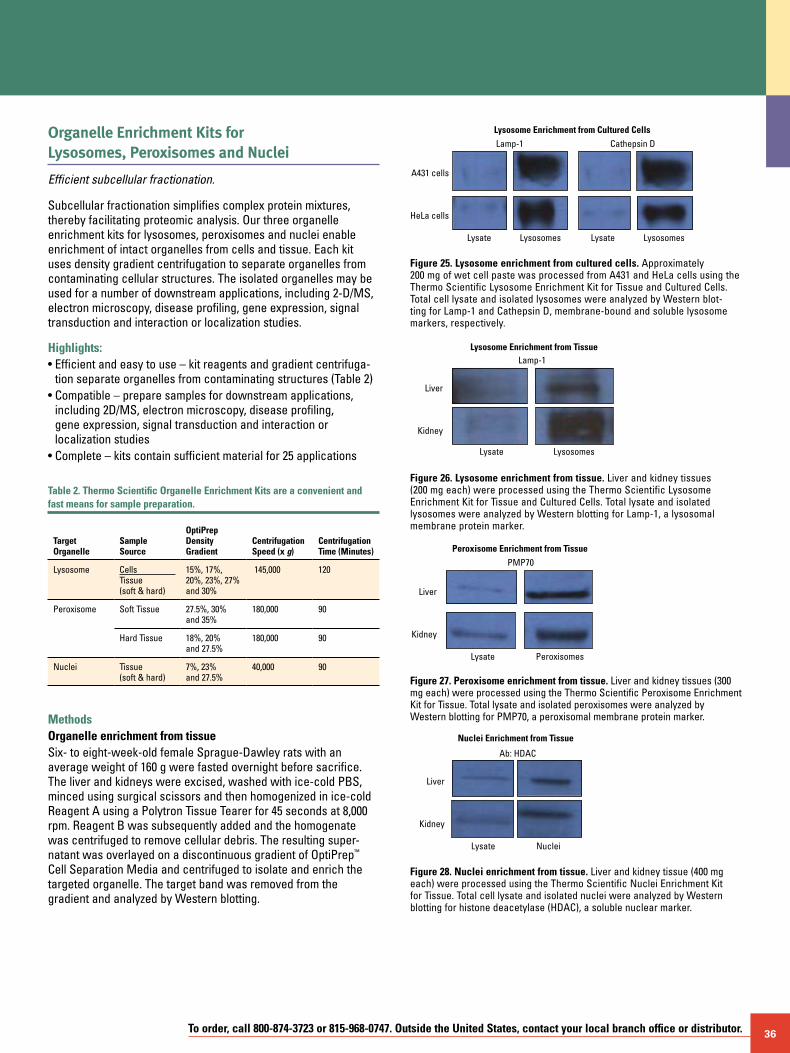
To order, call 800-874-3723 or 815-968-0747. Outside the United States, contact your local branch office or distributor. 36
Organelle Enrichment Kits for Lysosomes, Peroxisomes and Nuclei
Efficient subcellular fractionation.
Subcellular fractionation simplifies complex protein mixtures, thereby facilitating proteomic analysis. Our three organelle enrichment kits for lysosomes, peroxisomes and nuclei enable enrichment of intact organelles from cells and tissue. Each kit uses density gradient centrifugation to separate organelles from contaminating cellular structures. The isolated organelles may be used for a number of downstream applications, including 2-D/MS, electron microscopy, disease profiling, gene expression, signal transduction and interaction or localization studies.
Highlights:• Efficient and easy to use – kit reagents and gradient centrifuga-
tion separate organelles from contaminating structures (Table 2)• Compatible – prepare samples for downstream applications,
including 2D/MS, electron microscopy, disease profiling, gene expression, signal transduction and interaction or localization studies
• Complete – kits contain sufficient material for 25 applications
Table 2. Thermo Scientific Organelle Enrichment Kits are a convenient and fast means for sample preparation.
Target Organelle
Sample Source
OptiPrep DensityGradient
CentrifugationSpeed (x g)
CentrifugationTime (Minutes)
Lysosome CellsTissue (soft & hard)
15%, 17%, 20%, 23%, 27% and 30%
145,000 120
Peroxisome Soft Tissue 27.5%, 30% and 35%
180,000 90
Hard Tissue 18%, 20% and 27.5%
180,000 90
Nuclei Tissue(soft & hard)
7%, 23% and 27.5%
40,000 90
Methods Organelle enrichment from tissueSix- to eight-week-old female Sprague-Dawley rats with an average weight of 160 g were fasted overnight before sacrifice. The liver and kidneys were excised, washed with ice-cold PBS, minced using surgical scissors and then homogenized in ice-cold Reagent A using a Polytron Tissue Tearer for 45 seconds at 8,000 rpm. Reagent B was subsequently added and the homogenate was centrifuged to remove cellular debris. The resulting super- natant was overlayed on a discontinuous gradient of OptiPrep™ Cell Separation Media and centrifuged to isolate and enrich the targeted organelle. The target band was removed from the gradient and analyzed by Western blotting.
Lysate Lysosomes Lysate Lysosomes
A431 cells
Lamp-1 Cathepsin D
HeLa cells
Lysosome Enrichment from Cultured Cells
Figure 25. Lysosome enrichment from cultured cells. Approximately 200 mg of wet cell paste was processed from A431 and HeLa cells using the Thermo Scientific Lysosome Enrichment Kit for Tissue and Cultured Cells. Total cell lysate and isolated lysosomes were analyzed by Western blot-ting for Lamp-1 and Cathepsin D, membrane-bound and soluble lysosome markers, respectively.
Lysate Lysosomes
Liver
Lamp-1
Kidney
Lysosome Enrichment from Tissue
Figure 26. Lysosome enrichment from tissue. Liver and kidney tissues (200 mg each) were processed using the Thermo Scientific Lysosome Enrichment Kit for Tissue and Cultured Cells. Total lysate and isolated lysosomes were analyzed by Western blotting for Lamp-1, a lysosomal membrane protein marker.
Lysate Peroxisomes
Liver
PMP70
Kidney
Peroxisome Enrichment from Tissue
Figure 27. Peroxisome enrichment from tissue. Liver and kidney tissues (300 mg each) were processed using the Thermo Scientific Peroxisome Enrichment Kit for Tissue. Total lysate and isolated peroxisomes were analyzed by Western blotting for PMP70, a peroxisomal membrane protein marker.
Lysate Nuclei
Liver
Ab: HDAC
Kidney
Nuclei Enrichment from Tissue
Figure 28. Nuclei enrichment from tissue. Liver and kidney tissue (400 mg each) were processed using the Thermo Scientific Nuclei Enrichment Kit for Tissue. Total cell lysate and isolated nuclei were analyzed by Western blotting for histone deacetylase (HDAC), a soluble nuclear marker.
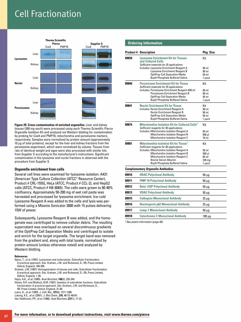
37 For more information, or to download product instructions, visit www.thermo.com/pierce
Figure 29. Cross-contamination of enriched organelles. Liver and kidney tissues (200 mg each) were processed using each Thermo Scientific Pierce Organelle Isolation Kit and analyzed via Western blotting for contamination by probing for Cox4 and PMP70, mitochondria and peroxisome markers, respectively. Samples were normalized by protein amount (approximately 10 µg of total proteins), except for the liver and kidney fractions from the peroxisome experiment, which were normalized by volume. Tissues from rats of identical weight and ages were also processed with similar kits from Supplier S according to the manufacturer’s instructions. Significant contamination in the lysosome and nuclei fractions is observed with the procedure from Supplier S.
Organelle enrichment from cellsSeveral cell lines were examined for lysosome isolation: A431 [American Type Culture Collection (ATCC® Resource Center), Product # CRL-1555], HeLa (ATCC, Product # CCL-2), and HepG2 cells (ATCC, Product # HB-8065). The cells were grown to 80-90% confluency. Approximately 50-200 mg of wet cell paste was harvested and processed for lysosome enrichment. Ice-cold Lysosome Reagent A was added to the cells and lysis was per-formed using a Misonix Sonicator 3000 with 15 pulses delivering 15W of power.
Subsequently, Lysosome Reagent B was added, and the homo-genate was centrifuged to remove cellular debris. The resulting supernatant was overlayed on several discontinuous gradients of the OptiPrep Cell Separation Media and centrifuged to isolate and enrich for the target organelle. The target band was removed from the gradient and, along with total lysate, normalized by protein amount (unless otherwise noted) and analyzed by Western blotting.
ReferencesGjoen, T., et al. (1997). Lysosomes and endocytosis. Subcellular fractionation:
A practical approach. Eds. Graham, J.M. and Rickwood, D., IRL Press Limited, Oxford, England, 169-200.
Graham, J.M. (1997). Homogenization of tissues and cells. Subcellular fractionation: A practical approach. Eds. Graham, J.M. and Rickwood, D., IRL Press Limited, Oxford, England, 1-28.
Hajra, A.K., et al. (1985). Anal Biochem, 148(2), 233-244. Hinton, R.H. and Mullock, B.M. (1997). Isolation of subcellular fractions. Subcellular
fractionation: A practical approach. Eds. Graham, J.M. and Rickwood, D., IRL Press Limited, Oxford, England, 31-69
Luers, G., et al. (1993). J. Cell. Bio., 121(6), 1271-1280. Lukong, K.E., et al. (2001). J. Biol Chem., 276, 46172-46181. Van Veldhoven, P.P., et al. (1996). Anal Biochem, 237(1), 17-23.
Ordering Information Product #
Description
Pkg. Size
89839
Lysosome Enrichment Kit for Tissues and Cultured Cells Sufficient materials for 25 applications.Includes: Lysosome Enrichment Reagent A
Lysosome Enrichment Reagent B OptiPrep Cell Separation Media BupH Phosphate Buffered Saline
Kit
90 ml90 ml50 ml 1 pack
89840 Peroxisome Enrichment Kit for TissueSufficient materials for 25 applications.Includes: Peroxisome Enrichment Reagent A90 ml
Peroxisome Enrichment Reagent B OptiPrep Cell Separation Media BupH Phosphate Buffered Saline
Kit
90 ml90 ml50 ml1 pack
89841 Nuclei Enrichment Kit for TissueIncludes: Nuclei Enrichment Reagent A
Nuclei Enrichment Reagent B OptiPrep Cell Separation Media BupH Phosphate Buffered Saline
Kit 90 ml 90 ml 50 ml 1 pack
89874 Mitochondria Isolation Kit for Cultured Cells†
Sufficient reagents for 50 applications.Includes: Mitochondria Isolation Reagent A
Mitochondria Isolation Reagent B Mitochondria Isolation Reagent C
Kit
50 ml 500 µl 70 ml
89801 Mitochondria Isolation Kit for Tissue†
Sufficient reagents for 50 applications.Includes: Mitochondria Isolation Reagent A
Mitochondria Isolation Reagent B Mitochondria Isolation Reagent C Bovine Serum Albumin BupH Phosphate Buffered Saline
Kit
50 ml 500 µl 65 ml 230 mg 1 pack
Complementary Organelle Antibodies
89910 HDAC Polyclonal Antibody 50 µg
89911 PMP 70 Polyclonal Antibody 50 µg
89912 Beta’-COP Polyclonal Antibody 50 µg
89913 VDAC Polyclonal Antibody 50 µg
89915 Cathepsin Monoclonal Antibody 25 µg
89916 Nucleoporin p62 Monoclonal Antibody 25 µg
89917 Lamp-1 Monoclonal Antibody 50 µg
89918 Cytochrome C Monoclonal Antibody 100 µg
† See patent information (page 50).
Liver
Thermo Scientific Pierce
Lysosomes
Nuclei
Peroxisomes
Supplier SCox4 PMP70 Cox4 PMP70
Kidney
Liver
Kidney
Liver
Kidney
Cell Fractionation
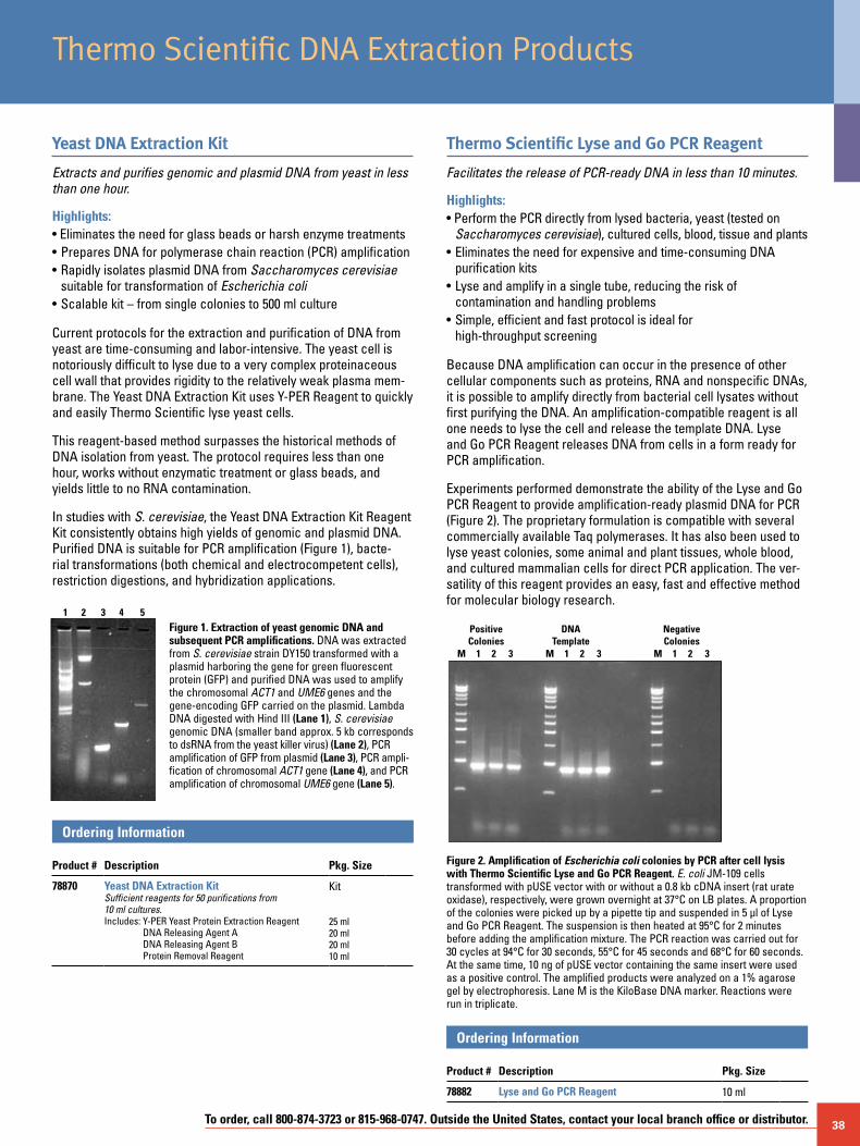
To order, call 800-874-3723 or 815-968-0747. Outside the United States, contact your local branch office or distributor. 38
Thermo Scientific DNA Extraction Products
Yeast DNA Extraction Kit
Extracts and purifies genomic and plasmid DNA from yeast in less than one hour.
Highlights:• Eliminates the need for glass beads or harsh enzyme treatments• Prepares DNA for polymerase chain reaction (PCR) amplification• Rapidly isolates plasmid DNA from Saccharomyces cerevisiae suitable for transformation of Escherichia coli• Scalable kit – from single colonies to 500 ml culture
Current protocols for the extraction and purification of DNA from yeast are time-consuming and labor-intensive. The yeast cell is notoriously difficult to lyse due to a very complex proteinaceous cell wall that provides rigidity to the relatively weak plasma mem-brane. The Yeast DNA Extraction Kit uses Y-PER Reagent to quickly and easily Thermo Scientific lyse yeast cells.
This reagent-based method surpasses the historical methods of DNA isolation from yeast. The protocol requires less than one hour, works without enzymatic treatment or glass beads, and yields little to no RNA contamination.
In studies with S. cerevisiae, the Yeast DNA Extraction Kit Reagent Kit consistently obtains high yields of genomic and plasmid DNA. Purified DNA is suitable for PCR amplification (Figure 1), bacte-rial transformations (both chemical and electrocompetent cells), restriction digestions, and hybridization applications.
Figure 1. Extraction of yeast genomic DNA and subsequent PCR amplifications. DNA was extracted from S. cerevisiae strain DY150 transformed with a plasmid harboring the gene for green fluorescent protein (GFP) and purified DNA was used to amplify the chromosomal ACT1 and UME6 genes and the gene-encoding GFP carried on the plasmid. Lambda DNA digested with Hind III (Lane 1), S. cerevisiae genomic DNA (smaller band approx. 5 kb corresponds to dsRNA from the yeast killer virus) (Lane 2), PCR amplification of GFP from plasmid (Lane 3), PCR ampli-fication of chromosomal ACT1 gene (Lane 4), and PCR amplification of chromosomal UME6 gene (Lane 5).
Ordering Information Product #
Description
Pkg. Size
78870 Yeast DNA Extraction KitSufficient reagents for 50 purifications from 10 ml cultures.Includes: Y-PER Yeast Protein Extraction Reagent
DNA Releasing Agent A DNA Releasing Agent B Protein Removal Reagent
Kit
25 ml20 ml20 ml 10 ml
Thermo Scientific Lyse and Go PCR Reagent
Facilitates the release of PCR-ready DNA in less than 10 minutes.
Highlights:• Perform the PCR directly from lysed bacteria, yeast (tested on
Saccharomyces cerevisiae), cultured cells, blood, tissue and plants• Eliminates the need for expensive and time-consuming DNA
purification kits• Lyse and amplify in a single tube, reducing the risk of
contamination and handling problems• Simple, efficient and fast protocol is ideal for
high-throughput screening
Because DNA amplification can occur in the presence of other cellular components such as proteins, RNA and nonspecific DNAs, it is possible to amplify directly from bacterial cell lysates without first purifying the DNA. An amplification-compatible reagent is all one needs to lyse the cell and release the template DNA. Lyse and Go PCR Reagent releases DNA from cells in a form ready for PCR amplification.
Experiments performed demonstrate the ability of the Lyse and Go PCR Reagent to provide amplification-ready plasmid DNA for PCR (Figure 2). The proprietary formulation is compatible with several commercially available Taq polymerases. It has also been used to lyse yeast colonies, some animal and plant tissues, whole blood, and cultured mammalian cells for direct PCR application. The ver-satility of this reagent provides an easy, fast and effective method for molecular biology research.
PositiveColonies
M 1 2 3 M 1 2 3 M 1 2 3
DNATemplate
NegativeColonies
Figure 2. Amplification of Escherichia coli colonies by PCR after cell lysis with Thermo Scientific Lyse and Go PCR Reagent. E. coli JM-109 cells transformed with pUSE vector with or without a 0.8 kb cDNA insert (rat urate oxidase), respectively, were grown overnight at 37°C on LB plates. A proportion of the colonies were picked up by a pipette tip and suspended in 5 µl of Lyse and Go PCR Reagent. The suspension is then heated at 95°C for 2 minutes before adding the amplification mixture. The PCR reaction was carried out for 30 cycles at 94°C for 30 seconds, 55°C for 45 seconds and 68°C for 60 seconds. At the same time, 10 ng of pUSE vector containing the same insert were used as a positive control. The amplified products were analyzed on a 1% agarose gel by electrophoresis. Lane M is the KiloBase DNA marker. Reactions were run in triplicate.
Ordering Information Product #
Description
Pkg. Size
78882 Lyse and Go PCR Reagent 10 ml
1 2 3 4 5
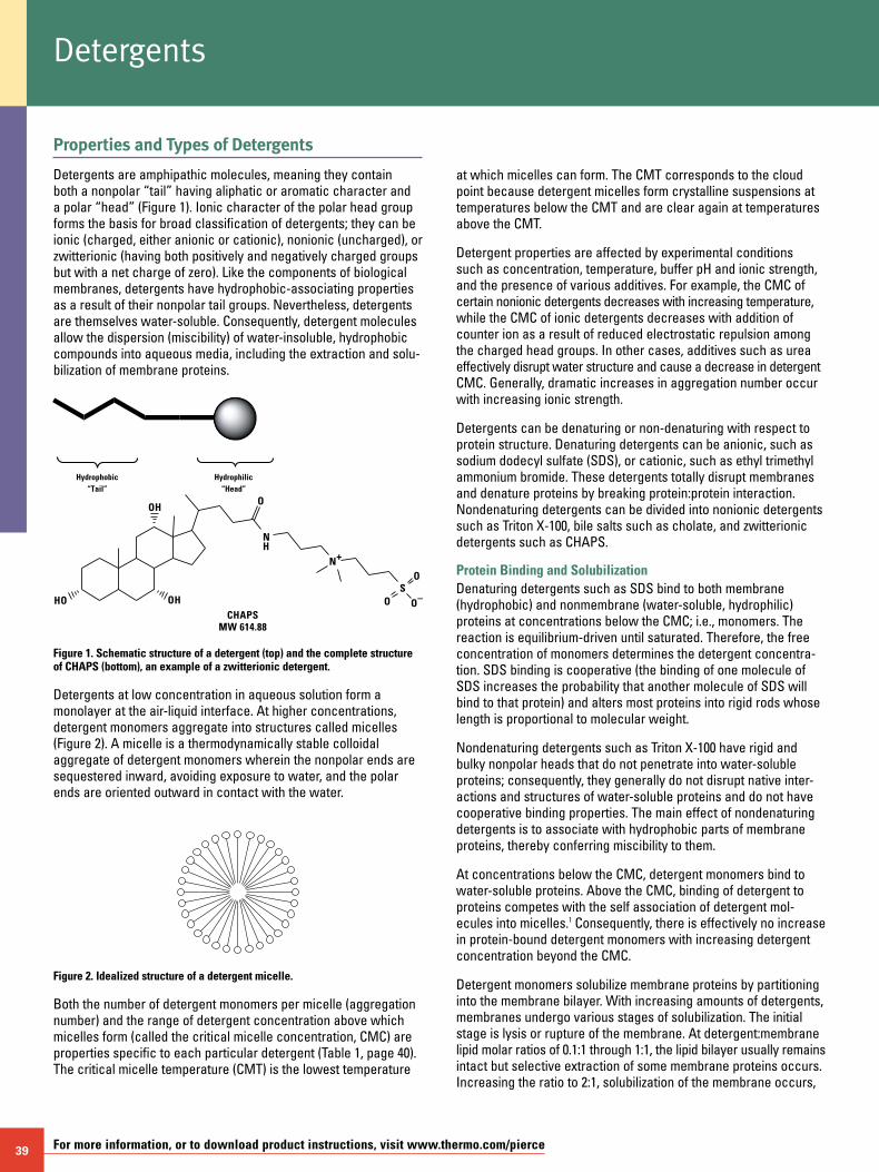
39 For more information, or to download product instructions, visit www.thermo.com/pierce
Properties and Types of Detergents
Detergents are amphipathic molecules, meaning they contain both a nonpolar “tail” having aliphatic or aromatic character and a polar “head” (Figure 1). Ionic character of the polar head group forms the basis for broad classification of detergents; they can be ionic (charged, either anionic or cationic), nonionic (uncharged), or zwitterionic (having both positively and negatively charged groups but with a net charge of zero). Like the components of biological membranes, detergents have hydrophobic-associating properties as a result of their nonpolar tail groups. Nevertheless, detergents are themselves water-soluble. Consequently, detergent molecules allow the dispersion (miscibility) of water-insoluble, hydrophobic compounds into aqueous media, including the extraction and solu-bilization of membrane proteins.
Hydrophobic“Tail”
Hydrophilic“Head”
Detergents
at which micelles can form. The CMT corresponds to the cloud point because detergent micelles form crystalline suspensions at temperatures below the CMT and are clear again at temperatures above the CMT.
Detergent properties are affected by experimental conditions such as concentration, temperature, buffer pH and ionic strength, and the presence of various additives. For example, the CMC of certain nonionic detergents decreases with increasing temperature, while the CMC of ionic detergents decreases with addition of counter ion as a result of reduced electrostatic repulsion among the charged head groups. In other cases, additives such as urea effectively disrupt water structure and cause a decrease in detergent CMC. Generally, dramatic increases in aggregation number occur with increasing ionic strength.
Detergents can be denaturing or non-denaturing with respect to protein structure. Denaturing detergents can be anionic, such as sodium dodecyl sulfate (SDS), or cationic, such as ethyl trimethyl ammonium bromide. These detergents totally disrupt membranes and denature proteins by breaking protein:protein interaction. Nondenaturing detergents can be divided into nonionic detergents such as Triton X-100, bile salts such as cholate, and zwitterionic detergents such as CHAPS.
Protein Binding and SolubilizationDenaturing detergents such as SDS bind to both membrane (hydrophobic) and nonmembrane (water-soluble, hydrophilic) proteins at concentrations below the CMC; i.e., monomers. The reaction is equilibrium-driven until saturated. Therefore, the free concentration of monomers determines the detergent concentra-tion. SDS binding is cooperative (the binding of one molecule of SDS increases the probability that another molecule of SDS will bind to that protein) and alters most proteins into rigid rods whose length is proportional to molecular weight.
Nondenaturing detergents such as Triton X-100 have rigid and bulky nonpolar heads that do not penetrate into water-soluble proteins; consequently, they generally do not disrupt native inter-actions and structures of water-soluble proteins and do not have cooperative binding properties. The main effect of nondenaturing detergents is to associate with hydrophobic parts of membrane proteins, thereby conferring miscibility to them.
At concentrations below the CMC, detergent monomers bind to water-soluble proteins. Above the CMC, binding of detergent to proteins competes with the self association of detergent mol-ecules into micelles.1 Consequently, there is effectively no increase in protein-bound detergent monomers with increasing detergent concentration beyond the CMC.
Detergent monomers solubilize membrane proteins by partitioning into the membrane bilayer. With increasing amounts of detergents, membranes undergo various stages of solubilization. The initial stage is lysis or rupture of the membrane. At detergent:membrane lipid molar ratios of 0.1:1 through 1:1, the lipid bilayer usually remains intact but selective extraction of some membrane proteins occurs. Increasing the ratio to 2:1, solubilization of the membrane occurs,
Figure 1. Schematic structure of a detergent (top) and the complete structure of CHAPS (bottom), an example of a zwitterionic detergent.
Detergents at low concentration in aqueous solution form a monolayer at the air-liquid interface. At higher concentrations, detergent monomers aggregate into structures called micelles (Figure 2). A micelle is a thermodynamically stable colloidal aggregate of detergent monomers wherein the nonpolar ends are sequestered inward, avoiding exposure to water, and the polar ends are oriented outward in contact with the water.
Figure 2. Idealized structure of a detergent micelle.
Both the number of detergent monomers per micelle (aggregation number) and the range of detergent concentration above which micelles form (called the critical micelle concentration, CMC) are properties specific to each particular detergent (Table 1, page 40). The critical micelle temperature (CMT) is the lowest temperature
HO OH
OHO
N+
SO
O
O–
CHAPSMW 614.88
NH
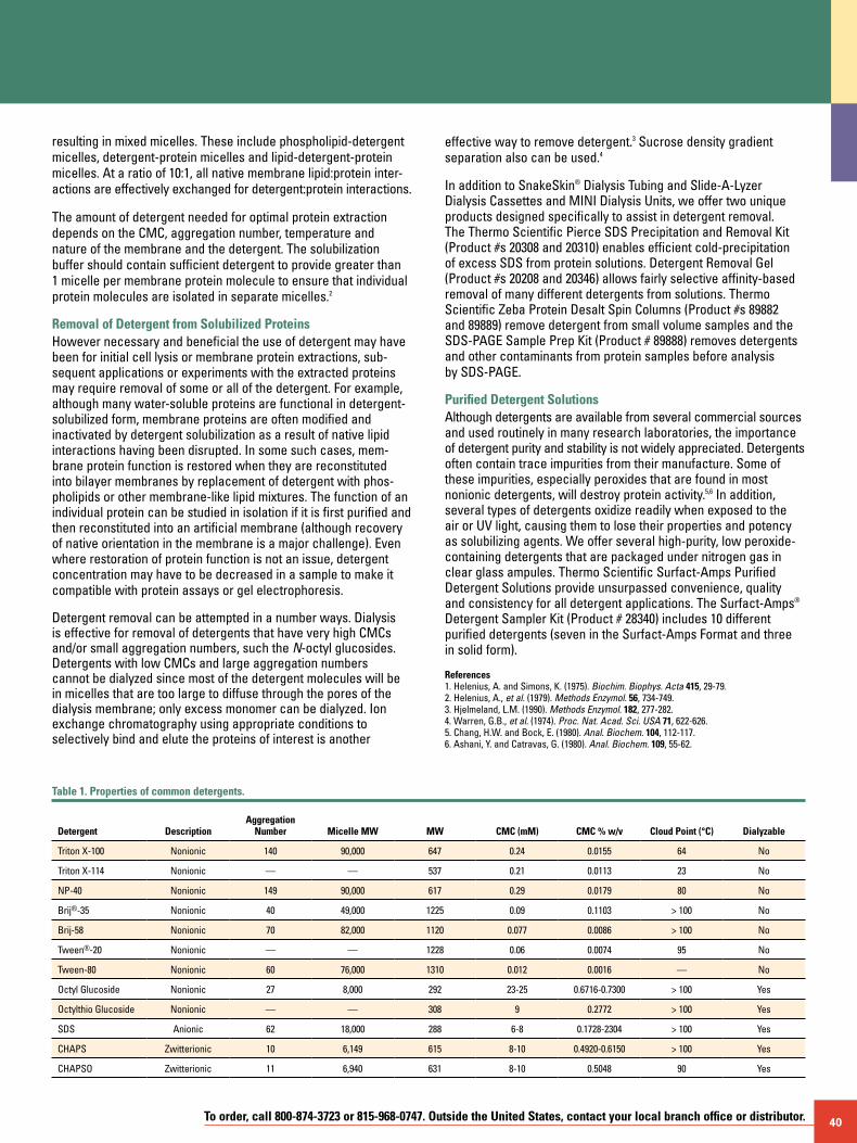
To order, call 800-874-3723 or 815-968-0747. Outside the United States, contact your local branch office or distributor. 40
resulting in mixed micelles. These include phospholipid-detergent micelles, detergent-protein micelles and lipid-detergent-protein micelles. At a ratio of 10:1, all native membrane lipid:protein inter-actions are effectively exchanged for detergent:protein interactions.
The amount of detergent needed for optimal protein extraction depends on the CMC, aggregation number, temperature and nature of the membrane and the detergent. The solubilization buffer should contain sufficient detergent to provide greater than 1 micelle per membrane protein molecule to ensure that individual protein molecules are isolated in separate micelles.2
Removal of Detergent from Solubilized ProteinsHowever necessary and beneficial the use of detergent may have been for initial cell lysis or membrane protein extractions, sub-sequent applications or experiments with the extracted proteins may require removal of some or all of the detergent. For example, although many water-soluble proteins are functional in detergent-solubilized form, membrane proteins are often modified and inactivated by detergent solubilization as a result of native lipid interactions having been disrupted. In some such cases, mem-brane protein function is restored when they are reconstituted into bilayer membranes by replacement of detergent with phos-pholipids or other membrane-like lipid mixtures. The function of an individual protein can be studied in isolation if it is first purified and then reconstituted into an artificial membrane (although recovery of native orientation in the membrane is a major challenge). Even where restoration of protein function is not an issue, detergent concentration may have to be decreased in a sample to make it compatible with protein assays or gel electrophoresis.
Detergent removal can be attempted in a number ways. Dialysis is effective for removal of detergents that have very high CMCs and/or small aggregation numbers, such the N-octyl glucosides. Detergents with low CMCs and large aggregation numbers cannot be dialyzed since most of the detergent molecules will be in micelles that are too large to diffuse through the pores of the dialysis membrane; only excess monomer can be dialyzed. Ion exchange chromatography using appropriate conditions to selectively bind and elute the proteins of interest is another
effective way to remove detergent.3 Sucrose density gradient separation also can be used.4
In addition to SnakeSkin® Dialysis Tubing and Slide-A-Lyzer Dialysis Cassettes and MINI Dialysis Units, we offer two unique products designed specifically to assist in detergent removal. The Thermo Scientific Pierce SDS Precipitation and Removal Kit (Product #s 20308 and 20310) enables efficient cold-precipitation of excess SDS from protein solutions. Detergent Removal Gel (Product #s 20208 and 20346) allows fairly selective affinity-based removal of many different detergents from solutions. Thermo Scientific Zeba Protein Desalt Spin Columns (Product #s 89882 and 89889) remove detergent from small volume samples and the SDS-PAGE Sample Prep Kit (Product # 89888) removes detergents and other contaminants from protein samples before analysis by SDS-PAGE.
Purified Detergent SolutionsAlthough detergents are available from several commercial sources and used routinely in many research laboratories, the importance of detergent purity and stability is not widely appreciated. Detergents often contain trace impurities from their manufacture. Some of these impurities, especially peroxides that are found in most nonionic detergents, will destroy protein activity.5,6 In addition, several types of detergents oxidize readily when exposed to the air or UV light, causing them to lose their properties and potency as solubilizing agents. We offer several high-purity, low peroxide- containing detergents that are packaged under nitrogen gas in clear glass ampules. Thermo Scientific Surfact-Amps Purified Detergent Solutions provide unsurpassed convenience, quality and consistency for all detergent applications. The Surfact-Amps® Detergent Sampler Kit (Product # 28340) includes 10 different purified detergents (seven in the Surfact-Amps Format and three in solid form).
References1. Helenius, A. and Simons, K. (1975). Biochim. Biophys. Acta 415, 29-79.2. Helenius, A., et al. (1979). Methods Enzymol. 56, 734-749.3. Hjelmeland, L.M. (1990). Methods Enzymol. 182, 277-282.4. Warren, G.B., et al. (1974). Proc. Nat. Acad. Sci. USA 71, 622-626.5. Chang, H.W. and Bock, E. (1980). Anal. Biochem. 104, 112-117.6. Ashani, Y. and Catravas, G. (1980). Anal. Biochem. 109, 55-62.
Table 1. Properties of common detergents.
Detergent DescriptionAggregation
Number Micelle MW MW CMC (mM) CMC % w/v Cloud Point (°C) Dialyzable
Triton X-100 Nonionic 140 90,000 647 0.24 0.0155 64 No
Triton X-114 Nonionic — — 537 0.21 0.0113 23 No
NP-40 Nonionic 149 90,000 617 0.29 0.0179 80 No
Brij®-35 Nonionic 40 49,000 1225 0.09 0.1103 > 100 No
Brij-58 Nonionic 70 82,000 1120 0.077 0.0086 > 100 No
Tween®-20 Nonionic — — 1228 0.06 0.0074 95 No
Tween-80 Nonionic 60 76,000 1310 0.012 0.0016 — No
Octyl Glucoside Nonionic 27 8,000 292 23-25 0.6716-0.7300 > 100 Yes
Octylthio Glucoside Nonionic — — 308 9 0.2772 > 100 Yes
SDS Anionic 62 18,000 288 6-8 0.1728-2304 > 100 Yes
CHAPS Zwitterionic 10 6,149 615 8-10 0.4920-0.6150 > 100 Yes
CHAPSO Zwitterionic 11 6,940 631 8-10 0.5048 90 Yes
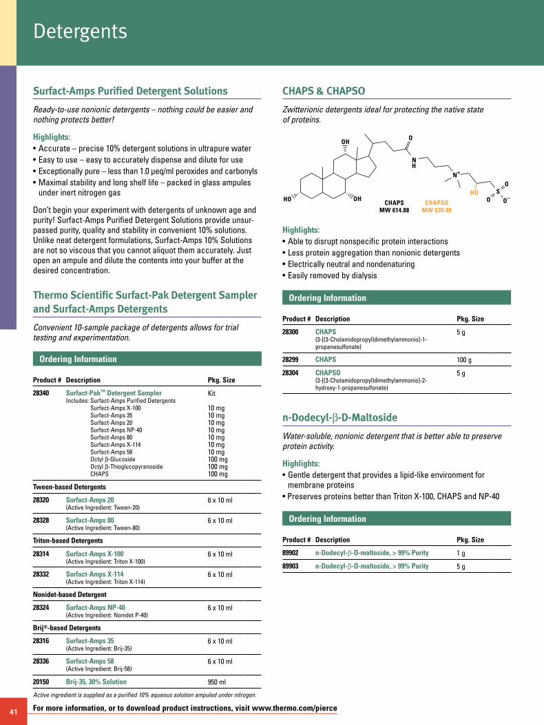
41 For more information, or to download product instructions, visit www.thermo.com/pierce
Surfact-Amps Purified Detergent Solutions
Ready-to-use nonionic detergents – nothing could be easier and nothing protects better!
Highlights:• Accurate – precise 10% detergent solutions in ultrapure water• Easy to use – easy to accurately dispense and dilute for use• Exceptionally pure – less than 1.0 µeq/ml peroxides and carbonyls• Maximal stability and long shelf life – packed in glass ampules under inert nitrogen gas
Don’t begin your experiment with detergents of unknown age and purity! Surfact-Amps Purified Detergent Solutions provide unsur-passed purity, quality and stability in convenient 10% solutions. Unlike neat detergent formulations, Surfact-Amps 10% Solutions are not so viscous that you cannot aliquot them accurately. Just open an ampule and dilute the contents into your buffer at the desired concentration.
Thermo Scientific Surfact-Pak Detergent Sampler and Surfact-Amps Detergents
Convenient 10-sample package of detergents allows for trial testing and experimentation.
Ordering Information Product #
Description
Pkg. Size
28340 Surfact-Pak™ Detergent SamplerIncludes: Surfact-Amps Purified Detergents
Surfact-Amps X-100 Surfact-Amps 35 Surfact-Amps 20 Surfact-Amps NP-40 Surfact-Amps 80 Surfact-Amps X-114 Surfact-Amps 58 Octyl b-Glucoside Octyl b-Thioglucopyranoside CHAPS
Kit 10 mg 10 mg 10 mg 10 mg 10 mg 10 mg 10 mg 100 mg 100 mg 100 mg
Tween-based Detergents
28320 Surfact-Amps 20(Active Ingredient: Tween-20)
6 x 10 ml
28328 Surfact-Amps 80(Active Ingredient: Tween-80)
6 x 10 ml
Triton-based Detergents
28314 Surfact-Amps X-100(Active Ingredient: Triton X-100)
6 x 10 ml
28332 Surfact-Amps X-114(Active Ingredient: Triton X-114)
6 x 10 ml
Nonidet-based Detergent
28324 Surfact-Amps NP-40(Active Ingredient: Nonidet P-40)
6 x 10 ml
Brij®-based Detergents
28316 Surfact-Amps 35(Active Ingredient: Brij-35)
6 x 10 ml
28336 Surfact-Amps 58(Active Ingredient: Brij-58)
6 x 10 ml
20150 Brij-35, 30% Solution 950 ml
Active ingredient is supplied as a purified 10% aqueous solution ampuled under nitrogen.
CHAPS & CHAPSO
Zwitterionic detergents ideal for protecting the native state of proteins.
CHAPSOMW 630.88
HO OH
OHO
N+
SO
O
O–
NH
HO
CHAPSMW 614.88
Highlights:• Able to disrupt nonspecific protein interactions• Less protein aggregation than nonionic detergents• Electrically neutral and nondenaturing• Easily removed by dialysis
Ordering Information Product #
Description
Pkg. Size
28300 CHAPS(3-[(3-Cholamidopropyl)dimethylammonio]-1-propanesulfonate)
5 g
28299 CHAPS 100 g
28304 CHAPSO(3-[(3-Cholamidopropyl)dimethylammonio]-2-hydroxy-1-propanesulfonate)
5 g
n-Dodecyl-b-D-Maltoside
Water-soluble, nonionic detergent that is better able to preserve protein activity.
Highlights:• Gentle detergent that provides a lipid-like environment for membrane proteins• Preserves proteins better than Triton X-100, CHAPS and NP-40
Ordering Information Product #
Description
Pkg. Size
89902 n-Dodecyl-b-D-maltoside, > 99% Purity 1 g
89903 n-Dodecyl-b-D-maltoside, > 99% Purity 5 g
Detergents
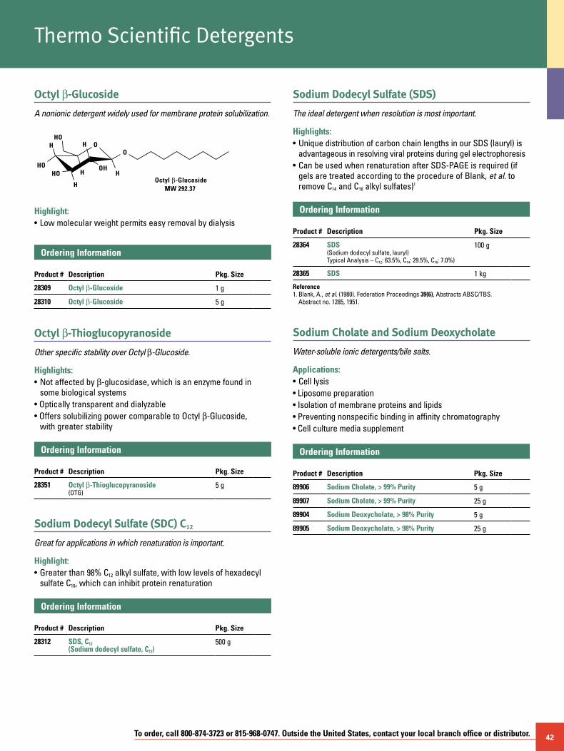
To order, call 800-874-3723 or 815-968-0747. Outside the United States, contact your local branch office or distributor. 42
Octyl b-Glucoside
A nonionic detergent widely used for membrane protein solubilization.
Highlight:• Low molecular weight permits easy removal by dialysis
Ordering Information Product #
Description
Pkg. Size
28309 Octyl b-Glucoside 1 g
28310 Octyl b-Glucoside 5 g
Octyl b-Thioglucopyranoside
Other specific stability over Octyl b-Glucoside.
Highlights:• Not affected by b-glucosidase, which is an enzyme found in some biological systems• Optically transparent and dialyzable• Offers solubilizing power comparable to Octyl b-Glucoside, with greater stability
Ordering Information Product #
Description
Pkg. Size
28351 Octyl b-Thioglucopyranoside (OTG)
5 g
Sodium Dodecyl Sulfate (SDC) C12
Great for applications in which renaturation is important.
Highlight:• Greater than 98% C12 alkyl sulfate, with low levels of hexadecyl sulfate C16, which can inhibit protein renaturation
Ordering Information Product #
Description
Pkg. Size
28312 SDS, C12 (Sodium dodecyl sulfate, C12)
500 g
Sodium Dodecyl Sulfate (SDS)
The ideal detergent when resolution is most important.
Highlights:• Unique distribution of carbon chain lengths in our SDS (lauryl) is advantageous in resolving viral proteins during gel electrophoresis• Can be used when renaturation after SDS-PAGE is required (if gels are treated according to the procedure of Blank, et al. to remove C14 and C16 alkyl sulfates)1
Ordering Information Product #
Description
Pkg. Size
28364 SDS(Sodium dodecyl sulfate, lauryl)Typical Analysis – C12: 63.5%, C14: 29.5%, C16: 7.0%)
100 g
28365 SDS 1 kg
Reference1. Blank, A., et al. (1980). Federation Proceedings 39(6), Abstracts ABSC/TBS.
Abstract no. 1285, 1951.
Sodium Cholate and Sodium Deoxycholate
Water-soluble ionic detergents/bile salts.
Applications:• Cell lysis • Liposome preparation • Isolation of membrane proteins and lipids• Preventing nonspecific binding in affinity chromatography• Cell culture media supplement
Ordering Information Product #
Description
Pkg. Size
89906 Sodium Cholate, > 99% Purity 5 g
89907 Sodium Cholate, > 99% Purity 25 g
89904 Sodium Deoxycholate, > 98% Purity 5 g
89905 Sodium Deoxycholate, > 98% Purity 25 g
OH
HO
H
HO
H
HOH
H
O
HO
Octyl b-GlucosideMW 292.37
Thermo Scientific Detergents
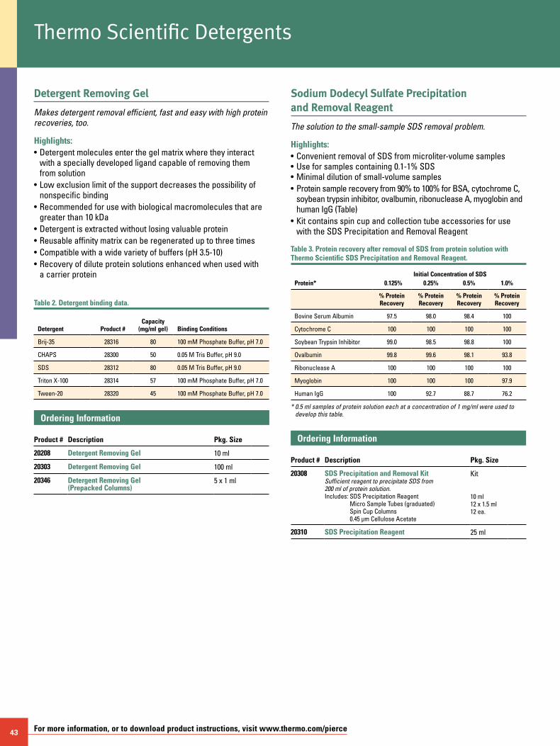
43 For more information, or to download product instructions, visit www.thermo.com/pierce
Detergent Removing Gel
Makes detergent removal efficient, fast and easy with high protein recoveries, too.
Highlights:• Detergent molecules enter the gel matrix where they interact with a specially developed ligand capable of removing them from solution • Low exclusion limit of the support decreases the possibility of nonspecific binding• Recommended for use with biological macromolecules that are greater than 10 kDa• Detergent is extracted without losing valuable protein• Reusable affinity matrix can be regenerated up to three times• Compatible with a wide variety of buffers (pH 3.5-10)• Recovery of dilute protein solutions enhanced when used with a carrier protein
Table 2. Detergent binding data.
Detergent
Product #
Capacity (mg/ml gel)
Binding Conditions
Brij-35 28316 80 100 mM Phosphate Buffer, pH 7.0
CHAPS 28300 50 0.05 M Tris Buffer, pH 9.0
SDS 28312 80 0.05 M Tris Buffer, pH 9.0
Triton X-100 28314 57 100 mM Phosphate Buffer, pH 7.0
Tween-20 28320 45 100 mM Phosphate Buffer, pH 7.0
Ordering Information Product #
Description
Pkg. Size
20208 Detergent Removing Gel 10 ml
20303 Detergent Removing Gel 100 ml
20346 Detergent Removing Gel (Prepacked Columns)
5 x 1 ml
Sodium Dodecyl Sulfate Precipitation and Removal Reagent
The solution to the small-sample SDS removal problem.
Highlights:• Convenient removal of SDS from microliter-volume samples • Use for samples containing 0.1-1% SDS • Minimal dilution of small-volume samples• Protein sample recovery from 90% to 100% for BSA, cytochrome C, soybean trypsin inhibitor, ovalbumin, ribonuclease A, myoglobin and human IgG (Table)• Kit contains spin cup and collection tube accessories for use with the SDS Precipitation and Removal Reagent
Table 3. Protein recovery after removal of SDS from protein solution with Thermo Scientific SDS Precipitation and Removal Reagent.
Initial Concentration of SDSProtein* 0.125% 0.25% 0.5% 1.0%
% Protein Recovery
% Protein Recovery
% Protein Recovery
% Protein Recovery
Bovine Serum Albumin 97.5 98.0 98.4 100
Cytochrome C 100 100 100 100
Soybean Trypsin Inhibitor 99.0 98.5 98.8 100
Ovalbumin 99.8 99.6 98.1 93.8
Ribonuclease A 100 100 100 100
Myoglobin 100 100 100 97.9
Human IgG 100 92.7 88.7 76.2
* 0.5 ml samples of protein solution each at a concentration of 1 mg/ml were used to develop this table.
Ordering Information Product #
Description
Pkg. Size
20308 SDS Precipitation and Removal Kit Sufficient reagent to precipitate SDS from 200 ml of protein solution. Includes: SDS Precipitation Reagent
Micro Sample Tubes (graduated) Spin Cup Columns 0.45 µm Cellulose Acetate
Kit 10 ml 12 x 1.5 ml 12 ea.
20310 SDS Precipitation Reagent 25 ml
Thermo Scientific Detergents
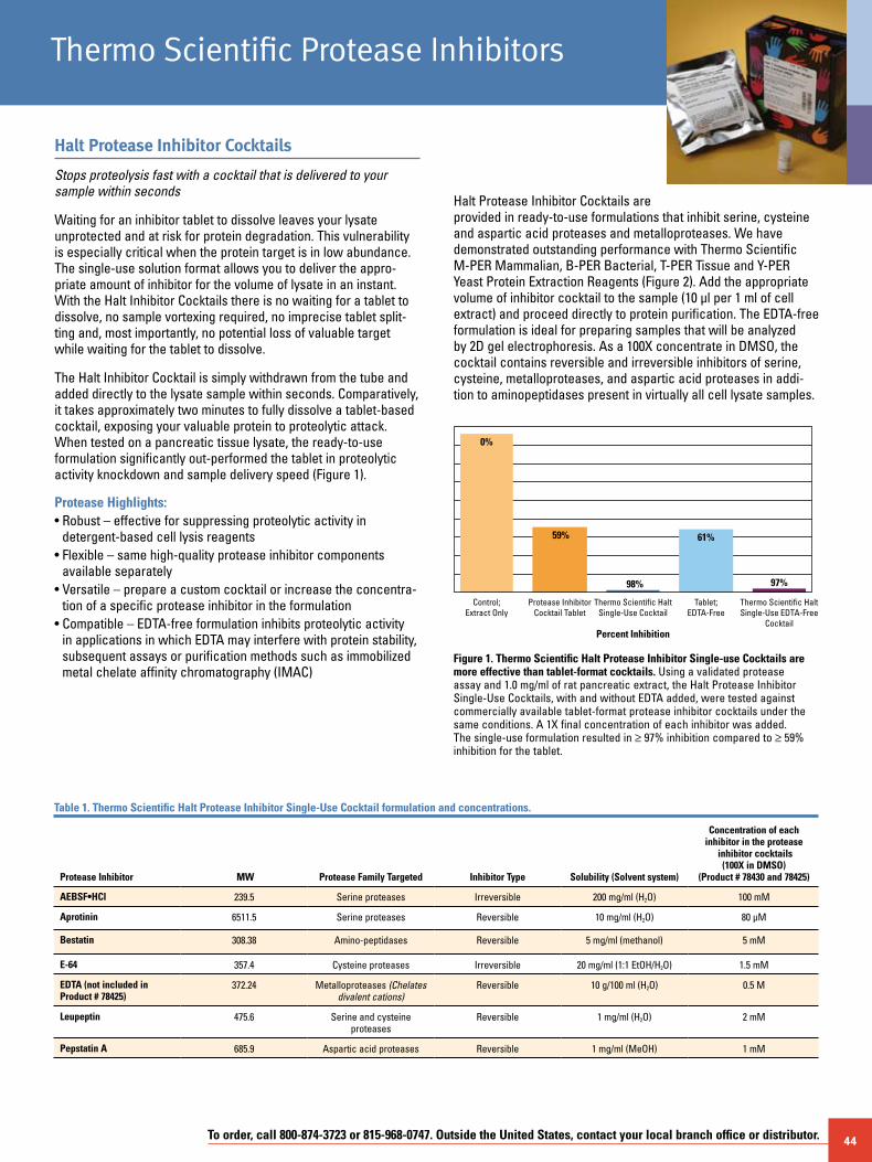
To order, call 800-874-3723 or 815-968-0747. Outside the United States, contact your local branch office or distributor. 44
Halt Protease Inhibitor Cocktails
Stops proteolysis fast with a cocktail that is delivered to your sample within seconds
Waiting for an inhibitor tablet to dissolve leaves your lysate unprotected and at risk for protein degradation. This vulnerability is especially critical when the protein target is in low abundance. The single-use solution format allows you to deliver the appro- priate amount of inhibitor for the volume of lysate in an instant. With the Halt Inhibitor Cocktails there is no waiting for a tablet to dissolve, no sample vortexing required, no imprecise tablet split-ting and, most importantly, no potential loss of valuable target while waiting for the tablet to dissolve.
The Halt Inhibitor Cocktail is simply withdrawn from the tube and added directly to the lysate sample within seconds. Comparatively, it takes approximately two minutes to fully dissolve a tablet-based cocktail, exposing your valuable protein to proteolytic attack. When tested on a pancreatic tissue lysate, the ready-to-use formulation significantly out-performed the tablet in proteolytic activity knockdown and sample delivery speed (Figure 1).
Protease Highlights:• Robust – effective for suppressing proteolytic activity in
detergent-based cell lysis reagents• Flexible – same high-quality protease inhibitor components
available separately• Versatile – prepare a custom cocktail or increase the concentra-
tion of a specific protease inhibitor in the formulation• Compatible – EDTA-free formulation inhibits proteolytic activity
in applications in which EDTA may interfere with protein stability, subsequent assays or purification methods such as immobilized metal chelate affinity chromatography (IMAC)
Halt Protease Inhibitor Cocktails are provided in ready-to-use formulations that inhibit serine, cysteine and aspartic acid proteases and metalloproteases. We have demonstrated outstanding performance with Thermo Scientific M-PER Mammalian, B-PER Bacterial, T-PER Tissue and Y-PER Yeast Protein Extraction Reagents (Figure 2). Add the appropriate volume of inhibitor cocktail to the sample (10 µl per 1 ml of cell extract) and proceed directly to protein purification. The EDTA-free formulation is ideal for preparing samples that will be analyzed by 2D gel electrophoresis. As a 100X concentrate in DMSO, the cocktail contains reversible and irreversible inhibitors of serine, cysteine, metalloproteases, and aspartic acid proteases in addi-tion to aminopeptidases present in virtually all cell lysate samples.
Control;Extract Only
Protease InhibitorCocktail Tablet
Thermo Scientific HaltSingle-Use Cocktail
Tablet;EDTA-Free
Thermo Scientific HaltSingle-Use EDTA-Free
Cocktail
97%
59%
0%
98%
61%
Percent Inhibition
Figure 1. Thermo Scientific Halt Protease Inhibitor Single-use Cocktails are more effective than tablet-format cocktails. Using a validated protease assay and 1.0 mg/ml of rat pancreatic extract, the Halt Protease Inhibitor Single-Use Cocktails, with and without EDTA added, were tested against commercially available tablet-format protease inhibitor cocktails under the same conditions. A 1X final concentration of each inhibitor was added. The single-use formulation resulted in ≥ 97% inhibition compared to ≥ 59% inhibition for the tablet.
Table 1. Thermo Scientific Halt Protease Inhibitor Single-Use Cocktail formulation and concentrations.
Protease Inhibitor MW Protease Family Targeted Inhibitor Type Solubility (Solvent system)
Concentration of each inhibitor in the protease
inhibitor cocktails (100X in DMSO)
(Product # 78430 and 78425)
AEBSF•HCl 239.5 Serine proteases Irreversible 200 mg/ml (H2O) 100 mM
Aprotinin 6511.5 Serine proteases Reversible 10 mg/ml (H2O) 80 µM
Bestatin 308.38 Amino-peptidases Reversible 5 mg/ml (methanol) 5 mM
E-64 357.4 Cysteine proteases Irreversible 20 mg/ml (1:1 EtOH/H2O) 1.5 mM
EDTA (not included in Product # 78425)
372.24 Metalloproteases (Chelates divalent cations)
Reversible 10 g/100 ml (H2O) 0.5 M
Leupeptin 475.6 Serine and cysteine proteases
Reversible 1 mg/ml (H2O) 2 mM
Pepstatin A 685.9 Aspartic acid proteases Reversible 1 mg/ml (MeOH) 1 mM
Thermo Scientific Protease Inhibitors

45 For more information, or to download product instructions, visit www.thermo.com/pierce
1 2 3 4 1 2 3 4
A. Thermo ScientificB-PER Bacterial Protein
Extraction Reagent
B. Thermo ScientificM-PER Mammalian
Protein Extraction Reagent
Figure 2. Thermo Scientific Halt Protease Inhibitor Single-Use Cocktails are compatible with Thermo Scientific M-PER Mammalian Protein Extraction and B-PER Bacterial Protein Extraction Reagents. Bovine serum albumin (BSA) was incubated overnight at 37°C in the presence of 0.1 mg/ml trypsin in B-PER Extraction Reagent and M-PER Extraction Reagent, respectively. Under extreme time and temperature conditions, substantial protection from degradation was observed as marginal to barely detectable cleavage products of BSA. BSA only (Lane 1); Trypsin (0.1 mg/ml) (+) BSA and no Halt Protease Inhibitor Single-Use Cocktail present (Lane 2); BSA (+) Trypsin with Halt Protease Inhibitor Single-Use Cocktail with EDTA (Lane 3); and BSA (+) Trypsin with Halt Protease Inhibitor Single-Use Cocktail (Lane 4).
Ordering Information Product #
Description
Pkg. Size
78425 Halt Protease Inhibitor Single-Use Cocktail EDTA-Free (100X)Each 100 µl microtube contains sufficient cocktail to treat 10 ml of lysate. Protease inhibitors prepared in DMSO solution.Protease Inhibitor Cocktail, 100 µl microtubes
1 ml
24
78430 Halt Protease Inhibitor Single-Use Cocktail (100X)Each 100 µl microtube contains sufficient cocktail to treat 10 ml of lysate. Protease inhibitors prepared in DMSO solution.Protease Inhibitor Cocktail, 100 µl microtubes 0.5 M EDTA Solution (100X), 2.5 ml
1 ea.
24
78429 Halt Protease Inhibitor Cocktail 5 ml
78437 Halt Protease Inhibitor Cocktail, EDTA Free 5 ml
78438 Halt Protease Inhibitor Cocktail 10 ml
78439 Halt Protease Inhibitor Cocktail, EDTA Free 10 ml
Halt Phosphatase Inhibitor Cocktails
Safeguards against serine, threonine and tyrosine phosphatase activities
Phosphorylation and dephosphorylation is a molecular on/off switch that regulates a number of key biological pathways within the cell, including signal transduction, cell division and apoptosis. The Halt Phosphatase Inhibitor Cocktail preserves protein phosphorylation in cell lysates and tissue extracts. The cocktail contains a mixture of four phosphatase inhibitors of broad specificity, including sodium fluoride, sodium orthovanadate, sodium pyrophosphate and b-glycerophosphate. Unlike other commercially available phosphatase inhibitor cocktails that
protect against either serine/threonine phosphatases or protein tyrosine phosphatases, the Halt Phosphatase Inhibitor Cocktail protects phosphoproteins from both families of phosphatases (Figure 3).
Phosphatase Highlights:• Complete protection from phosphatases – inhibits both serine/
threonine and protein tyrosine phosphatases• Compatible with standard protein assays – quantify treated
samples with the Thermo Scientific Pierce BCA Protein Assay Kit, Coomassie Plus (Bradford) Assay and Pierce 660 nm Protein Assay
• Efficient – 1 ml protects up to 100 ml of sample
A. MEK+– +–
PMEK
B. MAPK+– +–
PMAPK
C. STAT3+– +–
PSTAT3
Figure 3. Halt Phosphatase Inhibitor Cocktail preserves phosphorylation of MEK, MAP Kinase 42/44 and STAT3 in HeLa cell lysate. HeLa cells were lysed in the absence (-) and presence (+) of a 1X concentration of the Halt Phosphatase Inhibitor cocktail. Cell lysates were analyzed by Western blot for total and phosphorylated protein as indicated. Panel A. MEK and phosphorylated MEK (PMEK), Panel B. MAP Kinase and PMAP Kinase and Panel C. STAT3 and PSTAT3. The proteins are phosphorylated on serine, threonine/tyrosine and tyrosine, respectively.
Ordering Information Product #
Description
Pkg. Size
78420 Halt Phosphatase Inhibitor CocktailSufficient reagent to protect up to 100 ml of sample.
1 ml
78426 Halt Phosphatase Inhibitor Cocktail 5 x 1 ml
78427 Halt Phosphatase Inhibitor Cocktail 10 ml
78428 Halt Phosphatase Inhibitor Single-Use Cocktail Each 100 µl microtube contains sufficient cocktail to treat 10 ml of lysis
24 x 100 µl
Thermo Scientific Protease Inhibitors
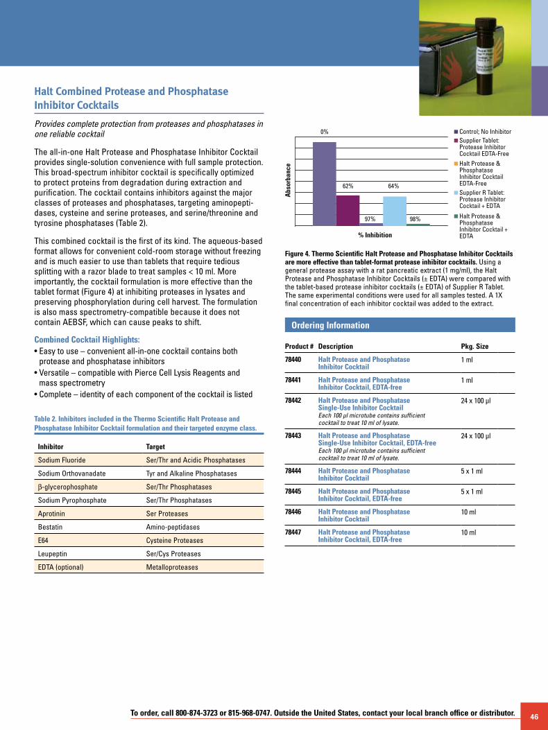
To order, call 800-874-3723 or 815-968-0747. Outside the United States, contact your local branch office or distributor. 46
Halt Combined Protease and Phosphatase Inhibitor Cocktails
Provides complete protection from proteases and phosphatases in one reliable cocktail
The all-in-one Halt Protease and Phosphatase Inhibitor Cocktail provides single-solution convenience with full sample protection. This broad-spectrum inhibitor cocktail is specifically optimized to protect proteins from degradation during extraction and purification. The cocktail contains inhibitors against the major classes of proteases and phosphatases, targeting aminopepti-dases, cysteine and serine proteases, and serine/threonine and tyrosine phosphatases (Table 2).
This combined cocktail is the first of its kind. The aqueous-based format allows for convenient cold-room storage without freezing and is much easier to use than tablets that require tedious splitting with a razor blade to treat samples < 10 ml. More importantly, the cocktail formulation is more effective than the tablet format (Figure 4) at inhibiting proteases in lysates and preserving phosphorylation during cell harvest. The formulation is also mass spectrometry-compatible because it does not contain AEBSF, which can cause peaks to shift.
Combined Cocktail Highlights:• Easy to use – convenient all-in-one cocktail contains both
protease and phosphatase inhibitors• Versatile – compatible with Pierce Cell Lysis Reagents and
mass spectrometry• Complete – identity of each component of the cocktail is listed
Table 2. Inhibitors included in the Thermo Scientific Halt Protease and Phosphatase Inhibitor Cocktail formulation and their targeted enzyme class.
Inhibitor Target
Sodium Fluoride Ser/Thr and Acidic Phosphatases
Sodium Orthovanadate Tyr and Alkaline Phosphatases
b-glycerophosphate Ser/Thr Phosphatases
Sodium Pyrophosphate Ser/Thr Phosphatases
Aprotinin Ser Proteases
Bestatin Amino-peptidases
E64 Cysteine Proteases
Leupeptin Ser/Cys Proteases
EDTA (optional) Metalloproteases
% Inhibition
Abs
orba
nce
Control; No InhibitorSupplier Tablet:Protease InhibitorCocktail EDTA-Free
Halt Protease &PhosphataseInhibitor CocktailEDTA-FreeSupplier R Tablet:Protease InhibitorCocktail + EDTA
Halt Protease &PhosphataseInhibitor Cocktail +EDTA
0%
62%
97%
64%
98%
Figure 4. Thermo Scientific Halt Protease and Phosphatase Inhibitor Cocktails are more effective than tablet-format protease inhibitor cocktails. Using a general protease assay with a rat pancreatic extract (1 mg/ml), the Halt Protease and Phosphatase Inhibitor Cocktails (± EDTA) were compared with the tablet-based protease inhibitor cocktails (± EDTA) of Supplier R Tablet. The same experimental conditions were used for all samples tested. A 1X final concentration of each inhibitor cocktail was added to the extract.
Ordering Information Product #
Description
Pkg. Size
78440 Halt Protease and Phosphatase Inhibitor Cocktail
1 ml
78441 Halt Protease and Phosphatase Inhibitor Cocktail, EDTA-free
1 ml
78442 Halt Protease and Phosphatase Single-Use Inhibitor CocktailEach 100 µl microtube contains sufficientcocktail to treat 10 ml of lysate.
24 x 100 µl
78443 Halt Protease and Phosphatase Single-Use Inhibitor Cocktail, EDTA-freeEach 100 µl microtube contains sufficientcocktail to treat 10 ml of lysate.
24 x 100 µl
78444 Halt Protease and Phosphatase Inhibitor Cocktail
5 x 1 ml
78445 Halt Protease and Phosphatase Inhibitor Cocktail, EDTA-free
5 x 1 ml
78446 Halt Protease and Phosphatase Inhibitor Cocktail
10 ml
78447 Halt Protease and Phosphatase Inhibitor Cocktail, EDTA-free
10 ml
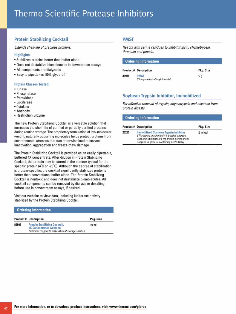
47 For more information, or to download product instructions, visit www.thermo.com/pierce
Protein Stabilizing Cocktail
Extends shelf-life of precious proteins.
Highlights:• Stabilizes proteins better than buffer alone• Does not destabilize biomolecules in downstream assays• All components are dialyzable• Easy to pipette (vs. 50% glycerol)
Protein Classes Tested:• Kinase • Phosphatase • Peroxidase • Luciferase • Cytokine • Antibody • Restriction Enzyme
The new Protein Stabilizing Cocktail is a versatile solution that increases the shelf-life of purified or partially purified proteins during routine storage. The proprietary formulation of low-molecular weight, naturally occurring molecules helps protect proteins from environmental stresses that can otherwise lead to enzyme inactivation, aggregation and freeze-thaw damage.
The Protein Stabilizing Cocktail is provided as an easily pipettable, buffered 4X concentrate. After dilution in Protein Stabilizing Cocktail, the protein may be stored in the manner typical for the specific protein (4˚C or -20˚C). Although the degree of stabilization is protein-specific, the cocktail significantly stabilizes proteins better than conventional buffer alone. The Protein Stabilizing Cocktail is nontoxic and does not destabilize biomolecules. All cocktail components can be removed by dialysis or desalting before use in downstream assays, if desired.
Visit our website to view data, including luciferase activity stabilized by the Protein Stabilizing Cocktail.
Ordering Information Product #
Description
Pkg. Size
89806 Protein Stabilizing Cocktail, 4X Concentrated SolutionSufficient reagent to make 40 ml of storage solution.
10 ml
PMSF
Reacts with serine residues to inhibit trypsin, chymotrypsin, thrombin and papain.
Ordering Information Product #
Description
Pkg. Size
36978 PMSF(Phenylmethylsulfonyl fluoride)
5 g
Soybean Trypsin Inhibitor, Immobilized
For effective removal of trypsin, chymotrypsin and elastase from protein digests.
Ordering Information Product #
Description
Pkg. Size
20235 Immobilized Soybean Trypsin InhibitorSTI coupled to spherical 4% beaded agarose.Capacity: Minimum of 6 mg trypsin per ml of gelSupplied in glycerol containing 0.05% NaN3
2 ml gel
Thermo Scientific Protease Inhibitors
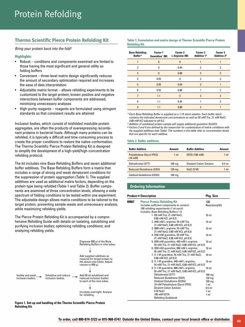
To order, call 800-874-3723 or 815-968-0747. Outside the United States, contact your local branch office or distributor. 48
Protein Refolding
Thermo Scientific Pierce Protein Refolding Kit
Bring your protein back into the fold!
Highlights:• Robust – conditions and components examined are limited to
those having the most significant and general utility as folding buffers
• Convenient – three-level matrix design significantly reduces the amount of secondary optimization required and increases the ease of data interpretation
• Adjustable matrix format – allows refolding experiments to be customized to the target protein; known positive and negative interactions between buffer components are addressed, minimizing unnecessary analyses
• High-purity reagents – reagents are formulated using stringent standards so that consistent results are attained
Inclusion bodies, which consist of misfolded insoluble protein aggregates, are often the products of overexpressing recombi-nant proteins in bacterial hosts. Although many proteins can be refolded, it is typically a difficult and time-consuming process to create the proper conditions to restore the native conformation. The Thermo Scientific Pierce Protein Refolding Kit is designed to simplify the development of a high-yield/high-concentration refolding protocol.
The kit includes nine Base Refolding Buffers and seven additional buffer additives. The Base Refolding Buffers form a matrix that includes a range of strong and weak denaturant conditions for the suppression of protein aggregation (Table 1). The supplied additives are used as additional matrix factors, depending on the protein type being refolded (Table 1 and Table 2). Buffer compo-nents are examined at three concentration levels, allowing a wide spectrum of folding conditions to be tested within one experiment. The adjustable design allows matrix conditions to be tailored to the target protein, preventing sample waste and unnecessary analysis, while maximizing refolding yields.
The Pierce Protein Refolding Kit is accompanied by a compre-hensive Refolding Guide with details on isolating, solubilizing and purifying inclusion bodies; optimizing refolding conditions; and analyzing refolding yields.
Isolate and washinclusion bodies.
Solubilize and reduceinclusion bodies.
Add 50 ml solubilized andreduced inclusion bodies to each of the nine tubes.
Add supplied additives as required for target protein to the above nine tubes. Adjust volume to 950 µl.
Dispense 900 µl of the BaseRefolding Buffers in nine tubes.
Incubate overnight. Analyze for refolding.
Figure 1. Set-up and handling of the Thermo Scientific Pierce Protein Refolding Kit.
Table 1. Formulation and matrix design of Thermo Scientific Pierce Protein Refolding Kit.
Base Refolding Buffer*
Factor 1 Guanidine† (M)
Factor 2 L-Arginine (M)
Factor 3 Additive 1‡
Factor 4 Additive 2‡
1 0 0 1 1
2 0 0.44 2 2
3 0 0.88 3 3
4 0.55 0 2 3
5 0.55 0.44 3 1
6 0.55 0.88 1 2
7 1.1 0 3 2
8 1.1 0.44 1 3
9 1.1 0.88 2 1
* Each Base Refolding Buffer is supplied as a 1.1X stock solution. Each buffer stock contains the indicated denaturant concentrations as well as 55 mM Tris, 21 mM NaCl, 0.88 mM KCl adjusted to pH 8.2.
† Addition of solubilized protein sample will supply additional guanidine (GuHCl).‡ Factors 3 and 4 are defined by the researcher for customization of matrix conditions with
the supplied additives (see Table). The numbers in the table refer to concentration levels that are specific for each additive.
Table 2. Buffer additives.
Buffer Additive Amount Buffer Additive Amount
Polyethylene Glycol (PEG) (10 mM)
1 ml EDTA (100 mM) 1 ml
Dithiothreitol (DTT) 100 mg Divalent Cation Solution 0.5 ml
Reduced Glutathione (GSH) 120 mg NaCl (5 M) 1 ml
Oxidized Glutathione (GSSG) 100 mg
Ordering Information Product #
Description
Pkg. Size
89867 Pierce Protein Refolding KitIncludes sufficient components to conduct100 refolding experiments (1 ml each).Includes: Base Refolding Buffers 1-9
1. (55 mM Tris, 21 mM NaCl, 0.88 mM KCl, pH 8.2)
2. (440 mM L-arginine, 55 mM Tris, 21 mM NaCl, 0.88 mM KCl, pH 8.2)
3. (880 mM L-arginine, 55 mM Tris, 21 mM NaCl, 0.88 mM KCl, pH 8.2)
4. (550 mM guanidine, 55 mM Tris, 21 mM NaCl, 0.88 mM KCl, pH 8.2)
5. (550 mM guanidine, 440 mM L-arginine, 55 mM Tris, 21 mM NaCl, 0.88 mM KCl, pH 8.2)
6. (550 mM guanidine, 880 mM L-arginine, 55 mM Tris, 21 mM NaCl, 0.88 mM KCl, pH 8.2)
7. (1.1 M guanidine, 55 mM Tris, 21 mM NaCl, 0.88 mM KCl, pH 8.2)
8. (1.1 M guanidine, 440 mM L-arginine, 55 mM Tris, 21 mM NaCl, 0.88 mM KCl, pH 8.2)
9. (1.1 M guanidine, 880 mM L-arginine, 55 mM Tris, 21 mM NaCl, 0.88 mM KCl, pH 8.2) Dithiothreitol (DTT) Reduced Glutathione (GSH) Oxidized Glutathione (GSSG) 10 mM Polyethylene Glycol (PEG) Divalent Cation Solution 5 M NaCl 100 mM EDTA Refolding Guidebook
100 Reactions/Kit 10 ml
10 ml 10 ml 10 ml 10 ml 10 ml 10 ml 10 ml 10 ml 100 mg 120 mg 100 mg 1 ml 0.5 ml 1 ml1 ml
49 For more information, or to download product instructions, visit www.thermo.com/pierce
Thermo Scientific Inclusion Body Solubilization Reagent
Effectively solubilizes insoluble protein.
Highlights:• Efficient – achieves the essential first step for subsequent
insoluble protein refolding procedures• Simple – one easy-to-use solution
Bacteria are widely used for recombinant protein expression. However, 70-80% of proteins produced by recombinant tech- niques in E. coli form inclusion bodies (i.e., protein aggregates). Once these aggregates are formed, it is very difficult to solubilize them. Pierce Inclusion Body Solubilization Reagent is designed to retrieve expressed protein in soluble form after lysis and extraction procedures.
A proprietary denaturant contained in this reagent provides the most effective means for solubilizing aggregated proteins. Additional components, such as a reducing agent and a chelating agent, may be added to the reagent, depending on the particular application. The following protocol is a generally applicable method useful in moving protein from the insoluble inclusion body state into solution in a gentle, phased approach. This is the first and essential step before proceeding with insoluble protein refolding procedures. (See Pierce Protein Refolding Kit, page 48.)
Inclusion body purification. Recombinant proteins expressed in bacteria often form inclusion bodies, especially when they are expressed at high levels. It is not known exactly how they are formed, but it is thought that the protein within the inclusion body is partially or incorrectly folded. The advantage of inclusion bodies is that they generally allow greater levels of expression, and they can be easily separated from a large proportion of bac-terial cytoplasmic proteins by centrifugation, giving an effective purification step. B-PER Reagent and its companion protocol have been optimized for inclusion body purification after soluble protein extraction.1 Although other methods may also be used, optimiza-tion is required to obtain inclusion body with good yield and purity.
Protein refolding. Because this reagent contains denaturant, the protein is denatured after solubilization. To obtain active protein, it is necessary to perform protein refolding. Several methods have been published describing protein refolding.2-4 A suggested protocol is provided for proceeding with refolding procedures. Because every protein possesses unique folding properties, the optimal refolding protocol for any given protein must be empirically determined.
Compatibility. The denaturant included in the Inclusion Body Solubilization Reagent will precipitate in SDS-PAGE sample buffer. However, this reagent is compatible with Pierce Coomassie Plus (Bradford) Protein Assay (Product # 23236). The denaturant can be removed by dialysis before performing SDS-PAGE analysis and other protein assays.
Protein Refolding
EXAMPLE SOLUBILIzATION PROTOCOL1. Purify the inclusion body using B-PER Bacterial Protein Extraction Reagent
(Product # 78248). Be sure to follow the manufacturer-supplied instruc-tions. Inclusion bodies purified by other methods are also applicable to the following protocols. The purity of the inclusion body prep may be analyzed by SDS-PAGE before performing the solubilization procedures. The purity of the inclusion body prep will not affect the solubilization efficiency. However, if a subsequent refolding procedure is desired, a greater than 90% purity is preferred.
2. Estimate the amount of inclusion body prep by subtracting the weight of the wet inclusion body pellet contained in a centrifuge tube from the weight of the centrifuge tube. Use 8 ml of the Inclusion Body Solubilization Reagent for 1 g of wet inclusion body pellet. More reagent may be used to dilute the protein further.
3. Resuspend the pellet in an appropriate amount of the Inclusion Body Solubilization Reagent by either vigorous vortex mixing or by pipetting until suspension is homogeneous. Shake the suspension for 30 minutes.
4. Remove cell debris by centrifugation at 27,000 x g (15,000 rpm for Beckman JA20 rotor) for 15 minutes.
5. Collect the supernatant that contains the solubilized protein from the inclu-sion body. If a protein assay is desired, Pierce Coomassie Plus (Bradford) Protein Assay (Product # 23236) is compatible. For SDS-PAGE analysis, it is necessary to remove the denaturant contained in the solubilization reagent by dialysis.
Protein Refolding Using Dialysis MethodMaterials required: • B-PER Bacterial Protein Extraction Reagent: for inclusion
purification• Inclusion Body Solubilization Reagent: For inclusion body
solubilization • DTT: Reducing agent (Product # 20290)• Urea: Mild denaturant• Slide-A-Lyzer Dialysis Cassette: Dialysis (Product # 66810)
SUGGESTED REFOLDING PROTOCOL1. Purify inclusion body from bacteria using B-PER Bacterial Protein
Extraction Reagent and solubilize inclusion body protein using Inclusion Body Solubilization Reagent following respective protocols. If disulfide bonds are involved in refolding, add DTT to 5 mM to the Inclusion Body Solubilization Reagent during the solubilization step.
2. Prepare 1 L of 6 M Urea solution in a 3.5 L glass or plastic beaker.3. Use an 18-gauge needle and a 10 ml syringe to transfer 8 ml of the
Inclusion Body Protein Solution to a 3-12 ml Slide-A-Lyzer Cassette. Dialyze the inclusion body protein against 6 M Urea for 6 hours.
4. Add 250 ml of 25 mM Tris•HCl (pH 7.5) to the beaker every 6-12 hours. Once the volume reaches 3 L, replace the dialysis solution with 2 L of 25 mM Tris•HCl (pH 7.5) and 150 mM NaCl. Dialyze for another 6 hours. To maintain protein stability, perform the dialysis in a cold room (4-8˚C). Precipitation may form during dialysis; however, there will always be some protein remaining in solution.
5. Recover the sample from the dialysis cassette using a similar-sized needle and syringe. Remove any visible insoluble material by centrifugation. Determine the folding status of the remaining soluble protein by activity assay or other methods.
Ordering Information Product #
Description
Pkg. Size
78115 Inclusion Body Solubilization Reagent 100 ml
ReferencesChu, R., et. al. (1998). Recombinant protein extraction from E. coli using B-PER Bacterial
Protein Extraction Reagent. Previews 2(1), 12-13.Mukhopadhyay, A. (1997). Inclusion bodies and purification of protein in biologically active
forms. Adv. Biochem. Eng. Biotechnol. 56, 61-109.Rudolph, R. and Lilie, H. (1996). In-vitro folding of inclusion body proteins. FASEB J. 10, 49-56.Marston, F.A.O. and Hartley, D.L. (1990). Solubilization of protein aggregates.
Meth. Enzymol. 182, 264-276.
To order, call 800-874-3723 or 815-968-0747. Outside the United States, contact your local branch office or distributor. 50
SuperSignal Technology is protected by U.S. Patent # 6,432,662.B-PER Technology is protected by U.S. Patent # 6,174,704. Slide-A-Lyzer Dialysis Cassette Technology is protected by U.S. Patent # 5,503,741 and 7,056,440; CA 2,170,738; and EP 0 720 508 B1.Slide-A-Lyzer MINI Dialysis Technology is protected by U.S. Patent # 6,039,871.U.S. Patent pending on Mitochondria Isolation Kit, P-PER Plant Protein Extraction Technology, Krypton Protein Stain Technology, GelCode Blue Stain Technology, Imperial Protein Stain Technology and Pierce 660 nm Protein Assay.Waring® is a trademark of Conair Corporation.Triton® is a trademark of Rohm and Haas Company.Sonifier® is a trademark of Branson Instruments, Inc.Biomasher® is a trademark of Nippi, Incorporated.Polytron® is a trademark of Kinematica AG Company.OptiPrep™ is a trademark of Axis-Shield plc.ATCC is a registered trademark of the American Type Culture Collection.Brij® and Tween® are trademarks of ICI Americas Inc. Corp.BugBuster® is a trademark of EMD Chemicals Inc.EasyLyse™ is a trademark of EPICENTRE Biotechnologies.CelLytic™ is a trademark of Sigma-Aldrich Co.
Contact Information
Belgium and Europe, the Middle East and Africa Distributors Tel: +32 53 85 71 84
France Tel: 0 800 50 82 15
The Netherlands Tel: 076 50 31 880
Germany Tel: 0228 9125650
United Kingdom Tel: 0800 252 185
Switzerland Tel: 0800 56 31 40
Email: [email protected] www.thermo.com/perbio
United States Tel: 815-968-0747 or 800-874-3723 Customer Assistance E-mail: [email protected] www.thermo.com
© 2009 Thermo Fisher Scientific Inc. All rights reserved. These products are supplied for laboratory or manufacturing applications only. Unless indicated otherwise on the inside back cover, all trademarks are property of Thermo Fisher Scientific Inc. and its subsidiaries. 16
0175
7 5
/09
Prin
ted
in th
e U.
S.