thermal interaction of short-pulsed laser focused …coe papers/jp_46.pdf · thermal interaction of...
TRANSCRIPT
IOP PUBLISHING PHYSICS IN MEDICINE AND BIOLOGY
Phys. Med. Biol. 54 (2009) 4225–4241 doi:10.1088/0031-9155/54/13/017
Thermal interaction of short-pulsed laser focusedbeams with skin tissues
Jian Jiao and Zhixiong Guo1
Department of Mechanical and Aerospace Engineering, Rutgers, The State University ofNew Jersey, Piscataway, NJ 08854, USA
E-mail: [email protected]
Received 22 February 2009, in final form 25 May 2009Published 17 June 2009Online at stacks.iop.org/PMB/54/4225
AbstractTime-dependent thermal interaction is developed in a skin tissue cylindersubjected to the irradiation of a train of short laser pulses. The skin embeddedwith a small tumor is stratified as three layers: epidermis, dermis andsubcutaneous fat with different optical, thermal and physiological properties.The laser beam is focused to the tumor site by an objective lens for thermaltherapy. The ultrafast radiation heat transfer of the focused beam is simulatedby the transient discrete ordinates method. The transient Pennes bio-heatequation is solved numerically by the finite volume method with alternatingdirection implicit scheme. Emphasis is placed on the characterization of thefocused beam propagation and absorption and the temperature rise in the focalspot. The effects of the focal spot size and location, the laser power, and thebio-heat equation are investigated. Comparisons with collimated irradiation areconducted. The focused beam can penetrate a greater depth and produce highertemperature rise at the target area, and thus reduce the possibility of thermaldamage to the surrounding healthy tissue. It is ideal for killing cancerous cellsand small tumors.
(Some figures in this article are in colour only in the electronic version)
1. Introduction
Basal cell carcinoma (BCC) is the most common skin cancer in humans. It develops from basalcells, which are in the deepest layer of the epidermis (Gilchrest et al 1999). Each year in theUnited States, about 900 000 people are diagnosed with basal cell carcinoma (550 000 male,350 000 female). The estimated lifetime risk of basal cell carcinoma in the white populationis 33–39% for men and 23–28% for women.
1 Author to whom any correspondence should be addressed.
0031-9155/09/134225+17$30.00 © 2009 Institute of Physics and Engineering in Medicine Printed in the UK 4225
4226 J Jiao and Z Guo
It has been well established that organic tissue responds strongly to temperature rise. Thethermal impact on tissue changes drastically when temperature exceeds 43 ◦C; the rate of ‘cellkill’ doubles for every 1 ◦C increase beyond 43 ◦C and decreases by a factor of 4–6 for every1 ◦C drops below 43 ◦C (Jeong et al 2003). It has also been noticed that tumor cells are moresensitive to temperature increase than normal tissues (Anghileri and Robert 1986). Moreover,Robinson et al (1998) found that a temperature evaluation to at least 56 ◦C for 1 s or more wassufficient to bring about cancer cell denaturation and death.
Lasers have been widely considered in clinical therapies (Anderson and Parrish 1983,Amin et al 1993, Manns et al 1999) and applications (Yamada 1995, Niemz 2002),including laser-induced hyperthermia, laser-induced interstitial thermotherapy, interstitial laserphotocoagulation therapy, laser microsurgery, as well as optical imaging (Hebden et al 1997,Yamada 2000, Gao et al 2004, Guo et al 2006). The objective of hyperthermia in cancertreatment is to raise the temperature in cancerous tissue above a therapeutic value whilemaintaining the surrounding healthy tissue at sub-lethal temperature values in cases wheresurgical intervention is dangerous. In order to more effectively destroy/detect canceroustissues, contrast agents such as absorbing and/or fluorescing dyes can be administrated forenhancement of light absorption (Ntziachristos et al 2000, Quan and Guo 2004).
Ultrafast lasers output ultra-short pulses with pulse width in the range from picosecondsdown to femtoseconds. Due to the extremely short pulse duration, which is much shorterthan the thermal relaxation time of many materials, beam interaction with a material occursbefore heat diffusion in the material ever takes place, leading to increased local temperature ina very short time period and minimization of the heat-affecting zone. This will lessen thermaldamage to the surroundings, a concern that should always be born in mind in laser applications.With the use of an ultrafast laser, significant improvement in the damage localization overcontinuous wave and general pulsed lasers has been attained (Zysset et al 1989).
Two characteristics in ultrafast radiation heat transfer (Kim and Guo 2004) are worthmentioning. One is that the transient effect is significant when the pulse duration is notsubstantially longer than the characteristic time of radiation propagation in the medium. Forapplications in tissues where the typical characteristic thickness is in the order of millimetersto centimeters, the corresponding characteristic time (length over the speed of light) is inthe range 0.01–1 ns. Hence, the propagation of an ultra-short pulse with the speed of lightmust be incorporated into the equation of radiative transfer (Guo and Kumar 2002). Anothercharacteristic is that the emission from the medium is generally negligible as compared withthe high laser intensity. Thus, the medium is cold in the modeling of radiative heat transfer.Many methods were considered for modeling photon transport and migration in turbid media(Yamada 1995, Sassaroli et al 1999, Binzoni et al 2006). Mitra and Kumar (1999) comparedseveral one-dimensional methods for simple plane wall problems. To accurately capturethe propagation of a short pulse in multi-dimensional geometries, the authors’ group hasdeveloped the transient Monte Carlo method (Guo et al 2000), the radiation element method(Guo and Kumar 2001a) and the transient discrete ordinates method (Guo and Kumar 2001b).Comparison with experiments has also been carried out (Guo and Kumar 2002, Wan et al2004).
Accurate prediction of temperature distributions and heat transfer rates in tissues (Pfeferet al 2000, Zhu et al 2002, Kim and Guo 2007) is also paramount for clinical laser therapiesand applications. The Pennes model that describes heat transfer in perfused tissues is a well-known bio-heat equation. It assumes that the thermal contribution of blood be modeled as if itentered an imaginary pool at equilibrium with the surrounding tissue. A recent review (Arkinet al 1994) compared several bio-heat models and concluded that the Pennes model is still the

Thermal interaction of short-pulsed laser focused beams with skin tissues 4227
most practical for fast prediction of transient temperature profiles such as those expected incertain hyperthermia conditions.
To maximize treatment efficacy and avoid undesirable reactions in normal tissues, thermaltherapies ideally deliver heat to the target while spare the surrounding healthy tissue. As anon-invasive means to deliver energy to a location inside the body, the technique of convergingbeam can eliminate the perforation of skin. Moreover, due to concentrating the energy at thefocal spot, a converging beam can penetrate a greater depth in the tissue without significantattenuation. A focused beam with a spot size comparable to cells is ideal for preciselykilling cancerous cells without damaging surrounding healthy cells. This makes it very usefulin the treatment of small tumors at the very early stage or as a supplemental method forkilling precisely any leftover cancerous cells that may exist after surgical procedures. Thetemperature at the focal zone is higher than that of the surroundings, and it is then consideredas the desired temperature for treatment or therapy. Therefore, accurate prediction of the focalzone temperature is critical. A literature survey reveals that complete thermal modeling of afocused beam in tissues has been rarely conducted.
In this work, a combined model of ultrafast radiative heat transfer and Pennes bio-heattransfer is developed to simulate the heat transfer processes in a model skin tissue subjectedto the irradiation of a train of short pulses. The skin tissue is stratified as three layers withdifferent properties. An inhomogeneity simulating a small skin tumor is embedded in thedermis layer beneath the skin surface. To effectively heat up the cancerous cells, a collimatedlaser beam is converged into the tumor site by a single objective lens. The laser beam isaxisymmetric and has temporal and spatial Gaussian distributions. A technique for treatingrealistic beam convergence is introduced. Comparisons between the converging beam andcollimated irradiation are conducted. The influences of the focal spot size, the laser powerand the bio-heat equation are also scrutinized.
2. Mathematical formulation
2.1. Governing equations
A laser beam is focused at the small tumor in the skin tissue as shown in figure 1. The intensityIl of the diffused radiation in a discrete ordinate −→sl is described by the time-dependent equationof radiative transfer in cylindrical coordinates (Kim and Guo 2004):
1
c
∂Il
∂t+
μl
r
∂
∂r(rIl)− 1
r
∂
∂φ(ηlIl)+ξl
∂Il
∂z+σeIl =σeSl, l = 1, 2, . . . ,M, (1)
where μl, ηl, ξ l are the three directional cosines, σ e is the extinction coefficient, which is thesummation of the absorption coefficient σ a and scattering coefficient σ s, c is the speed of lightin the tissue, Sl is the radiative source term, which is
Sl = (1 − ω)Ib +ω
4π
M∑j=1
wjjlIl + Sli , (2)
in which Ib is the black body emitting intensity of the tissue, ω = σ s/(σ s+σ a) is the scatteringalbedo, wj is the appropriate angular weight in the discrete direction �sj , jl represents thescattering phase function (�sj → �sl), and Sl
i is the source contribution of the laser irradiationand can be expressed as
Sli = 1
4πIi(�si → �sl), (3)
where the unit vector −→si represents the laser incident direction. Light scattering in tissuesis generally anisotropic (Binzoni et al 2006). However, it is reasonable to scaling down to

4228 J Jiao and Z Guo
HFFat
TumorHD
Epidermis
HT
RT DermisH
r
z
RLaser
HE
Figure 1. The skin geometry and the coordinates.
v(0)
θ
θ
r
Lf
L
Lens
Air
Tissue
2v0
d/2
O
z
r
Laser
i
Figure 2. The laser beam convergence.
isotropic scattering (Guo and Kumar 2000). When the reduced scattering coefficient is used,the scattering phase function is unity. In the region where no laser irradiation is passingthrough, Ii = Si = 0.
The intensity of a laser beam having a Gaussian profile both temporally and spatially canbe expressed as
Ii(r, z, t) = (1 − R0)I0(z) exp{−4 ln 2 × [(t − z/c)/tp − 1.5]2}× exp[−2r2/v(z)2] exp(−σez), (4)
where I0 is the amplitude of the beam radiation strength, v is the beam radius (1/e2), R0 is thereflectance on the tissue–air interface, and tp is the pulse width at half maximum. The totaltime duration of a whole pulse is set as 3 tp in this study so that the peak of the pulse arrivesat time 1.5 tp. The impinging area of the beam considered is r � ν(z).
Because of the mismatch of refractive indices of the air and tissue, the incident laser beamis refracted at the tissue–air interface and will not converged to the focus of the lens in freespace but rather to a deeper distance in the tissue as shown in figure 2. For a Gaussian beam,the radius of a diffraction-limit focal spot can be calculated as
v0 = 1.22(λLf /d), (5)

Thermal interaction of short-pulsed laser focused beams with skin tissues 4229
where λ is the laser wavelength, Lf is the focal length of the converging lens in free space, andd is the diameter of the collimated beam before the lens. The focal spot size is little affectedby the refractive index of the medium to be focused to. For a focusing depth L in the medium,the radius of the impinging area on the medium surface is then obtained as
v(0) = L · tan θr + v0, (6)
in which the refractive angle is obtainable by Snell’s law:
θr = sin−1
(sin θi
n
), (7)
where n is the refractive index of the tissue and it is assumed to be 1.40. The incident angleof the focusing beam is
θi = tan−1
(d/2 − v0
Lf
). (8)
Inside the tissue, the propagating beam radius varies with z:
v(z) = v(0) − v0
L|z − L| + v0. (9)
The amplitude of the beam radiation strength is correlated with the laser beam power Pat the tissue surface as
P = 0.46πv(z)2I0(z)f tp, (10)
where f is the pulse repetition rate.Once the intensity field is obtained, the incident radiation and the divergence of radiative
heat flux can be calculated as
G =n∑
l=1
wlIl + Ic, (11)
∇ · qrad = σa(4πIb − G), (12)
where Ic is normally incident laser intensity.Because the pulse duration considered is much shorter than the thermal relaxation time of
tissue, heat diffusion during the pulse irradiation period is negligible. Hence, the temperaturerise in the tissue is due to the irradiation of an ultra-short pulse is described as
ρCP
∂T (r, z, t)
∂t= −∇ · qrad(r, z, t). (13)
Between two successive pulses, heat diffusion is incorporated and Penne’s equation (Arkinet al 1994) is used for modeling the bio-heat transfer in the skin tissue:
ρCp
∂T (r, z, t)
∂t= k∇2T + (ρC)bωb(Ta − T ) + qm, (14)
where ρ, Cp, k,T denote the density, specific heat, thermal conductivity and temperature oftissue, respectively; Cb is the specific heat of blood, ωb is the blood perfusion rate, qm is themetabolic heat generation rate, and Ta is the supplying arterial blood temperature.

4230 J Jiao and Z Guo
2.2. Boundary conditions
For the radiative heat transfer modeling, three types of radiation boundary conditions areconsidered for the present problem. At the laser incident surface (z = 0) Fresnel reflectionmust be considered because of the mismatch of the refractive indices between the tissue andair. For internal radiation at the tissue–air interface, a critical angle is given by Snell’s law:
θcr = sin−1(1/n). (15)
Total reflection occurs when the angle of incidence θ i > θ cr. Otherwise, the reflection at theinterface is purely specular and the reflectance is calculated by Fresnel’s equation:
Rs = 1
2
[tan2(θi − θr)
tan2(θi + θr)+
sin2(θi − θr)
sin2(θi + θr)
]. (16)
All other surfaces are tissue–tissue interface. Since biological tissues are highly scattering,photons reaching the boundary of a tissue must have been undergone multiple scattering eventsand the possibilities of photons passing through the boundary or reflecting back are almostequal for a turbid medium. Thus, we specify a diffuse reflectance Rd = 0.5 on such surfaces.The reflecting boundary condition at the wall is given by
Iw = RsI−1w +
Rd
π
[Icw +
∑−→sl ·−→n <0
wlIw,l|−→sl · −→n |]. (17)
The centerline of the tissue cylinder (r = 0) is specified as an axisymmetric boundary.For the bio-heat transfer modeling, except for the laser incident surface which is exposed
to the ambient air at room temperature Tam = 25 ◦C with the heat transfer coefficient h = 15 W(m−2 K−1), all other surfaces of the tissue cylinder are surrounded by other tissue remainedat 37 ◦C. Again the centerline of the tissue cylinder is treated as the axisymmetric boundary.The initial and boundary conditions for the bio-heat transfer model are summarized below:
T = 37 ◦C, when t = 0, (18)
T = 37 ◦C, at r = R or z = H, (19)
∂T
∂r= 0, at r = 0, (20)
− k∂T
∂z= h(Tam − T ), at z = 0. (21)
2.3. Properties of the model skin tissue and the laser parameters
In this model, human skin (H = 10 mm, R = 10 mm) is organized in distinct layers, which areepidermis (HE = 1 mm), dermis (HD = 2 mm) and subcutaneous fat (HF = 7 mm), respectively.A small skin tumor (HT = 1, RT = 1 mm), either infiltrative basal cell carcinomas (IBCC) ornodular basal cell carcinomas (NBCC), is suited in the dermis layer. The IBCC-type tumorhas a higher absorption coefficient than that of the NBCC-type tumor. Their thermal andphysiological properties are assumed to be the same. The optical (at wavelengths 1200 nmand 1064 nm, respectively), thermal and physiological properties of the considered tissues arelisted in tables 1–3, respectively.
Unless specified otherwise, the laser and lens parameters used in this work are as follows.The focal length of the converging lens is 10 mm in free space. The collimated laser beamdiameter is 6 mm. The laser wavelength is centered at 1200 nm. At this wavelength light

Thermal interaction of short-pulsed laser focused beams with skin tissues 4231
Table 1. Optical parameters for different tissue layers at wavelengths 1200 nm and 1064 nm,respectively (Salomatina et al 2006).
Tissue type Epidermis Dermis Fat IBCC NBCC
λ (nm) 1200/1064σ s (mm−1) 2.6/3.0 1.7/1.83 1.5/1.69 1.05/1.22 1.0/1.11σ a (mm−1) 0.06/0.02 0.12/0.05 0.18/0.07 0.15/0.10 0.02/NA
Table 2. Thermal parameters for different tissue layers (Cohen 1977).
Tissue type Epidermis Dermis Fat Tumor
ρCp (J mm−3 K−1) 4.2 × 10⁻³ 4.2 × 10⁻³ 4.2 × 10⁻³ 4.2 × 10⁻³
k (W m−1 K−1) 0.21 0.30 0.21 0.59
Table 3. Physiological parameters for different skin tissue layers (Emery and Sekins 1982).
Tissue type Epidermis Dermis Fat Tumor
Perfusion ratio (ml ml−1 s−1) 0 1.63 × 10⁻³ 1.0 × 10⁻³ 5.0 × 10⁻³
Metabolic heat generation (W kg−1) 1.0 1.0 0.32 0.67
absorption of the epidermis is low (Salomatina et al 2006) and the incident light may penetrateto the cancerous region. The focal spot has a beam diameter of approximately 5 μm. Theaverage laser power is 0.065 W. The pulse width and repetition rate are 10 ps and 1 MHz,respectively. With these parametric values, the peak pulse power density at the focal spot1–2 mm beneath the skin surface after attenuation is two to three orders of magnitude lowerthan the ablation threshold (>1011 W cm−2) in tissues (Niemz 2002). Thus, laser-inducedablation mechanism does not apply and the current thermal analysis fits in.
2.4. Numerical schemes
The transient discrete ordinates method (TDOM) with S10 scheme was employed for thesolution of the present ultrafast radiative heat transfer problem. For detailed information onthe numerical scheme and accuracy, please refer to the author Guo’s previous publications(Guo and Kumar 2002, Kim and Guo 2004). As noted by Chai et al (1993) a ray effect existsbecause of limited number of discrete ordinates. In the literature, S4 (24 discrete ordinates)and S8 (80 discrete ordinates) schemes were commonly adopted. The strong directionality infocusing beam requires for high-order quadratures such as S10 (120 discrete ordinates). WithS10, the ray effect could be lessened (Guo and Kumar 2001b). The transient conductive heattransfer equations are solved numerically by using the alternating direction implicit (ADI)scheme (Anderson et al 1984) which is well known in computational physics, and thus, thedetails are not repeated here.
For both the radiation and conduction simulations, the same grid system is adopted toavoid interpolation. To improve the numerical calculation efficiency and to capture the rapidchange in the focus, a non-uniform grid system is employed, with a refined grid in the focusregion. For a typical skin tissue cylinder with R = 10 mm and H = 10 mm, the adoptedstaggered grid is 80 × 80 in the present calculations. The time step is 0.2 ps for the radiationcalculation and 0.25 μs for the bio-heat conduction, respectively. Several sets of different

4232 J Jiao and Z Guo
z*
Inci
dent
radi
atio
nG
0.2 0.4 0.6 0.8 1
0.3
0.6
0.9
1.2
1.5
t* = 0.5t* = 1.0t* = 3.0t* = 5.0
pure absorption
pure scattering
o Exact solution for pure scatteringΔ Exact solution for pure absorption
0
(a) Validation of the radiation model.
t (s)
Tem
per
atu
re(o
C)
100 200 300 400 500
40
45
50
60* 6080* 80100*100
o , Δ Exact solution
at (0,0)
at (5,5)
37
(b) Validation of the conduction model.
Figure 3. Comparison between the exact solutions and the numerical predictions. (a) Validationof the radiation model. (b) Validation of the conduction model.
grid sizes and time steps were considered, and they could all give satisfactory and convergentresults.
Since the optical and thermal properties of the tissue are assumed to be constant becausethe temperature is not dramatically changed during irradiation, the transient radiation heattransfer does not vary between pulses and it is only required to calculate the response of onepulse. The temperature rise obtained for this initial pulse can be applied to any subsequentpulse as a simple addition to the temperature field for the treatment of a pulse train. Thebio-heat transfer process is simulated in the whole period of the pulse train until the cut-off ofirradiation. For radiation transfer modeling, the computational time is about 30 min for onepulse irradiation using a DELL PC (Optiplex 755: 2.40 GHz CPU and 3.25 GB RAM).
3. Results and discussion
To validate the computational models, comparisons with exact solutions in simple situationsare conducted. Wu and Wu (1997) considered a cylinder of unity optical length and unityaspect ratio. The medium in the cylinder was assumed to be gray and homogeneous, andthe boundaries were assumed to be non-reflecting. A uniform collimated radiation with unityintensity was incident on the top of the cylinder. Two dimensionless variables were definedas z∗ = σez and t∗ = ct/L. The incident radiation profiles along the optical axis (r = 0)for both the pure absorption and purely scattering cases are shown in figure 3(a). For thepure scattering case, it is seen that the temporal profile of the incident radiation graduallyreaches to the steady-state exact solution (Wu and Wu 1997) as time proceeds to t∗ = 5 thatis much longer than the pulse duration. For the pure absorption case, the present transientsolution at t∗ = 5 matches perfectly with the steady-state exact solution obtained by using theBeer–Lambert law.
The ADI method for heat conduction modeling is validated in a tissue cylinder that isassumed to have a uniform initial temperature 50 ◦C. In figure 3(b), the numerically predictedtemperatures at the origin (0,0) and in a position (5 mm, 5 mm) are compared with the exactsolutions that the authors obtained by using the method of separation of variables. It is seen

Thermal interaction of short-pulsed laser focused beams with skin tissues 4233
r (mm)
z(m
m)
1 2 30
0.5
1
1.5
2
1.3E-111E-117E-124E-121E-12
r (mm)
z(m
m)
0 1 20
1
2
5.5E-094E-092.5E-091E-091E-12
(a) Collimated beam at t=4.7ps (b) Converging beam at t=4.7ps
r (mm)
z(m
m)
0 1 2 3
0.5
1
1.5
2
7E-115.5E-113.5E-112E-115E-12
r (mm)
z(m
m)
0 1 2
1
2
1E-061E-081E-105E-12
0.005 0.010.9
1
1.1
(c) Collimated beam at t=9.4ps (d) Converging beam at t=9.4ps
r (mm)
z(m
m)
0 1 2 30
1
2
1.7E-101.5E-101.3E-101.1E-109E-117E-115E-11
r (mm)
z(m
m)
0 1 2
1
2
3
5E-055E-065E-075E-085E-095E-105E-11
0.005 0.010.9
1
1.1
(e) Collimated beam at t=19.7ps (f) Converging beam at t=19.7ps
Figure 4. Comparison of radiation propagation between the collimated and focused beams:(a) collimated beam at t = 4.7 ps, (b) converging beam at t = 4.7 ps, (c) collimated beam at t =9.4 ps, (d) converging beam at t = 9.4 ps, (e) collimated beam at t = 19.7 ps, (f) converging beamat t = 19.7 ps.
that the numerical results calculated with three different grid systems converge to one curveand match well with the exact solutions.
Figure 4 shows the laser intensity contours at three time instants in the skin tissue subjectedto one single 10 ps pulse irradiation, in which the collimated and converging beams of the

4234 J Jiao and Z Guo
(t-z0 /c) / tp
Ab
sorb
eden
ergy
(μJ
/mm
2)
0 1 2 3 4 50
0.5
1
1.5
2
tp = 10pstp = 100pstp = 1ns
IBCC
NBCC
E= 65nJ
d= 6mm, 2v0= 5μm
Figure 5. The accumulation of the absorbed radiative energy at the focal spot due to the irradiationof a single pulse.
same power are considered for comparison. An IBCC tumor is located in the region 1–2 mmbeneath the skin surface. For the focused beam irradiation, the focal plane is at z = 1 mm.Since the speed of light in the skin tissue is approximately 0.21429 mm ps−1, the times forlight traveling 1 and 2 mm distances are 4.666 and 9.333 ps, respectively. Figures 4(b), (d)and (f) all show good convergence of the focused laser beam at the focal spot. At t = 4.7 ps,the wave front just passed the line z = 1 mm. At t = 9.4 ps, the wave front passed the line z =2 mm. At t = 19.7 ps that is 1.5 tp plus 4.7 ps, the peak power of the laser arrives at the focalspot. Comparing the focused beam results with the corresponding collimated beam results,it is seen that the focused beam can penetrate a great depth covering the whole tumor region,while the collimated beam can only penetrate a superficial layer to about 0.7 mm beneath theskin surface. The intensity of the focused beam is generally two to five orders of magnitudestronger than that of the collimated beam. In particular, it is seen that the intensity at the focalspot in figure 4(f) is over five orders of magnitude greater than the maximum intensity of thecollimated beam. More importantly, the maximum intensity in the collimated beam situationis always at the skin surface, while the focused beam can deliver the maximum power to thetarget (i.e., the focal spot) inside the tissue. The advantage of the focused beam is then obvious.
Taking the focal spot size as the characteristic length for heat diffusion (L =5.0 μm),the thermal relaxation time in the tumor region is estimated as (Niemz 2002) τ =L2/[4(k/ρC)tumor] = 45 μs. It is much longer than any laser pulse that could be consideredas a short pulse. Therefore, the thermal diffusion during a single pulse period is negligible.Then the temperature rise within a pulse irradiation period depends on the totally accumulatedenergy that is absorbed by the tissue. The profiles of the absorbed radiation energy at thefocal spot for the converging laser beam of different pulse widths (10 ps, 100 ps and 1 ns,respectively) are shown in figure 5. The time subtracted by the photon flight time from thesurface to the focal spot (z = 1 mm) is normalized by the respective laser pulse width. Itis found that the absorbed radiative energy accumulates rapidly within one tp period. Afterthe pulse is off at 3 tp, the accumulated energy almost saturates. It is reasonable to cut offthe transient calculation for one single pulse irradiation after 5tp plus the photon flight timeand assume a pseudosteady state is achieved. From figure 5, it is also observed that theradiative energy accumulated in the IBCC-type tumor is larger than that in the NBCC-typetumor because the IBCC has a higher absorption coefficient.

Thermal interaction of short-pulsed laser focused beams with skin tissues 4235
z (mm)
Tem
per
atu
reri
se(o C
)
0.5 1 1.5 2
10-7
10-6
10-5
10-4
10-3
10-2
r = 0 μmr = 1 μmr = 5 μmr = 500 μm
E= 65nJ, d= 6 mm
Focused beam
2v0= 5μm
Collimated beam
Figure 6. The temperature rise profiles at the end of the irradiation of a single pulse.
Figure 6 shows the temperature rise distributions along the axial direction with fourdifferent radial positions for the tissue cylinder subjected to one pulse of either the collimatedor converging irradiance. The collimated laser beam is 6 mm in diameter. The focal spot ofdiameter approximately 5 μm is at the plane z = 1 mm. The focused beam radius on theskin surface is then about 210 μm. It is seen that the temperature rise in the tissue along thebeam path is several orders of magnitude higher for the focused beam than for the collimatedbeam. There is an obvious peak rise around the focal spot (z = 1 mm, r < 2.5 μm). For thecollimated irradiance, a small temperature jump is also observed after z = 1 mm because ofthe higher absorption of the IBCC than the epidermis layer in front. However, this temperaturejump is still lower than that at the skin surface (z = 0). Thus, the skin surface is susceptibleto thermal damage during thermal therapy of the tumor if a collimated beam is adopted.
It should be noted that the temperature rise in figure 6 is due to the irradiation of asingle pulse. To achieve the target temperature for thermal therapy, continuously repetitivepulses will be needed. It has been discussed earlier that a temperature rise to 56 ◦C for 1 sor more could be sufficient to kill tumors. In the same time, the temperature cannot risetoo high to avoid cell necrosis in the surrounding healthy tissue. A critical temperature of62 ◦C corresponding to 25 ◦C temperature rise is selected as the onset of irreversible tissuedamage in this study. Therefore, it is crucial to consider the irradiation of a pulse train witha beam focused to the tumor region to realize a temperature rise in the range between 19 ◦Cand 25 ◦C for at least 1 s. The temperature used in figures 7 and 9–11 is the volumetricallyaveraged value over a volume of the focal spot size to facilitate the comparison of others in thefuture.
Figure 7 shows the transient profiles of temperature rise at four different axial locations inthe centerline of the skin tissue cylinder. The beam is focused at z = 1 mm. From figure 7(a),it is seen that the temperature rise at the focal spot is the largest. The temperature in the tissueincreases as time advances (i.e., with continuous pulse train incidence). For the consideredlaser power (0.065 W), the temperature rise in the focal spot reaches to19 ◦C at about 2.6 s andgoes over 25 ◦C at about 4.2 s. Thus, the total time duration in the therapeutic temperaturewindow (56–62 ◦C) is 1.6 s, longer than the required 1 s. The laser irradiation could be cut offany time between 3.6 and 4.2 s. In the case of t = 4.2 s, the skin surface temperature (z = 0) isabout 55 ◦C. No irreversible tissue damage occurs. Under the microscopic view in figure 7(b)for the temperature rise in the several initial pulses, it is observed that the temperature actually

4236 J Jiao and Z Guo
t (s)
Tem
per
atu
reri
se(o C
)
0 1 2 3 4
5
10
15
20
25
30
z= 0 mmz= 0.75mmz= 1.0mmz= 1.25mm
Δ t = 1.6sP= 0.065W, f= 1MHz
d= 6mm, 2ν0= 5μm
(a) Meso-time scale
t (μs)
Tem
per
atur
eri
se(o C
)
2 4 60
0.5
1
1.5
2
2.5
z=0.0mmz=0.75mmz=1.0mmz=1.25mm
(b) Micro-time scale
Figure 7. Transient profiles of the temperature rise due to the irradiation of a pulse train:(a) meso-time scale, (b) micro-time scale.
r (mm)
z(m
m)
0 2 4 6 8 10
2
4
6
8
10P= 0.065W, f= 1MHz
d= 6mm, 2ν0= 5μm
0.005 0.010.9
1
1.1
2423.623.2
22.822.42221.6
Figure 8. The temperature field at irradiation time instant 4 s.
increases like a staircase. At the onset of each new pulse, there is a clear temperature jump.Further, the temperature rise is substantially larger at the focal spot than other regions at theinitial time stage as shown in figure 7(b). This is because the laser energy is focused at the focalspot with negligible heat diffusion. As the time scale collapses, the effect of thermal diffusionis increasingly evident and thus the temperature difference between different locations narrowsas shown in figure 7(a).
The contour of temperature rise in the skin tissue with an IBCC-type tumor after 4 s pulsetrain irradiation (0.065 W) is plotted in figure 8. Inspecting the enlarged view in the focalspot, clearly the temperature is much higher in the focal spot than any other region, includingthe laser incident surface.
The focal spot size is inversely proportional to the collimated beam diameter before theconverging lens. Figure 9 shows the effects of the spot size on (a) the transient temperatureprofiles at two selected points (incident surface at z = 0 and focal plane at z = 1 mm) and(b) the temperature profiles along the optical axis at the end of a pulse train (t = 4 s). The three

Thermal interaction of short-pulsed laser focused beams with skin tissues 4237
t (s)
Tem
per
atu
re r
ise
(o C)
0 1 2 3 4
5
10
15
20
25
d=8 mm, 2ν0=3.6μm
d=6 mm, 2ν0=5.0μm
d=4 mm, 2ν0=7.2μm
z=1 mm
z=0 mm
P= 0.065W, f= 1MHz
(a)
z (mm)
Tem
per
atu
re r
ise
(o C)
0 0.5 1 1.5 214
16
18
20
22
24
26
28
d=8 mm, 2ν0=3.6μm
d=6 mm, 2ν0=5.0μm
d=4 mm, 2ν0=7.2μm
P= 0.065W, f= 1MHz, t=4s
ΔT = 19oC
ΔT = 25oC
(b)
Figure 9. (a) The transient profiles of the temperature rise with different focal spot sizes, and(b) the temperature rise profiles along the optical axis at irradiation time instant 4 s.
P (W)
t(s)
0.03 0.04 0.05 0.06 0.07 0.08 0.09
5
10
15
20
ΔT=25 oC
ΔT=19 oC
d= 6mm, 2ν0= 5μm
f=1MHz
Δt=4.9s
Δt=1.9sΔt=0.8s
Figure 10. The relationship between the laser power and the irradiation times to achieve the targettemperature rise at the focal spot.
selected collimated beams with diameter 4, 6 and 8 mm are converged to spots approximately7.2, 5.0 and 3.6 μm in diameter, respectively and the impinging areas on the skin surfaceare calculated as 141, 210 and 275 μm in diameter, respectively. From figure 9(a), it is seenthat the focal spot with the smallest diameter is preferred, because the temperature rise at thesmallest focal spot is the highest while the temperature rise at the incident skin surface is thelowest in this case. Figure 9(b) also demonstrates this preference because the temperaturein most of the epidermis layer before the tumor is the coldest for the smallest focal spot,while the temperature in the tumor region is the highest. Although the temperature rise goesover the 25 ◦C line for the small spots (3.6 and 5.0 μm), this problem can be resolved viareducing the laser power.
Figure 10 shows the incident laser powers and irradiation times required in order forthe temperature at the focal spot to raise 19 ◦C and 25 ◦C, respectively. The laser beam
4238 J Jiao and Z Guo
z (mm)
t(s)
P(W
)
1 1.2 1.4 1.6 1.8 210
0
101
102
0.25
0.5
0.75
NBCC
IBCC
λ =1200nm, d= 6mm, 2v0= 5 μm
ΔT(P=0.08W)= 19oC
ΔT(t=4s)= 19oC
(a)z (mm)
t(s)
P(W
)
1 1.2 1.4 1.6 1.8 2
0.25
0.5
0.75
1
0.8
1.2
1.6
2
IBCC
λ =1064nm,d =6mm, 2ν0= 50μm
ΔT(P=1W)=19oC
ΔT(t=0.22s)=19oC
(b)
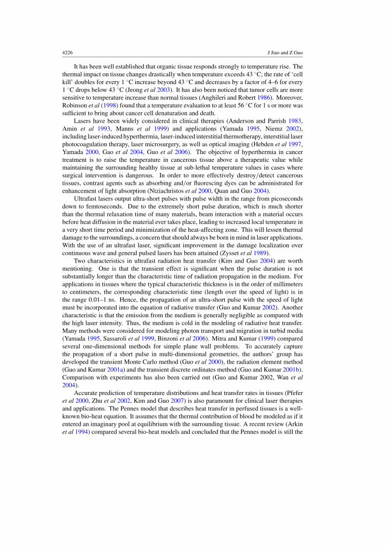
Figure 11. The required laser power or irradiation time for 19 ◦C temperature rise at the focal spotversus the focal plane location for irradiation: (a) with wavelength at 1200 nm and spot size 5 μmin diameter, and (b) with wavelength at 1064 nm and spot size 50 μm in diameter.
is converged to the tumor edge 1 mm beneath the skin surface, and the focal spot is 5 μmin diameter. The time gap between these two irradiation times is the time duration withinthe therapeutic temperature window. It is seen that the times required for reaching the twotarget temperatures decrease as the laser power increases. This decrease is faster for the targettemperature rise of 25 ◦C. Thus, the time period available for safe treatment decreases as thepower increases.
In order to completely kill a small skin tumor existing in the dermis layer, the focal spotmay have to vary in the axial direction to cover the whole cancerous region. Figure 11 showsthe relationships between the focal plane position and the incident laser power required for19 ◦C temperature rise at the focal spot at a fixed time instant or the irradiation time requiredfor 19 ◦C temperature rise at the focal spot with a fixed laser power. Two wavelengths areconsidered. In figure 11(a), the laser wavelength is 1200 nm and the focal spot size is 5 μmin diameter; in figure 11(b), the laser wavelength is 1064 nm and the focal spot size is 50 μmin diameter. The symbols in the figures represent the data which are calculated by the presentmodel while the lines are obtained by the exponential fitting of the calculated data. To betterillustrate the exponential effect, the logarithmic scale is adopted in the Y-coordinate. As shownin figure 11(a), as a result of the attenuation due to absorption and scattering in biologicaltissues, either the required laser power or irradiation time increases as the focus moves to deeptissue. The required power or irradiation time also depends on the absorption property of thetumor. It is seen that less laser power or irradiation time is required for the IBCC-type tumorbecause this type of tumor has a larger absorption coefficient than the NBCC-type tumor atthe considered laser wavelength. And this fact could also be found by observing the slopes oflines which are characterized by the extinction coefficients (σ e = σ s + σ a) of these two typesof tumor. Furthermore, there should be a balance of trade-off between the laser power andirradiation time. An increased power reduces the treatment time in the therapeutic temperaturewindow. For example, the power must be less than 0.09 W for the case considered in figure 9;otherwise, the treatment time will be less than 1 s. On the other side, an increased irradiationtime reduces the laser efficiency.
Comparing figure 11(b) with figure 11(a), it is found that when the focal spot size increases,the laser power required for the temperature rise increases and the irradiation time decreases.

Thermal interaction of short-pulsed laser focused beams with skin tissues 4239
r (mm)
Tem
pera
ture
rise
(o C)
0 1 20
5
10
15
20
25
30
z= 0 mm
z = 1.5mm
Beer-Lambert law + bioheat
Ultrafast radiation + bioheat
λ=1200nm, P=0.1W , f=1MHz, t=4s
IBCC, z0=1.5 mm,d= 6mm,2νo=5μm
(a)z (mm)
Tem
pera
ture
rise
(o C)
0 0.5 1 1.5 215
20
25
30
35
40Condution + ultrafast radiationBio-heat + ultrafast radiation
λ=1200nm, P=0.1W, f=1MHzt=40s, NBCC, z0=2mm
λ=1200nm, P=0.065W, f=1MHzt=4s, IBCC, z0=1mm
(b)
Figure 12. The effects of (a) ultrafast radiation transfer modeling and (b) bio-heat transfer modelingon the predicted temperature profile.
It is of practical interest to estimate how long it would take to destroy a small tumor of 1 mmin radius (RT ) and 1 mm in thickness (HT ). Considering the case with wavelength at 1064 nmand spot size 50 μm in diameter, it takes about 0.223 s to heat up the focal spot by 19 ◦C andretains for 1 s to completely kill the cancerous cells. The focal depth can be calculated as2(�z) = ±0.64πv2
0/λ ≈ 1 mm, covering the whole thickness of the tumor. Thus, the totaltreatment time is estimated as (RT /v0)
2 × 1.223 = 1957 s = 0.54 h.Figure 12(a) shows the radial profiles of the predicted temperatures in the focal plane
(z = z0 = 1.5 mm) and the upper surface of the skin tumor (z = 1 mm) at time instant 4 s,where the results predicted by the present model (ultrafast radiation + bio-heat) are comparedwith those calculated by the traditional method (Beer–Lambert law analysis + bio-heat). Thesimple Beer–Lambert law analysis along the beam path cannot predict accurately the strongscattering of light in biological tissues, resulting in an overestimated temperature rise in thefocus and a steeper slope in the temperature radial profile. When an appropriate radiationtransfer model is adopted like the present one, the scattering effect will distribute the incidentlaser power to a larger radial area and result in an extended radial profile. The predictedtemperature rise in the focus is over 10 ◦C low. Hence, the Beer–Lambert law is not a goodapproximation for light scattering in tissues. Instead the accurate ultrafast radiative heattransfer modeling is necessary.
Finally, the temperature rise along the optical axis predicted by the Pennes bio-heat transfermodeling with blood perfusion and metabolic heat generation is compared in figure 12(b) withthat predicted by the pure heat conduction modeling without blood perfusion and metabolicheat generation. The laser wavelength considered is 1200 nm. For the case with focus at theIBCC tumor at z0 = 1 mm, the therapeutic temperature can be realized with a short irradiationtime (4 s). It is seen that there is no difference between the two predictions, and thus, the effectof blood perfusion and metabolism is negligible. For the case with focus at the NBCC tumor atz0 = 2 mm, long irradiation time (40 s) is required in order to reach the therapeutic temperature,and there is a minor difference between the two predictions, about 0.2 ◦C difference in thetumor region. Moreover, the temperature is higher in the front epidermis layer than in theNBCC tumor region because the light absorption for the epidermis is stronger than that for

4240 J Jiao and Z Guo
the NBCC at the wavelength considered. Options to resolve this problem may include:(1) enhancement of the absorption coefficient of the NBCC via administrating absorbing dyesor selecting a different light wavelength and (2) augmentation of the skin surface heat transfercoefficient via cryogen spray cooling or other thermal management techniques.
4. Conclusions
A complete thermal analysis combining the ultrafast radiative heat transfer and transient bio-heat transfer is developed. The model is validated by comparison with exact solutions atsteady state for homogeneous cylinders. Thermal modeling of inhomogeneous model skintissues subjected to pulse train irradiation for thermal therapy is carried out. It is found thatpure heat conduction can be a good approximation for short irradiation time, but bio-heattransfer modeling is necessary for accurate prediction of tissue temperature particularly forlong irradiation time. The treatment of the focused beam is formulated. As compared withthe collimated irradiation, the converging beam can penetrate a greater depth to the desiredtarget region and produce a higher temperature rise at the focal spot than at the skin surface.It leads to localized heating for thermal therapy without concerns of thermal damage to thesurroundings.
The laser power or irradiation time required for the temperature in the focal spot rising tothe therapeutic temperatures depends on the tumor type, the axial location of the focal planeas well the size of the focal spot. A small focal spot is preferred because it reduces the laserpower needed and has a minimal temperature rise in the front epidermis layer. In order toretain at least 1 s irradiation time period in the therapeutic temperature window (56–62 ◦C)for killing the skin cancers, the current thermal modeling is significant for finding appropriatelaser parameters. The model can accurately predict the temperature distribution in the wholetissue cylinder at any time instant.
Acknowledgments
This material is based upon work supported by the National Science Foundation under grantno CBET-0827473.
References
Amin Z, Donald J J, Masters A, Kant R, Lees W and Brown S G 1993 Interstitial laser photocoagulation therapy forliver tumours: clinical results Proc. SPIE 1882 202–9
Anderson D A, Tannehill J C and Pletcher R H 1984 Computational Fluid Mechanics and Heat Transfer (Washington,DC: Hemisphere Publishing Corporation)
Anderson R R and Parrish J A 1983 Selective photothermolysis: precise microsurgery by selective absorption ofpulsed radiation Science 220 524–27
Anghileri L J and Robert J 1986 Hyperthermia in Cancer Treatment (Boca Raton, FL: CRC Press)Arkin H, Xu L X and Holmes K R 1994 Recent developments in modeling heat transfer in blood perfused tissues
IEEE Trans. Biomed. Eng. 41 97–107Binzoni T, Courvoisier C, Giust R, Tribilon G, Gharbi T, Hebden J C, Leung T S, Roux J and Deply D T 2006
Anisotropic photon migration in human skeletal muscle Phys. Med. Biol. 51 N79–90Chai J C, Lee H S and Patankar S V 1993 Ray effect and false scattering in the discrete ordinates method Numer.
Heat Transfer B 24 373–89Cohen M L 1977 Measurement of the thermal properties of human skin J. Invest. Dermatol. 69 333–8Emery A F and Sekins K M 1982 The use of heat transfer principles in designing optimal diathermy and cancer
treatment modalities Int. J. Heat Mass Transfer 25 823–34
Thermal interaction of short-pulsed laser focused beams with skin tissues 4241
Gao F, Zhao H J, Tanikawa Y and Yamada Y 2004 Optical tomographic mapping of cerebral haemodynamics bymeans of time-domain detection: methodology and phantom validation Phys. Med. Biol. 49 1055–78
Gilchrest B A, Eller M S, Geller A C and Yarr M 1999 The pathogenesis of melanoma induced by ultraviolet radiationN. Engl. J. Med. 340 1341–8
Guo Z and Kumar S 2000 Equivalent isotropic scattering formulation for transient radiative transfer in anisotropicscattering planar media Appl. Opt. 39 4411–7
Guo Z, Kumar S and San K-C 2000 Multi-dimensional Monte Carlo simulation of short pulse laser radiation transportin scattering media J. Thermophys. Heat Transfer 14 504–11
Guo Z and Kumar S 2001a Radiation element method for transient hyperbolic radiative transfer in plane-parallelinhomogeneous media Numer. Heat Transfer B 39 371–87
Guo Z and Kumar S 2001b Discrete-ordinates solution of short-pulsed laser transport in two-dimensional turbidmedia Appl. Opt. 40 3156–63
Guo Z and Kumar S 2002 Three-dimensional discrete ordinates method in transient radiative transfer J. Thermophys.Heat Transfer 16 289–96
Guo Z, Wan S K, August D A, Ying J, Dunn S M and Semmlow J L 2006 Optical imaging of breast tumor throughtemporal log-slope difference mappings Comput. Biol. Med. 36 209–23
Hebden J C, Arridge S R and Delpy D T 1997 Optical imaging in medicine: I. Experimental techniques Phys. Med.Biol. 42 825–40
Jeong S W, Liu H and Chen W R 2003 Temperature control in deep tumor treatment Proc. SPIE 5068 210–6Kim K H and Guo Z 2004 Ultrafast radiation heat transfer in laser tissue welding and soldering Numer. Heat Transfer
A 46 23–40Kim K and Guo Z 2007 Multi-time-scale heat transfer modeling of turbid tissues exposed to short-pulsed irradiations
Comput. Methods Programs Biomed. 86 112–23Manns F, Milne P J, Gonzalez-Cirre X, Denham D B, Parel J M and Robinson D S 1999 In situ temperature
measurements with thermocouple probes during laser interstitial thermometry (LITT): quantification andcorrection of a measurement artifact Lasers Surg. Med. 23 94–103
Mitra K and Kumar S 1999 Development and comparison of models for light pulse transport through scatteringabsorbing media Appl. Opt 38 188–96
Niemz M H 2002 Laser-Tissue Interactions: Fundamentals and Applications (Berlin: Springer)Ntziachristos V, Yodh A G, Schnall M and Chance B 2000 Concurrent MRI and diffuse optical tomography of breast
after indocyanine green enhancement Proc. Natl Acad. Sci. USA 97 2767–72Pfefer T J, Smithies D J, Milner T E, van Gemert M J C, Nelson J S and Welch A J 2000 Bioheat transfer analysis of
cryogen spray cooling during laser treatment of port wine stains Lasers Surg. Med. 26 145–57Quan H and Guo Z 2004 Fast 3-D optical imaging with transient fluorescence signals Opt. Exp. 12 449–57Robinson D S, Parel J M, Denham D B, Gonzalez-Cirre X, Manns F, Milne P J, Schachner R D, Herron A J,
Comander J and Hauptmann G 1998 Interstitial laser hyperthermia model development for minimally invasivetherapy of breast carcinoma J. Am. Coll. Surg. 186 284–92
Salomatina E, Jiang B, Novak J and Yaroslavsky A N 2006 Optical properties of normal and cancerous human skinin the visible and near infrared spectral range J. Biomed. Opt. 11 261–9
Sassaroli A, Martelli F, Imai D and Yamada Y 1999 Study on the propagation of ultra-short pulse light in cylindricaloptical phantoms Phys. Med. Biol. 44 2747–63
Wan S K, Guo Z, Kumar S, Aber J and Garetz B A 2004 Noninvasive detection of inhomogeneities in turbid mediawith time-resolved log-slope analysis J. Quant. Spectrosc. Radiat. Transfer 84 493–500
Wu S C and Wu C H 1997 Radiative heat transfer in a two-dimensional cylindrical medium exposed to collimatedradiation Int. Commun. Heat Mass Transfer 24 475–84
Yamada Y 1995 Light-tissue interaction and optical imaging in biomedicine Annu. Rev. Heat Transfer 6 1–59Yamada Y 2000 Fundamental studies of photon migration in biological tissues and their application to optical
tomography Opt. Rev. 7 366–74Zhu L, Xu L X, He Q and Weinbaum S A 2002 New fundamental bioheat equation for muscle tissue: part II.
Temperature of SAV vessels ASME J. Biomech. Eng. 124 121–32Zysset B, Fujimoto J G and Deutsch T F 1989 Time-resolved measurements of picosecond optical breakdown Appl.
Phys B 48 139–47