thejournal of immunology cutting edge · cutting edge the journal of immunology cutting edge: cure...
TRANSCRIPT

of September 17, 2018.This information is current as
Regulatory T Cells+CD25+Cutting Edge: Cure of Colitis by CD4
Christian Mottet, Holm H. Uhlig and Fiona Powrie
http://www.jimmunol.org/content/170/8/3939doi: 10.4049/jimmunol.170.8.3939
2003; 170:3939-3943; ;J Immunol
Referenceshttp://www.jimmunol.org/content/170/8/3939.full#ref-list-1
, 6 of which you can access for free at: cites 20 articlesThis article
average*
4 weeks from acceptance to publicationFast Publication! •
Every submission reviewed by practicing scientistsNo Triage! •
from submission to initial decisionRapid Reviews! 30 days* •
Submit online. ?The JIWhy
Subscriptionhttp://jimmunol.org/subscription
is online at: The Journal of ImmunologyInformation about subscribing to
Permissionshttp://www.aai.org/About/Publications/JI/copyright.htmlSubmit copyright permission requests at:
Email Alertshttp://jimmunol.org/alertsReceive free email-alerts when new articles cite this article. Sign up at:
Print ISSN: 0022-1767 Online ISSN: 1550-6606. Immunologists All rights reserved.Copyright © 2003 by The American Association of1451 Rockville Pike, Suite 650, Rockville, MD 20852The American Association of Immunologists, Inc.,
is published twice each month byThe Journal of Immunology
by guest on September 17, 2018
http://ww
w.jim
munol.org/
Dow
nloaded from
by guest on September 17, 2018
http://ww
w.jim
munol.org/
Dow
nloaded from

CUTTING EDGE
IMMUNOLOGY
THE OFJOURNAL
Cutting Edge: Cure of Colitis by CD4�CD25�
Regulatory T Cells1
Christian Mottet,2 Holm H. Uhlig,2 and Fiona Powrie3
CD4�CD25� regulatory T cells have been shown to pre-vent T cell-mediated immune pathology; however, theirability to ameliorate established inflammation has notbeen tested. Using the CD4�CD45RBhigh T cell transfermodel of inflammatory bowel disease, we show thatCD4�CD25� but not CD4�CD25�CD45RBlow T cellsare able to cure intestinal inflammation. Transfer ofCD4�CD25� T cells into mice with colitis led to resolutionof the lamina propria infiltrate in the intestine and reappear-ance of normal intestinal architecture. CD4�CD25� T cellswere found to proliferate in the mesenteric lymph nodes andinflamed colon. They were located between clusters ofCD11c� cells and pathogenic T cells and found to be in con-tact with both cell types. These studies suggest that manipu-lation of CD4�CD25� T cells may be beneficial in the treat-ment of chronic inflammatory diseases. The Journal ofImmunology, 2003, 170: 3939–3943.
T he inflammatory bowel diseases (IBD),4 which includeCrohn’s disease and ulcerative colitis, are chronic in-flammatory disorders affecting �0.3% of the Western
population (1). Understanding of the pathogenesis of IBD hasbeen aided by the development of animal models that mimicaspects of the human disease (2). A well-characterized model ofIBD is the transfer of predominantly naive CD4�CD45RBhigh
T cells into syngeneic immunodeficient mice (3). Four weekspost-T cell transfer, mice develop clinical signs of a progressiveand chronic IBD (4).
Cotransfer of CD4�CD45RBlow T cells together with po-tentially pathogenic CD4�CD45RBhigh T cells prevents devel-opment of colitis by mechanisms involving TGF-� and IL-10(5). Recently, regulatory T (TR) cells capable of inhibiting colitiswere found to enrich within the CD25� subset (6). This subset,which is present in the thymus and the periphery of mice, rats, andhumans, has been shown to suppress a number of additional T cell-mediated responses in vitro and in vivo, including autoimmunedisease, allograft rejection, and tumor immunity (7–9).
To be of use as therapeutic agents for inflammatory and au-toimmune diseases, TR cells must be able to inhibit ongoing T
cell responses and reverse established pathology. However, todate, CD4�CD25� TR cells have only been shown to preventimmune pathology. In this report, we assess the ability ofCD4�CD25� TR cells to reverse established colitis.
Materials and MethodsMice
BALB/cJ, C57BL/6J, congenic C57BL/6.SJL.CD45, C.B-17 SCID (SCID),and C57BL/6 recombinase-activating gene (rag)-1 deficient (rag1�/�) micewere bred under specific pathogen-free conditions. All mice used were �7 wkold.
Cell purification and flow cytometry
CD4� T cell subsets were isolated from spleens as described (10). For MACSsorting, CD4�-enriched cells were stained with biotinylated anti-CD25 (7D4),followed by streptavidin MACS beads, and sorted on an AutoMACS (MiltenyiBiotec, Bergisch Gladbach, Germany). The CD4�CD25� fraction was thenstained with anti-CD45RB-FITC (16A), followed by incubation with anti-FITC MACS beads, and the CD4�CD25�CD45RBlow fraction was isolated.For FACS sorting, CD4�-enriched cells were stained with anti-CD45RB, anti-CD25, and anti-CD4 (H129.19), and sorted on a MoFlo (Cytomation, FortCollins, CO). The purity of MACS- and FACS-sorted cells was �90% and�99%, respectively. Because similar results were obtained using MACS orFACS sorting, data were pooled.
T cell transfer experiments
SCID and rag1�/� mice were injected i.p. with 4 � 105 syngeneicCD4�CD45RBhigh T cells. Mice developed clinical signs of colitis 3.5–4.5 wk(wk 4) posttransfer. Mice with clinical signs of disease received either 106
CD4�CD25� or 106 CD4�CD25�CD45RBlow (CD4�CD25�) T cells i.p.,or no treatment, or were sacrificed to assess the severity of colitis. In some ex-periments, mice were injected i.p with 105 CD4�CD25� T cells at the sametime as CD4�CD45RBhigh cells. Mice were observed daily and weighedweekly. Any mice showing clinical signs of severe disease were sacrificed accord-ing to the United Kingdom Animals Scientific Procedures Act of 1986.
Enumeration of CD4� cells
Lymphocyte suspensions were prepared from spleen, mesenteric lymph node(MLN), and colon lamina propria (LP) (1), and analyzed for CD4 (H129.19),TCR-� (H57-597), and CD45.1 (A20) using a FACSCalibur or FACSort (BDBiosciences, San Jose, CA).
Histology
Tissue sections were stained with H&E as well as alcian blue and periodic acid-Schiff solution (11). Colitis severity was graded semiquantitatively from 0 to 4in a blinded fashion (6).
For CD4� cell enumeration, tissue samples were snap frozen. Acetone-fixedcryosection slides were blocked with donkey serum (Sigma-Aldrich, Poole,
Sir William Dunn School of Pathology, University of Oxford, Oxford, United Kingdom
Received for publication December 2, 2002. Accepted for publication January 29, 2003.
The costs of publication of this article were defrayed in part by the payment of page charges.This article must therefore be hereby marked advertisement in accordance with 18 U.S.C.Section 1734 solely to indicate this fact.1 This work was supported by the Swiss National Science Foundation, the Roche ResearchFoundation, the Novartis Foundation (to C.M.), European Union Grant QLRT-CT-1999-00050 (to H.H.U. and F.P.), and the Wellcome Trust (to F.P.).
2 C.M. and H.H.U. contributed equally to this work.3 Address correspondence and reprint requests to Dr. Fiona Powrie, Sir William DunnSchool of Pathology, University of Oxford, South Parks Road, Oxford OX1 3RE, U.K.E-mail address: [email protected] Abbreviations used in this paper: IBD, inflammatory bowel disease; TR, T regulatory;rag, recombinase-activating gene; MLN, mesenteric lymph node; LP, lamina propria;DAPI, 4�,6�-diamidino-2-phenylindole; POD, peroxidase.
Copyright © 2003 by The American Association of Immunologists, Inc. 0022-1767/03/$02.00
by guest on September 17, 2018
http://ww
w.jim
munol.org/
Dow
nloaded from
U.K.) and stained with anti-CD4 (clone RM4-5; BD Biosciences) followed bydonkey anti-rat IgG (Jackson ImmunoResearch, West Grove, PA). The muco-sal CD4 density represents the average of four areas per mouse.
For multicolor analysis, sections were sequentially stained for CD4,CD45.1, CD11c, and cell nuclei (4�,6�-diamidino-2-phenylindole (DAPI);Sigma-Aldrich). Endogenous peroxidase (POD) activity was inhibited. AfterCD4 staining and blocking with rat serum, binding of biotinylated anti-CD45.1 (A20; BD Biosciences) was revealed with avidin-POD (Vector Labo-ratories, Peterborough, U.K.), followed by tyramid-Cy3 amplification (NENLife Science Products, Zaventem, Belgium). POD activity was blocked, andsections were incubated with hamster anti-CD11c (HL3; BD Biosciences) anddonkey anti-hamster POD (Jackson ImmunoResearch), followed by Cy5 tyra-mide amplification (NEN Life Science Products).
To analyze the proliferative capacity, paraformaldehyde- and methanol-fixed frozen sections were stained for Ki67, CD4, CD45.1, and with DAPI.Ki67 expression was detected using mouse anti-Ki67 (B56) followed by anti-mouse Ig (Jackson ImmunoResearch).
Statistics
Two-tailed Mann-Whitney U test and Fisher exact test were performed usingGraphPad Prism 3.00 (GraphPad, San Diego, CA). Values of p � 0.05 wereregarded as significant. Data are presented as mean � SEM.
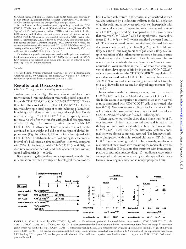
Results and DiscussionCD4�CD25� TR cells reverse wasting disease and colitis
To determine whether TR cells can ameliorate established coli-tis, we injected immunodeficient mice with clinical signs of co-litis with CD4�CD25� or CD4�CD45RBlowCD25� T cells(Fig. 1a). Three to 4 wk after CD4�CD45RBhigh T cell trans-fer, mice develop clinical signs of colitis including piloerection,hunching, anal inflammation, diarrhea, and weight loss. Coliticmice receiving 106 CD4�CD25� T cells typically startedto recover 2 wk after the transfer with gradual disappearanceof clinical signs. In contrast, mice injected with 106
CD4�CD25� T cells or control mice without second cell transfercontinued to lose weight and did not show signs of clinical im-provement (Fig. 1b). Overall, 9% of colitic mice injected withCD4�CD25� T cells had to be sacrificed during the experimentalinterval of 14 wk (median time to sacrifice, 14.0 wk), comparedwith 78% of mice injected with CD4�CD25� (p � 0.004; me-dian time to sacrifice, 5.7 wk) and 74% of control mice withoutsecond cell transfer (p � 0.002).
Because wasting disease does not always correlate with coloninflammation, we then investigated histological markers of co-
litis. Colonic architecture in the control mice sacrificed at wk 4was characterized by a leukocytic infiltrate in the LP, depletionof goblet cells, and a moderate epithelial cell hyperplasia withoccasional ulceration corresponding to an average colitis scoreof 3.1 � 0.2 (Figs. 1c and 2a). Compared with this group, micethat received CD4�CD25� cells had significantly lower colitisscores (1.5 � 0.9; p 0.01) when sacrificed between 5 and 14wk. There was restored colonic architecture, denoted by a re-duction of epithelial cell hyperplasia (Fig. 2a), rare LP infiltrates(Fig. 2, a and b), and reappearance of goblet cells (Fig. 2c). De-spite resolution of the inflammatory response, isolated clustersof leukocytes in the LP remained. These clusters were a featureof mice that had resolved colonic inflammation. Similar clustersoccurred in lower numbers in the LP of mice that were pre-vented from developing colitis by injection of CD4�CD25�
cells at the same time as the CD4�CD45RBhigh population. Inmice that received either CD4�CD25� cells (colitis score of3.0 � 0.7) or control mice receiving no second cell transfer(3.2 � 0.4), we did not see any histological improvement (Figs.1c and 2).
In accordance with the histology scores, mice that receivedCD4�CD25� cells had a 3-fold reduction in CD4� cell den-sity in the colon in comparison to control mice at wk 4 as wellas mice transferred with CD4�CD25� cells or untreated mice(p � 0.028). After recovery from colitis, mice had a similar CD4�
cell density in the colon as mice receiving an initial cotransfer ofCD4�CD45RBhigh and CD4�CD25� cells (Fig. 2b).
Taken together, our results show that a single transfer of TR
cells improves clinical status, survival rate, and intestinal pa-thology of mice with established colitis. Ten weeks afterCD4�CD25� T cell transfer, the histological colonic abnor-malities were almost completely resolved. The leukocytic infil-trate disappeared with only isolated clusters of CD11c� andCD4� T cells remaining in the LP. Interestingly, similar nor-malization of the mucosa with remaining leukocytic clusters hasbeen observed in IBD patients after treatment with immunosup-pressive or anti-inflammatory drugs (12). Additional experimentsare required to determine whether TR cell therapy will also be ef-fective at resolving inflammation in nonlymphopenic hosts.
FIGURE 1. Cure of colitis by CD4�CD25� TR cells. a, Experimental protocol. Immunodeficient mice received CD4�CD45RBhigh T cells.CD4�CD45RBlowCD25� or CD4�CD45RBlowCD25� T cells were transferred at the time points indicated. Mice were monitored for 14 wk, except for the controlgroup, which was sacrificed at wk 4. b, CD4�CD25� T cells reverse wasting disease. Data represent body weight as a percentage of the initial weight of individualmice. c, CD4�CD25� T cell transfer ameliorates established colitis. Colitis scores of individual mice are shown. In b and c, data of two experiments were pooled(SCID and rag1�/� recipients). Symbols represent individual mice. Three additional experiments with colitic mice receiving a second CD4�CD25� T cell transfergave similar results.
3940 CUTTING EDGE: CURE OF COLITIS BY TR CELLS
by guest on September 17, 2018
http://ww
w.jim
munol.org/
Dow
nloaded from

CD4�CD25� T cells home to the MLN and colon
The difference in the ability of CD4�CD25� andCD4�CD25� populations to resolve intestinal inflammationmay reflect differences in their homing or survival in vivo. Toexamine the distribution of these cells in vivo, colitis was in-duced in rag1�/� mice by transfer of CD4�CD45RBhigh
T cells from CD45.2� mice. Colitic mice were then injectedwith CD4�CD25� or CD4�CD25� T cells from congenicCD45.1� donors allowing detection of the progeny usingFACS and immunofluorescence.
Two to 3 wk after transfer of CD4�CD25� T cells, all micestill had marked inflammation in the colon. The frequency ofthe progeny of CD4�CD25� T cells was low in MLN, spleen,and LP (0.7–4.8% of total CD4� T cells; Fig. 3a). By 10 wkposttransfer, the mean frequency increased significantly to30.2% in the spleen, 40.7% in the MLN, and 17.7% in the LP(Fig. 3a). This increase in frequency was mirrored by an in-crease in the absolute numbers of CD4�CD25� progeny inspleen (from 7 � 5 to 33 � 18 � 104; NS), MLN (from 2 � 1to 32 � 7 � 104; p � 0.016), and LP (from 11 � 3 to 67 �
FIGURE 2. Histological sections of intermediate to distal colon. Initial cotransfer of CD4�CD45RBhigh and CD4�CD25� T cells into rag1�/� mice preventedthe development of colitis as illustrated by normal histology. In contrast, control mice developed moderate to severe colitis after CD4�CD45RBhigh T cell transfer.Transfer of CD4�CD25� but not of CD4�CD25� T cells into colitic mice ameliorates the colitis. a, H&E staining. b, CD4 staining and DAPI counterstaining.Insets show the CD4� cell density in cells per 10,000 �m2 (average of at least four mice per group � SEM). c, Alcian blue and periodic acid-Schiff staining to visualizegoblet cells and mucus deposition. Microphotographs were taken at �200 magnification.
FIGURE 3. Distribution of CD4�CD25� T cells after transfer into rag1�/� colitic mice. a, Percentage of CD4�CD25� T cell progeny among total CD4� Tcell in spleen, MLN, and LP after transfer of congenic CD4�CD25� cells. b, Immunostaining for CD4�CD45RBhigh, CD4�CD25�, or CD4�CD25� cellprogeny as well as CD11c� cells. CD4�CD25� and CD4�CD25� cells are distributed similarly in MLN and colon in the first 2 wk after transfer (wk 6). However,the CD4�CD25� progeny accumulates in the MLN and reverses colitis as indicated by reduction of the CD4� and CD11c� cell infiltrate. To illustrate thereduction of the LP infiltrate in CD4�CD25�-injected mice at the end of the experiment, the DAPI nucleus staining is shown to identify multiple epithelial cell areas(E). Microphotographs were taken at �400 magnification.
3941The Journal of Immunology
by guest on September 17, 2018
http://ww
w.jim
munol.org/
Dow
nloaded from

36 � 104; NS). During the first 2 wk, four of five of theCD4�CD25�-injected mice had to be sacrificed. The fre-quency of CD4�CD25� progeny was low (2% in spleen,MLN, and LP). Interestingly, although inconclusive, in the sur-viving mouse, the frequency remained low up to 10 wk aftertransfer (6% in MLN and 2% in spleen and LP). Histologicalanalysis of MLN and colon sections confirmed the FACS datawith a low but similar density of both CD4�CD25� andCD4�CD25� T cell progeny 2 wk after their transfer and anincreased density of CD4�CD25� but not CD4�CD25� Tcell progeny in the MLN and LP sections at 10 wk posttransfer(Fig. 3b).
The progeny of CD4�CD25� T cells proliferate in MLN and colon
To examine the influence of CD4�CD25� T cells on local Tcell proliferation and to identify where CD4�CD25� progenyproliferate in vivo, we examined the histological expression ofthe proliferation marker Ki67, which is specifically expressedand tightly regulated during cell proliferation (13) (Fig. 4a). Incolitic mice, CD4�CD45RBhigh progeny were found to be pro-liferating at wk 4 and 14 in both the MLN (35 vs 29%) andinflamed LP (17 vs 19%) (Fig. 4b). Two weeks after injection ofCD4�CD25� T cells, the frequency of Ki67 expression amongCD45RBhigh progeny was similar to that of mice that did notreceive TR cells. At this time point, a significant proportion ofCD4�CD25� T cell progeny also showed Ki67 expression inMLN (30 � 6%) and LP (33 � 6%), indicating an activeexpansion of this cell population in both compartments (Fig.4b). In contrast, 10 wk after transfer of CD4�CD25� T cells,when the inflammation in the colon had resolved, the fre-quency of proliferating cells among the progeny of bothCD4�CD45RBhigh as well as CD4�CD25� T cells was signif-icantly reduced in MLN and LP (Fig. 4b). Taken together,these data indicate that early after transfer into colitic mice,CD4�CD25� TR cells proliferate in MLN and colon and thatresolution of the inflammatory response correlates with a sub-stantially reduced number of proliferating pathogenic cells.
The finding that CD4�CD25� TR cells expand in the spleenand the MLN after transfer into immunodeficient mice is con-sistent with a previous report (14). However, our results showin addition that, under inflammatory conditions, not only clas-
sical effector T cells but also TR cells accumulate and proliferatein the intestinal mucosa. These results raise the possibility thatTR cells control effector T cell responses not only in the lymphnode but also in the inflamed tissue. Similar results have beendescribed in a model of transplantation tolerance (15). Induc-tion of TR cell proliferation in vitro via enhanced costimulationcoincides with the loss of their suppressor function (8, 9). Afterexpansion in vivo, CD4�CD25� TR cells were found to bemore potent suppressors in vitro (16). Our data also suggestthat, in vivo, under inflammatory conditions, vigorous prolif-eration does not lead to a loss of suppressor function as assessedby resolution of inflammation.
CD4�CD25� T cells are in contact with CD11c� cells and CD4� Tcells in vivo
To determine the localization of CD4�CD25� T cells in rela-tion to CD11c� cells and the progeny of CD4�CD45RBhigh Tcells, we analyzed the histological distribution of these cells inMLN and colon LP. The CD4�CD25� T cell progeny werefound to be in direct contact with the CD4�CD45RBhigh prog-eny as well as, in �90% of cases, with CD11c� cells (Fig. 3b),predominantly located between clusters of CD11c� cells andCD4�CD45RBhigh T cell progeny. In the colon, this localiza-tion pattern of the TR cells was seen 2 wk posttransfer in thepresence of the inflammatory infiltrate as well as in the remain-ing leukocytic clusters in the LP 10 wk posttransfer, indicatingthat direct physical contact of CD4�CD25� cells withCD11c� cells was a consistent pattern.
Previous studies have shown that interactions between patho-genic T cells and CD11c� cells within MLN and colon are impor-tant for the initiation of colitis in this model (17, 18). The observedlocation of TR cells at the interface of APC and effector T cells sup-ports a role for APC-TR interactions in TR function, i.e., in TR cellactivation and/or migration (19). In addition, TR cells may regu-late the activation state of the APC itself, which might thus inter-fere with their ability to activate effector T cell responses (20). Fur-thermore, the ability of TR cells to resolve established colitis mayalso involve direct TR-T effector cells interactions (7–9).
In summary, our data show that TR cell activity in vivo hasthe potential to reverse established inflammation leading tocure of colitis. Cell therapy with regulatory cells has some clear
FIGURE 4. CD4�CD25� T cells proliferate in MLN and LP after transfer into rag1�/� colitic mice. a, Ki67 expression in the colon LP. Staining for Ki67, CD4,CD45.1, and DAPI is shown for mice without second T cell transfer (wk 4), as well as early (wk 6) and late (wk 14) after CD4�CD25� T cell transfer. Inset showsKi67-positive cell of the CD4�CD25� progeny 2 wk after CD4�CD25� transfer. Microphotographs were taken at �630 magnification. b, Percentage of Ki67-positive in the progeny of CD4�CD45RBhigh or CD4�CD25� T cells in MLN and colon LP. Mean and SEM are shown for n � 4 mice.
3942 CUTTING EDGE: CURE OF COLITIS BY TR CELLS
by guest on September 17, 2018
http://ww
w.jim
munol.org/
Dow
nloaded from

advantages. These include their ability to migrate to inflamma-tory sites and to influence Th1 and Th2 responses (21) as wellas a potential for homeostatic and self-limited expansion.
AcknowledgmentsWe thank O. Annacker, K. Maloy, L. Fahlen, N. Rust, N. White, L. Darley,M. Coates, S. Laynes, and N. Barclay for help and critical comments.
References1. Podolsky, D. K. 2002. Inflammatory bowel disease. N. Engl. J. Med. 347:417.2. Strober, W., I. J. Fuss, and R. S. Blumberg. 2002. The immunology of mucosal mod-
els of inflammation. Annu. Rev. Immunol. 20:495.3. Powrie, F., M. W. Leach, S. Mauze, L. B. Caddle, and R. L. Coffman. 1993. Pheno-
typically distinct subsets of CD4� T cells induce or protect from chronic intestinalinflammation in C.B-17 scid mice. Int. Immunol. 5:1461.
4. Powrie, F., M. W. Leach, S. Mauze, S. Menon, L. B. Caddle, and R. L. Coffman.1994. Inhibition of Th1 responses prevents inflammatory bowel disease in scid micereconstituted with CD45RBhiCD4� T cells. Immunity 1:553.
5. Singh, B., S. Read, C. Asseman, V. Malmstrom, C. Mottet, L. A. Stephens,R. Stepankova, H. Tlaskalova, and F. Powrie. 2001. Control of intestinal inflamma-tion by regulatory T cells. Immunol. Rev. 182:190.
6. Read, S., V. Malmstrom, and F. Powrie. 2000. Cytotoxic T lymphocyte-associatedantigen 4 plays an essential role in the function of CD25�CD4� regulatory cells thatcontrol intestinal inflammation. J. Exp. Med. 192:295.
7. Maloy, K. J., and F. Powrie. 2001. Regulatory T cells in the control of immune pa-thology. Nat. Immunol. 2:816.
8. Shevach, E. M. 2002. CD4�CD25� suppressor T cells: more questions than answers.Nat. Rev. Immunol. 2:389.
9. Sakaguchi, S., N. Sakaguchi, J. Shimizu, S. Yamazaki, T. Sakihama, M. Itoh,Y. Kuniyasu, T. Nomura, M. Toda, and T. Takahashi. 2001. Immunologic tolerance
maintained by CD25�CD4� regulatory T cells: their common role in controlling auto-immunity, tumor immunity, and transplantation tolerance. Immunol. Rev. 182:18.
10. Asseman, C., S. Mauze, M. W. Leach, R. L. Coffman, and F. Powrie. 1999. An es-sential role for interleukin 10 in the function of regulatory T cells that inhibit intestinalinflammation. J. Exp. Med. 190:995.
11. Bancroft, J., and M. Gamble. 2002. Theory and Practice of Histological Techniques, 5thEd. Churchill Livingstone, New York.
12. Geboes, K., and I. Dalle. 2002. Influence of treatment on morphological features ofmucosal inflammation. Gut 50(Suppl. 3):III37.
13. Brown, D. C., and K. C. Gatter. 2002. Ki67 protein: the immaculate deception? His-topathology 40:2.
14. Annacker, O., R. Pimenta-Araujo, O. Burlen-Defranoux, T. C. Barbosa, A. Cumano,and A. Bandeira. 2001. CD25�CD4� T cells regulate the expansion of peripheralCD4 T cells through the production of IL-10. J. Immunol. 166:3008.
15. Graca, L., S. P. Cobbold, and H. Waldmann. 2002. Identification of regulatory T cellsin tolerated allografts. J. Exp. Med. 195:1641.
16. Gavin, M. A., S. R. Clarke, E. Negrou, A. Gallegos, and A. Rudensky. 2002. Ho-meostasis and anergy of CD4�CD25� suppressor T cells in vivo. Nat. Immunol. 3:33.
17. Leithauser, F., Z. Trobonjaca, P. Moller, and J. Reimann. 2001. Clustering of coloniclamina propria CD4� T cells to subepithelial dendritic cell aggregates precedes thedevelopment of colitis in a murine adoptive transfer model. Lab. Invest. 81:1339.
18. Malmstrom, V., D. Shipton, B. Singh, A. Al-Shamkhani, M. J. Puklavec,A. N. Barclay, and F. Powrie. 2001. CD134L expression on dendritic cells in themesenteric lymph nodes drives colitis in T cell-restored SCID mice. J. Immunol. 166:6972.
19. Bystry, R. S., V. Aluvihare, K. A. Welch, M. Kallikourdis, and A. G. Betz. 2001. B cellsand professional APCs recruit regulatory T cells via CCL4. Nat. Immunol. 2:1126.
20. Cederbom, L., H. Hall, and F. Ivars. 2000. CD4�CD25� regulatory T cells down-regulate co-stimulatory molecules on antigen-presenting cells. Eur. J. Immunol. 30:1538.
21. Cottrez, F., S. D. Hurst, R. L. Coffman, and H. Groux. 2000. T regulatory cells 1inhibit a Th2-specific response in vivo. J. Immunol. 165:4848.
3943The Journal of Immunology
by guest on September 17, 2018
http://ww
w.jim
munol.org/
Dow
nloaded from