theironchelatorsdp44mtanddfoinhibittgf- -induced ... · theironchelatorsdp44mtanddfoinhibittgf-...
TRANSCRIPT
The Iron Chelators Dp44mT and DFO Inhibit TGF-�-inducedEpithelial-Mesenchymal Transition via Up-Regulation ofN-Myc Downstream-regulated Gene 1 (NDRG1)□S
Received for publication, February 6, 2012, and in revised form, March 23, 2012 Published, JBC Papers in Press, March 27, 2012, DOI 10.1074/jbc.M112.350470
Zhiqiang Chen‡§, Daohai Zhang§, Fei Yue‡, Minhua Zheng‡1, Zaklina Kovacevic§, and Des R. Richardson‡§2
From the ‡General Surgery Department of Ruijin Hospital, Shanghai Jiao Tong University School of Medicine, Shanghai 200025,China and the §Iron Metabolism and Chelation Program, Department of Pathology, Bosch Institute, University of Sydney, NewSouth Wales 2006, Australia
Background: NDRG1 is an iron-regulated metastasis suppressor which is up-regulated by iron depletion and may beinvolved in the epithelial-mesenchymal transition (EMT).Results: NDRG1 is involved in the EMT through the SMAD andWnt pathways.Conclusion: Iron chelators could inhibit the TGF-�-induced EMT via NDRG1.Significance: The results are important for understanding the molecular roles of iron in proliferation and metastasis.
The epithelial-mesenchymal transition (EMT) is a key stepfor cancer cell migration, invasion, and metastasis. Transform-ing growth factor-� (TGF-�) regulates the EMT and the metas-tasis suppressor gene, N-myc downstream-regulated gene-1(NDRG1), could play a role in regulating the TGF-� pathway.NDRG1 expression is markedly increased after chelator-medi-ated iron depletion via hypoxia-inducible factor 1�-dependentand independent pathways (Le, N. T. and Richardson, D. R.(2004) Blood 104, 2967–2975). Moreover, novel iron chelatorsshow marked and selective anti-tumor activity and are a poten-tial new class of anti-metabolites. Considering this, the currentstudy investigated the relationship between NDRG1 and theEMT to examine if iron chelators can inhibit the EMT viaNDRG1 up-regulation. We demonstrated that TGF-� inducesthe EMT in HT29 and DU145 cells. Further, the chelators, des-ferrioxamine (DFO) and di-2-pyridylketone-4,4-dimethyl-3-thiosemicarbazone (Dp44mT), inhibited the TGF-�-inducedEMTbymaintainingE-cadherin and�-catenin, at the cellmem-brane. We then established stable clones with NDRG1 overex-pression and knock-down inHT29 andDU145 cells. These datashowed that NDRG1 overexpression maintained membraneE-cadherin and �-catenin and inhibited TGF-�-stimulated cellmigration and invasion. Conversely, NDRG1 knock-downcaused morphological changes from an epithelial- to fibroblas-tic-like phenotype and also increased migration and invasion,demonstrating NDRG1 knockdown induced the EMT andenhanced TGF-� effects. We also investigated the mechanismsinvolved and showed the TGF-�/SMAD and Wnt pathwayswere implicated in NDRG1 regulation of E-cadherin and�-catenin expression and translocation. This study demon-strates that chelators inhibit the TGF-�-induced EMT via a
process consistent with NDRG1 up-regulation and elucidatesthe mechanism of their activity.
Iron plays a crucial role in proliferation and DNA synthesisand neoplastic cells have an increased requirement for iron asshown by their markedly elevated expression of the transferrinreceptor 1 and enhanced uptake of iron (1). Recently, it hasbeen suggested that alteration in the regulation of iron metab-olism characterizes themalignant state, with an iron regulatorygene signature predicting outcome in breast cancer (2). How-ever, the precise molecular pathways involved remain unclearand are important to elucidate particularly in terms of themechanisms involved in metastasis, which is a major problemin cancer treatment.The epithelial-mesenchymal transition (EMT)3 is a highly
conserved process required for embryonic development, tissueremodeling and wound repair (3). In addition, there is increas-ing evidence that it is a key initial step for cancer cell migration,invasion and metastasis (4). During the process of the EMT,cells lose their epithelial characteristics such as epithelial mor-phology, cell polarity, cell-cell contact, and gain mesenchymalproperties such as fibroblastic morphology, increased migra-tion and invasion, thus causing cancer cell metastasis (5).Transforming growth factor-� (TGF-�) was first identified
as an inducer of the EMT in normal mammary epithelial cells(6) and subsequently recognized as a chief regulator of the EMTin a variety of cell-types and tissues including cancer cells (7).There are reports showing that TGF-�was increased in variouscancer cell types and that this plays an important role in theprocess of metastasis via the induction of the EMT (8, 9).Iron chelators are a relatively new class of potential anti-
metabolites that showmarked and selective anti-tumor activity□S This article contains supplemental Figs. S1–S6.1 To whom correspondence may be addressed: General Surgery Department
of Ruijin Hospital, Shanghai 200025, China. Tel.: 86-21-64458887; Fax:86-21-64333548; E-mail: [email protected].
2 To whom correspondence may be addressed: Iron Metabolism and Chela-tion Program, Discipline of Pathology and Bosch Institute, The University ofSydney, Sydney, New South Wales, 2006 Australia. Tel.: 61-2-9036-6548;Fax: 61-2-9351-3429; E-mail: [email protected].
3 The abbreviations used are: EMT, epithelial-mesenchymal transition; TGF-�,transforming growth factor-�; NDRG1, N-Myc downstream-regulatedgene 1; DFO, desferrioxamine; Dp44mT, di-2-pyridylketone-4,4-dimethyl-3-thiosemicarbazone; Dp2mT, di-2-pyridylketone 2-methyl-3-thiosemi-carbazone; DMSO, dimethyl sulfoxide; TJ, tight junction; ZO-1, zonulaoccludin-1.
THE JOURNAL OF BIOLOGICAL CHEMISTRY VOL. 287, NO. 21, pp. 17016 –17028, May 18, 2012© 2012 by The American Society for Biochemistry and Molecular Biology, Inc. Published in the U.S.A.
17016 JOURNAL OF BIOLOGICAL CHEMISTRY VOLUME 287 • NUMBER 21 • MAY 18, 2012
by guest on September 12, 2018
http://ww
w.jbc.org/
Dow
nloaded from
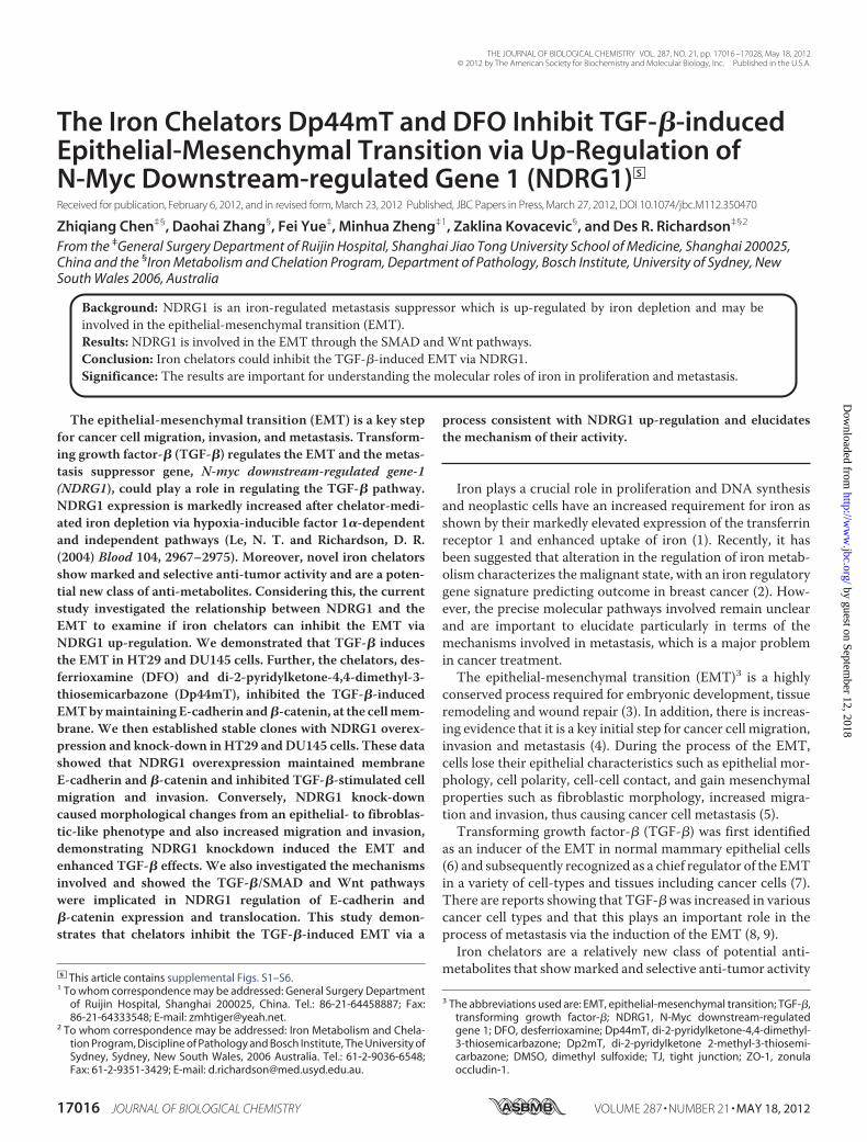
(1, 10), although their molecular targets and mechanisms ofaction remain to be completely elucidated.We reported a seriesof novel thiosemicarbazone iron chelators of the dipyridyl thio-semicarbazone class (e.g. di-2-pyridylketone-4,4-dimethyl-3-thiosemicarbazone, Dp44mT; Fig. 1A) as anti-tumor com-pounds which are effective against belligerent tumors in vivo bythe intravenous and oral routes (11, 12).This series of compounds were demonstrated to induce cell
cycle arrest and apoptosis through a mechanism involving irondepletion which affects a variety of targets including: (i) iron-dependent enzymes such as ribonucleotide reductase (RR),which is critical for DNA synthesis (13); (ii) the metastasis sup-pressor,N-myc downstream regulated gene 1 (NDRG1) (11); (iii)p53 (14); (iv) cyclinD1 (15); (v) p21CIP1/WAF1 (16) etc. The othermajor mechanism of thiosemicarbazone activity is mediatedthrough the redox cycling of their iron and copper complexes(17), causing cell death and apoptosis through targeting lyso-some integrity (18) and oxidizing glutathione and the sulfhydrylgroups of proteins involved in reducing systems (19).One of themost interesting genes regulated by iron chelators
isNDRG1 (11, 20), which is awell-knownmetastasis suppressorin various cancer cell types (21–26). Significantly, it has alsobeen reported that NDRG1 overexpression is correlated with alower metastatic rate and increased 5-year survival in clinicalstudies (21, 23, 27). Hence, NDRG1 is a promising moleculartarget for cancer therapy that is modulated by novel iron chela-tors (11, 12, 28). However, the detailed mechanisms for theanti-cancer effects of NDRG1 are not well elucidated and fur-ther investigation is required.Considering the potent anti-metastatic effect of NDRG1 in
various cancer types and the role TGF-� plays in cancer metas-tasis, we examined whether iron chelators could inhibit thecancer cell EMT induced by TGF-� and whether this effecttakes place via up-regulation ofNDRG1. In this study, we estab-lished four stable transfectants with NDRG1 overexpressionand knock-down in two cancer cell types, namely colon cancerHT29 and prostate cancer DU145. We then investigated therole and mechanism of NDRG1 in the TGF-�-induced EMTand its related biological functions. Our study shows that cellu-lar iron-depletion inhibits theTGF-�-inducedEMTvia up-reg-ulation of NDRG1.
EXPERIMENTAL PROCEDURES
Cell Culture and Cell Treatments—Human prostate cancerDU145 cells were grown in RPMI 1640 medium (Invitrogen)supplemented with 10% (v/v) fetal bovine serum (FBS, Invitro-gen). The HT29 human colon cancer cells were grown inMcCoy’s 5Amedium (Invitrogen) supplementedwith 10% (v/v)FBS. Cells were obtained from theAmerican Type Culture Col-
lection (ATCC) and used within 2 months of purchase afterresuscitation of frozen aliquots. Cell lines were authenticatedon the basis of viability, recovery, growth,morphology, and alsocytogenetic analysis, antigen expression, DNA profile and iso-enzymology by the provider. Human recombinant TGF-�1wasobtained from R&D Systems and used at a final concentrationof 5 ng/ml. The cells were incubated in serum-free mediumovernight, and then treated with TGF-� for 48 h and 96 h forDU145 and HT29 cells, respectively, to induce the EMT.The chelator, Dp44mT (Fig. 1A), and the negative control
compound, di-2-pyridylketone 2-methyl-3-thiosemicarbazone(Dp2mT; Fig. 1B), which cannot bind iron, were synthesizedand characterized using standardmethods (17, 29). Desferriox-amine (DFO; Fig. 1C) was purchased from Novartis. We uti-lized concentrations of 10�M forDp44mTandDp2mTand 100�M for DFO in 10% (v/v) FBS supplemented medium. Thegreater concentration of DFO was implemented due to its lim-ited ability to permeate the cell membrane (30). The chelatorDp44mTwas utilized at a lower concentration since this ligandshows far higher membrane permeability and demonstratesmarked iron chelation efficacy (29). BothDp2mT andDp44mTwere freshly dissolved in dimethyl sulfoxide (DMSO) anddiluted in culture media (final [DMSO]: � 0.1% (v/v)).Plasmid Construction and Transfection—For NDRG1 over-
expression, we used pCMV-tag2-FLAG-NDRG1 (GenHunter)and the empty pCMV-tag2-FLAG vector (Stratagene) as a neg-ative control. Both plasmids contained a G418 resistancemarker. The shRNA and negative control plasmids wereobtained from Qiagen (Cat. KH02202H) and contained aHygromycin resistancemarker. All cells were transfected usingLipofectamine 2000� (Invitrogen) following the manufactur-er’s protocol. The cells were selected for more than 4 weeks byincubation with G418 (400 ng/ml for DU145 and 1000 ng/mlfor HT29) for overexpression clones, or Hygromycin (500ng/ml for DU145 and 1000 ng/ml for HT29) for knock-downclones. Stable single clones were selected and NDRG1 expres-sion assessed using Western blot. Two single clones of eachstable transfectant were utilized in this study.Protein Extraction andWestern Blotting—Whole cell protein
lysates were extracted using lysis buffer with proteinase inhib-itor mixture (Cat. 11836170001; Roche Applied Science) andPhosSTOP (Cat. 04906845001; RocheApplied Science) accord-ing to standard methods (20). Western blotting was performedvia established protocols (20). Primary antibodies were against:ferritin (Cat. ab69090), NDRG1 (Cat. ab37897), vimentin (Cat.ab8978), occludin (Cat. ab31721), ZEB2 (Cat. ab25837), andtwist (Cat. ab50581) from Abcam; E-cadherin (Cat. 3195),�-catenin (Cat. 9562), ZO-1 (Cat. 8193), Snail (Cat. 3879), Slug
FIGURE 1. Line drawing of the chemical structures of: (A) Dp44mT, (B) Dp2mT, and (C) DFO.
Iron Chelators Inhibit the TGF-�-induced EMT via NDRG1
MAY 18, 2012 • VOLUME 287 • NUMBER 21 JOURNAL OF BIOLOGICAL CHEMISTRY 17017
by guest on September 12, 2018
http://ww
w.jbc.org/
Dow
nloaded from
(Cat. 9585), SMAD2 (Cat. 3122), pSMAD2 (serine 465/467)(Cat. 3101), pSMAD3 (Cat. 9520), and SMAD4 (Cat. 9515)werefrom Cell Signaling Technology and cyclin D1 (Cat. SC8396)was from Santa Cruz Biotechnology. The secondary antibodiesused were horseradish peroxidase (HRP)-conjugated anti-goat(Cat. A5420), anti-rabbit (Cat. A6154) and anti-mouse (Cat.A4416) fromSigma-Aldrich; Alexa Fluor� 555 conjugated anti-rabbit (Cat. 4413) andAlexa Fluor� 488 conjugated anti-mouse(Cat. 4408) from Cell Signaling Technology. Primary antibodyagainst �-actin (Cat. A1978) was from Sigma-Aldrich and usedas a loading control.Immunofluorescence—Immunofluorescence was performed
as described (31). Briefly, cells seeded on coverslips were fixedwith 4% (w/v) paraformaldehyde (Sigma-Aldrich) for 10 minand permeabilized with 0.1% (v/v) Triton X-100 for 5 min atroom temperature. The cells were then incubated overnightwith primary antibodies at 4 °C, followed by incubation withfluorescent secondary antibody for 1 h at room temperature.After final washes with PBS, the coverslips weremounted usingan anti-fade mounting solution containing 4�,6-diamidino-2-phenylindole (DAPI; Cat. P36935, Invitrogen) and images wereexamined and captured using an Olympus Zeiss AxioObserverZ1 fluorescence microscope (Olympus) with a 63� oil objec-tive. Raw images were analyzed usingOlympusAxiovision soft-ware (Olympus).Cell Migration and Invasion Assay—Cell migration was
assessed using established wound healing and transwell migra-tion assays (31). Briefly, for the wound healing assay, cellularmonolayers at 90–95% confluence were serum-starved for 24 hand scratched using a sterile 20 �l pipette tip. After washing(three times with complete medium) and removing thedetached cells, the plates were incubated at 37 °C for 12 h, andthe wounds were photographed and analyzed using the Olym-pus Zeiss AxioObserver Z1 fluorescence microscope. Thequantitative migration assay was performed using the Cyto-SelectTM 24-Well Cell Migration Assay kit from Cell Biolabs,according to the manufacturer’s protocol. Images were taken,and the migration abilities were quantified by optical absor-bance at 560 nm using a PerkinElmer 1420 multi-label platereader.Cell invasion was assessed using the CytoSelectTM 96-well
Cell Invasion Assay kit (Cell Biolabs) according to the man-ufacturer’s protocol. Invasion values were reported as meanrelative fluorescence units (RFUs) of quadruplicate samplesmeasured at 480 nm/520 nm using the PerkinElmer platereader.Statistical Analysis—Data are mean � S.D. and were statis-
tically analyzed using Student’s t test. Results were consideredsignificant when p � 0.05.
RESULTS
TGF-� Induces the EMT in HT29 and DU145 Cells—Todetermine whether TGF-� can induce a mesenchymal pheno-type consistent with the EMT in DU145 and HT29 cell-types,we incubated these cells with TGF-� at a physiological dose of 5ng/ml (32) for 48 h or 96 h, respectively. These different incu-bation periods were shown in preliminary experiments to dem-onstrate maximum efficacy at inducing the EMT in each cell
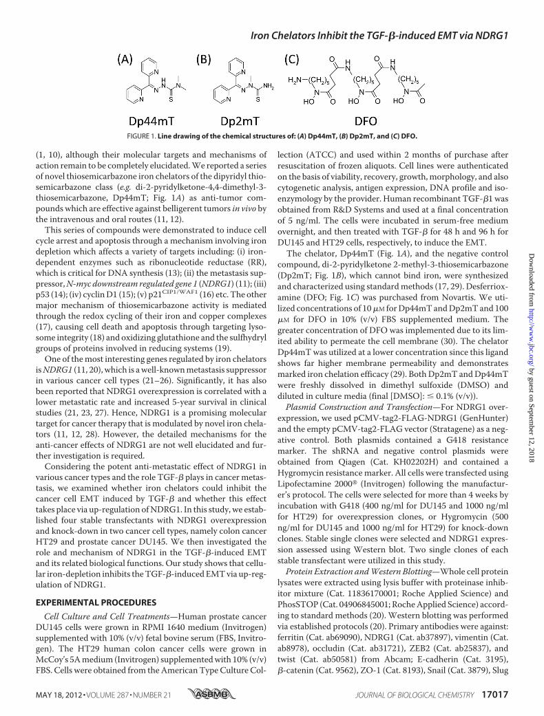
type. TreatmentwithTGF-� resulted inmarkedmorphologicalchanges in the HT29 and DU145 cell types as shown in Fig. 2A.In fact, the cells becamemore isolated and demonstratedmark-edly decreased intercellular contacts, with this being particu-larly notable for HT29 cells (Fig. 2A). Furthermore, for DU145cells, incubation with TGF-� resulted in more spindle-shaped,fibroblast-like cells. These morphological characteristics wereconsistent with cells undergoing the EMTafter incubationwithTGF-� (32, 33).We then investigated themolecular alterations in the expres-
sion of well-established EMT markers (4) by Western blottingand immunofluorescence. Consistent with the morphologicalchanges, there was a significant (p � 0.001) 2–4-fold decreasein the expression of the epithelial markers, E-cadherin and�-catenin (7, 32), and a significant (p� 0.001) 3–5-fold increasein the expression of the mesenchymal marker, vimentin (7, 32),after TGF-� treatment of HT29 andDU145 cells (Fig. 2B). Thiswas further confirmed by immunofluorescence staining, whichdemonstrated a pronounced reduction of membrane E-cad-herin and �-catenin levels (red fluorescence) and an inductionof mesenchymal vimentin expression (green fluorescence)upon incubation with TGF-� (Fig. 2C). Notably, E-cadherinand�-catenin form the cadherin complex at the cell membranethat is essential for formation of the adherens junction whichmaintains intercellular integrity (34).Considering that the EMT is characterized by enhanced cel-
lularmotility and invasion (4, 5), we then evaluated the changesto migratory capacity and invasive potential of HT29 andDU145 cells afterTGF-� treatment. As shown in Fig. 2,D andE,using the cell migration and invasion assays, TGF-� signifi-cantly (p � 0.001) increased migration and invasion of HT29and DU145 cells, when compared with untreated control cells.Collectively, these observations indicate that TGF-� inducesthe EMT in HT29 and DU145 cells.Iron Chelators Attenuate the TGF-�-induced EMT in HT29
and DU145 Cells—We have reported that novel series of ironchelators function as potent anti-tumor agents, among whichDp44mT is one of themost effective (12, 29). As the EMT playsan important role during cancer cell progression andmetastasis(4), we examined whether Dp44mT could act against the TGF-�-induced EMT. At the same time, in order to clarify whetherthis effect was dependent on iron depletion, we used Dp2mT(29). This compound has a similar chemical structure toDp44mT, but in contrast, cannot bind cellular iron and is thusan appropriate negative control (29) (Fig. 1, A and B). More-over, as a further control, we also examined the well-character-ized iron chelator, DFO (Fig. 1C), which binds iron with highaffinity, but does not redox cycle like Dp44mT (1, 17). Thislatter control was important to determine whether the effectsobserved were due to iron depletion or redox activity.As shown by immunofluorescence and Western blotting
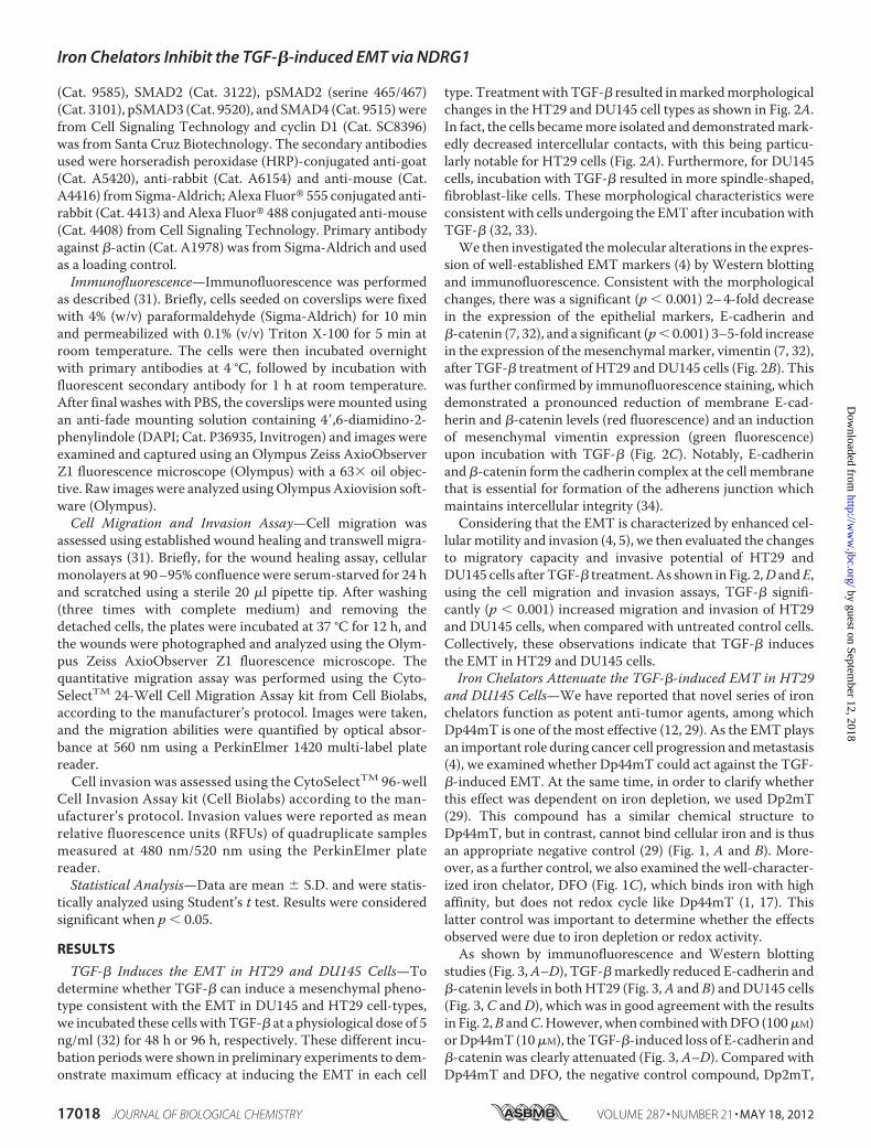
studies (Fig. 3,A–D), TGF-�markedly reduced E-cadherin and�-catenin levels in bothHT29 (Fig. 3,A and B) and DU145 cells(Fig. 3, C andD), which was in good agreement with the resultsin Fig. 2,B andC. However, when combinedwithDFO (100�M)orDp44mT (10�M), theTGF-�-induced loss of E-cadherin and�-catenin was clearly attenuated (Fig. 3, A–D). Compared withDp44mT and DFO, the negative control compound, Dp2mT,
Iron Chelators Inhibit the TGF-�-induced EMT via NDRG1
17018 JOURNAL OF BIOLOGICAL CHEMISTRY VOLUME 287 • NUMBER 21 • MAY 18, 2012
by guest on September 12, 2018
http://ww
w.jbc.org/
Dow
nloaded from
did not have any significant effect on attenuating the TGF-�-induced a decrease of E-cadherin and �-catenin (Fig. 3, A–D).This demonstrates the importance of iron-depletion on theability of chelators to inhibit the TGF-�-induced EMT. Thiswas further confirmed by pre-treating chelators with iron (asFeCl3) to form iron complexes that cannot bind cellular iron.These complexes prevented the iron depletion-mediated up-regulation of NDRG1, but also inhibited the ability of chelatorsto attenuate the TGF-�-induced EMT, as shown by the analysisof E-cadherin, �-catenin and vimentin expression (supplemen-tal Fig. S1). Furthermore, the fact that DFO and Dp44mT havesimilar effects indicates that iron depletion is key to the mech-anism involved, rather than the redox activity of Dp44mT (17,
29). Phenotypic analysis demonstrated that HT29 and DU145cells treatedwithTGF-�plusDp2mTshowed similarmorphol-ogy to those treated with TGF-� alone (supplemental Fig. S2).However, DFO or Dp44mT antagonized the effect of TGF-�(supplemental Fig. S2), indicating interruptedTGF-� signaling.
In agreementwith the ability ofDp44mTandDFO to attenuatethe TGF-�-induced reduction of E-cadherin and �-catenin, thesechelators also significantly (p � 0.001) decreased the TGF-�-in-duced up-regulation of the mesenchymal marker, vimentin, inHT29 (Fig. 3B) and DU145 cells (Fig. 3D). Together, these datademonstrate that chelators can block theTGF-�-induced EMT inHT29 and DU145 cells and that this is dependent on chelation ofcellular iron.
FIGURE 2. TGF-� induces characteristics of the epithelial-mesenchymal transition (EMT) in HT29 and DU145 cells. HT29 and DU145 cells were treated inthe presence or absence of TGF-� (5 ng/ml) for 96 h and 48 h, respectively, to induce the EMT. A, bright field images were taken to show cell morphologicalchanges after TGF-� treatment. Scale bars: 100 �m. B, whole cell lysates were extracted and Western blotting was performed to investigate changes tomolecular markers of the EMT. Densitometric analysis is expressed relative to the loading control, �-actin. C, merged images were taken to show immunoflu-orescence staining of E-cadherin (red), �-catenin (red), and vimentin (green) accompanied by the cell nucleus (blue) stained by DAPI. Scale bars: 20 �m. D, HT29and DU145 cells were pretreated for 72 h and 24 h, respectively, with TGF-� (5 ng/ml), then seeded at 100,000 cells/well and incubated for 12 h/37 °C in thepresence or absence of TGF-� (5 ng/ml). Migratory cells on the bottom of the polycarbonate membrane were stained (purple blue) and quantified at A560 afterextraction according to the manufacturer’s protocol (Cell Biolabs). Scale bars: 200 �m. E, cells were treated with TGF-� as described in D above, and 50,000cells/well were seeded in the transwell chamber coated with extra-cellular matrix and incubated for 12 h/37 °C. The invasion values were reported as meanrelative fluorescence unit (RFU) of quadruplicate samples. Data are typical of 3–5 experiments, and the histogram values are mean � S.D. (3–5 experiments).***, p � 0.001, relative to control cells without TGF-�.
Iron Chelators Inhibit the TGF-�-induced EMT via NDRG1
MAY 18, 2012 • VOLUME 287 • NUMBER 21 JOURNAL OF BIOLOGICAL CHEMISTRY 17019
by guest on September 12, 2018
http://ww
w.jbc.org/
Dow
nloaded from
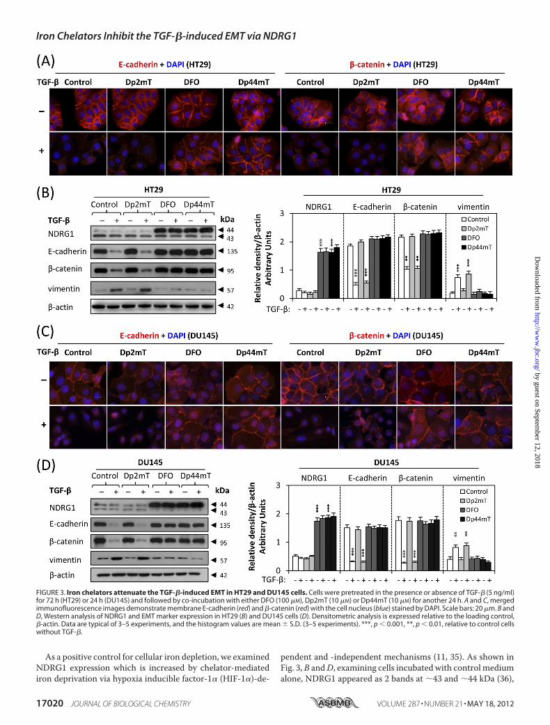
As a positive control for cellular iron depletion, we examinedNDRG1 expression which is increased by chelator-mediatediron deprivation via hypoxia inducible factor-1� (HIF-1�)-de-
pendent and -independent mechanisms (11, 35). As shown inFig. 3,B andD, examining cells incubated with control mediumalone, NDRG1 appeared as 2 bands at �43 and �44 kDa (36),
FIGURE 3. Iron chelators attenuate the TGF-�-induced EMT in HT29 and DU145 cells. Cells were pretreated in the presence or absence of TGF-� (5 ng/ml)for 72 h (HT29) or 24 h (DU145) and followed by co-incubation with either DFO (100 �M), Dp2mT (10 �M) or Dp44mT (10 �M) for another 24 h. A and C, mergedimmunofluorescence images demonstrate membrane E-cadherin (red) and �-catenin (red) with the cell nucleus (blue) stained by DAPI. Scale bars: 20 �m. B andD, Western analysis of NDRG1 and EMT marker expression in HT29 (B) and DU145 cells (D). Densitometric analysis is expressed relative to the loading control,�-actin. Data are typical of 3–5 experiments, and the histogram values are mean � S.D. (3–5 experiments). ***, p � 0.001, **, p � 0.01, relative to control cellswithout TGF-�.
Iron Chelators Inhibit the TGF-�-induced EMT via NDRG1
17020 JOURNAL OF BIOLOGICAL CHEMISTRY VOLUME 287 • NUMBER 21 • MAY 18, 2012
by guest on September 12, 2018
http://ww
w.jbc.org/
Dow
nloaded from
which may correspond to its different phosphorylation states(36, 37). The upper band is responsive to iron-depletion withchelators (Fig. 3, B andD) and is thought to represent the activeform of this molecule (37). Incubation with TGF-� alone didnot significantly (p � 0.05) alter NDRG1 expression relative tountreated control cells (Fig. 3,B andD). In addition,Dp2mTdidnot have any significant effect onNDRG1 expression relative tothe control, since it cannot bind cellular iron (29) (Fig. 3, B andD). However, incubation with either DFO or Dp44mT caused asignificant (p � 0.001) increase in the �44 kDa NDRG1 bandrelative to the control in the presence or absence of TGF-�,demonstrating that iron chelators induceNDRG1expression inthese cells. Apart from the up-regulation of NDRG1 after iron-depletion, we also demonstrated that under the same incuba-tion conditions, both DFO and Dp44mT down-regulate ferri-tin, while Dp2mT had no effect (supplemental Fig. S3). Ferritinis well known to be regulated in this manner by iron depletion(1, 13). Indeed, previous studies have clearly demonstrated theability of DFO and Dp44mT to reduce iron uptake from trans-ferrin by cells and induce cellular iron mobilization, leading toiron depletion (18, 29, 30).Considering the marked up-regulation of NDRG1 after iron
depletion and its functional role as ametastasis suppressor (21–26), we then hypothesized that NDRG1 may be involved in theprocess of the TGF-�-induced EMT. To examine this, weestablishedNDRG1overexpression and knock-downmodels inHT29 and DU145 cells.Overexpression of NDRG1 Attenuates the TGF-�-induced
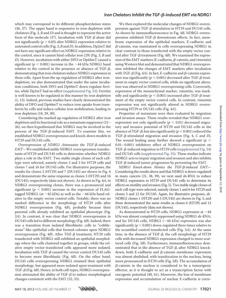
EMT—We established stable NDRG1 overexpression transfec-tants of HT29 and DU145 cells to determine whether NDRG1plays a role in the EMT. Two stable single clones of each cell-type were selected, namely clones 2 and 3 for HT29 cells andclones 7 and 16 for DU145 cells. For illustrative purposes, theresults for clones 2 (HT29) and 7 (DU145) are shown in Fig. 4and demonstrate the same response as clones 3 (HT29) and 16(DU145), respectively (data not shown). As shown in Fig. 4A, inNDRG1 overexpressing clones, there was a pronounced andsignificant (p � 0.001) increase in the expression of FLAG-tagged NDRG1 (at �45 kDa) as well as the �44 kDa band rel-ative to the empty vector control cells. Notably, there was nomarked difference in the morphology of HT29 cells afterNDRG1 overexpression (Fig. 4B), probably because theirparental cells already exhibited an epithelial phenotype (Fig.2A). In contrast, it was clear that NDRG1 overexpression inDU145 cells led to differentmorphology (Fig. 4B). Indeed, therewas a transition from isolated fibroblastic cells to “cobble-stone”-like epithelial cells that formed colonies upon NDRG1overexpression (Fig. 4B). After TGF-� treatment, HT29 cellstransfected with NDRG1 still exhibited an epithelial morphol-ogy where the cells clustered together in groups, while the rel-ative empty vector-transfected cells appeared more isolated.Incubation with TGF-� caused the vector control DU145 cellsto become more fibroblastic (Fig. 4B). On the other hand,DU145 cells overexpressing NDRG1 retained their epithelialmorphology, but appeared more isolated upon treatment withTGF-� (Fig. 4B). Hence, in both cell-types,NDRG1overexpres-sion attenuated the ability of TGF-� to induce morphologicalchanges consistent with the EMT (32, 33).
We then explored the molecular changes of NDRG1 overex-pression against TGF-� stimulation in HT29 and DU145 cells.As shown by immunofluorescence in Fig. 4B, NDRG1 overex-pression inhibited TGF-� downstream effects. In fact, mem-brane expression of the epithelial markers, E-cadherin and�-catenin, was maintained in cells overexpressing NDRG1 inclear contrast to those transfected with the empty vector con-trol after TGF-� treatment (Fig. 4B). We examined the expres-sion of the EMTmarkers (E-cadherin,�-catenin, and vimentin)usingWestern blot anddemonstrated thatNDRG1overexpres-sion inhibited the changes of EMT markers after incubationwith TGF-� (Fig. 4A). In fact, E-cadherin and�-catenin expres-sion was significantly (p � 0.001) decreased after TGF-� treat-ment in empty vector control cells, while no significant altera-tion was observed in NDRG1 overexpressing cells. Conversely,expression of the mesenchymal marker, vimentin, was mark-edly and significantly (p � 0.001) increased after TGF-� treat-ment of the empty vector control cells. In contrast, vimentinexpression was not significantly altered in NDRG1 overex-pressing HT29 or DU145 cells (Fig. 4A).Properties of metastasis were then evaluated by migration
and invasion assays. These results revealed that NDRG1 over-expression not only significantly (p � 0.01) decreased migra-tory and invasive potential of HT29 and DU145 cells in theabsence ofTGF-�, but also significantly (p� 0.001) reduced theTGF-�-stimulated migration and invasion (Fig. 4, C and D).The wound healing assay further showed a significant (p �0.01–0.001) inhibitory effect of NDRG1 overexpression onTGF-�-inducedmigration inHT29 cells (supplemental Fig. S4)and DU145 cells (supplemental Fig. S5). These studies indicateNDRG1 acts to impair migration and invasion and also inhibitsTGF-�-induced tumor progression by preventing the EMT.NDRG1 Knock-down Mimics the TGF-�-induced EMT—
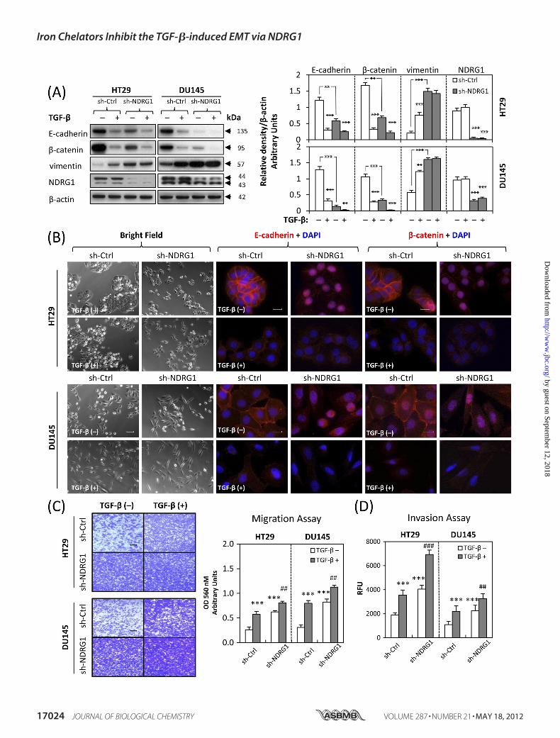
Considering the results above and thatNDRG1 is down-regulatedin many cancers (21, 38, 39), we next used sh-RNA to reduceNDRG1 expression in HT29 and DU145 cells to determine itseffects onmotility and invasion (Fig. 5). Two stable single clones ofeach cell-type were selected, namely clones 1 and 6 for HT29 andclones 5 and 12 for DU145. Again, for illustrative purposes, sh-NDRG1 clones 1 (HT29) and 5 (DU145) are shown in Fig. 5, andthese demonstrated the same results as clones 6 (HT29) and 12(DU145), respectively (data not shown).As demonstrated in HT29 cells, NDRG1 expression at �44
kDa was almost completely suppressed using NDRG1 sh-RNA,and for DU145 cells, NDRG1 (�44 kDa) expression was alsosignificantly (p � 0.001) down-regulated three times relative tothe scrambled control transfected cells (Fig. 5A). At the sametime, in the absence of TGF-�, the cell morphology of HT29cells with decreased NDRG1 expression changed to more scat-tered cells (Fig. 5B). Furthermore, immunofluorescence dem-onstrated that in the absence of TGF-�, after NDRG1 knock-down, both E-cadherin and �-catenin membrane expressionwas almost abolished, with translocation to the nucleus, beingmore pronounced in HT29 cells (Fig. 5B). The accumulation of�-catenin in the nucleus is considered to be a tumorigeniceffector, as it is thought to act as a transcription factor withoncogenic potential (40, 41). Moreover, the loss of membraneexpression and accumulation of nuclear E-cadherin is corre-
Iron Chelators Inhibit the TGF-�-induced EMT via NDRG1
MAY 18, 2012 • VOLUME 287 • NUMBER 21 JOURNAL OF BIOLOGICAL CHEMISTRY 17021
by guest on September 12, 2018
http://ww
w.jbc.org/
Dow
nloaded from
Iron Chelators Inhibit the TGF-�-induced EMT via NDRG1
17022 JOURNAL OF BIOLOGICAL CHEMISTRY VOLUME 287 • NUMBER 21 • MAY 18, 2012
by guest on September 12, 2018
http://ww
w.jbc.org/
Dow
nloaded from
lated with higher invasive and metastatic potential (42, 43).Nuclear translocationwas not observed in TGF-�-treated cells,indicating that this process occurred independently of the cyto-kine (Fig. 5B). As E-cadherin and�-catenin are key componentsof cell adherens junctions (34), their loss from the cell mem-brane will impair their ability to function in establishing inter-cellular contacts.We also examined the EMTmarkers usingWestern blotting
and revealed that NDRG1 knock-down markedly and signifi-cantly (p � 0.001) down-regulated E-cadherin and �-cateninexpression 4–5 times in both HT29 and DU145 cells in theabsence of TGF-�, while up-regulating vimentin 2–3 times,which was consistent with the EMT (Fig. 5A). Furthermore,these molecular changes for E-cadherin and �-catenin wereenhanced by the addition of TGF-� (Fig. 5A). We then investi-gated metastatic properties via migration and invasion assays.These results demonstrated that NDRG1 knock-down signifi-cantly (p � 0.001) increased migration and invasion for HT29and DU145 cells in the absence of TGF-� and these metastaticpotentials were enhanced after incubation with TGF-� (Fig. 5,C andD). Together, these studies indicate that NDRG1 knock-down mimics the EMT in HT29 and DU145 cells and alsoenhances the downstream effects induced by TGF-�.Expression of Cell Tight JunctionMarkers ZO-1 andOccludin
Are Not Affected by NDRG1 Expression—Cell morphology andpolarity are not only controlled by the adherens junctions, butalso by tight junctions (TJs) (44). Hence, we next examinedwhether cellular TJs were also affected by NDRG1 expression(overexpression and knockdown). In order to investigate this,we examined the expression of well established TJ proteins,including zonula occludin-1 (ZO-1) and occludin (44). How-ever, we saw no significant alterations in the expression of theseTJ markers by Western blotting and immunofluorescence inHT29 or DU145 cells in response to alterations in NDRG1expression (supplemental Fig. S6).Canonical SMAD/Snail/Slug and Wnt Pathways Are
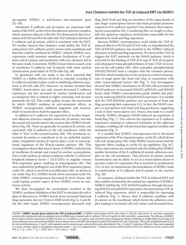
Involved in the NDRG1-mediated Inhibition of the EMT—Theabove data showed that NDRG1 was involved in the EMT ofcancer cells. We then assessed what molecular mechanismsmay be involved using the DU145 cell model, as it was moresusceptible to TGF-� stimulation and underwent more pro-nounced morphological changes relative to HT29 cells afterNDRG1 overexpression (Fig. 4B).We first examined the expression of nuclear transcriptional
repressors responsible for the down-regulation of E-cadherinduring the EMT, such as Snail, Slug, Twist and ZEB2 (45). ForTwist andZEB2, therewere no significant alterations in expres-sion after NDRG1 knock-down or overexpression (Fig. 6A). Incontrast, Snail was significantly (p � 0.001) increased afterNDRG1 knock-down, although there was no significant change
after NDRG1 overexpression (Fig. 6, A and B). However,NDRG1 knock-down markedly and significantly (p � 0.001)up-regulated the expression of Slug, and NDRG1 overexpres-sion significantly (p � 0.05) reduced Slug expression (Fig. 6, Aand B). Hence, the transcriptional E-cadherin repressors, Snail,and Slug, were involved in modulating E-cadherin expressionby NDRG1. As a positive control, we also showed that afterTGF-� treatment, Snail and Slug expression were significantly(p � 0.01) up-regulated, while there was no significant altera-tion to Twist or ZEB2 expression (Fig. 6A).As shown in this study, NDRG1 overexpression inhibited the
TGF-�-induced EMT in DU145 and HT29 cells (Fig. 4) andNDRG1 knock-downmimicked the TGF-�-induced EMT (Fig.5). We then hypothesized that the TGF-� pathway plays animportant role in this process. Hence, we probed for the SMADproteins thatmediateTGF-� signaling (46) to examinewhetherthey were affected by NDRG1. First, we demonstrated thatTGF-� significantly (p � 0.001) up-regulated SMAD2 andpSMAD3, although there was no significant difference inSMAD4 expression (Fig. 6C). Then we investigated whetherNDRG1 is involved in this pathway. Intriguingly, NDRG1knock-down in DU145 cells significantly (p � 0.001) increasedSMAD2, pSMAD3, and SMAD4 (Fig. 6C), although nopSMAD2 could be detected after exhaustive attempts (data notshown). Furthermore, NDRG1 overexpression significantly(p � 0.01) decreased SMAD2 and pSMAD3 (Fig. 6C), althoughthere was no alteration in SMAD4 expression. Hence, NDRG1knock-down in DU145 cells activates the TGF-�/SMAD path-way, while NDRG1 overexpression represses it.Next, we examined cyclin D1 expression during this process
as it is a Wnt-responsive gene that responds downstream ofnuclear �-catenin. As shown in Fig. 6C, cyclin D1 expressionwas significantly (p � 0.001) increased after NDRG1 knock-down and significantly (p � 0.01) decreased upon NDRG1overexpression. As shown previously, �-catenin was main-tained at the cell membrane by NDRG1 overexpression (Fig. 4,A and B), but down-regulated and translocated to the nucleusby sh-NDRG1 (Fig. 5, A and B). TGF-� alone acted similarly toNDRG1 knockdown, leading to significant (p � 0.001) up-reg-ulation of cyclin D1 and significant (p � 0.001) down-regula-tion of �-catenin (Fig. 6C).
DISCUSSION
Wedemonstrate that DFO andDp44mT attenuate the TGF-�-induced EMT in HT29 and DU145 cells. Furthermore,NDRG1 overexpression inhibited the TGF-�-induced EMTand conversely NDRG1 knock-down mimicked the down-streameffects ofTGF-�. Hence, the effect of chelators on inhib-iting the EMT was consistent with their ability to markedly
FIGURE 4. Overexpression of NDRG1 attenuates the TGF-�-induced EMT. NDRG1 overexpressing HT29 cells (clone 2), DU145 cells (clone 7), and their emptyvector control cells were treated in the presence or absence of TGF-� (5 ng/ml) for 96 h and 48 h, respectively. A, Western analysis of NDRG1 and EMT markerexpression. The FLAG-tagged NDRG1 was strongly expressed in both cell types (�45 kDa). In NDRG1-overexpressing clones, there was a marked increase in theexpression of FLAG-tagged NDRG1 (at �45 kDa) as well as the �44 kDa band. Densitometric analysis is expressed relative to the loading control, �-actin.B, bright field and immunofluorescence images were taken and E-cadherin (red), �-catenin (red), and nucleus (blue) were stained as described above. Scale bars:100 �m for bright field and 20 �m for immunofluorescence. C and D, the cell migration and invasion assays were performed as described in Fig. 2, D and E. Scalebars: 200 �m. Data were repeated for 3–5 times, and the values in histograms represent mean � S.D. (3–5 experiments). *, relative to vector control withoutTGF-�; #, relative to vector control with TGF-�. **, p � 0.01; ***, p � 0.001; ###, p � 0.001.
Iron Chelators Inhibit the TGF-�-induced EMT via NDRG1
MAY 18, 2012 • VOLUME 287 • NUMBER 21 JOURNAL OF BIOLOGICAL CHEMISTRY 17023
by guest on September 12, 2018
http://ww
w.jbc.org/
Dow
nloaded from
Iron Chelators Inhibit the TGF-�-induced EMT via NDRG1
17024 JOURNAL OF BIOLOGICAL CHEMISTRY VOLUME 287 • NUMBER 21 • MAY 18, 2012
by guest on September 12, 2018
http://ww
w.jbc.org/
Dow
nloaded from
up-regulate NDRG1, a well-known anti-metastatic gene(21–26).Membrane E-cadherin and �-catenin are important hall-
marks of the EMT, as they form the adherens junction complex,which connects adjacent cells (44). We demonstrate that incu-bation ofHT29 andDU145 cells with TGF-� leads to the loss ofE-cadherin and �-catenin, which is consistent with the EMT.We further showed that chelators could inhibit the TGF-�-induced loss of E-cadherin and �-catenin at themembrane andthat this could be mediated by NDRG1. Indeed, NDRG1 over-expression could maintain membrane localization of E-cad-herin and �-catenin, and incubation with iron chelators led tothe same result. Conversely, NDRG1 knock-down caused a lossof membrane E-cadherin and �-catenin with some nucleartranslocation, particularly in HT29 cells (Fig. 5B).In agreement with our study, it has been reported that
NDRG1 is a Rab4a effector involved in vesicular recycling ofE-cadherin and that it plays a role in stabilizing adherens junc-tions in DU145 cells (47). However, we further revealed thatNDRG1 knock-down not only caused decreased E-cadherinexpression, but also increased its nuclear translocation andaccumulation that is related to higher metastatic and invasivepotentials (42, 43). This could explain, in part, the mechanismby which NDRG1 mediates its anti-metastatic effects, asNDRG1 overexpression inhibited migration and invasion,while its knock-down had the opposite effect.In addition to E-cadherin, the expression of another impor-
tant adherens junction complex molecule, �-catenin, was alsodecreased and translocated to the nucleus after NDRG1 knock-down (Fig. 5B). There are generally two pools of �-catenin: oneassociated with E-cadherin at the cell membrane, while theother is “free” in the cytosol/nucleus (48). The membrane-as-sociated �-catenin is considered to be an epithelial marker,while cytoplasmic/nuclear �-catenin plays a role in transcrip-tional regulation of the Wnt/�-catenin pathway (49). Thisinvestigation shows that knock-down of NDRG1 induced lossof membrane �-catenin and caused its nuclear accumulation.Once in the nucleus, �-catenin combines with the T cell factor/lymphoid enhancer factor 1 (TCF/LEF1) to regulate variousWnt-responsive genes, resulting in tumorigenesis (49). Thiswas confirmed by probing for one of theWnt-responsive genes,cyclin D1, which is involved in proliferation (50). As shown inour study (Fig. 6C), NDRG1 knock-down increased cyclin D1,while NDRG1 overexpression decreased its level. Hence, thiscould explain another aspect of how NDRG1 exerts its anti-tumor activity.We then investigated the mechanisms involved in the
NDRG1-mediated inhibition of the EMT to elucidate the role itplays in this process. NDRG1 knock-down increased Snail andSlug expression, but not Twist or ZEB2 levels (Fig. 6, A and B).On the other hand, NDRG1 overexpression decreased only
Slug. Both Snail and Slug are members of the super-family ofzinc finger transcription factors that bind proximal promotersequences of E-cadherin, namely E-box elements, thus repress-ing its transcription (45). Considering this, we sought to eluci-date the upstream regulatory mechanisms responsible for thealteration in Snail and Slug expression.Because NDRG1 overexpression could inhibit the TGF-�-
induced effects in HT29 and DU145 cells, we hypothesized theTGF-�/SMAD pathway was involved in the NDRG1-inducedalteration in Snail and Slug expression.We showed that TGF-�triggers the EMT primarily via the SMAD pathway, which isactivated by the binding of TGF-� to type II TGF-� receptorsand subsequent trans-phosphorylation of type I TGF-� recep-tors on the cell surface (46). After activation, phosphorylatedSMAD2/SMAD3 form a complex with the common partner,SMAD4,which translocates to the nucleus to control transcrip-tion of target genes like Snail and Slug, in cooperation withother transcriptional factors (46) (Fig. 7). Our investigationshowed that NDRG1 knockdown in DU145 cells activated theSMAD pathway via increased SMAD2, pSMAD3, and SMAD4levels, while NDRG1 overexpression partly inhibits this path-way by reducing SMAD2 and pSMAD3 expression. It is notablethat the TGF-�/SMAD pathway acts up-stream of Snail andSlug, promoting their expression (51). In fact, the SMAD com-plex co-precipitates with Snail and Slug promoters, suggestinga role in their transcription (51). Hence, this leads to a modelwhereby NDRG1 abrogates SMAD-induced up-regulation ofSnail/Slug (Fig. 7). This relieves the repression on E-cadherinexpression resulting in enhanced formation of the adherenscomplex, enabling cell-cell junctions that suppressmotility andmetastasis (Fig. 7).It is notable that NDRG1 overexpression led to decreased
expression of theWnt-responsive gene, cyclin D1, which drivescell cycle progression (50), while NDRG1 knock-down had theopposite effect, leading to cyclin D1 up-regulation (Fig. 6C).These observations are consistentwith the finding thatNDRG1enables formation of the E-cadherin/�-catenin adherens com-plex on the cell membrane. This prevents �-catenin nucleartranslocation and its ability to act as a transcription factor toincrease cyclin D1 expression that is involved in proliferation(15). In fact, we demonstrate that knockdown of NDRG1 leadsto translocation of E-cadherin and �-catenin to the nucleus(Fig. 5B).In summary, chelators inhibit the TGF-�-induced EMT and
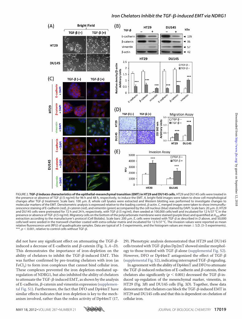
this could bemediated by themarked up-regulation ofNDRG1.NDRG1 inhibits the TGF-�/SMAD pathway through decreas-ing SMAD2 and pSMAD3 expression, thus preventing TGF-�-induced Slug expression, which is responsible for repressingE-cadherin (Fig. 7). Hence, then E-cadherin combines with�-catenin on the membrane which forms the adherens com-plex leading to increased cell-cell contact and decreased inva-
FIGURE 5. NDRG1 knock-down mimics the TGF-�-induced EMT. A, NDRG1 knock-down HT29 cells (clone 1), DU145 cells (clone 5), and their respectivecontrol cells transfected with scrambled control shRNA were treated with or without TGF-� (5 ng/ml) for 96 h and 48 h, respectively. The efficacy of NDRG1knock-down and the expression of EMT markers (i.e. E-cadherin, �-catenin, and vimentin) were analyzed by Western blotting. B, bright field microscopy andimmunofluorescence showed that NDRG1 knock-down mimics the EMT phenotype in both HT29 and DU145 cells. Scale bars: 100 �m for bright field and 20 �mfor immunofluorescence. C and D, cell migration and invasion assays were performed as described in Fig. 2, D and E. Scale bars: 200 �m. Data were repeated3–5 times, and the values in histograms represent mean � S.D. (3–5 experiments). *, relative to vector control without TGF-�; #, relative to vector control withTGF-�. **, p � 0.01; ***, p � 0.001; ##, p � 0.01; ###, p � 0.001.
Iron Chelators Inhibit the TGF-�-induced EMT via NDRG1
MAY 18, 2012 • VOLUME 287 • NUMBER 21 JOURNAL OF BIOLOGICAL CHEMISTRY 17025
by guest on September 12, 2018
http://ww
w.jbc.org/
Dow
nloaded from
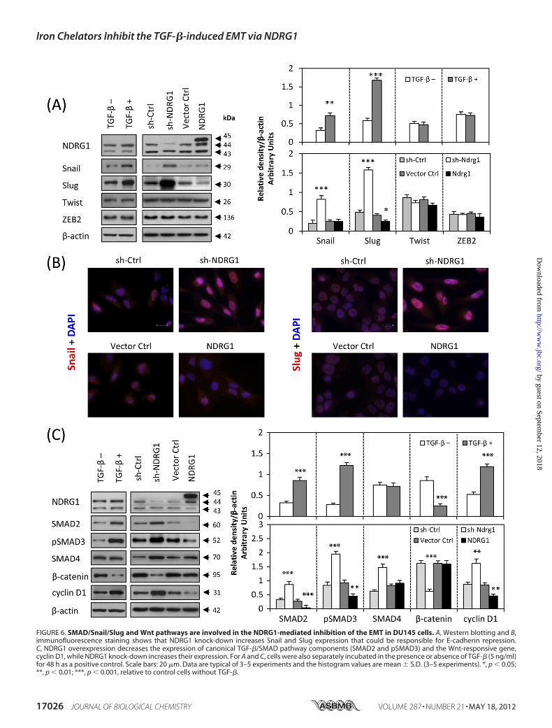
FIGURE 6. SMAD/Snail/Slug and Wnt pathways are involved in the NDRG1-mediated inhibition of the EMT in DU145 cells. A, Western blotting and B,immunofluorescence staining shows that NDRG1 knock-down increases Snail and Slug expression that could be responsible for E-cadherin repression.C, NDRG1 overexpression decreases the expression of canonical TGF-�/SMAD pathway components (SMAD2 and pSMAD3) and the Wnt-responsive gene,cyclin D1, while NDRG1 knock-down increases their expression. For A and C, cells were also separately incubated in the presence or absence of TGF-� (5 ng/ml)for 48 h as a positive control. Scale bars: 20 �m. Data are typical of 3–5 experiments and the histogram values are mean � S.D. (3–5 experiments). *, p � 0.05;**, p � 0.01; ***, p � 0.001, relative to control cells without TGF-�.
Iron Chelators Inhibit the TGF-�-induced EMT via NDRG1
17026 JOURNAL OF BIOLOGICAL CHEMISTRY VOLUME 287 • NUMBER 21 • MAY 18, 2012
by guest on September 12, 2018
http://ww
w.jbc.org/
Dow
nloaded from
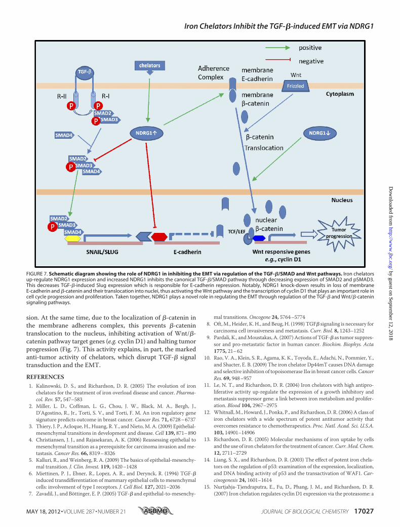
sion. At the same time, due to the localization of �-catenin inthe membrane adherens complex, this prevents �-catenintranslocation to the nucleus, inhibiting activation of Wnt/�-catenin pathway target genes (e.g. cyclin D1) and halting tumorprogression (Fig. 7). This activity explains, in part, the markedanti-tumor activity of chelators, which disrupt TGF-� signaltransduction and the EMT.
REFERENCES1. Kalinowski, D. S., and Richardson, D. R. (2005) The evolution of iron
chelators for the treatment of iron overload disease and cancer. Pharma-col. Rev. 57, 547–583
2. Miller, L. D., Coffman, L. G., Chou, J. W., Black, M. A., Bergh, J.,D’Agostino, R., Jr., Torti, S. V., and Torti, F. M. An iron regulatory genesignature predicts outcome in breast cancer. Cancer Res. 71, 6728–6737
3. Thiery, J. P., Acloque, H., Huang, R. Y., and Nieto, M. A. (2009) Epithelial-mesenchymal transitions in development and disease. Cell 139, 871–890
4. Christiansen, J. J., and Rajasekaran, A. K. (2006) Reassessing epithelial tomesenchymal transition as a prerequisite for carcinoma invasion andme-tastasis. Cancer Res. 66, 8319–8326
5. Kalluri, R., andWeinberg, R. A. (2009) The basics of epithelial-mesenchy-mal transition. J. Clin. Invest. 119, 1420–1428
6. Miettinen, P. J., Ebner, R., Lopez, A. R., and Derynck, R. (1994) TGF-�induced transdifferentiation of mammary epithelial cells to mesenchymalcells: involvement of type I receptors. J. Cell Biol. 127, 2021–2036
7. Zavadil, J., and Böttinger, E. P. (2005) TGF-� and epithelial-to-mesenchy-
mal transitions. Oncogene 24, 5764–57748. Oft,M., Heider, K. H., and Beug, H. (1998) TGF� signaling is necessary for
carcinoma cell invasiveness and metastasis. Curr. Biol. 8, 1243–12529. Pardali, K., andMoustakas, A. (2007) Actions of TGF-� as tumor suppres-
sor and pro-metastatic factor in human cancer. Biochim. Biophys. Acta1775, 21–62
10. Rao, V. A., Klein, S. R., Agama, K. K., Toyoda, E., Adachi, N., Pommier, Y.,and Shacter, E. B. (2009) The iron chelator Dp44mT causes DNA damageand selective inhibition of topoisomerase II� in breast cancer cells.CancerRes. 69, 948–957
11. Le, N. T., and Richardson, D. R. (2004) Iron chelators with high antipro-liferative activity up-regulate the expression of a growth inhibitory andmetastasis suppressor gene: a link between iron metabolism and prolifer-ation. Blood 104, 2967–2975
12. Whitnall,M., Howard, J., Ponka, P., and Richardson, D. R. (2006) A class ofiron chelators with a wide spectrum of potent antitumor activity thatovercomes resistance to chemotherapeutics. Proc. Natl. Acad. Sci. U.S.A.103, 14901–14906
13. Richardson, D. R. (2005) Molecular mechanisms of iron uptake by cellsand the use of iron chelators for the treatment of cancer.Curr.Med. Chem.12, 2711–2729
14. Liang, S. X., and Richardson, D. R. (2003) The effect of potent iron chela-tors on the regulation of p53: examination of the expression, localization,and DNA binding activity of p53 and the transactivation of WAF1. Car-cinogenesis 24, 1601–1614
15. Nurtjahja-Tjendraputra, E., Fu, D., Phang, J. M., and Richardson, D. R.(2007) Iron chelation regulates cyclin D1 expression via the proteasome: a
FIGURE 7. Schematic diagram showing the role of NDRG1 in inhibiting the EMT via regulation of the TGF-�/SMAD and Wnt pathways. Iron chelatorsup-regulate NDRG1 expression and increased NDRG1 inhibits the canonical TGF-�/SMAD pathway through decreasing expression of SMAD2 and pSMAD3.This decreases TGF-�-induced Slug expression which is responsible for E-cadherin repression. Notably, NDRG1 knock-down results in loss of membraneE-cadherin and �-catenin and their translocation into nuclei, thus activating the Wnt pathway and the transcription of cyclin D1 that plays an important role incell cycle progression and proliferation. Taken together, NDRG1 plays a novel role in regulating the EMT through regulation of the TGF-� and Wnt/�-cateninsignaling pathways.
Iron Chelators Inhibit the TGF-�-induced EMT via NDRG1
MAY 18, 2012 • VOLUME 287 • NUMBER 21 JOURNAL OF BIOLOGICAL CHEMISTRY 17027
by guest on September 12, 2018
http://ww
w.jbc.org/
Dow
nloaded from
link to iron deficiency-mediated growth suppression. Blood 109,4045–4054
16. Fu, D., and Richardson, D. R. (2007) Iron chelation and regulation of thecell cycle: 2 mechanisms of posttranscriptional regulation of the universalcyclin-dependent kinase inhibitor p21CIP1/WAF1 by iron depletion.Blood 110, 752–761
17. Richardson, D. R., Sharpe, P. C., Lovejoy, D. B., Senaratne, D., Kalinowski,D. S., Islam, M., and Bernhardt, P. V. (2006) Dipyridyl thiosemicarbazonechelators with potent and selective antitumor activity form iron com-plexes with redox activity. J. Med. Chem. 49, 6510–6521
18. Lovejoy, D. B., Jansson, P. J., Brunk, U. T., Wong, J., Ponka, P., and Rich-ardson, D. R. (2011) Antitumor activity of metal-chelating compoundDp44mT is mediated by formation of a redox-active copper complex thataccumulates in lysosomes. Cancer Res. 71, 5871–5880
19. Yu, Y., Suryo Rahmanto, Y., Hawkins, C. L., and Richardson, D. R. (2011)The potent and novel thiosemicarbazone chelators di-2-pyridylketone-4,4-dimethyl-3-thiosemicarbazone and 2-benzoylpyridine-4,4-dimethyl-3-thiosemicarbazone affect crucial thiol systems required for ribonucle-otide reductase activity.Mol. Pharmacol. 79, 921–931
20. Kovacevic, Z., Fu, D., and Richardson, D. R. (2008) The iron-regulatedmetastasis suppressor, Ndrg-1: identification of novel molecular targets.Biochim. Biophys. Acta 1783, 1981–1992
21. Bandyopadhyay, S., Pai, S. K., Gross, S. C., Hirota, S., Hosobe, S.,Miura, K.,Saito, K., Commes, T., Hayashi, S.,Watabe,M., andWatabe, K. (2003) TheDrg-1 gene suppresses tumor metastasis in prostate cancer. Cancer Res.63, 1731–1736
22. Guan, R. J., Ford, H. L., Fu, Y., Li, Y., Shaw, L. M., and Pardee, A. B. (2000)Drg-1 as a differentiation-related, putative metastatic suppressor gene inhuman colon cancer. Cancer Res. 60, 749–755
23. Bandyopadhyay, S., Pai, S. K., Hirota, S., Hosobe, S., Takano, Y., Saito, K.,Piquemal, D., Commes, T.,Watabe,M., Gross, S. C.,Wang, Y., Ran, S., andWatabe, K. (2004) Role of the putative tumor metastasis suppressor geneDrg-1 in breast cancer progression. Oncogene 23, 5675–5681
24. Maruyama, Y., Ono,M., Kawahara, A., Yokoyama, T., Basaki, Y., Kage,M.,Aoyagi, S., Kinoshita, H., and Kuwano, M. (2006) Tumor growth suppres-sion in pancreatic cancer by a putativemetastasis suppressor gene Cap43/NDRG1/Drg-1 through modulation of angiogenesis. Cancer Res. 66,6233–6242
25. Cangul, H. (2004) Hypoxia up-regulates the expression of the NDRG1gene leading to its overexpression in various human cancers. BMCGenet.5, 27
26. Ellen, T. P., Ke, Q., Zhang, P., and Costa, M. (2008) NDRG1, a growth andcancer-related gene: regulation of gene expression and function in normaland disease states. Carcinogenesis 29, 2–8
27. Shah, M. A., Kemeny, N., Hummer, A., Drobnjak, M., Motwani, M., Cor-don-Cardo, C., Gonen, M., and Schwartz, G. K. (2005) Drg1 expression in131 colorectal liver metastases: correlation with clinical variables and pa-tient outcomes. Clin. Cancer Res. 11, 3296–3302
28. Kovacevic, Z., Chikhani, S., Lovejoy, D. B., and Richardson, D. R. (2011)Novel thiosemicarbazone iron chelators induce up-regulation and phos-phorylation of the metastasis suppressor N-Myc downstream-regulatedgene 1: a new strategy for the treatment of pancreatic cancer.Mol. Phar-macol. 80, 598–609
29. Yuan, J., Lovejoy, D. B., and Richardson, D. R. (2004) Novel di-2-pyridyl-derived iron chelators with marked and selective antitumor activity: invitro and in vivo assessment. Blood 104, 1450–1458
30. Richardson, D., Ponka, P., and Baker, E. (1994) The effect of the iron(III)chelator, desferrioxamine, on iron and transferrin uptake by the humanmalignant melanoma cell. Cancer Res. 54, 685–689
31. Bambang, I. F., Lu, D., Li, H., Chiu, L. L., Lau,Q. C., Koay, E., and Zhang, D.(2009) Cytokeratin 19 regulates endoplasmic reticulum stress and inhibitsERp29 expression via p38 MAPK/XBP-1 signaling in breast cancer cells.Exp. Cell Res. 315, 1964–1974
32. Pino, M. S., Kikuchi, H., Zeng, M., Herraiz, M. T., Sperduti, I., Berger, D.,Park, D. Y., Iafrate, A. J., Zukerberg, L. R., and Chung, D. C. (2010) Epithe-lial to mesenchymal transition is impaired in colon cancer cells with mic-rosatellite instability. Gastroenterology 138, 1406–1417
33. Ellenrieder, V., Hendler, S. F., Boeck, W., Seufferlein, T., Menke, A., Ruh-land, C., Adler, G., andGress, T.M. (2001) Transforming growth factor�1treatment leads to an epithelial-mesenchymal transdifferentiation of pan-creatic cancer cells requiring extracellular signal-regulated kinase 2 acti-vation. Cancer Res. 61, 4222–4228
34. Wijnhoven, B. P., Dinjens, W. N., and Pignatelli, M. (2000) E-cadherin-catenin cell-cell adhesion complex and human cancer. Br J. Surg. 87,992–1005
35. Salnikow, K., Davidson, T., Zhang, Q., Chen, L. C., Su, W., and Costa, M.(2003) The involvement of hypoxia-inducible transcription factor-1-de-pendent pathway in nickel carcinogenesis. Cancer Res. 63, 3524–3530
36. Kovacevic, Z., Sivagurunathan, S., Mangs, H., Chikhani, S., Zhang, D., andRichardson, D. R. (2011) Themetastasis suppressor, N-Myc downstream-regulated gene 1 (NDRG1), up-regulates p21 via p53-independent mech-anisms. Carcinogenesis 32, 732–740
37. Sugiki, T., Murakami, M., Taketomi, Y., Kikuchi-Yanoshita, R., and Kudo,I. (2004) N-Myc down-regulated gene 1 is a phosphorylated protein inmast cells. Biol. Pharm. Bull. 27, 624–627
38. van Belzen, N., Dinjens, W. N., Diesveld, M. P., Groen, N. A., van derMade, A. C., Nozawa, Y., Vlietstra, R., Trapman, J., and Bosman, F. T.(1997) A novel gene which is up-regulated during colon epithelial celldifferentiation and down-regulated in colorectal neoplasms. Lab. Invest.77, 85–92
39. Bandyopadhyay, S., Pai, S. K., Hirota, S., Hosobe, S., Tsukada, T.,Miura, K.,Takano, Y., Saito, K., Commes, T., Piquemal, D., Watabe, M., Gross, S.,Wang, Y., Huggenvik, J., and Watabe, K. (2004) PTEN up-regulates thetumor metastasis suppressor gene Drg-1 in prostate and breast cancer.Cancer Res. 64, 7655–7660
40. Behrens, J., von Kries, J. P., Kühl, M., Bruhn, L., Wedlich, D., Grosschedl,R., and Birchmeier,W. (1996) Functional interaction of�-cateninwith thetranscription factor LEF-1. Nature 382, 638–642
41. Suzuki, H.,Masuda,N., Shimura, T., Araki, K., Kobayashi, T., Tsutsumi, S.,Asao, T., and Kuwano, H. (2008) Nuclear �-catenin expression at theinvasive front and in the vessels predicts liver metastasis in colorectalcarcinoma. Anticancer Res. 28, 1821–1830
42. Elston, M. S., Gill, A. J., Conaglen, J. V., Clarkson, A., Cook, R. J., Little,N. S., Robinson, B. G., Clifton-Bligh, R. J., and McDonald, K. L. (2009)Nuclear accumulation of e-cadherin correlates with loss of cytoplasmicmembrane staining and invasion in pituitary adenomas. J. Clin. Endocri-nol. Metab. 94, 1436–1442
43. Céspedes, M. V., Larriba, M. J., Pavón, M. A., Alamo, P., Casanova, I.,Parreño, M., Feliu, A., Sancho, F. J., Muñoz, A., and Mangues, R. (2010)Site-dependent E-cadherin cleavage and nuclear translocation in a meta-static colorectal cancer model. Am. J. Pathol. 177, 2067–2079
44. Hartsock, A., and Nelson, W. J. (2008) Adherens and tight junctions:structure, function, and connections to the actin cytoskeleton. Biochim.Biophys. Acta 1778, 660–669
45. Hotz, B., Arndt, M., Dullat, S., Bhargava, S., Buhr, H. J., and Hotz, H. G.(2007) Epithelial to mesenchymal transition: expression of the regulatorssnail, slug, and twist in pancreatic cancer. Clin. Cancer Res. 13,4769–4776
46. Heldin, C.H.,Miyazono, K., and tenDijke, P. (1997) TGF-� signaling fromcell membrane to nucleus through SMADproteins.Nature 390, 465–471
47. Kachhap, S. K., Faith, D., Qian, D. Z., Shabbeer, S., Galloway, N. L., Pili, R.,Denmeade, S. R., DeMarzo, A.M., andCarducci,M. A. (2007) TheN-Mycdown-regulated Gene1 (NDRG1) Is a Rab4a effector involved in vesicularrecycling of E-cadherin. PLoS One 2, e844
48. Wu, D., and Pan,W. (2010) GSK3: a multifaceted kinase inWnt signaling.Trends Biochem. Sci. 35, 161–168
49. Reya, T., and Clevers, H. (2005) Wnt signaling in stem cells and cancer.Nature 434, 843–850
50. Sherr, C. J. (1994) G1 phase progression: cycling on cue. Cell 79, 551–55551. Brandl, M., Seidler, B., Haller, F., Adamski, J., Schmid, R. M., Saur, D., and
Schneider, G. (2010) IKK(�) controls canonical TGF(�)-SMAD signalingto regulate genes expressing SNAIL and SLUGduring EMT in panc1 cells.J. Cell Sci. 123, 4231–4239
Iron Chelators Inhibit the TGF-�-induced EMT via NDRG1
17028 JOURNAL OF BIOLOGICAL CHEMISTRY VOLUME 287 • NUMBER 21 • MAY 18, 2012
by guest on September 12, 2018
http://ww
w.jbc.org/
Dow
nloaded from
RichardsonZhiqiang Chen, Daohai Zhang, Fei Yue, Minhua Zheng, Zaklina Kovacevic and Des R.
Downstream-regulated Gene 1 (NDRG1)Epithelial-Mesenchymal Transition via Up-Regulation of N-Myc
-inducedβThe Iron Chelators Dp44mT and DFO Inhibit TGF-
doi: 10.1074/jbc.M112.350470 originally published online March 27, 20122012, 287:17016-17028.J. Biol. Chem.
10.1074/jbc.M112.350470Access the most updated version of this article at doi:
Alerts:
When a correction for this article is posted•
When this article is cited•
to choose from all of JBC's e-mail alertsClick here
Supplemental material:
http://www.jbc.org/content/suppl/2012/03/27/M112.350470.DC1
http://www.jbc.org/content/287/21/17016.full.html#ref-list-1
This article cites 50 references, 23 of which can be accessed free at
by guest on September 12, 2018
http://ww
w.jbc.org/
Dow
nloaded from