the wobbler mouse, an als animal model - springer · the wobbler mouse, an als animal model ......
TRANSCRIPT
REVIEW
The wobbler mouse, an ALS animal model
Jakob Maximilian Moser • Paolo Bigini •
Thomas Schmitt-John
Received: 18 December 2012 / Accepted: 12 March 2013 / Published online: 29 March 2013
� The Author(s) 2013. This article is published with open access at Springerlink.com
Abstract This review article is focused on the research
progress made utilizing the wobbler mouse as animal
model for human motor neuron diseases, especially the
amyotrophic lateral sclerosis (ALS). The wobbler mouse
develops progressive degeneration of upper and lower
motor neurons and shows striking similarities to ALS. The
cellular effects of the wobbler mutation, cellular transport
defects, neurofilament aggregation, neuronal hyperexcit-
ability and neuroinflammation closely resemble human
ALS. Now, 57 years after the first report on the wobbler
mouse we summarize the progress made in understanding
the disease mechanism and testing various therapeutic
approaches and discuss the relevance of these advances for
human ALS. The identification of the causative mutation
linking the wobbler mutation to a vesicle transport factor
and the research focussed on the cellular basis and the
therapeutic treatment of the wobbler motor neuron degen-
eration has shed new light on the molecular pathology of
the disease and might contribute to the understanding the
complexity of ALS.
Keywords ALS �Wobbler �Motor neuron degeneration �GARP complex � Vesicle tethering
Introduction
The wobbler mouse has successfully been used as animal
model for human motor neuron diseases, especially ALS in
the investigation of both, pathology and therapeutic inter-
vention, as reviewed by (Boillee et al. 2003). In the recent
years substantial progress has been made in understanding
the molecular basis of the ALS-like wobbler motor neuron
degeneration and testing therapeutic interventions. This
article will briefly introduce the general progress in ALS
research, will review the specific progress made on the
understanding of molecular and cellular basis of the wob-
bler motor neuron disease and will provide an overview
over therapeutic treatments tested in wobbler mice, which
in itself have contributed to the understanding of the
disease.
Amyotrophic lateral sclerosis
Amyotrophic lateral sclerosis (ALS) was first described in
1869 by Jean-Martin Charcot and is the most common
motor neuron disease (MND) among adults (Bruijn et al.
2004). Age of onset is typically between 50 and 60 years
although earlier onset is not uncommon (Bruijn et al.
2004). Roughly 2 out of 100,000 individuals develop ALS
and men have a slightly elevated risk compared to women
(Ferraiuolo et al. 2011). 5–10 % of ALS cases can be
attributed to familial forms (fALS), which predominantly
are inherited in an autosomal dominant way. However, the
vast majority of cases occur sporadically with no clear
inheritance and thus, are termed sALS (Ferraiuolo et al.
2011). sALS is thought to be caused by a combination of
genetic susceptibility and possible environmental factors
(Ferraiuolo et al. 2011). Even with the emerging wider
Communicated by J. Graw .
J. M. Moser � T. Schmitt-John (&)
Molecular Biology and Genetics Department, Aarhus University,
C. F. Møllers Alle 3, 8000 Aarhus C, Denmark
e-mail: [email protected]
P. Bigini
Instituto di Ricerche Farmacologiche ‘‘Mario Negri’’ – IRCCS,
Via La Masa 19, Milano 20156, Italy
123
Mol Genet Genomics (2013) 288:207–229
DOI 10.1007/s00438-013-0741-0
knowledge of the primary cause for at least a subset of ALS
cases, the molecular and cellular pathomechanism of ALS
is still poorly understood and was recently reviewed by
(Ferraiuolo et al. 2011).
Clinically ALS can be described by loss of motor neu-
rons at all levels of the motor system; from motor cortex to
the ventral horn of the spinal cord (Mitchell and Borasio
2007). Previously, ALS was considered to be a disease,
only affecting motor neurons, but recent evidence suggests
the involvement of both sensory and spinocerebellar
pathways and other neurons in the brain (Ferraiuolo et al.
2011). ALS is progressive and always fatal, leading to
death within 3–5 years after onset of disease symptoms,
resulting from failure of the respiratory muscles, being the
most common cause of death (Bruijn et al. 2004; Wood-
Allum and Shaw 2010). The loss of motor neurons leads to
spasticity, hyperreflexia (upper motor neurons), general-
ized weakness, paralysis and muscle atrophy (lower motor
neurons) (Bruijn et al. 2004). Pharmacological treatment
options for ALS patients are very limited. Currently, only
one FDA-approved drug, Riluzole, is available. The glu-
tamate release inhibitor has a mild disease modifying effect
and prolongs survival for about 3–4 months (Wood-Allum
and Shaw 2010). Understanding the mechanisms underly-
ing ALS and the effects preceding the onset of clinical
symptoms is paramount in order to develop an effective
treatment for ALS patients.
Pathomechanisms of ALS
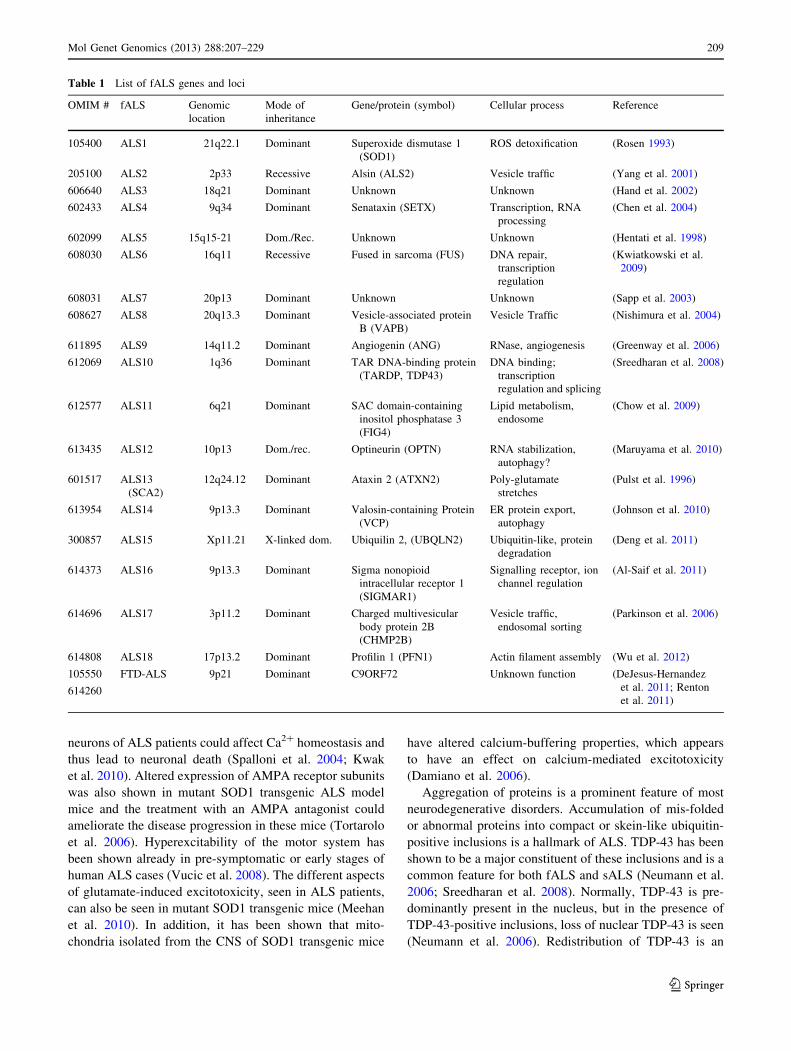
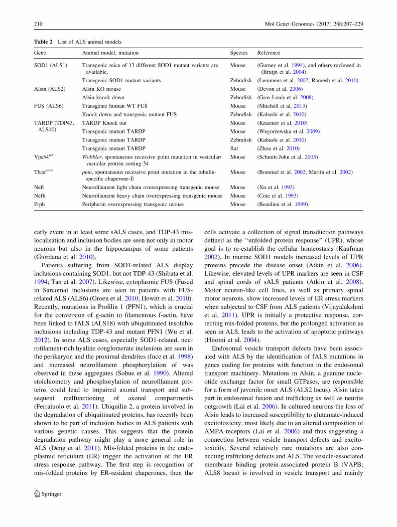
The identification of affected genes in some of the familial
ALS cases (Table 1) and the generation and investigation
of ALS animal models (Table 2) has shed some light on the
causes of ALS pathology and the contributing factors. ALS
has been linked to oxidative stress, excitotoxicity, abnor-
mal aggregation of protein and defects of vesicle- and
axonal transport systems as potential causes of the neuro-
degeneration, but also the neuroinflammatory processes are
considered to contribute to the disease progression (Fer-
raiuolo et al. 2011).
ALS has initially been linked to oxidative stress through
mutations in the superoxide dismutase 1 (SOD1) gene
(ALS1 locus), coding for a major antioxidant protein.
SOD1 is an enzyme involved in the detoxification of free
superoxide radicals and more than 100 different ALS-
associated mutations have been described in the SOD1
gene so far (Ferraiuolo et al. 2011). Mutations are found
both in fALS cases, where they constitute around 20 % of
all cases, as well as in sALS cases (Mitchell and Borasio
2007). These mutations are dominantly inherited indicating
that a gain of toxic function rather than loss-of-SOD1
function underlies the disease mechanism (Ferraiuolo et al.
2011). This is supported by the finding that abolishing the
activity of the copper chaperone for SOD1, rendering
SOD1 enzymatically inactive, no change in disease onset
or progression was seen in SOD1 transgenic model mice
(Subramaniam et al. 2002). However, markers for oxida-
tive stress have been found in cerebrospinal fluid (CSF),
serum and urine isolated from ALS patients (Smith et al.
1998; Simpson et al. 2004; Mitsumoto et al. 2008) and
increased oxidative damage to proteins, DNA, lipids and
mRNA species have been reported in tissue from both
f- and sALS-patients (Shaw et al. 1995b; Fitzmaurice et al.
1996; Shibata et al. 2001; Chang et al. 2008). A further
indication that oxidative stress plays a general role in motor
neuron injury is supported by the findings that the fALS-
associated TAR DNA-binding protein 43 (TDP-43;
ALS10), a protein involved in the RNA machinery, is able
to induce oxidative stress in a motor neuron-like cell line
(Duan et al. 2010). Oxidative stress might be not a direct
cause of ALS, but instead might aggravate other cellular
effects of ALS, such as glutamate-induced excitotoxicity,
mitochondrial impairment, protein aggregation, ER stress
and signalling from neuronal support cells, thus aggravat-
ing motor neuron injury (Duffy et al. 2011; Wood et al.
2003; Kanekura et al. 2009; Blackburn et al. 2009; Sar-
gsyan et al. 2005).
Mitochondrial dysfunction seems to be involved in at
least some ALS cases. Mitochondria are involved in cal-
cium homeostasis, intracellular energy production and
control of apoptosis. SOD1-mutant mice display protein
aggregates in the mitochondrial intermembrane space
(Wong et al. 1995). Dysregulation of energy metabolism
has been shown in both ALS patients and murine SOD1
models (Wiedemann et al. 2002; Mattiazzi et al. 2002).
Likewise, altered morphology of mitochondria has been
observed in skeletal muscle and spinal motor neurons in
both ALS patients and in mouse models (Sasaki and Iwata
2007; Wong et al. 1995).
Excitotoxicity is thought to play a role in ALS. Gluta-
mate is the main excitatory neurotransmitter in the central
nervous system (CNS). Some ALS patients show increased
levels of glutamate in the cerebrospinal fluid (CSF) (Shaw
et al. 1995a). In affected areas of the CNS of sALS and
fALS patients, decreased expression and reduced activity
of the excitatory amino acid transporter 2 (EAAT2), an
astroglial glutamate re-uptake transporter, was reported
(Rothstein et al. 1992, 1995; Fray et al. 1998). The over-
expression of EAAT2 (Guo et al. 2003) and the pharma-
cologically induced up-regulation of EAAT2 transcription
could show beneficial effects in ALS model mice (Roth-
stein et al. 2005); suggesting that decreased EAAT2
expression is rather a cause than a consequence of ALS.
Altered composition of the a-amino-3-hydroxy-5-methyl-
4-isoxazolepropionic acid (AMPA) receptor in motor
208 Mol Genet Genomics (2013) 288:207–229
123
neurons of ALS patients could affect Ca2? homeostasis and
thus lead to neuronal death (Spalloni et al. 2004; Kwak
et al. 2010). Altered expression of AMPA receptor subunits
was also shown in mutant SOD1 transgenic ALS model
mice and the treatment with an AMPA antagonist could
ameliorate the disease progression in these mice (Tortarolo
et al. 2006). Hyperexcitability of the motor system has
been shown already in pre-symptomatic or early stages of
human ALS cases (Vucic et al. 2008). The different aspects
of glutamate-induced excitotoxicity, seen in ALS patients,
can also be seen in mutant SOD1 transgenic mice (Meehan
et al. 2010). In addition, it has been shown that mito-
chondria isolated from the CNS of SOD1 transgenic mice
have altered calcium-buffering properties, which appears
to have an effect on calcium-mediated excitotoxicity
(Damiano et al. 2006).
Aggregation of proteins is a prominent feature of most
neurodegenerative disorders. Accumulation of mis-folded
or abnormal proteins into compact or skein-like ubiquitin-
positive inclusions is a hallmark of ALS. TDP-43 has been
shown to be a major constituent of these inclusions and is a
common feature for both fALS and sALS (Neumann et al.
2006; Sreedharan et al. 2008). Normally, TDP-43 is pre-
dominantly present in the nucleus, but in the presence of
TDP-43-positive inclusions, loss of nuclear TDP-43 is seen
(Neumann et al. 2006). Redistribution of TDP-43 is an
Table 1 List of fALS genes and loci
OMIM # fALS Genomic
location
Mode of
inheritance
Gene/protein (symbol) Cellular process Reference
105400 ALS1 21q22.1 Dominant Superoxide dismutase 1
(SOD1)
ROS detoxification (Rosen 1993)
205100 ALS2 2p33 Recessive Alsin (ALS2) Vesicle traffic (Yang et al. 2001)
606640 ALS3 18q21 Dominant Unknown Unknown (Hand et al. 2002)
602433 ALS4 9q34 Dominant Senataxin (SETX) Transcription, RNA
processing
(Chen et al. 2004)
602099 ALS5 15q15-21 Dom./Rec. Unknown Unknown (Hentati et al. 1998)
608030 ALS6 16q11 Recessive Fused in sarcoma (FUS) DNA repair,
transcription
regulation
(Kwiatkowski et al.
2009)
608031 ALS7 20p13 Dominant Unknown Unknown (Sapp et al. 2003)
608627 ALS8 20q13.3 Dominant Vesicle-associated protein
B (VAPB)
Vesicle Traffic (Nishimura et al. 2004)
611895 ALS9 14q11.2 Dominant Angiogenin (ANG) RNase, angiogenesis (Greenway et al. 2006)
612069 ALS10 1q36 Dominant TAR DNA-binding protein
(TARDP, TDP43)
DNA binding;
transcription
regulation and splicing
(Sreedharan et al. 2008)
612577 ALS11 6q21 Dominant SAC domain-containing
inositol phosphatase 3
(FIG4)
Lipid metabolism,
endosome
(Chow et al. 2009)
613435 ALS12 10p13 Dom./rec. Optineurin (OPTN) RNA stabilization,
autophagy?
(Maruyama et al. 2010)
601517 ALS13
(SCA2)
12q24.12 Dominant Ataxin 2 (ATXN2) Poly-glutamate
stretches
(Pulst et al. 1996)
613954 ALS14 9p13.3 Dominant Valosin-containing Protein
(VCP)
ER protein export,
autophagy
(Johnson et al. 2010)
300857 ALS15 Xp11.21 X-linked dom. Ubiquilin 2, (UBQLN2) Ubiquitin-like, protein
degradation
(Deng et al. 2011)
614373 ALS16 9p13.3 Dominant Sigma nonopioid
intracellular receptor 1
(SIGMAR1)
Signalling receptor, ion
channel regulation
(Al-Saif et al. 2011)
614696 ALS17 3p11.2 Dominant Charged multivesicular
body protein 2B
(CHMP2B)
Vesicle traffic,
endosomal sorting
(Parkinson et al. 2006)
614808 ALS18 17p13.2 Dominant Profilin 1 (PFN1) Actin filament assembly (Wu et al. 2012)
105550
614260
FTD-ALS 9p21 Dominant C9ORF72 Unknown function (DeJesus-Hernandez
et al. 2011; Renton
et al. 2011)
Mol Genet Genomics (2013) 288:207–229 209
123
early event in at least some sALS cases, and TDP-43 mis-
localisation and inclusion bodies are seen not only in motor
neurons but also in the hippocampus of some patients
(Giordana et al. 2010).
Patients suffering from SOD1-related ALS display
inclusions containing SOD1, but not TDP-43 (Shibata et al.
1994; Tan et al. 2007). Likewise, cytoplasmic FUS (Fused
in Sarcoma) inclusions are seen in patients with FUS-
related ALS (ALS6) (Groen et al. 2010; Hewitt et al. 2010).
Recently, mutations in Profilin 1 (PFN1), which is crucial
for the conversion of g-actin to filamentous f-actin, have
been linked to fALS (ALS18) with ubiquitinated insoluble
inclusions including TDP-43 and mutant PFN1 (Wu et al.
2012). In some ALS cases, especially SOD1-related, neu-
rofilament-rich hyaline conglomerate inclusions are seen in
the perikaryon and the proximal dendrites (Ince et al. 1998)
and increased neurofilament phosphorylation of was
observed in these aggregates (Sobue et al. 1990). Altered
stoichiometry and phosphorylation of neurofilament pro-
teins could lead to impaired axonal transport and sub-
sequent malfunctioning of axonal compartments
(Ferraiuolo et al. 2011). Ubiquilin 2, a protein involved in
the degradation of ubiquitinated proteins, has recently been
shown to be part of inclusion bodies in ALS patients with
various genetic causes. This suggests that the protein
degradation pathway might play a more general role in
ALS (Deng et al. 2011). Mis-folded proteins in the endo-
plasmic reticulum (ER) trigger the activation of the ER
stress response pathway. The first step is recognition of
mis-folded proteins by ER-resident chaperones, then the
cells activate a collection of signal transduction pathways
defined as the ‘‘unfolded protein response’’ (UPR), whose
goal is to re-establish the cellular homeostasis (Kaufman
2002). In murine SOD1 models increased levels of UPR
proteins precede the disease onset (Atkin et al. 2006).
Likewise, elevated levels of UPR markers are seen in CSF
and spinal cords of sALS patients (Atkin et al. 2008).
Motor neuron-like cell lines, as well as primary spinal
motor neurons, show increased levels of ER stress markers
when subjected to CSF from ALS patients (Vijayalakshmi
et al. 2011). UPR is initially a protective response, cor-
recting mis-folded proteins, but the prolonged activation as
seen in ALS, leads to the activation of apoptotic pathways
(Hitomi et al. 2004).
Endosomal vesicle transport defects have been associ-
ated with ALS by the identification of fALS mutations in
genes coding for proteins with function in the endosomal
transport machinery. Mutations in Alsin, a guanine nucle-
otide exchange factor for small GTPases, are responsible
for a form of juvenile onset ALS (ALS2 locus). Alsin takes
part in endosomal fusion and trafficking as well as neurite
outgrowth (Lai et al. 2006). In cultured neurons the loss of
Alsin leads to increased susceptibility to glutamate-induced
excitotoxicity, most likely due to an altered composition of
AMPA-receptors (Lai et al. 2006) and thus suggesting a
connection between vesicle transport defects and excito-
toxicity. Several relatively rare mutations are also con-
necting trafficking defects and ALS. The vesicle-associated
membrane binding protein-associated protein B (VAPB;
ALS8 locus) is involved in vesicle transport and mainly
Table 2 List of ALS animal models
Gene Animal model, mutation Species Reference
SOD1 (ALS1) Transgenic mice of 13 different SOD1 mutant variants are
available.
Mouse (Gurney et al. 1994), and others reviewed in
(Bruijn et al. 2004)
Transgenic SOD1 mutant variants Zebrafish (Lemmens et al. 2007; Ramesh et al. 2010)
Alsin (ALS2) Alsin KO mouse Mouse (Devon et al. 2006)
Alsin knock down Zebrafish (Gros-Louis et al. 2008)
FUS (ALS6) Transgenic human WT FUS Mouse (Mitchell et al. 2013)
Knock down and transgenic mutant FUS Zebrafish (Kabashi et al. 2010)
TARDP (TDP43,
ALS10)
TARDP Knock out Mouse (Kraemer et al. 2010)
Transgenic mutant TARDP Mouse (Wegorzewska et al. 2009)
Transgenic mutant TARDP Zebrafish (Kabashi et al. 2010)
Transgenic mutant TARDP Rat (Zhou et al. 2010)
Vps54wr Wobbler, spontaneous recessive point mutation in vesicular/
vacuolar protein sorting 54
Mouse (Schmitt-John et al. 2005)
Tbcepmn pmn, spontaneous recessive point mutation in the tubulin-
specific chaperone-E
Mouse (Bommel et al. 2002; Martin et al. 2002)
Nefl Neurofilament light chain overexpressing transgenic mouse Mouse (Xu et al. 1993)
Nefh Neurofilament heavy chain overexpressing transgenic mouse Mouse (Cote et al. 1993)
Prph Peripherin overexpressing transgenic mouse Mouse (Beaulieu et al. 1999)
210 Mol Genet Genomics (2013) 288:207–229
123
located in the ER. VAPB aggregation is speculated to lead
to ER stress and disruption of proteasome function and thus
might contribute to an altered protein homeostasis and
ultimately to motor neuron death (Suzuki et al. 2009). In
addition, VAPB is involved in the UPR response
(Nishimura et al. 2004; Moumen et al. 2011; Chen et al. 2010).
Mutations in optineurin (ALS12 locus), a protein involved
in the maintenance of the Golgi complex, exocytosis and
various aspects of Golgi trafficking, lead to increased
NF-jB activation, which is also upregulated in sALS and
thus, might be connected with motor neuronal cell death
(Maruyama et al. 2010). Charged multivesicular body
protein 2B (CHMP2B), a component of the endosomal
sorting complex, causes vacuolisation, lysosomal mis-
localisation and impaired autophagy in cultured cells and
has been associated with frontotemporal dementia (FTD;
FTD3) and ALS (ALS17) (Parkinson et al. 2006; Cox et al.
2010). FIG4 gene (ALS11 locus) encodes a polyphos-
phoinositide phosphatase, which regulates the level of
phosphatidylinositol 3,5 biphosphate, a signal lipid regu-
lating the retrograde vesicle transport from lysosomes and
late endosomes to the Golgi apparatus (Chow et al. 2009).
FIG4 mutations found in human fALS cases have shown to
induce vacuolization in yeast (Chow et al. 2009).
Impaired axonal transport is a key feature of ALS
(Ferraiuolo et al. 2011). Both anterograde and retrograde
axonal transport are impaired in mutant SOD1 transgenic
mice early in the disease progression. The mechanism
behind axonal transport defects is unknown, but is likely to
be cargo-specific (Williamson and Cleveland 1999; De Vos
et al. 2007; Bilsland et al. 2010). Anterograde transport
along microtubules depends on kinesin motor proteins,
while retrograde transport depends on cytoplasmic dynein.
It is possible that impaired mitochondrial function could be
caused to some extent by a decreased mitochondrial
transport along axons, which would lead to an increased
mitochondrial aging, because the charging of mitochondria
with nuclear-encoded mitochondrial proteins in the peri-
karyon is decreased. This in turn could reduce the energy
available for general axonal transport and thereby might
aggravate the transport defect and also affect other cargos
(De Vos et al. 2007; Miller and Sheetz 2004). However, by
increasing the axonal mobility of mitochondria in mutant
SOD1 transgenic mice, no beneficial effect on the neuro-
degeneration could be observed (Zhu and Sheng 2011). In
mutant SOD1 transgenic mice, levels of tumor necrosis
factor (TNF) are elevated, which can disrupt kinesin
function through a mechanism involving p38-MAPK
(Kiaei et al. 2007; De Vos et al. 2007) and glutamate has
been shown to reduce axonal transport of neurofilament
medium chain (NF-M) by activating protein kinases, which
phosphorylate NFM proteins (Ackerley et al. 2000). Neu-
rofilament hyperphosphorylation leads to decreased
neurofilament transport and might also aggravate neuro-
filament aggregation in the perikaryon.
Transcription and RNA processing is altered in ALS
(Ferraiuolo et al. 2011). TDP-43 (ALS10 locus) is involved
in transcriptional regulation, alternative splicing and
miRNA processing and FUS (ALS6 locus) is involved in
transcriptional regulation, RNA and miRNA processing,
and mRNA transport (Mackenzie et al. 2010). Under nor-
mal circumstances both are located in the nucleus (Mac-
kenzie et al. 2010). Mutations in TDP-43 and FUS both
account for 4 % of fALS cases. In sALS TDP-43 mutations
are found in 1.5 % of the cases, while FUS mutations are
found in \1 % (Mackenzie et al. 2010). ALS-associated
mutations in TDP-43 and FUS lead to loss of nuclear
localization, re-localization to the cytoplasm, and inclusion
in cytoplasmic stress granules—a rapid, reversible cellular
response to stress, controlling RNA metabolism (Liu-
Yesucevitz et al. 2010; Ito et al. 2011; Dormann et al.
2010). How altered mRNA transcription and processing
leads to motor neuron injury is unknown. It is possible that
TDP-43 and FUS could be part of an RNA transport
complex and that mutations in either could lead to the loss
of axonal transport (Ferraiuolo et al. 2011). Another pos-
sibility is that the loss of nuclear expression leads to partial
disruption of the RNA machinery, such as pre-mRNA
splicing, nuclear mRNA export, mRNA sorting and pro-
cessing of non-coding RNA (Ferraiuolo et al. 2011). A
third option is that mutated TDP-43 and FUS-induced
stress granules revert more slowly, leading to abnormal
accumulation (Liu-Yesucevitz et al. 2010; Ito et al. 2011;
Dormann et al. 2010). Angiogenin (ANG; ALS9) and
Senataxin (SETX, ALS4) both have been associated with
fALS (Greenway et al. 2006; Chen et al. 2004) and are also
involved in RNA metabolism.
Neuroinflammation is a hallmark of ALS and involves
glia activation and infiltration of peripheral immune cells,
recently reviewed by (Papadimitriou et al. 2010). Similar to
the ER stress response, the neuroinflammatory processes
appear to have both protective and harmful effects on the
neurodegeneration (Liao et al. 2012). Proinflammatory
cytokines have been reported in the CSF of ALS patients
(Kuhle et al. 2009) and evidence from mutant SOD1
transgenic mice lacking CD4, which develop an aggravated
neurodegeneration (Beers et al. 2008), indicate that the
inflammatory reactions have an impact on the ALS neu-
rodegeneration. In chimeric mutant SOD1 transgenic mice,
normal motor neurons display signs of ALS pathology
when surrounded by mutant SOD1 expressing glial cells
(Clement et al. 2003). Astrocytes expressing mutant SOD1
exhibit toxic effects on cultured primary motor neurons and
motor neurons derived from both human and murine stem
cells (Di Giorgio et al. 2008; Nagai et al. 2007). However,
it is still under debate to what extent astrogliosis and
Mol Genet Genomics (2013) 288:207–229 211
123
microgliosis have beneficial and/or harmful effects on ALS
pathology (Papadimitriou et al. 2010).
ALS appears to be a complex disorder and many factors
contribute to the pathology. So far, mutations in 18 ALS
genes/loci have been found to cause familial ALS
(Table 1) and further genetic risk factors might contribute
to sporadic ALS. Most of the ALS genes are ubiquitously
expressed and involved in fundamental cellular processes,
which raises the question why motor neurons are more
vulnerable than other cells. The intuitive explanation that
motor neurons are specifically vulnerable due to their
extraordinary axon lengths is probably much too simple,
since motor neurons degenerating first are not necessarily
those with the longest axons and sensory neurons with
similar axon lengths are not affected. However, motor
neurons have shown to be particularly sensitive to gluta-
mate excitotoxicity (Williams et al. 1997; Ince et al. 1993)
as well as ER stress (Saxena et al. 2009). Motor neurons
also seem to have a high threshold for mounting a heat
shock response, a high sensitivity to oxidative damage and
calcium overload via mitochondria (Sullivan et al. 2004;
Panov et al. 2011). However, it is still poorly understood
what makes motor neurons more susceptible than other
cells.
The comprehension of the molecular processes under-
lying ALS is crucial for the development of an efficient
treatment of the disease. Thus, the generation and investi-
gation of suitable animal models (Table 2) contributes to
both understanding the pathomechanisms and the devel-
oping therapeutic intervention.
The wobbler mouse
Several mouse models for studying ALS exist (Table 2).
The most commonly used is the SOD1G93A, which over-
expresses a mutated human SOD1-gene (Gurney et al.
1994), but several animal models exist, where the SOD1
gene is mutated at different positions (Tovar et al. 2009).
Recently, transgenic mouse models of Alsin- (knock out)
and hTDP-43-associated fALS (transgenic) have been
created (Tovar et al. 2009; Shan et al. 2010; Wils et al.
2010; Wegorzewska et al. 2009). However, the majority of
ALS cases are sporadic with unknown cause; even though
clinically undistinguishable from fALS this fact indicates
the need for further animal models.
The wobbler mouse arose spontaneously in a C57BL/Fa
strain and was first described by Falconer in the late 1950s
(Falconer 1956). The autosomal recessive-mutation was
soon linked to motor neuron degeneration and is caused by
a point mutation in the Vps54 gene (Schmitt-John et al.
2005). The wobbler mouse is the best-characterized spon-
taneous mutant with motor neuron degeneration, which
mimics several of the features seen in ALS patients, but
where the comparable mutation of VPS54 has not been
found in ALS patients so far, although only a limited
number of patients have been examined for the mutation
(Meisler et al. 2008).
The wobbler phenotype
When describing the disease symptoms of the wobbler
mouse it is convenient to divide them into three different
phases, based on the development of physical symptoms:
the pre-symptomatic, the evolutionary and the stabilized
phase (Boillee et al. 2003). The pre-symptomatic phase
lasts from birth to around 3 weeks of age. At this point
homozygous wobbler mice exhibit little to no clinical
symptoms; in homozygous wobbler mice body weight, grip
strength, righting reflexes and grid-walking tests all appear
normal (Boillee et al. 2003). During the evolutionary
phase, which lasts from around 3 weeks to 3 months of
age, clinical, morphological and molecular symptoms
develop. Homozygous wobbler mice display reduced
bodyweight and muscle strength; develop a wobbly gait
and head tremor. Muscle atrophy leads to a pointed muzzle,
and suspension by the tail results in flexed instead of
extended hind limbs (Boillee et al. 2003). The stabilized
phase, from 3 months of age to death, is characterized by
an arrest in the progression of motor neuron degeneration
(Boillee et al. 2003). In addition, male homozygous wob-
bler mice are infertile due to failed spermatogenesis
(Leestma and Sepsenwol 1980; Heimann et al. 1991),
which will be discussed later.
First morphological changes appear already at the pre-
symptomatic stage. Affected motor neurons are character-
ized by weak staining of Nissl bodies and enlarged somas
(Duchen and Strich 1968; Blondet et al. 2002), although
the number of motor neurons remains unaffected in the
median nerve nuclei of the cervical spinal cord at this stage
(Blondet et al. 2002). In the brainstem, the ventral reticular
magnocellular nucleus, and the motor nuclei of the central
nerves V and VII, cells with diverse anomalies are found
but rarely elsewhere in the spinal cord or brain (Duchen
and Strich 1968). Schwann cells display intra-axonal in-
vaginations, dilation of the ER is seen and motor neurons
start to display vacuolization (Mitsumoto and Bradley
1982). Affected motor neurons are characterized by
microtubule segregation and the presence of large, dense
secondary lysozymes (Mitsumoto and Bradley 1982). First
signs of neurodegeneration are observed on day 13 post
natal (p.n.) in the thalamus (N. ventralis), deep cerebellar
nuclei, brain stem (N. vestibularis) and spinal inter neu-
rons, while spinal motor neurons follow around day 15 p.n.
(Rathke-Hartlieb et al. 1999). Neuroinflammation is seen
212 Mol Genet Genomics (2013) 288:207–229
123
as consequence of the neurodegeneration; from day 17 p.n.
onwards, reactive astrocytes are seen and around day 23
p.n. microgliosis is observed (Rathke-Hartlieb et al. 1999).
In the motor cortex of wobbler mice a reduced number of
parvalbumin-positive GABAergic interneurons is seen at
the pre-symptomatic phase between day 15 and 25 (Nieto-
Gonzalez et al. 2011). Thus, neurodegeneration is not
restricted to motor neurons in the wobbler mouse. Inter-
estingly, a reduced release of GABA is seen in synapto-
somes isolated from the cervical spinal cord of 12-week-
old wobbler mice (Bonanno et al. 2009), indicating that
GABAergic interneurons might play a critical role in the
wobbler motor neurodegeneration.
During the evolutionary phase numerous signs of
degeneration and a marked loss of motor neurons become
apparent (Pollin et al. 1990). In the brain degenerating
motor neurons show weak staining of Nissl bodies without
any vacuoles being present, as well as eccentric localiza-
tion of nuclei (LaVail et al. 1987). At this point, the
3-month-old wobbler mouse displays signs of muscular
denervation in the forelimbs, though not in the hind limbs
(Andrews et al. 1974). There is a reduction of motor nerve
terminals and axonal sprouts (Duchen and Strich 1968;
LaVail et al. 1987) together with reduced diameter or even
loss of myelin sheaths, resulting in large non-myelinated
fibres (Bird et al. 1971; Biscoe and Lewkowicz 1982;
Lewkowicz 1979). At the end of the evolutionary phase,
reactive astrocytes and microglial cells are present
throughout the dorsal and ventral horn of the spinal cord
(Boillee et al. 2001). Muscles in wobbler mice show a shift
from a slow oxidative to fast glycolytic myosin heavy
chain isoform pattern expression (Agbulut et al. 2004;
Staunton et al. 2011).
On the molecular level, abnormal aggregation of pro-
teins such as neurofilaments occurs in the cytoplasm
(Pernas-Alonso et al. 2001). As a result of the decrease in
chaperone protein number, reactive-ubiquitin deposits are
seen (Boillee et al. 2003). During the evolutionary phase,
large and numerous vacuoles appear in the cell body of
affected motor neurons until they fill up the soma. The
vacuoles are thought to originate from dilated ER, but are
of otherwise unknown composition (Mitsumoto and
Bradley 1982). Recent ultrastructural analyses suggest that
the vacuoles first appear in the proximity of the Golgi
apparatus and are in later stages ER-derived (Palmisano
et al. 2011). Enlarged APP- and Rab7-positive endosomal
structures become apparent, which might be identical with
the early vacuoles seen on electron micrographs (Palmi-
sano et al. 2011). Similar effects were found in a subset of
sALS patients and might indicate endosomal vesicle
transport defects (Palmisano et al. 2011).
Proteomic profiling has been conducted on both spinal
cord and muscles from wobbler mice. At 4 weeks of age,
the cervical part of the spinal cord is affected while the
lumbar part remains unaffected. In the cervical- and lumbar
spinal cord of individuals, homozygous for the wild-type
allele of Vps54, a divergent protein pattern was found,
while in wobbler individuals the protein pattern of cervical
and lumbar spinal cord was similar (Bastone et al. 2009).
The wobbler cervical spinal cord shows alterations of
proteins involved in the glutamate–glutamine cycle, energy
transduction, astrogliosis or redox functions, when com-
pared to wild-type cervical spinal cords. In the lumbar
spinal cord of wobbler mice, proteins involved in vesicle
trafficking are upregulated when compared to wild-type
lumbar spinal cords (Bastone et al. 2009).
Impaired axonal transport, a key feature of ALS, is
observed in wobbler mice as well. Slow- and fast antero-
grade- as well as fast retrograde axonal transport of pro-
teins is affected, both with respect to speed and the quantity
of proteins transported (Bird et al. 1971; Mitsumoto and
Gambetti 1986; Mitsumoto et al. 1990, 1993).
As mentioned earlier, alterations in mitochondria and
mitochondrial dysfunction play a critical role in ALS. In
wobbler mice mitochondrial abnormalities have been
reported from onset of disease symptoms, comprising
altered oxygen consumption rates and morphological
abnormalities (Xu et al. 2001; Santoro et al. 2004; Dave
et al. 2003a). It is possible that an increased activation of
protein kinase C delta (PKC-delta) could induce mito-
chondrial apoptosis in wobbler motor neurons (Dave et al.
2005).
TDP-43 expression is increased in the spinal cord of
wobbler mice. Like in ALS, displacement of TDP-43 from
the nucleus to the cytoplasm is seen, as well as TDP-43
inclusion in ubiquitin-positive aggregates (Dennis and
Citron 2009). In general, motor neurons display an
abnormal RNA metabolism with a reduced RNA content
(Murakami et al. 1981), indicating alterations in tran-
scription and RNA processing. In accordance with this, an
altered protein expression profile is seen not only in
affected motor neurons, but also in completely healthy
motor neurons (Murakami et al. 1980).
The involvement of glutamate excitotoxicity in the
wobbler disease presents a murky picture. Glutamate levels
in the brain of wobbler mice were found to be similar to
levels in controls and even slightly downregulated in the
spinal cord (Krieger et al. 1991). On the other hand, neural
precursor cell (NPC)-derived astrocytes were found to
release excessive levels of glutamate into the medium in
cell culture while at the same time having lower concen-
trations of intracellular glutamate (Diana et al. 2010).
Binding to the metabotropic glutamate receptor was found
to be unaltered whereas the binding to NMDA receptors,
Kainate-receptors and AMPA-receptors was increased in
the wobbler cervical spinal cord (Tomiyama et al. 1994). In
Mol Genet Genomics (2013) 288:207–229 213
123
the cervical spinal cord from wobbler mice a reduced level
of neuronal glutamate receptor EAAC1 has been shown,
while the glial glutamate transporters GLT-1 and GLAST
were shown in normal levels (Bigini et al. 2001). In vivo,
grafted astrocytes derived from NPCs showed reduced
immunoreactivity for both GLT-1 and GLAST (Diana et al.
2010). These astrocytes also displayed a reduced uptake of
D-[2,3-3H]-aspartic acid most likely due to a lower number
of glutamate receptors (Diana et al. 2010). In synaptosomes
isolated from the cervical spinal cords of 4- and 12-week-
old wobbler mice, the glutamate uptake was shown not to
be altered while the release of [3H]-D-aspartate was
increased (Bonanno et al. 2009). However, the role of
glutamate excitotoxicity in the wobbler MND remains
largely unclear.
The neurological phenotype of the wobbler mouse
resembles ALS in many ways as summarized in Table 3;
and thus, the wobbler mouse is a valuable animal model to
investigate the molecular and cellular mechanisms under-
lying the pathologic changes.
Towards understanding the disease mechanism
In order to understand the disease mechanism, it has been
crucial to identify the gene affected by the wobbler
mutation. The recessive inheritance indicates a loss-of-
function effect and the investigation of chimeric mice
pointed to a cell-autonomous effect of the wobbler muta-
tion (Augustin et al. 1997).
Table 3 Phenotypic aspects of wobbler mice compared to sALS and fALS
Effects, cellular effects/affected processes Wobbler/ref ALS/ref
Effects on the
organism
Motor defects 1 (Duchen and Strich
1968)
1 (Bruijn et al. 2004)a
Tremor, hyperreflexia, spasticity 1 (Duchen and Strich
1968)
1 (Bruijn et al. 2004)a
Muscle weakness, cramps 1 (Duchen and Strich
1968)
1 (Bruijn et al. 2004)a
Cognitive defects, frontotemporal dementia – Not tested – In some cases (Achi and Rudnicki
2012)a
Death due to respiratory failure 1 (Duchen and Strich
1968)
1 (Bruijn et al. 2004)a
Effects on cells Degeneration of upper and lower motor
neurons
1 (Duchen and Strich
1968)
1 (Ferraiuolo et al. 2011)a
Astrogliosis 1 (Duchen and Strich
1968)
1 (Ferraiuolo et al. 2011)a
Microgliosis 1 (Duchen and Strich
1968)
1 (Ferraiuolo et al. 2011)a
Muscle atrophy 1 (Duchen and Strich
1968)
1 (Ferraiuolo et al. 2011)a
Hyperexcitability, decreased GABAergic
inhibition
1 (Nieto-Gonzalez et al.
2011)
1 (Vucic et al. 2008)
Spermatogenesis defect 1 (Heimann et al. 1991) – Not reported
Effects in motor
neurons
Vesicle traffic defects 1 (Palmisano et al. 2011) 1 (Ferraiuolo et al. 2011)a
Enlarged endosomes vacuolization 1 (Palmisano et al. 2011) 1 (Palmisano et al. 2011)
Impaired axonal transport 1 (Mitsumoto et al. 1990) 1 (Williamson and Cleveland 1999)
Protein missorting 1 (Perez-Victoria et al.
2008)
1 (Yang et al. 2001; Nishimura et al.
2004)
Ubiquitin-positive protein aggregates 1 (Dennis and Citron
2009)
1 (Neumann et al. 2006)
TDP-43-positive protein aggregates 1 (Dennis and Citron
2009)
1 (Neumann et al. 2006)
Neurofilament aggregations (perinuclear) 1 (Pernas-Alonso et al.
2001)
1 (Hirano et al. 1984)
Mitochondrial dysfunction 1 (Santoro et al. 2004) 1 (Wiedemann et al. 2002)
Cortical hyperexcitability/excitotoxicity 1 (Nieto-Gonzalez et al.
2011)
1 (Vucic et al. 2008)
a Effects, which were several times reported, but recently reviewed in
214 Mol Genet Genomics (2013) 288:207–229
123
Identification of the wobbler gene, Vps54
The wobbler mutation was mapped to the proximal mouse
chromosome 11 by backcrossing to various mouse strains
(Kaupmann et al. 1992; Resch et al. 1998; Fuchs et al.
2002). The homologous region in the human genome was
found to be on chromosome 2p13 (Korthaus et al. 1997)
and physical maps of the proximal mouse Chr 11 and the
corresponding region on 2p13 were established (Resch
et al. 1998; Fuchs et al. 2002). The critical interval was
narrowed down to less than 1 cM, corresponding to 894 kb
(Schmitt-John et al. 2005). All genes in the critical interval
were sequenced and finally a single base pair exchange was
found in the last exon of the Vps54 gene (Schmitt-John
et al. 2005).
Vps54 (vacuolar protein sorting 54) is a component of
the Golgi-associated retrograde protein (GARP) complex
(Fig. 1). The Vps54 gene is located on proximal chromo-
some (11 21,139,284-21,221,236 [Ensembl; ENS-
MUSG00000020128]). Four splice isoforms of the gene
were detected, where the predominant encodes a protein of
977 amino acids (Schmitt-John et al. 2005). The wobbler
mutation was identified in exon 23; here, an A ? T
transversion at the second position of codon 967 was found
(Schmitt-John et al. 2005). This transversion results in an
amino acid substitution, where the conserved leucine at
position 967 is exchanged by a glutamate (Schmitt-John
et al. 2005).
To ascertain that the Vps54 gene indeed is affected by
the wobbler mutation, a transgenic rescue experiment was
conducted. A transgene covering the full genomic sequence
of wild-type Vps54 including sequences 63 kb upstream
and 18 kb downstream was used to generate transgenic
mice, which were bred into the wobbler strain. This wild-
type Vps54 transgene could compensate the wobbler phe-
notype (Schmitt-John et al. 2005), wild-type Vps54 trans-
genic wr/wr individuals had normal grip strength, mobility
and bodyweight. Likewise, histological appearance of
motor neurons and astrogliosis were comparable to wild-
type control mice. Male rescued mice were also able to
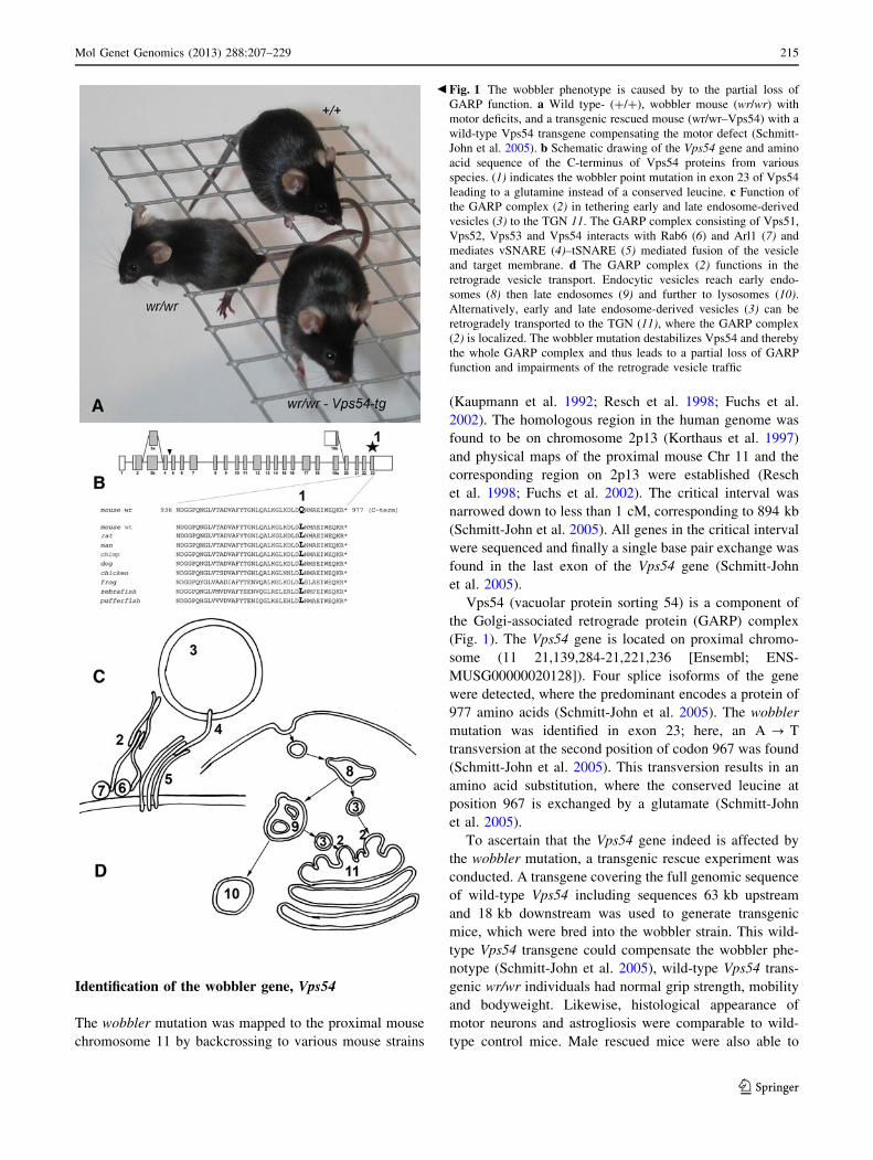
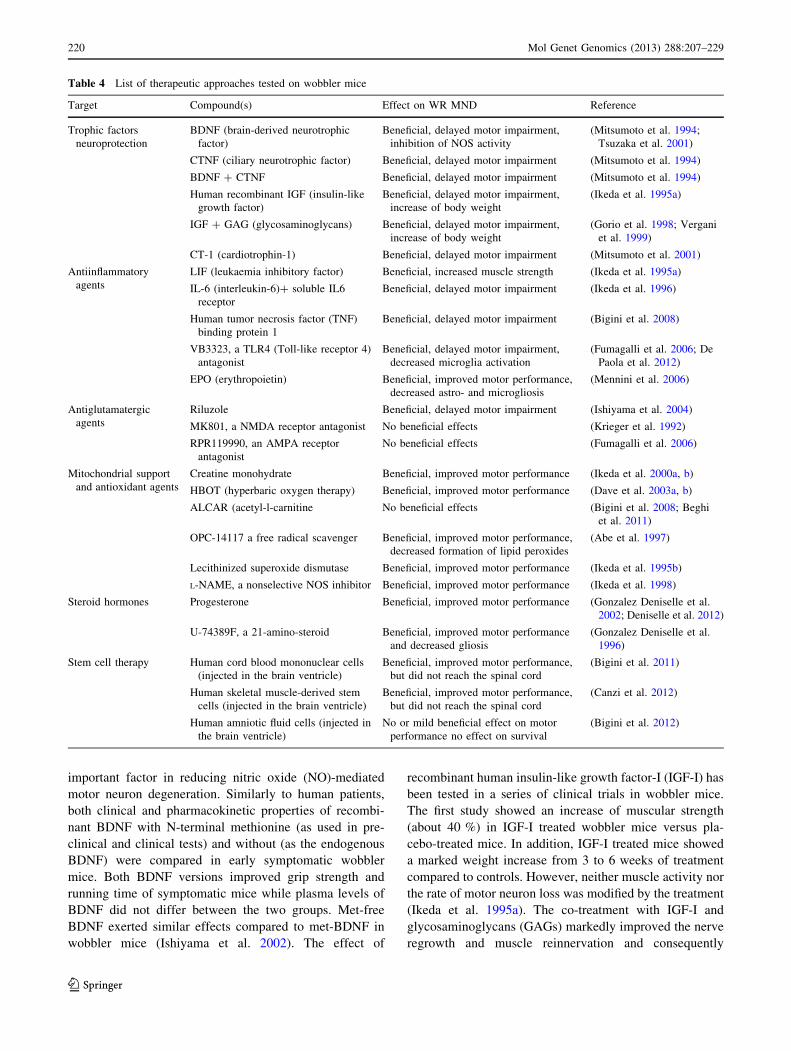
Fig. 1 The wobbler phenotype is caused by to the partial loss of
GARP function. a Wild type- (?/?), wobbler mouse (wr/wr) with
motor deficits, and a transgenic rescued mouse (wr/wr–Vps54) with a
wild-type Vps54 transgene compensating the motor defect (Schmitt-
John et al. 2005). b Schematic drawing of the Vps54 gene and amino
acid sequence of the C-terminus of Vps54 proteins from various
species. (1) indicates the wobbler point mutation in exon 23 of Vps54
leading to a glutamine instead of a conserved leucine. c Function of
the GARP complex (2) in tethering early and late endosome-derived
vesicles (3) to the TGN 11. The GARP complex consisting of Vps51,
Vps52, Vps53 and Vps54 interacts with Rab6 (6) and Arl1 (7) and
mediates vSNARE (4)–tSNARE (5) mediated fusion of the vesicle
and target membrane. d The GARP complex (2) functions in the
retrograde vesicle transport. Endocytic vesicles reach early endo-
somes (8) then late endosomes (9) and further to lysosomes (10).Alternatively, early and late endosome-derived vesicles (3) can be
retrogradely transported to the TGN (11), where the GARP complex
(2) is localized. The wobbler mutation destabilizes Vps54 and thereby
the whole GARP complex and thus leads to a partial loss of GARP
function and impairments of the retrograde vesicle traffic
b
Mol Genet Genomics (2013) 288:207–229 215
123
reproduce normally and father viable offspring (Schmitt-
John et al. 2005). This proves that Vps54 is the wobbler
gene and demonstrates that the wobbler point mutation of
Vps54 is responsible for both the neurological and the
spermatogenesis phenotype.
The wobbler point mutation of Vps54 was expected to
be a hypomorphic allele of Vps54, but not a complete loss-
of-function. Thus, a null-mutant allele was generated using
a gene trap embryonic stem cell line (Schmitt-John et al.
2005). The Vps54 gene trap clone, Vps54gt(pGT10)2841Ucd,
hence termed as Vps54b-geo, has a b-geo (b-galactosidase-
neomycin resistance fusion protein) cassette inserted
between exon 4 and 5, and the non-functional fusion pro-
tein product contains the first 152 amino acids of Vps54
fused with b-geo. The embryonic stem cells were used to
generate chimeric mice and among their offspring hetero-
zygous Vps54b-geo/? mice were obtained, which generally
had a normal phenotype (Schmitt-John et al. 2005). Matings
of heterozygous Vps54b-geo/? mice failed to produce any
homozygous Vps54b-geo/b-geo pups. Vps54b-geo/b-geo embryos
were found severely developmentally retarded at E11.5, and
at E12.5 only resorption sites with Vps54b-geo/b-geo genotype
were found, thus, Vps54-null mutation causes embryonic
lethality around day E11.5 (Schmitt-John et al. 2005). When
examining retarded Vps54b-geo/b-geo embryos at E11.5 it is
seen that the spinal cord is underdeveloped, the dorsal root
ganglia are nearly absent and severe hypoplasia is seen in the
atrial and ventricular myocardium (Schmitt-John et al.
2005). Vps54b-geo/wr compound heterozygotes display a
standard wobbler phenotype with MND and spermatogene-
sis defect, indicating that one point mutant Vps54 allele is
sufficient to rescue embryonic lethality (Schmitt-John et al.
2005).
The GARP complex in vesicle tethering
The positional cloning of the wobbler gene has connected
the ALS-like wobbler motor neurodegeneration to the par-
tial loss-of-Vps54 function (Schmitt-John et al. 2005) and
thereby to the GARP complex, of which Vps54 is a com-
ponent (Fig. 1). The function of the GARP complex has
recently been reviewed by Bonifacino and Hierro (2011).
In yeast the GARP complex was described as a vesicle-
tethering factor (Conibear and Stevens 2000; Whyte and
Munro 2002),. The GARP complex is a member of the
family of multisubunit tethering complexes (MTC) and
consists of the four subunits Vps51p, Vps52p, Vps53p and
Vps54p in a 1:1:1:1 stoichiometric ratio (Conibear et al.
2003). Due to sequence similarities and structural relation
the GARP complex belongs to the CATCHR (complexes
associated with tethering containing helical rods) group of
MTCs also comprising Dsl1-, COG- and Exocyst
complexes [reviewed in Bonifacino and Hierro (2011)].
The complex is involved in intracellular vesicular traf-
ficking and tethers vesicles derived from both early and late
endosomes to the trans Golgi network (TGN) (Quenneville
et al. 2006; Conibear et al. 2003). In yeast the C- and
N-terminal domains of Vps54p were shown to have dif-
ferent functions. The N-terminal domain is important for
GARP complex assembly and stabilization, while the
C-terminal domain facilitates localization to an early
endocytic compartment (Quenneville et al. 2006). Point
mutations of highly conserved nucleotides in the C-termi-
nal region of Vps54 do not affect retrograde transport from
late endosomes, but block early endosome recycling by
preventing localization to the polarized early endosome
(Quenneville et al. 2006).
There is a high degree of conservation between yeast
and mammalian GARP complexes. However, in yeast,
knock out of any of the subunits is non-lethal (Conibear
et al. 2003), while in mice at least-null mutation of either
Vps54 (Schmitt-John et al. 2005) or Vps53 (Schmitt-John
and Moser, unpublished results) causes embryonic lethality
around day 11 of the embryonic development and thus
might be considered a GARP-null mutation. Even though
Vps54- and Vps53-null mutant embryos die around day 11
of the embryonic development, blastocyst-derived embry-
onic stem cells and embryonic fibroblasts from 9.5-day-old
embryos are able to grow in cell culture and can be
maintained over several passages in cell culture (Schmitt-
John and Moser, unpublished results).
Interestingly, Vps52-null mutation was recently associ-
ated with a recessive t-complex mutation (tw5-lethal),
which causes a gastrulation defect and thus earlier
embryonic lethality (Sugimoto et al. 2012). This might
argue for an additional function of murine Vps52 in gas-
trulation, perhaps independent of the GARP complex.
The human GARP complex consists of VPS52, VPS53,
VPS54 (Liewen et al. 2005) and ANG2, the latter corre-
sponding to Vps51 in yeast (Perez-Victoria et al. 2010)
making the tetrameric, 1:1:1:1 complex evolutionary con-
served. RAB6, which targets vesicles between organelles,
is an interaction partner of human VPS52 just like its yeast
homolog Ypt6 interacts with Vps52 (Liewen et al. 2005).
Syntaxin 10 also interacts with the GARP complex like
Tlg1P in yeast (Liewen et al. 2005). Other SNAREs, syn-
taxin 6, syntaxin 16 and Vamp4 have been shown to
interact with the mammalian GARP complex in a direct
manner (Perez-Victoria and Bonifacino 2009). The GARP
complex seems not only to bind to these SNAREs, but also
to promote their assembly into complexes and regulating
their correct localization (Perez-Victoria and Bonifacino
2009). It was also shown that the tethering function of the
GARP complex is not dependent on the interaction of the
GARP complex with SNAREs (Perez-Victoria and
216 Mol Genet Genomics (2013) 288:207–229
123

Bonifacino 2009). It has been shown that the human GARP
complex is required for the mannose 6-phosphate-receptor-
dependent sorting of cathepsin D (CatD) to lysosomes
(Perez-Victoria et al. 2008). At the TGN mannose-6-
phosphate modified hydrolases bind to cation-dependent-
(CD-MPR) and cation-independent mannose-6-phosphate
receptors (CI-MPR) (Ghosh et al. 2003) and the complexes
are delivered to endosomes by transport carriers, where the
hydrolases are released and continue to lysosomes, while
the MPRs returned to the TGN (Ghosh et al. 2003). By
RNA interference-based depletion of subunits of the GARP
complex, it was shown that due to the impaired MPR
recycling, CatD was released into the medium of cultured
cells and leading to swollen lysosomes (Perez-Victoria
et al. 2008). Vps54 with the wobbler point mutation was
shown to be able to assemble into the GARP complex and
restore the mis-distribution of CatD and CI-MPR (Perez-
Victoria et al. 2008). The recently determined structure of
the C-terminal part of the murine Vps54-protein covering
145 amino acids (from residue 836 to 977) including the
residue where the wobbler mutation is located shows
similarities to specific domains seen in other tethering
complexes, such as the exocyst-, Dsl1- and COG-com-
plexes (Perez-Victoria et al. 2010). The critical residue,
967, seems to be involved in several hydrophobic inter-
actions with other residues. The wobbler mutation from
leucine to glutamate does not change the volume of the
residue, but it alters the hydrophobicity of the side chain
and thus is thought to cause a destabilization of Vps54 and
probably the whole GARP complex (Perez-Victoria et al.
2010). The wobbler amino acid exchange has been shown
to cause a decreased level of Vps54 protein in several
tissues of wobbler mice along with decreased levels of
Vps53 protein (Perez-Victoria et al. 2010), indicating a
higher rate of degradation of not only Vps54, but the whole
GARP complex and thus, limiting the availability of the
GARP complex for tethering endosome-derived vesicles to
the TGN (Perez-Victoria et al. 2010). Since wobbler mice
are viable, it is not surprising that the wobbler version of
Vps54 is able to maintain some functionality or that even a
low level of the GARP complex is able to uphold retro-
grade vesicle transport to some degree. However, in wob-
bler cells impairments of the retrograde vesicle transport
and missorting of proteins are seen (Schmitt-John, Moser,
unpublished data).
Cellular consequences of the wobbler mutation
The cellular effects of the wobbler mutation are summa-
rized in Fig. 2. As reported above, the ALS-like wobbler
phenotype is caused by a point mutation of Vps54, which
leads to destabilization of Vps54 protein and the whole
GARP complex and thereby to impairments of the retro-
grade vesicle transport. Thus, we have a relatively clear
conception of the primary cause of the disease and the final
effects like muscle atrophy, astrogliosis and microgliosis
can be seen as a logical consequence of the progressive
motor neuron degeneration. However, the link between
GARP malfunction and motor neuron degeneration is still
missing.
One of the obvious questions is: Why does the mutation
of a ubiquitously expressed gene, Vps54, affect predomi-
nantly motor neurons and perhaps a few other types of
neurons? Ubiquitous- or at least widespread expression is a
feature shared with most of the human fALS disease genes
and it was frequently speculated that the extraordinary
length of motor neurons might make them more susceptible
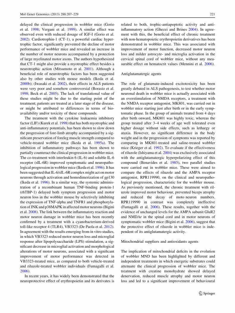
Fig. 2 Cellular effects of the wobbler mutation. The schematic
drawing show a lower motor neuron connected to skeletal muscle
cells and associated with interneurons and glial cells, such as
astrocytes, microglial cells, oligodendrocytes and Schwann cells. The
effects on the different cells, motor neuron degeneration muscle
atrophy, astrogliosis, microgliosis and the loss of GABAergic
interneurons are indicated. The effects of the dysfunction of cellular
processes are given with numbers: (1) the formation of APP- and
Rab7-positive vacuoles, (2) neurofilament aggregations, (3) impaired
axonal transport, (4) further ubiquitin-positive protein aggregates and
mitochondrial dysfunction (5)
Mol Genet Genomics (2013) 288:207–229 217
123
to minor impairments of cellular processes. However,
sensory neurons can have similar lengths and are not
affected and in wobbler mice cranial and cervical motor
neurons are the first affected ones, but they are not nec-
essarily the longest. Thus, we have to expect a more
complex explanation for this hitherto open question.
Starting from the solid ground of Vps54 being affected
by the wobbler mutation (Schmitt-John et al. 2005), leading
to a decreased stability and abundance of the GARP com-
plex (Perez-Victoria et al. 2010) in all cells of wobbler
mice, we can assume an impairment of the retrograde ves-
icle transport from endosomal compartments to the TGN.
This surely affects the sorting of proteins like MPRs and
lysosomal proteins (Perez-Victoria and Bonifacino 2009)
and probably several other proteins. This vesicle transport
defect might also explain the accumulation of enlarged
APP-positive endosomal structures, or vacuoles seen in
degenerating wobbler motor neurons and in a subset of
human sALS cases (Palmisano et al. 2011). The impaired
retrograde vesicle transport might be responsible for the
increasing size of the endosomal compartments, probably
leading to a critical imbalance in the membrane distribution
and thereby probably affecting all kinds of intracellular
transport. Defects in the fast anterograde (Mitsumoto et al.
1993) and retrograde axonal transport (Mitsumoto et al.
1990) have been reported for wobbler neurons, perhaps as a
consequence of general transport problems. Impairments of
the cellular transport systems could also explain perinuclear
neurofilament aggregation seen in wobbler mice (Pernas-
Alonso et al. 1996; Schmitt-John, Moser, unpublished data)
but also in ALS patients (Hirano et al. 1984).
Impairments of the cellular vesicle transport processes
are also expected for fALS (ALS2, ALS8, ALS11, and
ALS17) and thus might be a more general hallmark of
ALS. Transport defects might also explain the mitochon-
drial dysfunction seen in wobbler cells (Santoro et al.
2004). Impaired transport of mitochondria to the cell body
and subsequently decreased re-loading with newly syn-
thesized nucleus-encoded mitochondrial proteins might
lead to dysfunction and premature aging of mitochondria.
This might be especially critical in the distant periphery at
the neuro-muscular junction. Mitochondrial dysfunction,
transport deficits and perhaps lysosomal degradation
defects (due to missorted lysosomal proteins) all might
contribute to the accumulation of ubiquitinylated protein
aggregates including TDP-43 found wobbler motor neu-
rons (Dennis and Citron 2009).
Protein missorting might be most critical for motor
neuron survival when survival factor receptors like tyrosine
receptor kinases (TRK) or ion channels on the neuronal
surface are affected, which would directly affect the elec-
trical properties of the neurons. Recent electrophysiologi-
cal recordings have demonstrated a hyperexcitability of
layer V pyramidal neurons of the wobbler motor cortex
already prior to the onset of disease symptoms, due to
decreased GABAergic inhibition (Nieto-Gonzalez et al.
2011). Similar cortical hyperexcitability has also been
reported as an early feature of human ALS (Vucic et al.
2008, 2009) and recently decreased GABA levels have
been found in the motor cortex of ALS patients (Foerster
et al. 2012). Intuitively one might expect that the cortical
hyperexcitability in wobbler mice is caused by missorting
of GABA receptors in wobbler principal neurons, but in
contrast a decreased number of GABAergic interneurons in
the wobbler motor cortex was found (Nieto-Gonzalez et al.
2011). Thus, the decreasing numbers of GABAergic
interneurons are a very early event in the progression of the
wobbler MND, already evident in the pre-symptomatic
phase; an effect not easily connected to the reported vesicle
transport defects. However, the loss of short interval in-
tracortical inhibition seen in ALS patients has been
attributed to the degeneration of inhibitory GABAergic
interneurons (Vucic et al. 2009), which suggests the wob-
bler mouse for the investigation of this aspect.
Taken together, the wobbler motor neuron degeneration
shows most of the features reported for sALS and fALS
(Table 3), such as, mitochondrial dysfunction, transport
defects, protein aggregation but also cortical hyperexcit-
ability making it a valuable ALS animal model.
Muscle atrophy (Duchen and Strich 1968) and the
neuroinflammatory processes (Rathke-Hartlieb et al. 1999)
like astrogliosis and microgliosis are well described for the
wobbler mouse and closely resemble human ALS. These
effects are thought to be secondary effects of the neurode-
generation. However, it is still under debate to what extent
neuroinflammation is beneficial and/or harmful for motor
neuron survival in ALS (Philips and Robberecht 2011) and
the wobbler mouse. Muscle atrophy is doubtless a logical
consequence of the loss of motor neurons. Recent proteome
analysis of wobbler muscle suggests a transition from slow
to fast muscles and thus resembles muscle atrophy caused
by denervation or disuse (Staunton et al. 2011).
The cellular consequences of the wobbler mutation, like
those of sALS and fALS, are still unclear. How exactly the
vesicle transport defects, directly or indirectly cause the
observed effects like protein missorting, protein aggrega-
tion, vacuolization, mitochondrial dysfunction and hyper-
excitability and how these finally lead to motor neuron
death and in which way other cells like astrocytes,
microglial cells or interneurons contribute remains elusive.
Wobbler spermatogenesis defect
When discussing the cellular effects of the wobbler muta-
tion, spermatogenesis cannot be excluded. Unlike human
218 Mol Genet Genomics (2013) 288:207–229
123
MNDs, the wobbler mutation has a pleiotropic effect on the
spermatogenesis. Homozygous wobbler males are sterile
and produce decreased numbers (oligospermia) and
abnormally shaped round-headed sperm cells (globozoo-
spermia) with decreased motility (Heimann et al. 1991).
The positional cloning of the wobbler gene and the sub-
sequent transgenic rescue of the wobbler phenotype (Sch-
mitt-John et al. 2005) clearly demonstrated that the
wobbler point mutation of Vps54 causes both, the motor
neurodegeneration and the spermatogenesis defect and thus
suggests a role for Vps54 and probably the GARP complex
in spermatogenesis.
Spermatogenesis is the formation of haploid sperm cells,
spermatozoa from spermatogonia including meiosis and
final differentiation of the haploid spermatids to the very
specialized mature spermatozoa. The latter process, called
spermiogenesis, is affected in wobbler males. Wobbler
spermatozoa show defective acrosomes and incomplete
condensation of the nucleus, similar to the defects seen in
human globozoospermia (Heimann et al. 1991). In early
spermiogenesis, the Golgi apparatus is extremely impor-
tant. The proper formation of an acrosome depends on
vesicles and granules produced from the Golgi apparatus.
The Golgi-derived pro-acrosomal vesicles attach to the
nuclear envelope and fuse to an acrosomic granule, which
starts to flatten into a small cap over the nuclear surface.
During the acrosome formation the spermatids start to
become elongated and nuclei start to condense. In the case
of wobbler males fusion of pro-acrosomal vesicles and
nuclear condensation fails, leading to round-headed sperms
with defective acrosome, reduced motility and midpiece
defects (Heimann et al. 1991; Paiardi et al. 2011).
Vps54 has recently been shown to be involved in
acrosome formation (Berruti et al. 2010); in spermatocytes
the distribution of Vps54 is diffuse while during acrosome
formation Vps54 concentrates at the developing acrosomal
cap (Berruti et al. 2010). In this respect, Vps54 follows the
route of the endosomal sorting complex, ESCRT-0 and
USP8, a de-ubiquitinating enzyme. Male wobbler individ-
uals fail to develop Vps54-positive vesicles during acro-
some formation, Vps54 remains scattered in the cytoplasm
and no USP8-positive acrosomal structures were found in
wobbler testes (Paiardi et al. 2011). USP8 has been shown
to be involved in endosomal sorting and is highly expres-
sed not only in male germ cells, but also neuronal cells
(Berruti and Martegani 2005; Bruzzone et al. 2008). This
might suggest that endosomal traffic and acrosome for-
mation share common features and that USP8, Vps54 and
perhaps the GARP complex are important for both
processes.
Globozoospermia is a rare male infertility disorder
where in vitro fertilization fails due to the inability of the
sperms to interact with the oocyte. Even intracytoplasmic
sperm injection (ICSI) has little success, because the
injected sperm cells fail to activate the oocyte. Since
wobbler sperm resembles the human sperm defects and
also fail to activate oocytes after ICSI, the wobbler mouse
was used as an animal model to test methods for assisted
oocyte activation (AOA). After injection of wobbler sper-
matozoa in mouse oocytes, AOA leads to successful fer-
tilization (Heytens et al. 2010). Furthermore, it could be
shown that the oocyte-activating factor PLCzeta is mis-
localized on the surface of wobbler sperms, which could
explain why wobbler sperms fail to activate oocytes after
ICSI (Heytens et al. 2010). Thus, the wobbler mouse
appears to be a useful animal model not only for ALS, but
also globozoospermia and indicates shared features
between acrosome formation and retrograde vesicle trans-
port in neurons.
Towards the treatment of ALS
The rapid and reproducible progression of symptoms and
the possibility to easily monitor the evolution of motor
dysfunction makes the wobbler mouse a reliable model to
evaluate the efficacy of different pharmacological
treatments.
In the last decades many different compounds have been
tested in wobbler mice, summarized in Table 4. The ben-
eficial effects achieved by treatments with a wide range of
compounds further enforce the hypothesis of the hetero-
geneity of the ‘‘wobbler mouse disease’’. This character-
istic is an important point shared with human ALS (Beghi
et al. 2007).
To better describe the different trials approached in the
wobbler mouse, treatments will be clustered by associating
them with the different classes of drugs.
Trophic factors and anti-inflammatory agents
In the last 20 years, several pharmacological treatments
were performed with neurotrophic molecules, cytokines,
soluble receptors and/or antiinflammatory compounds in
wobbler mice. Mitsumoto and colleagues demonstrated
that co-treatment with the ciliary neurotrophic factor
(CNTF) and the brain-derived neurotrophic factor (BDNF)
hugely delayed motor impairment by directly acting on the
survival of the cervical spinal cord motor neurons, and that
even the administration of CNTF or BDNF alone was able
to slow down symptom progression, even though to at a
lesser extent (Mitsumoto et al. 1994). Further treatments
demonstrated that exogenous BDNF had a beneficial effect
by partially inhibiting the increase of nitric oxide synthase
(NOS) activity in spinal cord of wobbler mice (Tsuzaka
et al. 2001). These results suggest that the BDNF is an
Mol Genet Genomics (2013) 288:207–229 219
123
important factor in reducing nitric oxide (NO)-mediated
motor neuron degeneration. Similarly to human patients,
both clinical and pharmacokinetic properties of recombi-
nant BDNF with N-terminal methionine (as used in pre-
clinical and clinical tests) and without (as the endogenous
BDNF) were compared in early symptomatic wobbler
mice. Both BDNF versions improved grip strength and
running time of symptomatic mice while plasma levels of
BDNF did not differ between the two groups. Met-free
BDNF exerted similar effects compared to met-BDNF in
wobbler mice (Ishiyama et al. 2002). The effect of
recombinant human insulin-like growth factor-I (IGF-I) has
been tested in a series of clinical trials in wobbler mice.
The first study showed an increase of muscular strength
(about 40 %) in IGF-I treated wobbler mice versus pla-
cebo-treated mice. In addition, IGF-I treated mice showed
a marked weight increase from 3 to 6 weeks of treatment
compared to controls. However, neither muscle activity nor
the rate of motor neuron loss was modified by the treatment
(Ikeda et al. 1995a). The co-treatment with IGF-I and
glycosaminoglycans (GAGs) markedly improved the nerve
regrowth and muscle reinnervation and consequently
Table 4 List of therapeutic approaches tested on wobbler mice
Target Compound(s) Effect on WR MND Reference
Trophic factors
neuroprotection
BDNF (brain-derived neurotrophic
factor)
Beneficial, delayed motor impairment,
inhibition of NOS activity
(Mitsumoto et al. 1994;
Tsuzaka et al. 2001)
CTNF (ciliary neurotrophic factor) Beneficial, delayed motor impairment (Mitsumoto et al. 1994)
BDNF ? CTNF Beneficial, delayed motor impairment (Mitsumoto et al. 1994)
Human recombinant IGF (insulin-like
growth factor)
Beneficial, delayed motor impairment,
increase of body weight
(Ikeda et al. 1995a)
IGF ? GAG (glycosaminoglycans) Beneficial, delayed motor impairment,
increase of body weight
(Gorio et al. 1998; Vergani
et al. 1999)
CT-1 (cardiotrophin-1) Beneficial, delayed motor impairment (Mitsumoto et al. 2001)
Antiinflammatory
agents
LIF (leukaemia inhibitory factor) Beneficial, increased muscle strength (Ikeda et al. 1995a)
IL-6 (interleukin-6)? soluble IL6
receptor
Beneficial, delayed motor impairment (Ikeda et al. 1996)
Human tumor necrosis factor (TNF)
binding protein 1
Beneficial, delayed motor impairment (Bigini et al. 2008)
VB3323, a TLR4 (Toll-like receptor 4)
antagonist
Beneficial, delayed motor impairment,
decreased microglia activation
(Fumagalli et al. 2006; De
Paola et al. 2012)
EPO (erythropoietin) Beneficial, improved motor performance,
decreased astro- and microgliosis
(Mennini et al. 2006)
Antiglutamatergic
agents
Riluzole Beneficial, delayed motor impairment (Ishiyama et al. 2004)
MK801, a NMDA receptor antagonist No beneficial effects (Krieger et al. 1992)
RPR119990, an AMPA receptor
antagonist
No beneficial effects (Fumagalli et al. 2006)
Mitochondrial support
and antioxidant agents
Creatine monohydrate Beneficial, improved motor performance (Ikeda et al. 2000a, b)
HBOT (hyperbaric oxygen therapy) Beneficial, improved motor performance (Dave et al. 2003a, b)
ALCAR (acetyl-l-carnitine No beneficial effects (Bigini et al. 2008; Beghi
et al. 2011)
OPC-14117 a free radical scavenger Beneficial, improved motor performance,
decreased formation of lipid peroxides
(Abe et al. 1997)
Lecithinized superoxide dismutase Beneficial, improved motor performance (Ikeda et al. 1995b)
L-NAME, a nonselective NOS inhibitor Beneficial, improved motor performance (Ikeda et al. 1998)
Steroid hormones Progesterone Beneficial, improved motor performance (Gonzalez Deniselle et al.
2002; Deniselle et al. 2012)
U-74389F, a 21-amino-steroid Beneficial, improved motor performance
and decreased gliosis
(Gonzalez Deniselle et al.
1996)
Stem cell therapy Human cord blood mononuclear cells
(injected in the brain ventricle)
Beneficial, improved motor performance,
but did not reach the spinal cord
(Bigini et al. 2011)
Human skeletal muscle-derived stem
cells (injected in the brain ventricle)
Beneficial, improved motor performance,
but did not reach the spinal cord
(Canzi et al. 2012)
Human amniotic fluid cells (injected in
the brain ventricle)
No or mild beneficial effect on motor
performance no effect on survival
(Bigini et al. 2012)
220 Mol Genet Genomics (2013) 288:207–229
123
delayed the clinical progression in wobbler mice (Gorio
et al. 1998; Vergani et al. 1999). A similar effect was
observed even with reduced dosage of IGF-I (Gorio et al.
2002). Cardiotrophin-1 (CT-1), a powerful cardiac hyper-
trophic factor, significantly prevented the decline of motor
performance of wobbler mice and revealed an increase in
the number of motor neurons accompanied by a protection
of large myelinated motor axons. The authors hypothesized
that CT-1 might also provide a myotrophic effect besides a
neurotrophic action (Mitsumoto et al. 2001). Although a
beneficial role of neurotrophic factors has been suggested
also by other studies with mouse models (Ikeda et al.
2000b); (Iwasaki et al. 2002), their effects in ALS patients
were very poor and somehow controversial (Borasio et al.
1998; Beck et al. 2005). The lack of translational value of
these studies might be due to the starting point of the
treatment; patients are treated at a later stage of the disease,
or might be attributed to differences in terms of bio-
availability and/or toxicity of these compounds.
The treatment with the cytokine leukaemia inhibitory
factor (LIF) (Kurek et al. 1998) that has both myotrophic and
anti-inflammatory potentials, has been shown to slow down
the progression of fore-limb atrophy accompanied by a sig-
nificant preservation of foreleg muscle strength compared to
vehicle-treated wobbler mice (Ikeda et al. 1995a). The
inhibition of inflammatory pathways has been shown to
partially counteract the clinical progression in wobbler mice.
The co-treatment with interleukin 6 (IL-6) and soluble IL-6
receptor (sIL-6R) improved symptomatic and neuropatho-
logical progression in wobbler mice (Ikeda et al. 1996). It has
been suggested that IL-6/sIL-6R complex might act on motor
neurons through activation and homodimerization of gp130
(Ikeda et al. 1996). In a similar way, the systemic adminis-
tration of a recombinant human TNF-binding protein-1
(rhTBP-1) delayed both symptom progression and motor
neuron loss in the wobbler mouse by selectively inhibiting
the expression of TNF-alpha and TNFR1 and phosphoryla-
tion of JNK and p38MAPK in affected motor neurons (Bigini
et al. 2008). The link between the inflammatory reaction and
motor neuron damage in wobbler mice has been recently
confirmed by a treatment with a cyanobacterium-derived
toll-like receptor 4 (TLR4), VB3323 (De Paola et al. 2012).
In agreement with the results emerging from in vitro studies,
in which VB3323 reduced motor neuron loss and microglial
response after lipopolysaccharide (LPS) stimulation, a sig-
nificant decrease in microglial activation and morphological
alterations of motor neurons, associated with a significant
improvement of motor performance was detected in
VB3323-treated mice, as compared to both vehicle-treated
and riluzole-treated wobbler individuals (Fumagalli et al.
2006).
In recent years, it has widely been demonstrated that the
neuroprotective effect of erythropoietin and its derivates is
related to both, trophic-antiapoptotic activity and anti-
inflammatory action (Ghezzi and Brines 2004). In agree-
ment with this, the beneficial effect of chronic treatment
with non-hematopoietic erythropoietin derivatives has been
demonstrated in wobbler mice. This was associated with
improvement of motor function, decreased motor neuron
loss and milder astrocyte- and microglia activation in the
cervical spinal cord of wobbler mice, without any mea-
surable effect on hematocrit values (Mennini et al. 2006).
Antiglutamatergic agents
The role of glutamate-induced excitotoxicity has been
greatly debated in ALS pathogenesis, to test whether motor
neuronal death in wobbler mice is actually associated with
an overstimulation of NMDA receptors, a treatment with
the NMDA receptor antagonist, MK801, was carried out in
wobbler mice starting just after birth or in the early symp-
tomatic phase. In the group of animals treated from 4 days
after birth onward, MK801 was highly toxic, whereas the
group treated after 3–4 weeks of age well tolerated even
higher dosage without side effects, such as lethargy or
ataxia. However, no significant difference in the body
weight and in the progression of symptoms was reported by
comparing in MK801-treated and saline-treated wobbler
mice (Krieger et al. 1992). To evaluate if the effectiveness
of riluzole (Ishiyama et al. 2004) was exclusively associated
with the antiglutamatergic hyperpolarizing effect of this
compound (Benavides et al. 1985), two parallel studies
were carried out in wobbler mice. These were aimed to
compare the effects of riluzole and the AMPA receptor
antagonist, RPR119990, on the clinical and neuropatho-
logical progression, characteristic for the wobbler mouse.
As previously mentioned, the chronic treatment with ril-
uzole improved motor behaviour, prevented biceps atrophy
and reduced the decay of moto neuron numbers.
RPR119990 in contrast was completely ineffective
(Fumagalli et al. 2006). These results, together with the
evidence of unchanged levels for the AMPA subunit GluR2
and NMDAr in the spinal cord and in motor neurons of
symptomatic wobbler mice (Bigini et al. 2006), suggest that
the protective effect of riluzole in wobbler mice is inde-
pendent of its antiglutamatergic activity.
Mitochondrial suppliers and antioxidants agents
The implication of mitochondrial deficits in the evolution
of wobbler MND has been highlighted by different and
independent treatments in which energetic substrates could
attenuate the clinical progression of wobbler mice. The
treatment with creatine monohydrate showed delayed
denervation, reduced muscle atrophy and motor neuron
loss and led to a significant improvement of behavioural
Mol Genet Genomics (2013) 288:207–229 221
123
performance (Ikeda et al. 2000a). The link between the
efficiency of mitochondrial activity and the beneficial
effect on wobbler mice was confirmed by the evidence that
early symptomatic wobbler mice exposed to hyperbaric
oxygen therapy (HBOT) showed both delayed onset of
symptoms and significantly improved motor performance
(Dave et al. 2003b). Based on these results, the effect of
acetyl-L-carnitine (ALCAR) was tested in wobbler mice by
two different approaches. The rationale of these studies
was to connect the well-known effect of ALCAR on
mitochondrial activity (Bertamini et al. 2002) with the
neuroprotective and neurotrophic activity seen in cultured
primary motor neurons from rat embryos (Bigini et al.
2002). Unexpectedly, neither the systemic treatment
(intraperitoneal injection) and the application with mini-
pumps had any beneficial effect, nor on the clinical
symptoms and neuropathological effects in early symp-
tomatic wobbler mice (Bigini et al. 2002); (Beghi et al.
2011).
Since an impaired electron transport chain is often
accompanied by an overproduction of reactive oxygen
species, the possible beneficial effects of scavengers were
evaluated in wobbler mice. The treatment with the free
radical scavenger, OPC-14117, improved motor activity
and reduced forelimb weakness in a dose-dependent man-
ner. The mechanism appears to be associated with a
decreased formation of lipid peroxides, which might
diminish motor neuron death (Abe et al. 1997). A similar
effect was observed when treating wobbler mice with the
antioxidant lecithinized superoxide dismutase. In particu-
lar, the latter compound prevented denervation, muscle
atrophy and delayed degeneration of spinal motor neurons
in wobbler mice (Ikeda et al. 1995b). A further study,
conducted in early symptomatic wobbler mice with a
nonselective NOS inhibitor, L-NAME, sustained grip
strength and attenuated the loss of motor neurons and
disease progression (Ikeda et al. 1998).
Steroid hormones
Since the early 1990s an important contribution to the
understanding of the potential role of progesterone, steroid
receptors and glucocorticoids in enhancing degenerative
processes or in protecting motor neurons in wobbler mice
has been provided by De Nicola and colleagues. In par-
ticular, a significant improvement of muscle function was
observed in wobbler mice with a single progesterone pellet
implanted. The beneficial effect on motor performance was
associated with a marked reduction of cytoplasmic vacu-
olization in the degenerating motor neurons, a decrease of
growth-associated protein (GAP-43) mRNA expression
and mitigation of reactive gliosis (Gonzalez Deniselle et al.
2002; Deniselle et al. 2012). Another effect of progesterone
treatment was a reduced expression of nicotinamide ade-
nine dinucleotide phosphate-diaphorase (NADPHD) and
NOS in spinal cords of symptomatic wobbler mice
(Gonzalez Deniselle et al. 2005). A similar neuroprotective
function has been suggested for the 21-aminosteroid,
U-74389F, leading to increased muscle trophism, reduced
oxidative stress and decreased glial response (Gonzalez
Deniselle et al. 1996, 1999).
One of the most interesting results emerging from the
treatment with riluzole in wobbler mice was the selective
increase of BDNF immunoreactivity in cervical motor
neurons (Fumagalli et al. 2006). The evidence that the
treatment with progesterone could increase the mRNA
levels of BDNF in spinal cords of early and late symp-
tomatic wobbler mice (Gonzalez Deniselle et al. 2007)
confirmed the therapeutic value of BDNF in MND and
supports the hypothesis that the beneficial effect of riluzole
is not exclusively dependent on its antiglutamatergic
activity.
Stem cell therapy
Many further trials with different types of pharmacological
compounds, such as gangliosides, plasminogen activators,
anticholesteremic agents have been reported to be effective
in wobbler mice (Blondet et al. 1992; Bose et al. 1999;
Lisovoski et al. 1997; Iwamoto et al. 2009). However,
despite these encouraging results, the clinical trials of some
of these compounds in ALS patients had no significant effect.
Thus, new innovative strategies are required and have been
developed to the preclinical level. Among those, stem cell
(SC) therapy is considered one of the most promising
approaches for the treatment of ALS and other neurode-
generative disorders. In recent years, the wobbler mouse has
been successfully employed as an ALS model to test the
efficacy of different types of stem cells and to determine the
interaction between transplanted cells and host tissues
(biodistribution, half-life, clearance, accumulation).
Among the cell therapy approaches, the systemic
injection of human cord blood mononuclear cells (HuCB-
MNCs) has proven to reproducibly increase, the life span
of SOD1G93A mice in a dose-dependent manner (Gar-
buzova-Davis et al. 2008). In order to verify their potential
in other models of motor neuron degeneration and by other
routes of administration, HuCB-MNCs, labelled with the
nuclear dye
Hoechst 33258, were directly implanted in the lateral
brain ventricles of early symptomatic wobbler mice.
HuCB-MNCs transplantation significantly ameliorated the
symptoms and reduced the rate of motor neuron death and
gliosis (Bigini et al. 2011). The lack of Hoechst
33258-positive cells at the spinal cord level suggested that
the beneficial role of transplanted cells is not due to the cell
222 Mol Genet Genomics (2013) 288:207–229
123
replacement, but is rather associated with the production
and release of circulating protective factors. The potential
of fetal cell transplantation has also been demonstrated.
‘‘Human skeletal muscle-derived stem cells (SkmSCs)’’
showed a positive effect, after implantation in the brain
lateral ventricles of wobbler mice. Like HuCB-MNCs,
transplanted SkmSCs did not reach the ventral horns of
cervical spinal cords, but magnetic resonance imaging
(MRI) showed that some clusters of cells migrated in the
spinal parenchyma and the signal was maintained up to
18 weeks after transplantation (Canzi et al. 2012). The
evidence of a marked expression of specific human anti-
inflammatory cytokines in the brain of transplanted wob-
bler mice seems to confirm the hypothesis that a bystander
effect of soluble factors, released by SkmSCs, even far
from the affected areas, might play a neuroprotective role.
Finally, the combination of super paramagnetic iron
oxide nanoparticles (SPIOn) and Hoechst 33258 was used
to track human amniotic fluid cells (hAFCs) after trans-
plantation in the lateral ventricles of wobbler- and healthy
mice. In vivo and ex vivo approaches have shown that
hAFCs rapidly spread to the whole ventricular system, but
did not migrate into the brain parenchyma independently
from the health status of the individual mouse. The trans-
planted cells survive for a long time in the ventricles of
both wobbler and healthy mice, but had no influence on the
survival of mice and did not cause inflammation or pleo-
cytosis (Bigini et al. 2012).
Conclusions
The phenotype of the wobbler mouse closely resembles
human ALS regarding motor neuron degeneration, muscle
atrophy, reactive gliosis as well as the cellular effects like
ubiquitinated protein aggregation, oxidative stress, cortical
hyperexcitability and cellular transport defects. Thus, the
wobbler mouse models the complexity of ALS pathology
with a number of interdependent cellular defects, the pre-
cise contribution of which is currently unclear. The ALS-
like wobbler phenotype is caused by a point mutation in
Vps54, a component of the GARP vesicle-tethering com-
plex. The point mutation leads to a decreased stability of
the GARP complex and thereby to impairments in the
retrograde vesicle transport from endosomes to the TGN.
These impairments consequently lead to missorting of
proteins, accumulation of abnormally enlarged APP-posi-
tive endosomal compartments as it is also seen in a subset
of human ALS patients. Even though there is no gap-less
chain of evidence, it appears plausible that the impairment
of the retrograde vesicle traffic leads to disturbance of
several cellular processes such axonal protein- and orga-
nelle transport and the final consequences could be protein
aggregation and mitochondrial dysfunction. The therapeu-
tic interventions tested on wobbler mice have also con-
tributed to the understanding of the complex pathology of
ALS. The beneficial effects seen for anti-inflammatory-
and antioxidant agents indicate that neuroinflammation and
oxidative stress also plays a critical role in the wobbler
motor neuron degeneration, while the poor effect of an-
tiglutamatergic compounds might argue that glutamate-
associated excitotoxicity plays a minor role. Our recent
finding of hyperexcitability due to a reduced number of
GABAergic interneurons in wobbler CNS, even prior to the
disease onset appears to be more difficult to explain.
However, similar cortical hyperexcitability has been
reported in human ALS cases, also already prior to the
onset of disease symptoms and has been attributed to the
degeneration of inhibitory circuits (Vucic et al. 2009). Loss
or degeneration of inhibitory neurons appears to be an early
event in the pathology and thus might have a diagnostic
and a therapeutic impact, both of which could be addressed
using the wobbler mouse. Thus, the wobbler mouse is, even
57 years after its first description, still a valuable animal
model for human MNDs such as ALS, in order to unravel
the disease mechanism and in order to develop and test new
diagnostic and therapeutic approaches.
Acknowledgments Authors are grateful to Dr. Ester Zito (Institute
Mario Negri) and Associate Professor Ernst-Martin Fuchtbauer
(Molecular Biology and Genetics, Aarhus University) for careful
reading and helpful comments on the manuscript and Tobias John for
help with the figures.
Open Access This article is distributed under the terms of the
Creative Commons Attribution License which permits any use, dis-
tribution, and reproduction in any medium, provided the original
author(s) and the source are credited.
References
Abe K, Morita S, Kikuchi T, Itoyama Y (1997) Protective effect of a
novel free radical scavenger, OPC-14117, on wobbler mouse
motor neuron disease. J Neurosci Res 48:63–70
Achi EY, Rudnicki SA (2012) ALS and frontotemporal dysfunction: a
review. Neurol Res Int 2012:806306
Ackerley S, Grierson AJ, Brownlees J, Thornhill P, Anderton BH et al
(2000) Glutamate slows axonal transport of neurofilaments in
transfected neurons. J Cell Biol 150:165–176
Agbulut O, Noirez P, Butler-Browne G, Jockusch H (2004) Specific
isomyosin proportions in hyperexcitable and physiologically
denervated mouse muscle. FEBS Lett 561:191–194
Al-Saif A, Al-Mohanna F, Bohlega S (2011) A mutation in sigma-1
receptor causes juvenile amyotrophic lateral sclerosis. Ann
Neurol 70:913–919
Andrews JM, Gardner MB, Wolfgram FJ, Ellison GW, Porter DD,
Brandkamp WW (1974) Studies on a murine form of sponta-
neous lower motor neuron degeneration—the wobbler (wa)
mouse. Am J Pathol 76:63–78
Atkin JD, Farg MA, Turner BJ, Tomas D, Lysaght JA et al (2006)
Induction of the unfolded protein response in familial
Mol Genet Genomics (2013) 288:207–229 223
123
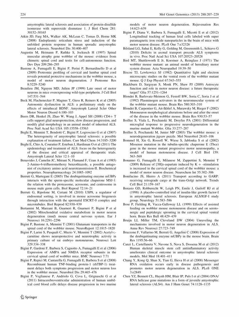
amyotrophic lateral sclerosis and association of protein-disulfide
isomerase with superoxide dismutase 1. J Biol Chem 281:
30152–30165
Atkin JD, Farg MA, Walker AK, McLean C, Tomas D, Horne MK
(2008) Endoplasmic reticulum stress and induction of the
unfolded protein response in human sporadic amyotrophic
lateral sclerosis. Neurobiol Dis 30:400–407
Augustin M, Heimann P, Rathke S, Jockusch H (1997) Spinal
muscular atrophy gene wobbler of the mouse: evidence from
chimeric spinal cord and testis for cell-autonomous function.
Dev Dyn 209:286–295
Bastone A, Fumagalli E, Bigini P, Perini P, Bernardinello D et al
(2009) Proteomic profiling of cervical and lumbar spinal cord
reveals potential protective mechanisms in the wobbler mouse, a
model of motor neuron degeneration. J Proteome Res
8:5229–5240
Beaulieu JM, Nguyen MD, Julien JP (1999) Late onset of motor
neurons in mice overexpressing wild-type peripherin. J Cell Biol
147:531–544
Beck M, Flachenecker P, Magnus T, Giess R, Reiners K et al (2005)
Autonomic dysfunction in ALS: a preliminary study on the
effects of intrathecal BDNF. Amyotroph Lateral Scler Other
Motor Neuron Disord 6:100–103
Beers DR, Henkel JS, Zhao W, Wang J, Appel SH (2008) CD4? T
cells support glial neuroprotection, slow disease progression, and
modify glial morphology in an animal model of inherited ALS.
Proc Natl Acad Sci USA 105:15558–15563
Beghi E, Mennini T, Bendotti C, Bigini P, Logroscino G et al (2007)
The heterogeneity of amyotrophic lateral sclerosis: a possible
explanation of treatment failure. Curr Med Chem 14:3185–3200
Beghi E, Chio A, Couratier P, Esteban J, Hardiman O et al (2011) The
epidemiology and treatment of ALS: focus on the heterogeneity
of the disease and critical appraisal of therapeutic trials.
Amyotroph Lateral Scler 12:1–10
Benavides J, Camelin JC, Mitrani N, Flamand F, Uzan A et al (1985)
2-Amino-6-trifluoromethoxy benzothiazole, a possible antago-
nist of excitatory amino acid neurotransmission-II. Biochemical
properties. Neuropharmacology 24:1085–1092
Berruti G, Martegani E (2005) The deubiquitinating enzyme mUBPy
interacts with the sperm-specific molecular chaperone MSJ-1:
the relation with the proteasome, acrosome, and centrosome in
mouse male germ cells. Biol Reprod 72:14–21
Berruti G, Ripolone M, Ceriani M (2010) USP8, a regulator of
endosomal sorting, is involved in mouse acrosome biogenesis
through interaction with the spermatid ESCRT-0 complex and
microtubules. Biol Reprod 82:930–939
Bertamini M, Marzani B, Guarneri R, Guarneri P, Bigini P et al
(2002) Mitochondrial oxidative metabolism in motor neuron
degeneration (mnd) mouse central nervous system. Eur J
Neurosci 16:2291–2296
Bigini P, Bastone A, Mennini T (2001) Glutamate transporters in the
spinal cord of the wobbler mouse. NeuroReport 12:1815–1820
Bigini P, Larini S, Pasquali C, Muzio V, Mennini T (2002) Acetyl-L-
carnitine shows neuroprotective and neurotrophic activity in
primary culture of rat embryo motoneurons. Neurosci Lett
329:334–338
Bigini P, Gardoni F, Barbera S, Cagnotto A, Fumagalli E et al (2006)
Expression of AMPA and NMDA receptor subunits in the
cervical spinal cord of wobbler mice. BMC Neurosci 7:71
Bigini P, Repici M, Cantarella G, Fumagalli E, Barbera S et al (2008)
Recombinant human TNF-binding protein-1 (rhTBP-1) treat-
ment delays both symptoms progression and motor neuron loss
in the wobbler mouse. Neurobiol Dis 29:465–476
Bigini P, Veglianese P, Andriolo G, Cova L, Grignaschi G et al
(2011) Intracerebroventricular administration of human umbil-
ical cord blood cells delays disease progression in two murine
models of motor neuron degeneration. Rejuvenation Res
14:623–639
Bigini P, Diana V, Barbera S, Fumagalli E, Micotti E et al (2012)
Longitudinal tracking of human fetal cells labeled with super
paramagnetic iron oxide nanoparticles in the brain of mice with
motor neuron disease. PLoS One 7:e32326
Bilsland LG, Sahai E, Kelly G, Golding M, Greensmith L, Schiavo G
(2010) Deficits in axonal transport precede ALS symptoms
in vivo. Proc Natl Acad Sci USA 107:20523–20528
Bird MT, Shuttleworth E Jr, Koestner A, Reinglass J (1971) The
wobbler mouse mutant: an animal model of hereditary motor
system disease. Acta Neuropathol 19:39–50
Biscoe TJ, Lewkowicz SJ (1982) Quantitative light and electron
microscopic studies on the ventral roots of the wobbler mutant
mouse. Q J Exp Physiol 67:543–555
Blackburn D, Sargsyan S, Monk PN, Shaw PJ (2009) Astrocyte
function and role in motor neuron disease: a future therapeutic
target? Glia 57:1251–1264
Blondet B, Barlovatz-Meimon G, Festoff BW, Soria C, Soria J et al
(1992) Plasminogen activators in the neuromuscular system of
the wobbler mutant mouse. Brain Res 580:303–310
Blondet B, Carpentier G, Ait-Ikhlef A, Murawsky M, Rieger F (2002)
Motoneuron morphological alterations before and after the onset
of the disease in the wobbler mouse. Brain Res 930:53–57
Boillee S, Viala L, Peschanski M, Dreyfus PA (2001) Differential
microglial response to progressive neurodegeneration in the
murine mutant Wobbler. Glia 33:277–287
Boillee S, Peschanski M, Junier MP (2003) The wobbler mouse: a
neurodegeneration jigsaw puzzle. Mol Neurobiol 28:65–106
Bommel H, Xie G, Rossoll W, Wiese S, Jablonka S et al (2002)
Missense mutation in the tubulin-specific chaperone E (Tbce)
gene in the mouse mutant progressive motor neuronopathy, a
model of human motoneuron disease. J Cell Biol 159:
563–569
Bonanno G, Fumagalli E, Milanese M, Zappettini S, Mennini T
(2009) Release of [3H]D-aspartate induced by K ? -stimulation
is increased in the cervical spinal cord of the wobbler mouse: a
model of motor neuron disease. Neurochem Int 55:302–306
Bonifacino JS, Hierro A (2011) Transport according to GARP:
receiving retrograde cargo at the trans-Golgi network. Trends
Cell Biol 21:159–167
Borasio GD, Robberecht W, Leigh PN, Emile J, Guiloff RJ et al
(1998) A placebo-controlled trial of insulin-like growth factor-I
in amyotrophic lateral sclerosis. European ALS/IGF-I study
group. Neurology 51:583–586
Bose P, Fielding R, Vacca-Galloway LL (1999) Effects of assisted
feeding on wobbler mouse motoneuron disease and on seroto-
nergic and peptidergic sprouting in the cervical spinal ventral
horn. Brain Res Bull 48:429–439
Bruijn LI, Miller TM, Cleveland DW (2004) Unraveling the
mechanisms involved in motor neuron degeneration in ALS.
Annu Rev Neurosci 27:723–749
Bruzzone F, Vallarino M, Berruti G, Angelini C (2008) Expression of
the deubiquitinating enzyme mUBPy in the mouse brain. Brain
Res 1195:56–66
Canzi L, Castellaneta V, Navone S, Nava S, Dossena M et al (2012)
Human skeletal muscle stem cell antiinflammatory activity
ameliorates clinical outcome in amyotrophic lateral sclerosis
models. Mol Med 18:401–411
Chang Y, Kong Q, Shan X, Tian G, Ilieva H et al (2008) Messenger
RNA oxidation occurs early in disease pathogenesis and
promotes motor neuron degeneration in ALS. PLoS ONE
3:e2849
Chen YZ, Bennett CL, Huynh HM, Blair IP, Puls I et al (2004) DNA/
RNA helicase gene mutations in a form of juvenile amyotrophic
lateral sclerosis (ALS4). Am J Hum Genet 74:1128–1135
224 Mol Genet Genomics (2013) 288:207–229
123
Chen HJ, Anagnostou G, Chai A, Withers J, Morris A et al (2010)
Characterization of the properties of a novel mutation in VAPB
in familial amyotrophic lateral sclerosis. J Biol Chem
285:40266–40281
Chow CY, Landers JE, Bergren SK, Sapp PC, Grant AE et al (2009)
Deleterious variants of FIG4, a phosphoinositide phosphatase, in
patients with ALS. Am J Hum Genet 84:85–88
Clement AM, Nguyen MD, Roberts EA, Garcia ML, Boillee S et al
(2003) Wild-type nonneuronal cells extend survival of SOD1
mutant motor neurons in ALS mice. Science 302:113–117
Conibear E, Stevens TH (2000) Vps52p, Vps53p, and Vps54p form a
novel multisubunit complex required for protein sorting at the
yeast late Golgi. Mol Biol Cell 11:305–323
Conibear E, Cleck JN, Stevens TH (2003) Vps51p mediates the
association of the GARP (Vps52/53/54) complex with the late
Golgi t-SNARE Tlg1p. Mol Biol Cell 14:1610–1623
Cote F, Collard JF, Julien JP (1993) Progressive neuronopathy in
transgenic mice expressing the human neurofilament heavy gene:
a mouse model of amyotrophic lateral sclerosis. Cell 73:35–46
Cox LE, Ferraiuolo L, Goodall EF, Heath PR, Higginbottom A et al
(2010) Mutations in CHMP2B in lower motor neuron predom-
inant amyotrophic lateral sclerosis (ALS). PLoS One 5:e9872
Damiano M, Starkov AA, Petri S, Kipiani K, Kiaei M et al (2006)
Neural mitochondrial Ca2? capacity impairment precedes the
onset of motor symptoms in G93A Cu/Zn-superoxide dismutase
mutant mice. J Neurochem 96:1349–1361
Dave KR, Bradley WG, Perez-Pinzon MA (2003a) Early mitochon-
drial dysfunction occurs in motor cortex and spinal cord at the
onset of disease in the Wobbler mouse. Exp Neurol 182:412–420
Dave KR, Prado R, Busto R, Raval AP, Bradley WG et al (2003b)
Hyperbaric oxygen therapy protects against mitochondrial
dysfunction and delays onset of motor neuron disease in
Wobbler mice. Neuroscience 120:113–120
Dave KR, Raval AP, Purroy J, Kirkinezos IG, Moraes CT et al (2005)
Aberrant deltaPKC activation in the spinal cord of Wobbler
mouse: a model of motor neuron disease. Neurobiol Dis
18:126–133
De Paola M, Mariani A, Bigini P, Peviani M, Ferrara G et al (2012)
Neuroprotective effects of toll-like receptor 4 antagonism in
spinal cord cultures and in a mouse model of motor neuron
degeneration. Mol Med 18:971–981
De Vos KJ, Chapman AL, Tennant ME, Manser C, Tudor EL et al
(2007) Familial amyotrophic lateral sclerosis-linked SOD1
mutants perturb fast axonal transport to reduce axonal mito-
chondria content. Hum Mol Genet 16:2720–2728
DeJesus-Hernandez M, Mackenzie IR, Boeve BF, Boxer AL, Baker
M et al (2011) Expanded GGGGCC hexanucleotide repeat in
noncoding region of C9ORF72 causes chromosome 9p-linked
FTD and ALS. Neuron 72:245–256
Deng HX, Chen W, Hong ST, Boycott KM, Gorrie GH et al (2011)
Mutations in UBQLN2 cause dominant X-linked juvenile and
adult-onset ALS and ALS/dementia. Nature 477:211–215
Deniselle MC, Carreras MC, Garay L, Gargiulo-Monachelli G, Meyer
M et al (2012) Progesterone prevents mitochondrial dysfunction
in the spinal cord of wobbler mice. J Neurochem 122:185–195
Dennis JS, Citron BA (2009) Wobbler mice modeling motor neuron
disease display elevated transactive response DNA binding
protein. Neuroscience 158:745–750
Devon RS, Orban PC, Gerrow K, Barbieri MA, Schwab C et al (2006)
Als2-deficient mice exhibit disturbances in endosome trafficking
associated with motor behavioral abnormalities. Proc Natl Acad
Sci USA 103:9595–9600
Di Giorgio FP, Boulting GL, Bobrowicz S, Eggan KC (2008) Human
embryonic stem cell-derived motor neurons are sensitive to the
toxic effect of glial cells carrying an ALS-causing mutation. Cell
Stem Cell 3:637–648
Diana V, Ottolina A, Botti F, Fumagalli E, Calcagno E et al (2010)
Neural precursor-derived astrocytes of wobbler mice induce
apoptotic death of motor neurons through reduced glutamate
uptake. Exp Neurol 225:163–172
Dormann D, Rodde R, Edbauer D, Bentmann E, Fischer I et al (2010)
ALS-associated fused in sarcoma (FUS) mutations disrupt
Transportin-mediated nuclear import. EMBO J 29:2841–2857
Duan W, Li X, Shi J, Guo Y, Li Z, Li C (2010) Mutant TAR DNA-
binding protein-43 induces oxidative injury in motor neuron-like
cell. Neuroscience 169:1621–1629
Duchen LW, Strich SJ (1968) An hereditary motor neuron disease
with progressive denervation of muscle in mouse: the mutant
‘‘wobbler’’. J Neurol Neurosurg Psychiat 31:535–542
Duffy LM, Chapman AL, Shaw PJ, Grierson AJ (2011) Review: the
role of mitochondria in the pathogenesis of amyotrophic lateral
sclerosis. Neuropathol Appl Neurobiol 37:336–352
Falconer DS (1956) Wobbler (wr). Mouse News Lett 15:22
Ferraiuolo L, Kirby J, Grierson AJ, Sendtner M, Shaw PJ (2011)
Molecular pathways of motor neuron injury in amyotrophic
lateral sclerosis. Nat Rev Neurol 7:616–630
Fitzmaurice PS, Shaw IC, Kleiner HE, Miller RT, Monks TJ et al
(1996) Evidence for DNA damage in amyotrophic lateral
sclerosis. Muscle Nerve 19:797–798
Foerster BR, Callaghan BC, Petrou M, Edden RA, Chenevert TL,
Feldman EL (2012) Decreased motor cortex gamma-aminobu-
tyric acid in amyotrophic lateral sclerosis. Neurology
78:1596–1600
Fray AE, Ince PG, Banner SJ, Milton ID, Usher PA et al (1998) The
expression of the glial glutamate transporter protein EAAT2 in
motor neuron disease: an immunohistochemical study. Eur J
Neurosci 10:2481–2489
Fuchs S, Resch K, Thiel C, Ulbrich M, Platzer M et al (2002)
Comparative transcription map of the wobbler critical region on
mouse chromosome 11 and the homologous region on human
chromosome 2p13-14. BMC Genet 3:14
Fumagalli E, Bigini P, Barbera S, De Paola M, Mennini T (2006)
Riluzole, unlike the AMPA antagonist RPR119990, reduces
motor impairment and partially prevents motoneuron death in
the wobbler mouse, a model of neurodegenerative disease. Exp
Neurol 198:114–128
Garbuzova-Davis S, Sanberg CD, Kuzmin-Nichols N, Willing AE,
Gemma C et al (2008) Human umbilical cord blood treatment in
a mouse model of ALS: optimization of cell dose. PLoS One
3:e2494
Ghezzi P, Brines M (2004) Erythropoietin as an antiapoptotic, tissue-
protective cytokine. Cell Death Differ 11(Suppl 1):S37–S44
Ghosh P, Griffith J, Geuze HJ, Kornfeld S (2003) Mammalian GGAs
act together to sort mannose 6-phosphate receptors. J Cell Biol
163:755–766
Giordana MT, Piccinini M, Grifoni S, De Marco G, Vercellino M et al
(2010) TDP-43 redistribution is an early event in sporadic
amyotrophic lateral sclerosis. Brain Pathol 20:351–360
Gonzalez Deniselle MC, Gonzalez SL, Piroli GG, Lima AE, De
Nicola AF (1996) The 21-aminosteroid U-74389F increases the
number of glial fibrillary acidic protein-expressing astrocytes in
the spinal cord of control and Wobbler mice. Cell Mol Neurobiol
16:61–72
Gonzalez Deniselle MC, Gonzalez SL, Lima AE, Wilkin G, De
Nicola AF (1999) The 21-aminosteroid U-74389F attenuates
hyperexpression of GAP-43 and NADPH-diaphorase in the
spinal cord of wobbler mouse, a model for amyotrophic lateral
sclerosis. Neurochem Res 24:1–8
Gonzalez Deniselle MC, Lopez-Costa JJ, Saavedra JP, Pietranera L,
Gonzalez SL et al (2002) Progesterone neuroprotection in the
wobbler mouse, a genetic model of spinal cord motor neuron
disease. Neurobiol Dis 11:457–468
Mol Genet Genomics (2013) 288:207–229 225
123
Gonzalez Deniselle MC, Garay L, Gonzalez S, Guennoun R,
Schumacher M, De Nicola AF (2005) Progesterone restores
retrograde labeling of cervical motoneurons in Wobbler mouse
motoneuron disease. Exp Neurol 195:518–523
Gonzalez Deniselle MC, Garay L, Gonzalez S, Saravia F, Labom-
barda F et al (2007) Progesterone modulates brain-derived
neurotrophic factor and choline acetyltransferase in degenerating
Wobbler motoneurons. Exp Neurol 203:406–414
Gorio A, Vergani L, Lesma E, Di Giulio AM (1998) Neuroprotection,
neuroregeneration, and interaction with insulin-like growth
factor-I: novel non-anticoagulant action of glycosaminoglycans.
J Neurosci Res 51:559–562
Gorio A, Lesma E, Madaschi L, Di Giulio AM (2002) Co-
administration of IGF-I and glycosaminoglycans greatly delays
motor neurone disease and affects IGF-I expression in the
wobbler mouse: a long-term study. J Neurochem 81:194–202
Greenway MJ, Andersen PM, Russ C, Ennis S, Cashman S et al
(2006) ANG mutations segregate with familial and ‘sporadic’
amyotrophic lateral sclerosis. Nat Genet 38:411–413
Groen EJ, van Es MA, van Vught PW, Spliet WG, van Engelen-Lee J
et al (2010) FUS mutations in familial amyotrophic lateral
sclerosis in the Netherlands. Arch Neurol 67:224–230
Gros-Louis F, Kriz J, Kabashi E, McDearmid J, Millecamps S et al
(2008) Als2 mRNA splicing variants detected in KO mice rescue
severe motor dysfunction phenotype in Als2 knock-down
zebrafish. Hum Mol Genet 17:2691–2702
Guo H, Lai L, Butchbach ME, Stockinger MP, Shan X et al (2003)
Increased expression of the glial glutamate transporter EAAT2
modulates excitotoxicity and delays the onset but not the
outcome of ALS in mice. Hum Mol Genet 12:2519–2532
Gurney ME, Pu H, Chiu AY, Dal Canto MC, Polchow CY et al (1994)
Motor neuron degeneration in mice that express a human Cu, Zn
superoxide dismutase mutation. Science 264:1772–1775
Hand CK, Khoris J, Salachas F, Gros-Louis F, Lopes AA et al (2002)
A novel locus for familial amyotrophic lateral sclerosis, on
chromosome 18q. Am J Hum Genet 70:251–256
Heimann P, Laage S, Jockusch H (1991) Defect of sperm assembly in
a neurological mutant of the mouse, wobbler (WR). Differen-
tiation 47:77–83
Hentati A, Ouahchi K, Pericak-Vance MA, Nijhawan D, Ahmad A
et al (1998) Linkage of a commoner form of recessive
amyotrophic lateral sclerosis to chromosome 15q15–q22 mark-
ers. Neurogenetics 2:55–60
Hewitt C, Kirby J, Highley JR, Hartley JA, Hibberd R et al (2010)
Novel FUS/TLS mutations and pathology in familial and
sporadic amyotrophic lateral sclerosis. Arch Neurol 67:455–461
Heytens E, Schmitt-John T, Moser JM, Mandøe Jensen N, Soleimani
R et al (2010) Reduced fertilization after ICSI and PLCzeta in
sperm from the Wobbler mouse. RBM-online 21:742–749
Hirano A, Donnenfeld H, Sasaki S, Nakano I (1984) Fine structural
observations of neurofilamentous changes in amyotrophic lateral
sclerosis. J Neuropathol Exp Neurol 43:461–470
Hitomi J, Katayama T, Eguchi Y, Kudo T, Taniguchi M et al (2004)
Involvement of caspase-4 in endoplasmic reticulum stress-
induced apoptosis and Abeta-induced cell death. J Cell Biol
165:347–356
Ikeda K, Iwasaki Y, Tagaya N, Shiojima T, Kinoshita M (1995a)
Neuroprotective effect of cholinergic differentiation factor/
leukemia inhibitory factor on wobbler murine motor neuron
disease. Muscle Nerve 18:1344–1347
Ikeda K, Kinoshita M, Iwasaki Y, Tagaya N, Shiojima T (1995b)
Lecithinized superoxide dismutase retards wobbler mouse
motoneuron disease. Neuromuscul Disord 5:383–390
Ikeda K, Kinoshita M, Tagaya N, Shiojima T, Taga T et al (1996)
Coadministration of interleukin-6 (IL-6) and soluble IL-6
receptor delays progression of wobbler mouse motor neuron
disease. Brain Res 726:91–97
Ikeda K, Iwasaki Y, Kinoshita M (1998) Neuronal nitric oxide
synthase inhibitor, 7-nitroindazole, delays motor dysfunction and
spinal motoneuron degeneration in the wobbler mouse. J Neurol
Sci 160:9–15
Ikeda K, Iwasaki Y, Kinoshita M (2000a) Oral administration of
creatine monohydrate retards progression of motor neuron
disease in the wobbler mouse. Amyotroph Lateral Scler Other
Motor Neuron Disord 1:207–212
Ikeda K, Iwasaki Y, Kinoshita M, Marubuchi S, Ono S (2000b)
T-588, a novel neuroprotective agent, delays progression of
neuromuscular dysfunction in wobbler mouse motoneuron
disease. Brain Res 858:84–91
Ince P, Stout N, Shaw P, Slade J, Hunziker W et al (1993)
Parvalbumin and Calbindin D-28k in the human motor system
and in motor neuron disease. Neuropathol Appl Neurobiol
19:291–299
Ince PG, Tomkins J, Slade JY, Thatcher NM, Shaw PJ (1998)
Amyotrophic lateral sclerosis associated with genetic abnormal-
ities in the gene encoding Cu/Zn superoxide dismutase: molec-
ular pathology of five new cases, and comparison with previous
reports and 73 sporadic cases of ALS. J Neuropathol Exp Neurol
57:895–904
Ishiyama T, Ogo H, Wong V, Klinkosz B, Noguchi H et al (2002)
Methionine-free brain-derived neurotrophic factor in wobbler
mouse motor neuron disease: dose-related effects and compar-
ison with the methionyl form. Brain Res 944:195–199
Ishiyama T, Okada R, Nishibe H, Mitsumoto H, Nakayama C (2004)
Riluzole slows the progression of neuromuscular dysfunction in
the wobbler mouse motor neuron disease. Brain Res 1019:
226–236
Ito D, Seki M, Tsunoda Y, Uchiyama H, Suzuki N (2011) Nuclear
transport impairment of amyotrophic lateral sclerosis-linked
mutations in FUS/TLS. Ann Neurol 69:152–162
Iwamoto K, Yoshii Y, Ikeda K (2009) Atorvastatin treatment
attenuates motor neuron degeneration in wobbler mice. Amyo-
troph Lateral Scler 10:405–409
Iwasaki Y, Ikeda K, Ichikawa Y, Igarashi O, Kinoshita M et al (2002)
T-588 enhances neurite outgrowth and choline acetyltransferase
in cultured rat spinal ventral horn neurons. Neurochem Res
27:225–228
Johnson JO, Mandrioli J, Benatar M, Abramzon Y, Van Deerlin VM
et al (2010) Exome sequencing reveals VCP mutations as a cause
of familial ALS. Neuron 68:857–864
Kabashi E, Lin L, Tradewell ML, Dion PA, Bercier V et al (2010)
Gain and loss of function of ALS-related mutations of TARDBP
(TDP-43) cause motor deficits in vivo. Hum Mol Genet
19:671–683
Kanekura K, Suzuki H, Aiso S, Matsuoka M (2009) ER stress and
unfolded protein response in amyotrophic lateral sclerosis. Mol
Neurobiol 39:81–89
Kaufman RJ (2002) Orchestrating the unfolded protein response in
health and disease. J Clin Invest 110:1389–1398
Kaupmann K, Simon-Chazottes D, Guenet JL, Jockusch H (1992)
Wobbler, a mutation affecting motoneuron survival and gonadal
functions in the mouse, maps to proximal chromosome 11.
Genomics 13:39–43
Kiaei M, Kipiani K, Calingasan NY, Wille E, Chen J et al (2007)
Matrix metalloproteinase-9 regulates TNF-alpha and FasL
expression in neuronal, glial cells and its absence extends life
in a transgenic mouse model of amyotrophic lateral sclerosis.
Exp Neurol 205:74–81
Korthaus D, Wedemeyer N, Lengeling A, Ronsiek M, Jockusch H,
Schmitt-John T (1997) Integrated radiation hybrid map of human
226 Mol Genet Genomics (2013) 288:207–229
123
chromosome 2p13: possible involvement of dynactin in neuro-
muscular diseases. Genomics 43:242–244
Kraemer BC, Schuck T, Wheeler JM, Robinson LC, Trojanowski JQ
et al (2010) Loss of murine TDP-43 disrupts motor function and
plays an essential role in embryogenesis. Acta Neuropathol
119:409–419
Krieger C, Perry TL, Hansen S, Mitsumoto H (1991) The wobbler
mouse: amino acid contents in brain and spinal cord. Brain Res
551:142–144
Krieger C, Perry TL, Hansen S, Mitsumoto H, Honore T (1992)
Excitatory amino acid receptor antagonist in murine motoneuron
disease (the wobbler mouse). Can J Neurol Sci 19:462–465
Kuhle J, Lindberg RL, Regeniter A, Mehling M, Steck AJ et al (2009)
Increased levels of inflammatory chemokines in amyotrophic
lateral sclerosis. Eur J Neurol 16:771–774
Kurek JB, Radford AJ, Crump DE, Bower JJ, Feeney SJ et al (1998)
LIF (AM424), a promising growth factor for the treatment of
ALS. J Neurol Sci 160(Suppl 1):S106–S113
Kwak S, Hideyama T, Yamashita T, Aizawa H (2010) AMPA
receptor-mediated neuronal death in sporadic ALS. Neuropa-
thology 30:182–188
Kwiatkowski TJ Jr, Bosco DA, Leclerc AL, Tamrazian E, Vander-
burg CR et al (2009) Mutations in the FUS/TLS gene on
chromosome 16 cause familial amyotrophic lateral sclerosis.
Science 323:1205–1208
Lai C, Xie C, McCormack SG, Chiang HC, Michalak MK et al (2006)
Amyotrophic lateral sclerosis 2-deficiency leads to neuronal
degeneration in amyotrophic lateral sclerosis through altered
AMPA receptor trafficking. J Neurosci 26:11798–11806
LaVail JH, Koo EH, Dekker NP (1987) Motoneuron loss in the
abducens nucleus of wobbler mice. Brain Res 404:127–132
Leestma JE, Sepsenwol S (1980) Sperm tail axoneme alterations in
the wobbler mouse. J Reprod Fertil 58:267–270
Lemmens R, Van Hoecke A, Hersmus N, Geelen V, D’Hollander I
et al (2007) Overexpression of mutant superoxide dismutase 1
causes a motor axonopathy in the zebrafish. Hum Mol Genet
16:2359–2365
Lewkowicz SJ (1979) The relationship of Schwann cell migration
in vitro to injury, using normal, wobbler, and dystrophic mice.
Brain Res 169:443–454
Liao B, Zhao W, Beers DR, Henkel JS, Appel SH (2012) Transfor-
mation from a neuroprotective to a neurotoxic microglial
phenotype in a mouse model of ALS. Exp Neurol 237:147–152
Liewen H, Meinhold-Heerlein I, Oliveira V, Schwarzenbacher R, Luo
G et al (2005) Characterization of the human GARP (Golgi
associated retrograde protein) complex. Exp Cell Res 306:24–34
Lisovoski F, Blot S, Lacombe C, Bellier JP, Dreyfus PA, Junier MP
(1997) Transforming growth factor alpha expression as a
response of murine motor neurons to axonal injury and
mutation-induced degeneration. J Neuropathol Exp Neurol
56:459–471
Liu-Yesucevitz L, Bilgutay A, Zhang YJ, Vanderweyde T, Citro A
et al (2010) Tar DNA binding protein-43 (TDP-43) associates
with stress granules: analysis of cultured cells and pathological
brain tissue. PLoS One 5:e13250
Mackenzie IR, Rademakers R, Neumann M (2010) TDP-43 and FUS
in amyotrophic lateral sclerosis and frontotemporal dementia.
Lancet Neurol 9:995–1007
Martin N, Jaubert J, Gounon P, Salido E, Haase G et al (2002) A
missense mutation in Tbce causes progressive motor neuronop-
athy in mice. Nat Genet 32:443–447
Maruyama H, Morino H, Ito H, Izumi Y, Kato H et al (2010)
Mutations of optineurin in amyotrophic lateral sclerosis. Nature
465:223–226
Mattiazzi M, D’Aurelio M, Gajewski CD, Martushova K, Kiaei M
et al (2002) Mutated human SOD1 causes dysfunction of
oxidative phosphorylation in mitochondria of transgenic mice.
J Biol Chem 277:29626–29633
Meehan CF, Moldovan M, Marklund SL, Graffmo KS, Nielsen JB,
Hultborn H (2010) Intrinsic properties of lumbar motor neurones
in the adult G127insTGGG superoxide dismutase-1 mutant
mouse in vivo: evidence for increased persistent inward currents.
Acta Physiol (Oxf) 200:361–376
Meisler MH, Russ C, Montgomery KT, Greenway M, Ennis S et al
(2008) Evaluation of the Golgi trafficking protein VPS54
(wobbler) as a candidate for ALS. Amyotroph Lateral Scler
9:141–148
Mennini T, De Paola M, Bigini P, Mastrotto C, Fumagalli E et al
(2006) Nonhematopoietic erythropoietin derivatives prevent
motoneuron degeneration in vitro and in vivo. Mol Med
12:153–160
Miller KE, Sheetz MP (2004) Axonal mitochondrial transport and
potential are correlated. J Cell Sci 117:2791–2804
Mitchell JD, Borasio GD (2007) Amyotrophic lateral sclerosis.
Lancet 369:2031–2041
Mitchell JC, McGoldrick P, Vance C, Hortobagyi T, Sreedharan J
et al (2013) Overexpression of human wild-type FUS causes
progressive motor neuron degeneration in an age- and dose-
dependent fashion. Acta Neuropathol 125:273–288
Mitsumoto H, Bradley WG (1982) Murine motor neuron disease (the
wobbler mouse): degeneration and regeneration of the lower
motor neuron. Brain 105(Pt 4):811–834
Mitsumoto H, Gambetti P (1986) Impaired slow axonal transport in
wobbler mouse motor neuron disease. Ann Neurol 19:36–43
Mitsumoto H, Ferut AL, Kurahashi K, McQuarrie IG (1990)
Impairment of retrograde axonal transport in wobbler mouse
motor neuron disease. Muscle Nerve 13:121–126
Mitsumoto H, Kurahashi K, Jacob JM, McQuarrie IG (1993)
Retardation of fast axonal transport in wobbler mice. Muscle
Nerve 16:542–547
Mitsumoto H, Ikeda K, Klinkosz B, Cedarbaum JM, Wong V,
Lindsay RM (1994) Arrest of motor neuron disease in wobbler
mice cotreated with CNTF and BDNF. Science 265:1107–1110
Mitsumoto H, Klinkosz B, Pioro EP, Tsuzaka K, Ishiyama T et al
(2001) Effects of cardiotrophin-1 (CT-1) in a mouse motor
neuron disease. Muscle Nerve 24:769–777
Mitsumoto H, Santella RM, Liu X, Bogdanov M, Zipprich J et al
(2008) Oxidative stress biomarkers in sporadic ALS. Amyotroph
Lateral Scler 9:177–183
Moumen A, Virard I, Raoul C (2011) Accumulation of wildtype and
ALS-linked mutated VAPB impairs activity of the proteasome.
PLoS One 6:e26066
Murakami T, Mastaglia FL, Bradley WG (1980) Reduced protein
synthesis in spinal anterior horn neurons in wobbler mouse
mutant. Exp Neurol 67:423–432
Murakami T, Mastaglia FL, Mann DM, Bradley WG (1981)
Abnormal RNA metabolism in spinal motor neurons in the
wobbler mouse. Muscle Nerve 4:407–412
Nagai M, Re DB, Nagata T, Chalazonitis A, Jessell TM et al (2007)
Astrocytes expressing ALS-linked mutated SOD1 release factors
selectively toxic to motor neurons. Nat Neurosci 10:615–622
Neumann M, Sampathu DM, Kwong LK, Truax AC, Micsenyi MC et al
(2006) Ubiquitinated TDP-43 in frontotemporal lobar degenera-
tion and amyotrophic lateral sclerosis. Science 314:130–133
Nieto-Gonzalez J, Moser JM, Laurtizen M, Schmitt-John T, Jensen K
(2011) Reduced GABAergic inhibition underlies cortical hyper-
excitability in the wobbler mouse model of ALS. Cereb Cortex
21:625–635
Nishimura AL, Mitne-Neto M, Silva HC, Richieri-Costa A, Middle-
ton S et al (2004) A mutation in the vesicle-trafficking protein
VAPB causes late-onset spinal muscular atrophy and amyotro-
phic lateral sclerosis. Am J Hum Genet 75:822–831
Mol Genet Genomics (2013) 288:207–229 227
123
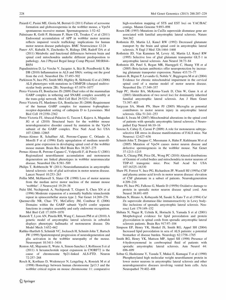
Paiardi C, Pasini ME, Gioria M, Berruti G (2011) Failure of acrosome
formation and globozoospermia in the wobbler mouse, a Vps54
spontaneous recessive mutant. Spermatogenesis 1:52–62
Palmisano R, Golfi P, Heimann P, Shaw CE, Troakes C et al (2011)
Endosomal accumulation of APP in wobbler motor neurons
reflects impaired vesicle trafficking: implications for human
motor neuron disease pathologies. BMC Neuroscience 12:24
Panov AV, Kubalik N, Zinchenko N, Ridings DM, Radoff DA et al
(2011) Metabolic and functional differences between brain and
spinal cord mitochondria underlie different predisposition to
pathology. Am J Physiol Regul Integr Comp Physiol 300:R844–
R854
Papadimitriou D, Le Verche V, Jacquier A, Ikiz B, Przedborski S, Re
DB (2010) Inflammation in ALS and SMA: sorting out the good
from the evil. Neurobiol Dis 37:493–502
Parkinson N, Ince PG, Smith MO, Highley R, Skibinski G et al (2006)
ALS phenotypes with mutations in CHMP2B (charged multive-
sicular body protein 2B). Neurology 67:1074–1077
Perez-Victoria FJ, Bonifacino JS (2009) Dual roles of the mammalian
GARP complex in tethering and SNARE complex assembly at
the trans-golgi network. Mol Cell Biol 29:5251–5263
Perez-Victoria FJ, Mardones GA, Bonifacino JS (2008) Requirement
of the human GARP complex for mannose 6-phosphate-
receptor-dependent sorting of cathepsin D to Lysosomes. Mol
Biol Cell 19:2350–2362
Perez-Victoria FJ, Abascal-Palacios G, Tascon I, Kajava A, Magadan
JG et al (2010) Structural basis for the wobbler mouse
neurodegenerative disorder caused by mutation in the Vps54
subunit of the GARP complex. Proc Natl Acad Sci USA
107:12860–12865
Pernas-Alonso R, Schaffner AE, Perrone-Capano C, Orlando A,
Morelli F et al (1996) Early upregulation of medium neurofil-
ament gene expression in developing spinal cord of the wobbler
mouse mutant. Brain Res Mol Brain Res 38:267–275
Pernas-Alonso R, Perrone-Capano C, Volpicelli F, di Porzio U (2001)
Regionalized neurofilament accumulation and motoneuron
degeneration are linked phenotypes in wobbler neuromuscular
disease. Neurobiol Dis 8:581–589
Philips T, Robberecht W (2011) Neuroinflammation in amyotrophic
lateral sclerosis: role of glial activation in motor neuron disease.
Lancet Neurol 10:253–263
Pollin MM, McHanwell S, Slater CR (1990) Loss of motor neurons
from the median nerve motor nucleus of the mutant mouse
‘wobbler’. J Neurocytol 19:29–38
Pulst SM, Nechiporuk A, Nechiporuk T, Gispert S, Chen XN et al
(1996) Moderate expansion of a normally biallelic trinucleotide
repeat in spinocerebellar ataxia type 2. Nat Genet 14:269–276
Quenneville NR, Chao TY, McCaffery JM, Conibear E (2006)
Domains within the GARP subunit Vps54 confer separate
functions in complex assembly and early endosome recognition.
Mol Biol Cell 17:1859–1870
Ramesh T, Lyon AN, Pineda RH, Wang C, Janssen PM et al (2010) A
genetic model of amyotrophic lateral sclerosis in zebrafish
displays phenotypic hallmarks of motoneuron disease. Dis
Model Mech 3:652–662
Rathke-Hartlieb S, Schmidt VC, Jockusch H, Schmitt-John T, Bartsch
JW (1999) Spatiotemporal progression of neurodegeneration and
glia activation in the wobbler neuropathy of the mouse.
Neuroreport 10:3411–3416
Renton AE, Majounie E, Waite A, Simon-Sanchez J, Rollinson S et al
(2011) A hexanucleotide repeat expansion in C9ORF72 is the
cause of chromosome 9p21-linked ALS-FTD. Neuron
72:257–268
Resch K, Korthaus D, Wedemeyer N, Lengeling A, Ronsiek M et al
(1998) Homology between human chromosome 2p13.3 and the
wobbler critical region on mouse chromosome 11: comparative
high-resolution mapping of STS and EST loci on YAC/BAC
contigs. Mamm Genome 9:893–898
Rosen DR (1993) Mutations in Cu/Zn superoxide dismutase gene are
associated with familial amyotrophic lateral sclerosis. Nature
364:362
Rothstein JD, Martin LJ, Kuncl RW (1992) Decreased glutamate
transport by the brain and spinal cord in amyotrophic lateral
sclerosis. N Engl J Med 326:1464–1468
Rothstein JD, Van Kammen M, Levey AI, Martin LJ, Kuncl RW
(1995) Selective loss of glial glutamate transporter GLT-1 in
amyotrophic lateral sclerosis. Ann Neurol 38:73–84
Rothstein JD, Patel S, Regan MR, Haenggeli C, Huang YH et al
(2005) Beta-lactam antibiotics offer neuroprotection by increas-
ing glutamate transporter expression. Nature 433:73–77
Santoro B, Bigini P, Levandis G, Nobile V, Biggiogera M et al (2004)
Evidence for chronic mitochondrial impairment in the cervical
spinal cord of a murine model of motor neuron disease.
Neurobiol Dis 17:349–357
Sapp PC, Hosler BA, McKenna-Yasek D, Chin W, Gann A et al
(2003) Identification of two novel loci for dominantly inherited
familial amyotrophic lateral sclerosis. Am J Hum Genet
73:397–403
Sargsyan SA, Monk PN, Shaw PJ (2005) Microglia as potential
contributors to motor neuron injury in amyotrophic lateral
sclerosis. Glia 51:241–253
Sasaki S, Iwata M (2007) Mitochondrial alterations in the spinal cord
of patients with sporadic amyotrophic lateral sclerosis. J Neuro-
pathol Exp Neurol 66:10–16
Saxena S, Cabuy E, Caroni P (2009) A role for motoneuron subtype-
selective ER stress in disease manifestations of FALS mice. Nat
Neurosci 12:627–636
Schmitt-John T, Drepper C, Mussmann A, Hahn P, Kuhlmann M et al
(2005) Mutation of Vps54 causes motor neuron disease and
defective spermiogenesis in the wobbler mouse. Nat Genet
37:1213–1215
Shan X, Chiang PM, Price DL, Wong PC (2010) Altered distributions
of Gemini of coiled bodies and mitochondria in motor neurons of
TDP-43 transgenic mice. Proc Natl Acad Sci USA
107:16325–16330
Shaw PJ, Forrest V, Ince PG, Richardson JP, Wastell HJ (1995a) CSF
and plasma amino acid levels in motor neuron disease: elevation
of CSF glutamate in a subset of patients. Neurodegeneration
4:209–216
Shaw PJ, Ince PG, Falkous G, Mantle D (1995b) Oxidative damage to
protein in sporadic motor neuron disease spinal cord. Ann
Neurol 38:691–695
Shibata N, Hirano A, Kobayashi M, Sasaki S, Kato T et al (1994) Cu/
Zn superoxide dismutase-like immunoreactivity in Lewy body-
like inclusions of sporadic amyotrophic lateral sclerosis. Neu-
rosci Lett 179:149–152
Shibata N, Nagai R, Uchida K, Horiuchi S, Yamada S et al (2001)
Morphological evidence for lipid peroxidation and protein
glycoxidation in spinal cords from sporadic amyotrophic lateral
sclerosis patients. Brain Res 917:97–104
Simpson EP, Henry YK, Henkel JS, Smith RG, Appel SH (2004)
Increased lipid peroxidation in sera of ALS patients: a potential
biomarker of disease burden. Neurology 62:1758–1765
Smith RG, Henry YK, Mattson MP, Appel SH (1998) Presence of
4-hydroxynonenal in cerebrospinal fluid of patients with
sporadic amyotrophic lateral sclerosis. Ann Neurol 44:
696–699
Sobue G, Hashizume Y, Yasuda T, Mukai E, Kumagai T et al (1990)
Phosphorylated high molecular weight neurofilament protein in
lower motor neurons in amyotrophic lateral sclerosis and other
neurodegenerative diseases involving ventral horn cells. Acta
Neuropathol 79:402–408
228 Mol Genet Genomics (2013) 288:207–229
123
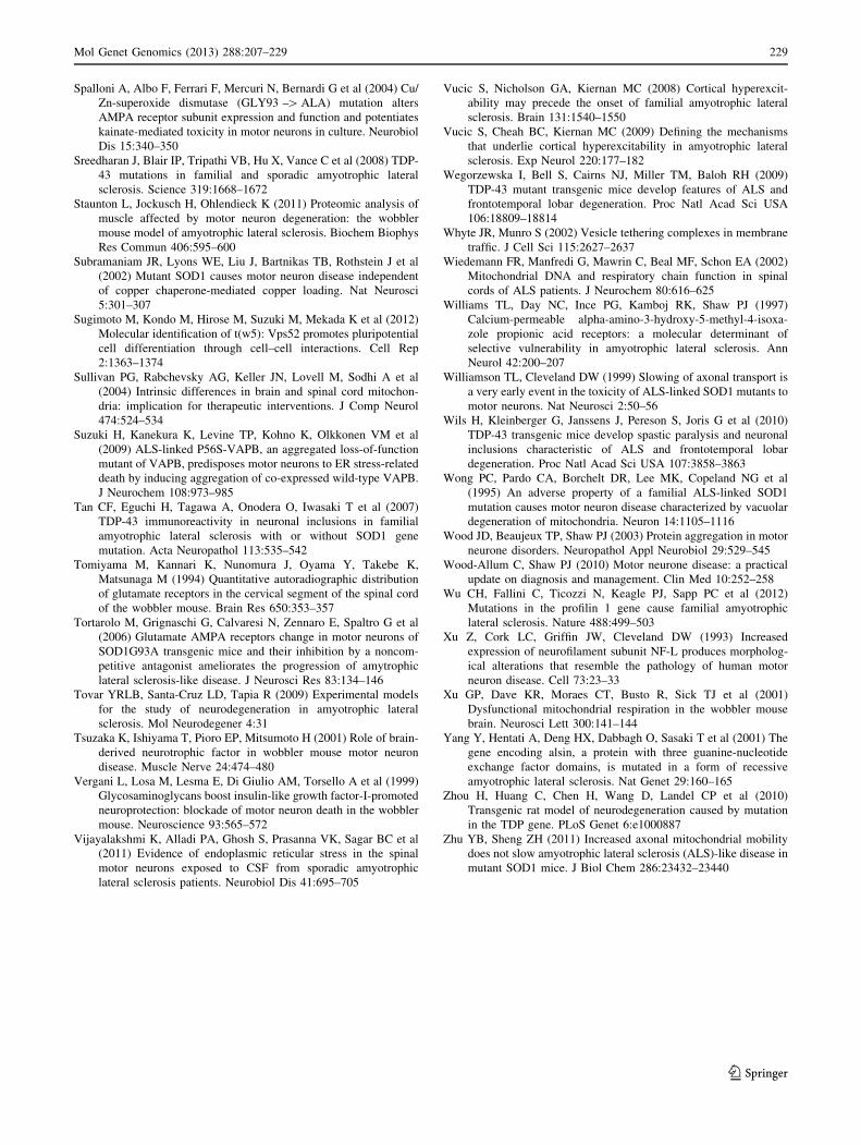
Spalloni A, Albo F, Ferrari F, Mercuri N, Bernardi G et al (2004) Cu/
Zn-superoxide dismutase (GLY93 –[ ALA) mutation alters
AMPA receptor subunit expression and function and potentiates
kainate-mediated toxicity in motor neurons in culture. Neurobiol
Dis 15:340–350
Sreedharan J, Blair IP, Tripathi VB, Hu X, Vance C et al (2008) TDP-
43 mutations in familial and sporadic amyotrophic lateral
sclerosis. Science 319:1668–1672
Staunton L, Jockusch H, Ohlendieck K (2011) Proteomic analysis of
muscle affected by motor neuron degeneration: the wobbler
mouse model of amyotrophic lateral sclerosis. Biochem Biophys
Res Commun 406:595–600
Subramaniam JR, Lyons WE, Liu J, Bartnikas TB, Rothstein J et al
(2002) Mutant SOD1 causes motor neuron disease independent
of copper chaperone-mediated copper loading. Nat Neurosci
5:301–307
Sugimoto M, Kondo M, Hirose M, Suzuki M, Mekada K et al (2012)
Molecular identification of t(w5): Vps52 promotes pluripotential
cell differentiation through cell–cell interactions. Cell Rep
2:1363–1374
Sullivan PG, Rabchevsky AG, Keller JN, Lovell M, Sodhi A et al
(2004) Intrinsic differences in brain and spinal cord mitochon-
dria: implication for therapeutic interventions. J Comp Neurol
474:524–534
Suzuki H, Kanekura K, Levine TP, Kohno K, Olkkonen VM et al
(2009) ALS-linked P56S-VAPB, an aggregated loss-of-function
mutant of VAPB, predisposes motor neurons to ER stress-related
death by inducing aggregation of co-expressed wild-type VAPB.
J Neurochem 108:973–985
Tan CF, Eguchi H, Tagawa A, Onodera O, Iwasaki T et al (2007)
TDP-43 immunoreactivity in neuronal inclusions in familial
amyotrophic lateral sclerosis with or without SOD1 gene
mutation. Acta Neuropathol 113:535–542
Tomiyama M, Kannari K, Nunomura J, Oyama Y, Takebe K,
Matsunaga M (1994) Quantitative autoradiographic distribution
of glutamate receptors in the cervical segment of the spinal cord
of the wobbler mouse. Brain Res 650:353–357
Tortarolo M, Grignaschi G, Calvaresi N, Zennaro E, Spaltro G et al
(2006) Glutamate AMPA receptors change in motor neurons of
SOD1G93A transgenic mice and their inhibition by a noncom-
petitive antagonist ameliorates the progression of amytrophic
lateral sclerosis-like disease. J Neurosci Res 83:134–146
Tovar YRLB, Santa-Cruz LD, Tapia R (2009) Experimental models
for the study of neurodegeneration in amyotrophic lateral
sclerosis. Mol Neurodegener 4:31
Tsuzaka K, Ishiyama T, Pioro EP, Mitsumoto H (2001) Role of brain-
derived neurotrophic factor in wobbler mouse motor neuron
disease. Muscle Nerve 24:474–480
Vergani L, Losa M, Lesma E, Di Giulio AM, Torsello A et al (1999)
Glycosaminoglycans boost insulin-like growth factor-I-promoted
neuroprotection: blockade of motor neuron death in the wobbler
mouse. Neuroscience 93:565–572
Vijayalakshmi K, Alladi PA, Ghosh S, Prasanna VK, Sagar BC et al
(2011) Evidence of endoplasmic reticular stress in the spinal
motor neurons exposed to CSF from sporadic amyotrophic
lateral sclerosis patients. Neurobiol Dis 41:695–705
Vucic S, Nicholson GA, Kiernan MC (2008) Cortical hyperexcit-
ability may precede the onset of familial amyotrophic lateral
sclerosis. Brain 131:1540–1550
Vucic S, Cheah BC, Kiernan MC (2009) Defining the mechanisms
that underlie cortical hyperexcitability in amyotrophic lateral
sclerosis. Exp Neurol 220:177–182
Wegorzewska I, Bell S, Cairns NJ, Miller TM, Baloh RH (2009)
TDP-43 mutant transgenic mice develop features of ALS and
frontotemporal lobar degeneration. Proc Natl Acad Sci USA
106:18809–18814
Whyte JR, Munro S (2002) Vesicle tethering complexes in membrane
traffic. J Cell Sci 115:2627–2637
Wiedemann FR, Manfredi G, Mawrin C, Beal MF, Schon EA (2002)
Mitochondrial DNA and respiratory chain function in spinal
cords of ALS patients. J Neurochem 80:616–625
Williams TL, Day NC, Ince PG, Kamboj RK, Shaw PJ (1997)
Calcium-permeable alpha-amino-3-hydroxy-5-methyl-4-isoxa-
zole propionic acid receptors: a molecular determinant of
selective vulnerability in amyotrophic lateral sclerosis. Ann
Neurol 42:200–207
Williamson TL, Cleveland DW (1999) Slowing of axonal transport is
a very early event in the toxicity of ALS-linked SOD1 mutants to
motor neurons. Nat Neurosci 2:50–56
Wils H, Kleinberger G, Janssens J, Pereson S, Joris G et al (2010)
TDP-43 transgenic mice develop spastic paralysis and neuronal
inclusions characteristic of ALS and frontotemporal lobar
degeneration. Proc Natl Acad Sci USA 107:3858–3863
Wong PC, Pardo CA, Borchelt DR, Lee MK, Copeland NG et al
(1995) An adverse property of a familial ALS-linked SOD1
mutation causes motor neuron disease characterized by vacuolar
degeneration of mitochondria. Neuron 14:1105–1116
Wood JD, Beaujeux TP, Shaw PJ (2003) Protein aggregation in motor
neurone disorders. Neuropathol Appl Neurobiol 29:529–545
Wood-Allum C, Shaw PJ (2010) Motor neurone disease: a practical
update on diagnosis and management. Clin Med 10:252–258
Wu CH, Fallini C, Ticozzi N, Keagle PJ, Sapp PC et al (2012)
Mutations in the profilin 1 gene cause familial amyotrophic
lateral sclerosis. Nature 488:499–503
Xu Z, Cork LC, Griffin JW, Cleveland DW (1993) Increased
expression of neurofilament subunit NF-L produces morpholog-
ical alterations that resemble the pathology of human motor
neuron disease. Cell 73:23–33
Xu GP, Dave KR, Moraes CT, Busto R, Sick TJ et al (2001)
Dysfunctional mitochondrial respiration in the wobbler mouse
brain. Neurosci Lett 300:141–144
Yang Y, Hentati A, Deng HX, Dabbagh O, Sasaki T et al (2001) The
gene encoding alsin, a protein with three guanine-nucleotide
exchange factor domains, is mutated in a form of recessive
amyotrophic lateral sclerosis. Nat Genet 29:160–165
Zhou H, Huang C, Chen H, Wang D, Landel CP et al (2010)
Transgenic rat model of neurodegeneration caused by mutation
in the TDP gene. PLoS Genet 6:e1000887
Zhu YB, Sheng ZH (2011) Increased axonal mitochondrial mobility
does not slow amyotrophic lateral sclerosis (ALS)-like disease in
mutant SOD1 mice. J Biol Chem 286:23432–23440
Mol Genet Genomics (2013) 288:207–229 229
123