the viral replication complex is associated with the ... · nome to form the nucleocapsid, which...
TRANSCRIPT

JOURNAL OF VIROLOGY, Oct. 2010, p. 10113–10120 Vol. 84, No. 190022-538X/10/$12.00 doi:10.1128/JVI.00097-10Copyright © 2010, American Society for Microbiology. All Rights Reserved.
The Viral Replication Complex Is Associated with the Virulence ofNewcastle Disease Virus�
J. C. F. M. Dortmans,1,2 P. J. M. Rottier,2 G. Koch,1 and B. P. H. Peeters1*Central Veterinary Institute of Wageningen UR, Lelystad, Netherlands,1 and Virology Division, Department of Infectious Diseases and
Immunology, Faculty of Veterinary Medicine, Utrecht University, Netherlands2
Received 15 January 2010/Accepted 2 July 2010
Virulent strains of Newcastle disease virus ([NDV] also known as avian paramyxovirus type 1) can bediscriminated from low-virulence strains by the presence of multiple basic amino acid residues at the proteo-lytic cleavage site of the fusion (F) protein. However, some NDV variants isolated from pigeons (pigeonparamyxovirus type 1 [PPMV-1]) have low levels of virulence, despite the fact that their F protein cleavage sitescontain a multibasic amino acid sequence and have the same functionality as that of virulent strains. Todetermine the molecular basis of this discrepancy, we examined the role of the internal proteins in NDVvirulence. Using reverse genetics, the genes encoding the nucleoprotein (NP), phosphoprotein (P), matrixprotein (M), and large polymerase protein (L) were exchanged between the nonvirulent PPMV-1 strain AV324and the highly virulent NDV strain Herts. Recombinant viruses were evaluated for their pathogenicities andreplication levels in day-old chickens, and viral genome replication and plaque sizes were examined in cellculture monolayers. We also tested the contributions of the individual NP, P, and L proteins to the activity ofthe viral replication complex in an in vitro replication assay. The results showed that the replication proteinsof Herts are more active than those of AV324 and that the activity of the viral replication complex is directlyrelated to virulence. Although the M protein affected viral replication in vitro, it had only a minor effect onvirulence.
Newcastle disease is a severe infectious disease of birdscaused by Newcastle disease virus (NDV), or avian paramyxo-virus type 1 (APMV-1). NDV is classified in the genus Avula-virus of the family Paramyxoviridae (32) and has a single-stranded, negative-sense RNA genome consisting of six genesin the order 3�-NP-P-M-F-HN-L-5� (28) that encode at leastseven proteins: the nucleocapsid protein (NP), the phospho-protein (P), the matrix protein (M), the fusion protein (F), thehemagglutinin-neuraminidase protein (HN), and the polymer-ase protein (L). During P gene transcription, an additional,nonstructural protein (V) is produced by means of mRNAediting (55).
The M, F, and HN proteins are associated with the viralenvelope. The F and HN proteins mediate entry and release,and the M protein is involved in the morphogenesis and bud-ding of NDV (28). The V protein is involved in interferonantagonism (42). The NP protein encapsidates the RNA ge-nome to form the nucleocapsid, which serves as the templatefor viral transcription and replication. The P protein is essen-tial for viral RNA synthesis and has multiple roles (10, 12). Itforms separate complexes with the NP and L proteins and thenucleocapsid (22). Transcription of the viral genomic RNAoccurs by way of the viral polymerase (P-L complex); the cat-alytic activities of the polymerase are functions of the L pro-tein, and the P protein is responsible for the binding of the P-Lcomplex to the nucleocapsid. Once sufficient viral proteins aregenerated, NP starts to bind to the leader chain, a process in
which the P protein acts as a chaperone to deliver NP to thenascent RNA (11). The NP-P complex is believed to regulatethe switch from transcription to replication (59), but severalfindings also show an important role for the M protein in thisprocess. Because the M protein associates with the nucleocap-sid (19, 29, 56), it may also affect transcription and/or replica-tion (17, 19, 25, 38). The P-L complex is responsible for ge-nome replication, i.e., the synthesis of full-length plus-strandantigenomic RNA, which in turn serves as the template for thesynthesis of minus-strand genomic RNA, which is ultimatelypackaged into progeny virions. The L protein performs post-transcriptional modification activities such as capping, methyl-ation, and polyadenylation of mRNAs (47, 53). The NP, P, andL proteins together constitute the viral replication complex(28).
Based on the mean times in which they kill inoculatedchicken embryos and their virulences for day-old chickens,NDV strains can be categorized into one of four pathotypes,i.e., nonvirulent, lentogenic (low virulence), mesogenic (inter-mediate), or velogenic (highly virulent) (3). Cleavage of the Fprotein is required for the initiation of infection and is themajor virulence determinant. The cleavage site of the F proteinof virulent NDV strains contains multiple basic amino acidresidues and is recognized by ubiquitous intracellular furin-likeproteases, resulting in a systemic infection. The cleavage site ofthe F protein of low-virulence strains does not contain thesemultiple basic amino acids and is recognized by extracellulartrypsin-like proteases found in a limited number of tissues,predominantly in the respiratory and intestinal tracts (36, 37),thereby limiting the replication of low-virulence strains tothese tissues.
Pigeon paramyxovirus type 1 (PPMV-1) strains are variant
* Corresponding author. Mailing address: P.O. Box 65, 8200 ABLelystad, Netherlands. Phone: 31 320 238693. Fax: 31 320 238668.E-mail: [email protected].
� Published ahead of print on 21 July 2010.
10113

strains of NDV associated with infections of pigeons. SomePPMV-1 strains behave as lentogenic viruses, i.e., they show alow intracerebral pathogenicity index (ICPI) in chickens, de-spite the presence of an F protein cleavage site motif that isgenerally associated with virulent viruses (34). In a previousstudy, we showed that the exchange of the F gene between alow-virulence PPMV-1 virus and a highly virulent virus did notsignificantly affect the virulences of the chimeric viruses rela-tive to those of their respective parental viruses (16). Thus, thelow virulence of some PPMV-1 strains must be determined byother factors. The V, HN, and L proteins of NDV have all beenshown to be involved in the virulence of NDV (14, 23, 24, 33,41, 42, 50, 51). However, little is still known about the mech-anisms underlying their function as virulence determinants inNDV strains and especially in PPMV-1 strains.
In this study, we examined the contributions of the NP, P, M,and L proteins to the virulence of NDV. Using reverse genet-ics, the genes encoding these internal proteins were exchangedbetween the low-virulence PPMV-1 strain AV324 and thehighly virulent NDV strain Herts. The pathogenicities andlevels of in vivo replication of the chimeric viruses were deter-mined in 1-day-old chickens. Furthermore, we investigated thereplication kinetics and plaque sizes of the different chimericviruses in cell culture monolayers, and we developed an in vitroreplication assay using cotransfection of plasmids encoding aminigenome that expresses luciferase in the presence of theNP, P, and L proteins. Our results indicate that the virulenceof NDV is directly related to the activity of the viral replicationcomplex.
MATERIALS AND METHODS
Cells, viruses, and animals. QM5 cells (4) were grown in QT35 medium(Gibco-BRL/Life Technologies), and DF-1 cells were grown in Dulbecco’s mod-ified Eagle’s medium (DMEM) plus GlutaMAX (Invitrogen). Both media were
supplemented with 5% fetal bovine serum and 1% of an antibiotic stock con-sisting of penicillin (100 units/ml), streptomycin (100 �g/ml), and fungizone (2.5�g/ml). Both cell lines were grown at 37°C in a 5% CO2 incubator. The fowlpoxrecombinant virus fpE-FLT7pol (6) (hereafter called FPV-T7), which expressesthe bacteriophage T7 RNA polymerase, was used as recently described (15). ThecDNA clone rgAV324 was derived from the low-virulence PPMV-1 strainAV324/96, and the cDNA clone FL-Herts was derived from the virulent NDVstrain Herts/33, as previously described (14, 16). In this study, specific-pathogen-free (SPF) chickens were used. Animal experiments were approved by the EthicsCommittee for Animal Experiments of the Central Veterinary Institute ofWageningen UR and comply with Dutch law on animal experiments.
Construction of full-length chimeric AV324/Herts antigenomic cDNAs. Chi-meric viruses in which either the individual NP, P, M, or L genes, or combina-tions thereof, were exchanged between the AV324 and Herts strains were gen-erated (Fig. 1). The published nucleotide sequences of the Herts/33 (GenBankaccession no. AY741404) and AV324/96 (GenBank accession no. GQ429292)strains were used as guides for the construction of the chimeric viruses.
To swap the replication genes between the plasmids FL-Herts and rgAV324,the unique restriction sites SfiI, PacI, AscI, SpeI, and SgfI were used (Fig. 1). Tointroduce a unique restriction site for SpeI (position 8101) in the rgAV324AF
cDNA (16), site-directed mutagenesis was performed, resulting in rgAV324AFS.The superscript AF represents the introduced restriction sites AscI and FseI, andthe superscript AFS represents the introduced restriction sites AscI, FseI, andSpeI. Plasmids FL-HertsAF and rgAV324AFS were digested with SgfI and PacI tosimultaneously exchange the NP and P genes, resulting in rgAV324(NP-P�)Herts
and FL-Herts(NP-P�)AV324. Because the PacI site is positioned at nucleotide (nt)2900 in Herts and nt 2906 in AV324, 57 amino acids of the C terminus of the Pproteins were not exchanged. Of these 57 amino acids, 7 differ between the twoviruses. To exchange the L gene, both full-length cDNA clones were digestedwith SpeI and SgfI and reciprocally cloned, resulting in rgAV324(L)Herts andFL-Herts(L)AV324. The PacI and SpeI sites were used to simultaneously ex-change the NP, P, and L genes, resulting in rgAV324(NP-P�-L)Herts and FL-Herts(NP-P�-L)AV324. To exchange the NP and P genes individually, site-di-rected mutagenesis was performed to introduce a unique SfiI site in FL-HertsAF
(this site is present in AV324 but is lacking in Herts). To exchange the NP gene,the SgfI and SfiI sites were used, resulting in rgAV324(NP)Herts and FL-Herts(NP)AV324, and to exchange the P gene, the SfiI and PacI sites were used,resulting in rgAV324(P�)Herts and FL-Herts(P�)AV324. Finally, the M genes wereexchanged, using the PacI and AscI sites, resulting in rgAV324(M)Herts andFL-Herts(M)AV324.
FIG. 1. Schematic illustration of the cloning strategy used to exchange the NP, P, M, and L genes between FL-HertsAF and rgAV324AFS. Thevirulences of the different recombinant viruses were determined by measuring the intracerebral pathogenicity index (ICPI) in day-old chickens. Themaximum ICPI score was 2.0. KM, kanamycin resistance gene positioned in the vector plasmid.
10114 DORTMANS ET AL. J. VIROL.
Rescue of virus from cDNA. QM5 cells were infected with FPV-T7 for 1 h andsubsequently cotransfected with full-length cDNA constructs and helper plas-mids expressing P and L, as previously described (16, 45). The respective helperplasmids of either rgAV324 or FL-Herts were used along with their respectivefull-length cDNAs to prevent potential heterologous recombination in the trans-fected cells. After 3 days, the culture supernatant was harvested and inoculatedinto 9- to 11-day-old embryonated SPF eggs to obtain a virus stock.
Pathogenicity test, HI assay, and reisolation of virus. Determination of theintracerebral pathogenicity index (ICPI) in 1-day-old chickens and the hemag-glutination inhibition (HI) assay were performed as described in EuropeanCouncil Directive 92/66/EEC (9). Sera of the chickens that survived the ICPI testwere tested in an HI assay to verify that the animals had been infected. In orderto check the sequences of the different recombinant viruses after the ICPI tests,brains, livers, and lungs of dead chickens were collected, and virus reisolation inembryonated SPF eggs was performed as previously described (13).
RNA isolation, reverse transcriptase PCR, and sequencing. RNA of the re-combinant viruses was isolated, using a High Pure viral RNA kit (Roche Diag-nostics). First-strand DNA synthesis was carried out using a SuperScript IIIreverse transcriptase kit (Invitrogen), and PCR fragments were purified using aHigh Pure PCR purification kit (Roche Diagnostics). Nucleotide sequencing(primer sequences are available upon request) was carried out using a BigDyeTerminator v1.1 cycle sequencing kit and a 3130 genetic analyzer instrument(Applied Biosystems).
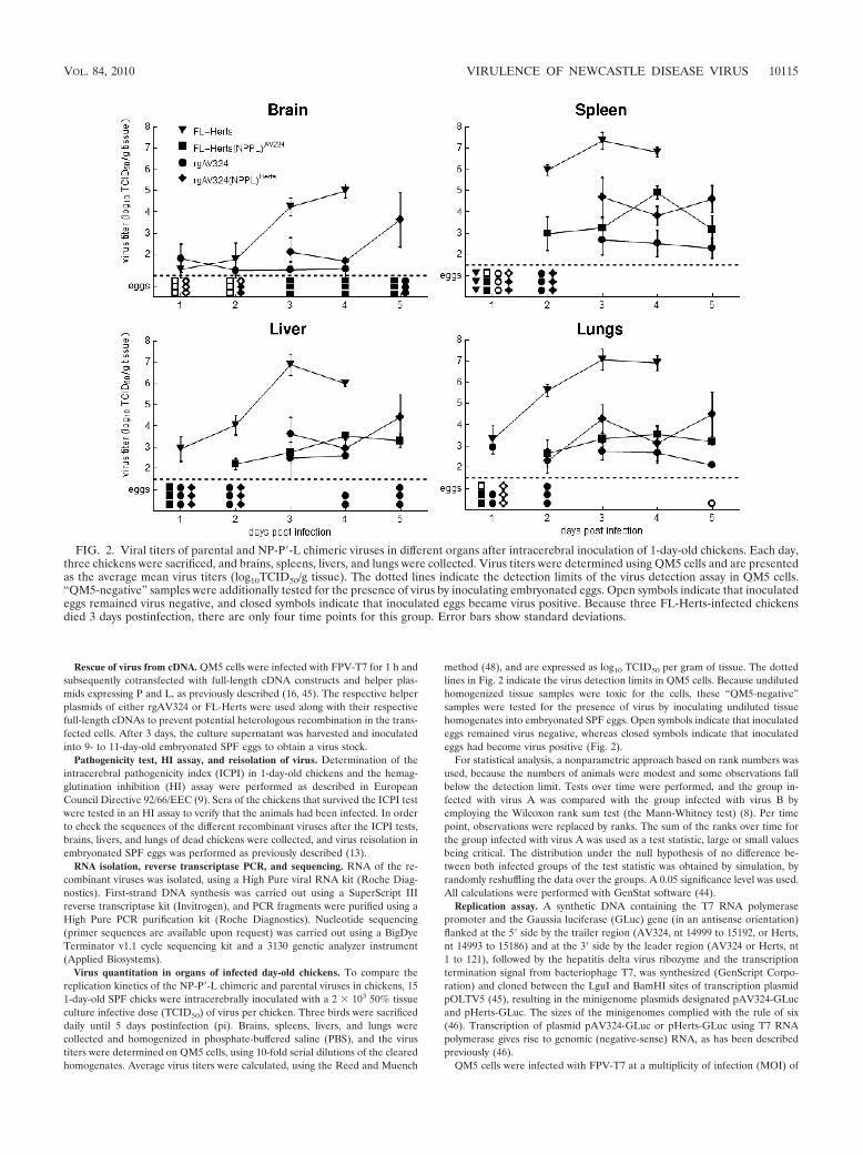
Virus quantitation in organs of infected day-old chickens. To compare thereplication kinetics of the NP-P�-L chimeric and parental viruses in chickens, 151-day-old SPF chicks were intracerebrally inoculated with a 2 � 103 50% tissueculture infective dose (TCID50) of virus per chicken. Three birds were sacrificeddaily until 5 days postinfection (pi). Brains, spleens, livers, and lungs werecollected and homogenized in phosphate-buffered saline (PBS), and the virustiters were determined on QM5 cells, using 10-fold serial dilutions of the clearedhomogenates. Average virus titers were calculated, using the Reed and Muench
method (48), and are expressed as log10 TCID50 per gram of tissue. The dottedlines in Fig. 2 indicate the virus detection limits in QM5 cells. Because undilutedhomogenized tissue samples were toxic for the cells, these “QM5-negative”samples were tested for the presence of virus by inoculating undiluted tissuehomogenates into embryonated SPF eggs. Open symbols indicate that inoculatedeggs remained virus negative, whereas closed symbols indicate that inoculatedeggs had become virus positive (Fig. 2).
For statistical analysis, a nonparametric approach based on rank numbers wasused, because the numbers of animals were modest and some observations fallbelow the detection limit. Tests over time were performed, and the group in-fected with virus A was compared with the group infected with virus B byemploying the Wilcoxon rank sum test (the Mann-Whitney test) (8). Per timepoint, observations were replaced by ranks. The sum of the ranks over time forthe group infected with virus A was used as a test statistic, large or small valuesbeing critical. The distribution under the null hypothesis of no difference be-tween both infected groups of the test statistic was obtained by simulation, byrandomly reshuffling the data over the groups. A 0.05 significance level was used.All calculations were performed with GenStat software (44).
Replication assay. A synthetic DNA containing the T7 RNA polymerasepromoter and the Gaussia luciferase (GLuc) gene (in an antisense orientation)flanked at the 5� side by the trailer region (AV324, nt 14999 to 15192, or Herts,nt 14993 to 15186) and at the 3� side by the leader region (AV324 or Herts, nt1 to 121), followed by the hepatitis delta virus ribozyme and the transcriptiontermination signal from bacteriophage T7, was synthesized (GenScript Corpo-ration) and cloned between the LguI and BamHI sites of transcription plasmidpOLTV5 (45), resulting in the minigenome plasmids designated pAV324-GLucand pHerts-GLuc. The sizes of the minigenomes complied with the rule of six(46). Transcription of plasmid pAV324-GLuc or pHerts-GLuc using T7 RNApolymerase gives rise to genomic (negative-sense) RNA, as has been describedpreviously (46).
QM5 cells were infected with FPV-T7 at a multiplicity of infection (MOI) of
FIG. 2. Viral titers of parental and NP-P�-L chimeric viruses in different organs after intracerebral inoculation of 1-day-old chickens. Each day,three chickens were sacrificed, and brains, spleens, livers, and lungs were collected. Virus titers were determined using QM5 cells and are presentedas the average mean virus titers (log10TCID50/g tissue). The dotted lines indicate the detection limits of the virus detection assay in QM5 cells.“QM5-negative” samples were additionally tested for the presence of virus by inoculating embryonated eggs. Open symbols indicate that inoculatedeggs remained virus negative, and closed symbols indicate that inoculated eggs became virus positive. Because three FL-Herts-infected chickensdied 3 days postinfection, there are only four time points for this group. Error bars show standard deviations.
VOL. 84, 2010 VIRULENCE OF NEWCASTLE DISEASE VIRUS 10115

1 for 1 h and subsequently cotransfected with either pAV324-GLuc or pHerts-GLuc and helper plasmids expressing NP, P, and L originating either from theAV324 strain (16) or from the Herts strain (14). The minigenome and itsexpression plasmids containing NP, P, and L were cotransfected at a ratio of1.0:1.6:0.8:0.8, respectively, by using FuGENE HD (Roche). For normalization,a plasmid containing the firefly luciferase gene under the control of the humancytomegalovirus (hCMV) immediate-early promoter (a kind gift of Erik de Vriesand Xander de Haan, Faculty of Veterinary Medicine, Utrecht, Netherlands)was cotransfected. After 24 h, the expression levels of the secreted (Gaussia) andinternal (firefly) luciferase activities were measured, using a luciferase assay kit(Promega) and a GloMax luminometer (Promega). One experiment comprises atriplicate measurement of luciferase expression. A total of six separate experi-ments were performed. Differences in luciferase expression were statisticallyanalyzed using the Wilcoxon test. Mean differences were considered significantwhen the P value was less than 0.05.
Analysis of genome replication by quantitation of negative-sense genomicRNA. To investigate the onset and kinetics of virus replication, the relativeamounts of negative-sense genomic RNA were determined by quantitative real-time reverse transcriptase PCR (qRRT-PCR) at different time points pi. Toset up a qRRT-PCR, the sequences of Herts and AV324 were aligned, usingthe EMBL-EBI web-based software ClustalW2 (http://www.ebi.ac.uk/Tools/clustalw2/index.html). A PCR primer pair was selected in a homologous regionof the L genes such that the primers had no mismatches in either virus. Thesequence of the forward primer is 5�-CCCGACCGACTGTGATCTAT-3�, andthat of the reverse primer is 5�-GCAGCAAGTTGGATTGCAG-3�. Subse-quently, a perfectly matching probe within the same region was chosen andlabeled at the 5� end with 6-carboxyfluorescein (FAM) and at the 3� end withBHQ1, as follows: 5�-FAM-TGCTAGAGGGGGCATTGAGGGA-BHQ1-3�(Tib Mobiol).
DF-1 cells were seeded in 24-well plates (Greiner) and infected in duplicatewith virus at an MOI of 10. At 2, 4, 6, 8, and 10 h pi (hpi), plates were frozen at�70°C. RNA was extracted from the combined infected cells and the superna-tants, using a MagNA Pure LC total nucleic acid isolation kit and a MagNA PureLC instrument (Roche Applied Science) according to the manufacturer’s instruc-tions. Cycling conditions for the qRRT-PCR, using the Mx3005P system (Strat-agene), were one cycle at 50°C for 30 min, one cycle at 95°C for 15 min, 45 cyclesat 95°C for 15 s, 55°C for 30 s, and 72°C for 30 s. For data analysis, MxPro qPCRsoftware, version 4.10 (Stratagene), was used. The correlation coefficients (R2) ofthe standard curves were 0.999 (FL-Herts) and 0.993 (rgAV324). All PCRamplification efficiencies (E) were �0.95. Details of the PCR protocol are avail-able upon request.
Determination of plaque size. Monolayers of QM5 cells and DF-1 cells wereinfected with the parental and chimeric viruses and incubated for 2 days underan overlay of the Glasgow modification of Eagle medium/Eagle’s minimal es-sential medium (ASG-Lelystad) containing 1% methylcellulose, without the ad-dition of exogenous trypsin. Plaques were visualized by immunological staining,using monoclonal antibody (MAb) Fusie 133 8E12A8C3 (CVI of WageningenUR) against the NDV F protein and horseradish peroxidase (HRPO)-conju-gated polyclonal rabbit anti-mouse Ig (Dako). The average plaque size wasdetermined by measuring the areas of digital images of 12 to 16 discrete plaquesper virus (photographed at a magnification of 6.3) using Image-Pro Plus software(MediaCybernetics, Inc.). Differences in plaque size were statistically analyzedusing the Wilcoxon test. Mean differences were considered significant when theP value was less than 0.05.
RESULTS
Construction and recovery of recombinant viruses. To studythe molecular basis of the low pathogenicities of PPMV-1strains that contain a fully functional multiple basic amino acidmotif in their F protein cleavage sites, we exchanged the genesencoding the internal viral proteins NP, P, M, and L betweenthe nonvirulent PPMV-1 strain AV324 and the highly virulentNDV strain Herts. The amino acid sequence identity of theseproteins is 94% for NP, 84% for P, 93% for M, and 95% for L.The cloning strategy that was used to construct the chimericcDNAs is described in the Materials and Methods section andis illustrated in Fig. 1. Sequence analysis of the chimericcDNAs confirmed the intended gene exchanges and the ab-sence of any undesired mutations. For virus recovery by means
of cotransfection, helper plasmids expressing P and L of eitherrgAV324 or FL-Herts were used along with their respectivefull-length cDNAs. All chimeric viruses could be rescued, in-dicating that the biological functions of these proteins of theHerts and AV324 strains are compatible.
Pathogenicity in day-old chickens. The virulences of thechimeric viruses were assessed by a standard intracerebralpathogenicity test (ICPI) in 1-day-old chickens. Replacementof the internal genes of the Herts strain, either individually orin combination, by those of the AV324 strain resulted in adecrease in virulence. The simultaneous replacement of thegenes encoding the complete NP-P�-L replication complex hada significant effect on virulence, as shown by a decrease in theICPI value from 1.54 for FL-Herts to 0.55 for FL-Herts(NP-P�-L)AV324 (Fig. 1). In contrast, although not all replacementsof the internal genes of AV324 by those of Herts resulted in anincrease in virulence, again the simultaneous replacement ofthe NP, P, and L genes had a large effect. The exchangeresulted in a significant increase in ICPI values from 0.10 forrgAV324 to 1.03 for rgAV324(NP-P�-L)Herts (Fig. 1). Theseresults show that the origin of the complete replication com-plex from either AV324 or Herts has a major effect on deter-mining the virulence of the chimeric viruses.
In vivo replication in day-old chickens. Because exchangingthe complete viral replication complex had the greatest effecton virulence, the levels of in vivo replication of these chimericviruses and their parental viruses were determined in day-oldchickens after intracerebral inoculation (Fig. 2). The resultsshowed that the AV324 strain replicated to much lower levelsthan the Herts strain in all organs examined. In brain tissue, nosignificant difference in the replication levels of the AV324recombinant virus that expresses the replication complex of theHerts strain [rgAV324(NP-P�-L)Herts] and that of the parentalAV324 strain was observed (P � 0.05). However, in liver,lungs, and spleen, replication of rgAV324(NP-P�-L)Herts wassignificantly enhanced compared to that of rgAV324. Con-versely, providing the Herts virus with the AV324 replicationcomplex [FL-Herts(NP-P�-L)AV324] significantly reduced thereplicative abilities of the chimeric virus in all tissues examined(Fig. 2). These results strongly suggest that the pathogenicity ofavian paramyxoviruses is directly correlated with the level ofvirus replication in organs of infected animals.
In vitro replication assay. To test whether in vivo virus rep-lication correlated with the intrinsic activity of the viral repli-cation complex, we developed an in vitro replication assay,using a minigenome that expresses the Gaussia luciferase(GLuc) gene in the presence of the NP, P, and L proteins. Twoslightly different minigenomes were used, one based on theleader and trailer sequences of the Herts strain (pHerts-GLuc)and the other on the leader and trailer sequences of the AV324strain (pAV324-GLuc). Transcription of pAV324-GLuc orpHerts-GLuc using T7 RNA polymerase generates genomic(negative-sense) minigenome RNA. Hence, expression of theGLuc gene is dependent on the conversion of the negative-sense RNA into positive-sense RNA. This process is com-pletely dependent on the viral NP, P, and L proteins, providinga suitable assay to test the roles of the individual proteins inminigenome replication activity.
The in vitro replication assay revealed that the two minige-nomes behaved similarly, exhibiting comparable levels of re-
10116 DORTMANS ET AL. J. VIROL.

porter gene expression when driven by both the AV324 repli-cation proteins and those of the Herts virus (data not shown).This indicates that the nucleotide differences in the leader andtrailer sequences that code for the genomic (3-nt difference)and antigenomic (4-nt difference) promoter (31) of AV324 andHerts did not affect the efficiency of viral replication. There-fore, only the results obtained with the pHerts-GLuc minige-nome are shown. The results revealed that the activity of theAV324 replication complex is significantly lower than that ofthe Herts replication complex (Fig. 3). Furthermore, none ofthe possible combinations of the NP, P, and L proteinsof AV324 and Herts reached the 100% level obtained with theHerts replication proteins only. These observations suggestthat optimal replication is not determined by one or two pro-teins but by the combined action of all three replication pro-teins.
Viral genome replication. To investigate the onset and ki-netics of viral genome replication of the recombinant viruses incell culture monolayers, a qRRT-PCR that specifically detectsnegative-strand viral RNA was used. To this end, DF-1 cellswere infected at an MOI of 10, and at different time points upto 10 hpi, the amount of genomic RNA was determined. Theresults showed that the genome of FL-Herts replicated at ahigher rate than that of rgAV324 (Fig. 4). When the Hertsvirus was provided with the AV324 replication complex, thereplication kinetics were similar to those of rgAV324. Unex-pectedly, however, the recombinant virus in which the replica-tion genes of AV324 were replaced by those of Herts[rgAV324(NP-P�-L)Herts] showed the lowest replication effi-ciency of all viruses tested. Because several studies have shownthat the M protein may affect transcription and/or replication,we also examined the recombinant viruses in which only the Mgene was exchanged. The results showed that the kinetics ofFL-Herts(M)AV324 were very similar to those of rgAV324(NP-P�-L)Herts (Fig. 4), suggesting that the specific combination of
the M protein of AV324 and the NP-P�-L complex of Hertsresults in a decreased replication rate. This effect also seems toaffect virulence, since the introduction of the AV324 M proteinin Herts resulted in a reduction of the ICPI value from 1.54 to1.18 (Fig. 1). The kinetics of genome replication of rgAV324 wasnot increased by the M protein of Herts, and no significant effect onvirulence was noted [cf. rgAV324 and rgAV324(M)Herts], indicatingthat the effect is nonreciprocal.
Plaque size. In order to examine the replication of the dif-ferent viruses in tissue culture cells in another way, we deter-mined their plaque sizes in monolayers of quail-derived QM5cells and chicken-derived DF-1 cells at 48 hpi (Fig. 5). Relativeto FL-Herts, rgAV324 showed a significantly smaller plaquesize in QM5 and in DF-1 cells. Furthermore, smaller plaquesizes in both cell types were also observed when the Herts viruswas provided with the M protein of AV324 or the replicationcomplex of AV324. When the AV324 virus expressed the Mprotein or the replication complex of Herts, QM5 cells andDF-1 cells showed contrasting results with regard to plaquesize. While the plaque sizes were smaller in QM5 cells, largerplaques were observed in DF-1 cells (Fig. 5).
DISCUSSION
The results of this study show that all three proteins thatmake up the viral replication complex (NP, P, and L) play asignificant role in determining the virulence of NDV. By ex-changing the replication genes simultaneously, the virulentHerts virus was significantly attenuated, whereas the low-viru-lence AV324 strain became much more virulent. All individualreplication proteins have their own contribution but act syner-gistically when all three together are exchanged. The matrix(M) protein of AV324 showed a distinct effect on virulence inthe Herts background, probably by interacting with the viralreplication complex. However, this effect was not reciprocal,since the M protein of Herts lacked the ability to increase thevirulence of AV324.
The difference in virulence between AV324 and Herts seemsto be directly related to the efficiency of in vivo viral replica-
FIG. 3. Relative luciferase expression levels after cotransfection ofFPV-T7-infected QM5 cells with the viral minigenome plasmid (con-taining the leader and trailer sequences of Herts that flank the Glucreporter gene) and plasmids expressing NP, P, and L of Herts (gray) orAV324 (black). The background level of luciferase activity was deter-mined by omitting the L plasmid (X). The graph shows the averagevalues obtained in six separate experiments. On the left y axis, valuesfor luciferase activities relative to Herts are given, and on the right yaxis, values for luciferase activities relative to AV324 are shown. Errorbars represents standard errors of the means (SEMs).
FIG. 4. Onset and kinetics of negative-sense genomic RNA repli-cation in the first 10 h of infection. DF-1 cells were infected at an MOIof 10, and replication was determined by qRRT-PCR. Results areaverages for duplicate infections.
VOL. 84, 2010 VIRULENCE OF NEWCASTLE DISEASE VIRUS 10117

tion. After the genes encoding the entire NP-P-L replicationcomplex were exchanged, the virus titers of the virulent Hertsstrain in day-old chickens were decreased in all organs exam-ined, whereas those of the avirulent AV324 strain were in-creased in three of the four organs tested (Fig. 2). LentogenicNDV strains are generally unable to spread systemically wheninoculated intracerebrally. Because these viruses lack themultibasic cleavage motif in their F proteins, activation re-quires trypsin-like proteases that are apparently not present inneuronal tissue. Furthermore, replication of these viruses isoften limited to the inoculation site (39, 51, 60). The titers ofthe AV324 strain in brain tissue gradually decreased over time,but the virus was still able to spread to secondary organs (Fig.2). However, this systemic infection did not sicken its hostdrastically, as evidenced by the virus’s ICPI of 0.10 (Fig. 1) andthe absence of clinical signs (data not shown). Thus, low-virulence PPMV-1 isolates may be perfectly adapted to theirhosts by being able to spread systemically due to their typicalvelogenic F cleavage site motif while replicating at a relativelylow level.
The differences in efficiencies of the viral replication com-
plexes were confirmed in an in vitro replication assay (Fig. 3).Optimal replication was observed when all three replicationproteins originated from the Herts strain. This finding matchesthe results of the ICPI tests (Fig. 1). However, none of thepossible combinations of the NP, P, and L proteins of AV324and Herts reached the level of replication obtained with onlyHerts replication proteins. One of the possible explanationsmay be that the individual Herts replication proteins are in-herently more active but that optimal activity is dependent onthe presence of the cognate interaction partners. The aminoacid sequences of the protein domains responsible for theinteraction of the NP and P proteins (26) differ between Hertsand AV324. Furthermore, although not well identified forNDV, the interaction domains responsible for the P-L inter-action presumably also differ between the two viruses, as theyhave been shown to differ among several other paramyxovi-ruses (20, 22, 27, 43, 58). Since these complexes are essentialfor transcription and replication of the viral genome (28), theyprobably do not function optimally in a heterologous constitu-tion.
The discrepancy between the in vivo results (Fig. 1 and 2)
FIG. 5. Relative plaque sizes of the recombinant viruses in QM5 (a) and DF-1 (b) cells 48 h postinfection. Plaques were visualized byimmunological staining using a MAb against the NDV F protein. The average plaque size was determined by measuring the areas of at least 12plaques per virus with Image-Pro Plus software (Media Cybernetics, Inc.). On the left y axis, plaque sizes relative to those of FL-Herts are given,and on the right y axis, plaque sizes relative to those of rgAV324 are shown. Error bars represent standard deviations.
10118 DORTMANS ET AL. J. VIROL.

and the in vitro results (Fig. 4 and 5) might be explained by therole of the M protein. While the M protein is considered to bethe central organizer of viral morphogenesis (28), several stud-ies have shown that in addition, it is involved in regulating viralRNA synthesis (19, 25, 49). With NP as its most likely bindingpartner (25, 40), the M protein associates with the nucleocap-sid (19, 29, 56). Upon viral entry into the target cell, thenucleocapsid dissociates from the M protein and is releasedinto the cytoplasm, where transcription can occur. The inter-action of the M protein with the nucleocapsid might affecttranscription and have a subsequent effect on replication. Fur-thermore, this interaction may be strain specific and nonrecip-rocal and might explain the relatively low replication rate (Fig.4) and small plaque size (Fig. 5) of rgAV324(NP-P�-L)Herts,since similar results were found for FL-Herts(M)AV324. An-other possible explanation for our results is that the associationof the M protein with host cell factors might differ between thetwo strains. During the infection cycle, the M protein of NDVand other paramyxoviruses traffics between the cytoplasm andthe nucleus (21). Early during infection, the M protein residesprimarily in the nucleus, while later during infection M islocalized mainly in the cytoplasm (7, 21). It has been suggestedthat the function of M in the nucleus relates to inhibition ofhost cell functions (1, 18), although this has not yet beenconfirmed for NDV. The nuclear localization signals (7) andthe proposed viral late-domain core sequence necessary forbudding, 24FPIV27 (52), do not differ between Herts andAV324 M proteins.
The involvement of the NP, P, and L proteins in NDVvirulence has been examined before (51). In that study, chi-meric viruses were generated by exchanging genes between thelentogenic strain LaSota and the mesogenic strain BeaudetteC, both classified as members of lineage 2 or genotype II in theavian paramyxovirus type 1 group (2). Surprisingly, a recom-binant Beaudette C virus that contained the L gene of LaSotareplicated at a higher level and was more virulent than itsparental virus. However, no effect was found for the NP and Pproteins. These results differ from our findings, which showthat all three replication proteins are associated with virulence.An explanation for this difference might be that LaSota andBeaudette C belong to the same phylogenetic lineage andgenotype, whereas the strains used in this study belong todifferent lineages and genotypes. Herts is classified as a mem-ber of lineage 3b (or genotype IV), and AV324 belongs tolineage 4b (or genotype VI) (2). Furthermore, Herts is a chicken-derived strain, and AV324 is of pigeon origin.
The molecular mechanism for the relationship between thelevel of replication of a virus and its pathogenesis is not fullyunderstood. It is conceivable that higher levels of RNA syn-thesis lead to higher levels of viral replication and thus to morevirus production. This may overwhelm the host immune re-sponse, causing enhanced pathogenesis. A correlation betweenvirulence and the efficiency of viral replication has been ob-served before. It has, for instance, been reported that reducedlevels of RNA synthesis are associated with reduced virulenceof NDV (30). For several other paramyxoviruses, such as mea-sles virus (5, 57), respiratory syncytial virus, and parainfluenzavirus (35, 54), it has been described that determinants of virusattenuation are associated with mutations in the P and L genes.
Due to the use of the PacI cloning site, the exchange of the
P genes actually resulted in the exchange of chimeric P pro-teins in which 7 of the C-terminal 57 amino acids that differbetween the P proteins of Herts and AV324 are still similar tothose of the backbone strain. Exchanging the chimeric P geneshad only a limited effect on virulence [ICPI � 1.33 for FL-Herts(P)�AV324 and ICPI � 0.25 for rgAV324(P�)Herts]. How-ever, we cannot completely exclude the possibility that ex-changing the complete P protein may have had a differenteffect on its function and virulence.
In conclusion, this study shows that in addition to the F, V,HN, and L proteins, the NP, P, and L protein complex con-tributes to the virulence of NDV. Additional studies will berequired to elucidate whether the proteins have an effect onviral transcription, replication, or both. Altogether, these ob-servations illustrate that the virulence of NDV is a complextrait determined by multiple genetic factors. Furthermore, thedegree to which these factors are involved in NDV virulenceseems to be strain and cell type dependent.
ACKNOWLEDGMENTS
We thank Alan Rigter and Olav de Leeuw for helpful discussions,Rene Heutink for excellent technical assistance, and the animal tech-nicians for performing the pathogenicity tests. We thank JantienBacker for her help with preparing Fig. 2 and Willem Buist and BasEngel for statistical analyses. We also thank Erik de Vries and Xanderde Haan, Faculty of Veterinary Medicine, Utrecht, Netherlands, forproviding the firefly luciferase plasmid.
This work was supported by the Dutch Ministry of Agriculture,Nature and Food Quality.
REFERENCES
1. Ahmed, M., M. O. McKenzie, S. Puckett, M. Hojnacki, L. Poliquin, and D. S.Lyles. 2003. Ability of the matrix protein of vesicular stomatitis virus tosuppress beta interferon gene expression is genetically correlated with theinhibition of host RNA and protein synthesis. J. Virol. 77:4646–4657.
2. Aldous, E. W., J. K. Mynn, J. Banks, and D. J. Alexander. 2003. A molecularepidemiological study of avian paramyxovirus type 1 (Newcastle diseasevirus) isolates by phylogenetic analysis of a partial nucleotide sequence of thefusion protein gene. Avian Pathol. 32:239–256.
3. Alexander, D. J., and R. E. Gough. 2003. Newcastle disease, other avianparamyxoviruses and pneumovirus infections, p. 63–92. In Y. M. Saif, H. J.Barnes, A. M. Fadly, J. R. Glisson, L. R. McDougald, and D. E. Swayne(ed.), Diseases of poultry, 11 ed. Iowa State University Press, Ames, IA.
4. Antin, P. B., and C. P. Ordahl. 1991. Isolation and characterization of anavian myogenic cell line. Dev. Biol. 143:111–121.
5. Bankamp, B., S. P. Kearney, X. Liu, W. J. Bellini, and P. A. Rota. 2002.Activity of polymerase proteins of vaccine and wild-type measles virus strainsin a minigenome replication assay. J. Virol. 76:7073–7081.
6. Britton, P., P. Green, S. Kottier, K. L. Mawditt, Z. Penzes, D. Cavanagh, andM. A. Skinner. 1996. Expression of bacteriophage T7 RNA polymerase inavian and mammalian cells by a recombinant fowlpox virus. J. Gen. Virol.77:963–967.
7. Coleman, N. A., and M. E. Peeples. 1993. The matrix protein of Newcastledisease virus localizes to the nucleus via a bipartite nuclear localizationsignal. Virology 195:596–607.
8. Conover, W. J. 1980. Practical nonparametric statistics, 2 ed. John Wileyand Sons, New York, NY.
9. Council of the European Communities. 1992. Council directive 92/66/EEC of14 July 1992 introducing community measures for the control of Newcastledisease. Off. J. Eur. Union L260:1–20.
10. Curran, J. 1996. Reexamination of the Sendai virus P protein domainsrequired for RNA synthesis: a possible supplemental role for the P protein.Virology 221:130–140.
11. Curran, J., J. B. Marq, and D. Kolakofsky. 1995. An N-terminal domain ofthe Sendai paramyxovirus P protein acts as a chaperone for the NP proteinduring the nascent chain assembly step of genome replication. J. Virol.69:849–855.
12. Curran, J., J. B. Marq, and D. Kolakofsky. 1992. The Sendai virus nonstruc-tural C proteins specifically inhibit viral mRNA synthesis. Virology 189:647–656.
13. de Leeuw, O. S., L. Hartog, G. Koch, and B. P. Peeters. 2003. Effect of fusionprotein cleavage site mutations on virulence of Newcastle disease virus:
VOL. 84, 2010 VIRULENCE OF NEWCASTLE DISEASE VIRUS 10119

non-virulent cleavage site mutants revert to virulence after one passage inchicken brain. J. Gen. Virol. 84:475–484.
14. de Leeuw, O. S., G. Koch, L. Hartog, N. Ravenshorst, and B. P. Peeters.2005. Virulence of Newcastle disease virus is determined by the cleavage siteof the fusion protein and by both the stem region and globular head of thehaemagglutinin-neuraminidase protein. J. Gen. Virol. 86:1759–1769.
15. Dortmans, J. C., C. M. Fuller, E. W. Aldous, P. J. Rottier, and B. P. Peeters.2010. Two genetically closely related pigeon paramyxovirus type 1 (PPMV-1)variants with identical velogenic fusion protein cleavage sites but withstrongly contrasting virulence. Vet. Microbiol. 143:139–144.
16. Dortmans, J. C., G. Koch, P. J. M. Rottier, and B. P. H. Peeters. 2009.Virulence of pigeon paramyxovirus type 1 does not always correlate with thecleavability of its fusion protein. J. Gen. Virol. 90:2746–2750.
17. Finke, S., R. Mueller-Waldeck, and K. K. Conzelmann. 2003. Rabies virusmatrix protein regulates the balance of virus transcription and replication.J. Gen. Virol. 84:1613–1621.
18. Ghildyal, R., A. Ho, and D. A. Jans. 2006. Central role of the respiratorysyncytial virus matrix protein in infection. FEMS Microbiol. Rev. 30:692–705.
19. Ghildyal, R., J. Mills, M. Murray, N. Vardaxis, and J. Meanger. 2002.Respiratory syncytial virus matrix protein associates with nucleocapsids ininfected cells. J. Gen. Virol. 83:753–757.
20. Hamaguchi, M., T. Yoshida, K. Nishikawa, H. Naruse, and Y. Nagai. 1983.Transcriptive complex of Newcastle disease virus. I. Both L and P proteinsare required to constitute an active complex. Virology 128:105–117.
21. Harrison, M. S., T. Sakaguchi, and A. P. Schmitt.14 April 2010, posting date.Paramyxovirus assembly and budding: building particles that transmit infec-tions. Int. J. Biochem. Cell Biol.[Epub ahead of print.] doi:10.1016/j.bio-cel.2010.04.005.
22. Horikami, S. M., J. Curran, D. Kolakofsky, and S. A. Moyer. 1992. Com-plexes of Sendai virus NP-P and P-L proteins are required for defectiveinterfering particle genome replication in vitro. J. Virol. 66:4901–4908.
23. Huang, Z., S. Krishnamurthy, A. Panda, and S. K. Samal. 2003. Newcastledisease virus V protein is associated with viral pathogenesis and functions asan alpha interferon antagonist. J. Virol. 77:8676–8685.
24. Huang, Z., A. Panda, S. Elankumaran, D. Govindarajan, D. D. Rockemann,and S. K. Samal. 2004. The hemagglutinin-neuraminidase protein of New-castle disease virus determines tropism and virulence. J. Virol. 78:4176–4184.
25. Iwasaki, M., M. Takeda, Y. Shirogane, Y. Nakatsu, T. Nakamura, and Y.Yanagi. 2009. The matrix protein of measles virus regulates viral RNAsynthesis and assembly by interacting with the nucleocapsid protein. J. Virol.83:10374–10383.
26. Jahanshiri, F., M. Eshaghi, and K. Yusoff. 2005. Identification of phospho-protein:phosphoprotein and phosphoprotein:nucleocapsid protein interac-tion domains of the Newcastle disease virus. Arch. Virol. 150:611–618.
27. Kingston, R. L., D. J. Hamel, L. S. Gay, F. W. Dahlquist, and B. W. Mat-thews. 2004. Structural basis for the attachment of a paramyxoviral polymer-ase to its template. Proc. Natl. Acad. Sci. U. S. A. 101:8301–8306.
28. Lamb, R. A., and G. D. Parks. 2007. Paramyxoviridae: the viruses and theirreplication, p. 1449–1496. In D. M. Knipe, P. M. Howley, D. E. Griffin, R. A.Lamb, M. A. Martin, B. Roizman, and S. E. Straus (ed.), Fields virology, 5thed. Lippincott Williams & Wilkins, Philadelphia, PA.
29. Li, D., D. A. Jans, P. G. Bardin, J. Meanger, J. Mills, and R. Ghildyal. 2008.Association of respiratory syncytial virus M protein with viral nucleocapsidsis mediated by the M2-1 protein. J. Virol. 82:8863–8870.
30. Madansky, C. H., and M. A. Bratt. 1981. Noncytopathic mutants of New-castle disease virus are defective in virus-specific RNA synthesis. J. Virol.37:317–327.
31. Marcos, F., L. Ferreira, J. Cros, M. S. Park, T. Nakaya, A. Garcia-Sastre,and E. Villar. 2005. Mapping of the RNA promoter of Newcastle diseasevirus. Virology 331:396–406.
32. Mayo, M. A. 2002. A summary of taxonomic changes recently approved byICTV. Arch. Virol. 147:1655–1663.
33. Mebatsion, T., S. Verstegen, L. T. De Vaan, A. Romer-Oberdorfer, and C. C.Schrier. 2001. A recombinant Newcastle disease virus with low-level V pro-tein expression is immunogenic and lacks pathogenicity for chicken embryos.J. Virol. 75:420–428.
34. Meulemans, G., T. P. van den Berg, M. Decaesstecker, and M. Boschmans.2002. Evolution of pigeon Newcastle disease virus strains. Avian Pathol.31:515–519.
35. Murphy, B. R., and P. L. Collins. 2002. Live-attenuated virus vaccines forrespiratory syncytial and parainfluenza viruses: applications of reverse ge-netics. J. Clin. Invest. 110:21–27.
36. Nagai, Y., H. D. Klenk, and R. Rott. 1976. Proteolytic cleavage of the viralglycoproteins and its significance for the virulence of Newcastle disease virus.Virology 72:494–508.
37. Ogasawara, T., B. Gotoh, H. Suzuki, J. Asaka, K. Shimokata, R. Rott, and Y.
Nagai. 1992. Expression of factor X and its significance for the determinationof paramyxovirus tropism in the chick embryo. EMBO J. 11:467–472.
38. Ogino, T., M. Iwama, Y. Ohsawa, and K. Mizumoto. 2003. Interaction ofcellular tubulin with Sendai virus M protein regulates transcription of viralgenome. Biochem. Biophys. Res. Commun. 311:283–293.
39. Panda, A., Z. Huang, S. Elankumaran, D. D. Rockemann, and S. K. Samal.2004. Role of fusion protein cleavage site in the virulence of Newcastledisease virus. Microb. Pathog. 36:1–10.
40. Pantua, H. D., L. W. McGinnes, M. E. Peeples, and T. G. Morrison. 2006.Requirements for the assembly and release of Newcastle disease virus-likeparticles. J. Virol. 80:11062–11073.
41. Park, M. S., A. Garcia-Sastre, J. F. Cros, C. F. Basler, and P. Palese. 2003.Newcastle disease virus V protein is a determinant of host range restriction.J. Virol. 77:9522–9532.
42. Park, M. S., M. L. Shaw, J. Munoz-Jordan, J. F. Cros, T. Nakaya, N.Bouvier, P. Palese, A. Garcia-Sastre, and C. F. Basler. 2003. Newcastledisease virus (NDV)-based assay demonstrates interferon-antagonist activityfor the NDV V protein and the Nipah virus V, W, and C proteins. J. Virol.77:1501–1511.
43. Parks, G. D. 1994. Mapping of a region of the paramyxovirus L proteinrequired for the formation of a stable complex with the viral phosphoproteinP. J. Virol. 68:4862–4872.
44. Payne, R. W., S. A. Harding, D. A. Murray, D. M. Soutar, D. B. Baird, A. I.Glaser, I. C. Channing, S. J. Welham, A. R. Gilmour, R. Thompson, and R.Webster. 2009. GenStat release 12 reference manual. VSN International,Hemel Hempstead, United Kingdom.
45. Peeters, B. P., O. S. de Leeuw, G. Koch, and A. L. Gielkens. 1999. Rescue ofNewcastle disease virus from cloned cDNA: evidence that cleavability of thefusion protein is a major determinant for virulence. J. Virol. 73:5001–5009.
46. Peeters, B. P., Y. K. Gruijthuijsen, O. S. de Leeuw, and A. L. Gielkens. 2000.Genome replication of Newcastle disease virus: involvement of the rule-of-six. Arch. Virol. 145:1829–1845.
47. Poch, O., B. M. Blumberg, L. Bougueleret, and N. Tordo. 1990. Sequencecomparison of five polymerases (L proteins) of unsegmented negative-strandRNA viruses: theoretical assignment of functional domains. J. Gen. Virol.71:1153–1162.
48. Reed, L. J., and H. A. Muench. 1938. A simple method of estimating fiftypercent endpoints. Am. J. Hyg. 27:493–497.
49. Reuter, T., B. Weissbrich, S. Schneider-Schaulies, and J. Schneider-Schaulies. 2006. RNA interference with measles virus N, P, and L mRNAsefficiently prevents and with matrix protein mRNA enhances viral transcrip-tion. J. Virol. 80:5951–5957.
50. Romer-Oberdorfer, A., J. Veits, O. Werner, and T. C. Mettenleiter. 2006.Enhancement of pathogenicity of Newcastle disease virus by alteration ofspecific amino acid residues in the surface glycoproteins F and HN. AvianDis. 50:259–263.
51. Rout, S. N., and S. K. Samal. 2008. The large polymerase protein is associ-ated with the virulence of Newcastle disease virus. J. Virol. 82:7828–7836.
52. Schmitt, A. P., G. P. Leser, E. Morita, W. I. Sundquist, and R. A. Lamb.2005. Evidence for a new viral late-domain core sequence, FPIV, necessaryfor budding of a paramyxovirus. J. Virol. 79:2988–2997.
53. Sidhu, M. S., J. P. Menonna, S. D. Cook, P. C. Dowling, and S. A. Udem.1993. Canine distemper virus L gene: sequence and comparison with relatedviruses. Virology 193:50–65.
54. Skiadopoulos, M. H., A. P. Durbin, J. M. Tatem, S. L. Wu, M. Paschalis, T.Tao, P. L. Collins, and B. R. Murphy. 1998. Three amino acid substitutionsin the L protein of the human parainfluenza virus type 3 cp45 live attenuatedvaccine candidate contribute to its temperature-sensitive and attenuationphenotypes. J. Virol. 72:1762–1768.
55. Steward, M., I. B. Vipond, N. S. Millar, and P. T. Emmerson. 1993. RNAediting in Newcastle disease virus. J. Gen. Virol. 74:2539–2547.
56. Suryanarayana, K., K. Baczko, V. ter Meulen, and R. R. Wagner. 1994.Transcription inhibition and other properties of matrix proteins expressed byM genes cloned from measles viruses and diseased human brain tissue.J. Virol. 68:1532–1543.
57. Takeda, M., A. Kato, F. Kobune, H. Sakata, Y. Li, T. Shioda, Y. Sakai, M.Asakawa, and Y. Nagai. 1998. Measles virus attenuation associated withtranscriptional impediment and a few amino acid changes in the polymeraseand accessory proteins. J. Virol. 72:8690–8696.
58. Tarbouriech, N., J. Curran, C. Ebel, R. W. Ruigrok, and W. P. Burmeister.2000. On the domain structure and the polymerization state of the Sendaivirus P protein. Virology 266:99–109.
59. Vidal, S., and D. Kolakofsky. 1989. Modified model for the switch fromSendai virus transcription to replication. J. Virol. 63:1951–1958.
60. Wakamatsu, N., D. J. King, B. S. Seal, B. P. Peeters, and C. C. Brown. 2006.The effect on pathogenesis of Newcastle disease virus LaSota strain from amutation of the fusion cleavage site to a virulent sequence. Avian Dis.50:483–488.
10120 DORTMANS ET AL. J. VIROL.