the use of lasers in veterinary ophthalmology ... · 96 b.m. spiess: the use of lasers in...
TRANSCRIPT
Zurich Open Repository andArchiveUniversity of ZurichMain LibraryStrickhofstrasse 39CH-8057 Zurichwww.zora.uzh.ch
Year: 2012
The use of lasers in veterinary ophthalmology: Recommendations based onliterature
Spiess, B M
Abstract: Lasers are routinely used in veterinary ophthalmology for the treatment of a number of oph-thalmic conditions and diseases. In veterinary ophthalmology gas lasers and semiconductor lasers areprimarily used. The most common indications for the use of diode and carbon dioxide lasers are pre-sented and discussed. Diode lasers are primarily used in the treatment of glaucoma, either by transscleralcyclophotocoagulation or by endoscopic cyclophotocoagulation. Other indications are various forms ofretinal detachments, as well as pigmented neoplasms of the uvea. An emerging field in veterinary oph-thalmology is photodynamic therapy of periocular tumors. Carbon dioxide lasers are used for pars planavitrectomies and surgical excision of ocular tumors.
DOI: https://doi.org/10.1515/plm-2012-0003
Other titles: Laseranwendungen in der Veterinär-Ophthalmologie: Empfehlungen auf Basis der Literatur
Posted at the Zurich Open Repository and Archive, University of ZurichZORA URL: https://doi.org/10.5167/uzh-67673Journal ArticlePublished Version
Originally published at:Spiess, B M (2012). The use of lasers in veterinary ophthalmology: Recommendations based on literature.Photonics Lasers in Medicine, 1(2):95-102.DOI: https://doi.org/10.1515/plm-2012-0003
Photon Lasers Med 1 (2012): 95–102 © 2012 by Walter de Gruyter • Berlin • Boston. DOI 10.1515/plm-2012-0003
Review
The use of lasers in veterinary ophthalmology: Recommendations based on literature
Laseranwendungen in der Veterin ä r-Ophthalmologie: Empfehlungen auf Basis der Literatur
Bernhard M. Spiess
Equine Department , Vetsuisse Faculty, University of Zurich, Winterthurerstrasse 260, CH-8057 Zurich , Switzerland , e-mail: [email protected]
Abstract
Lasers are routinely used in veterinary ophthalmology for the treatment of a number of ophthalmic conditions and dis-eases. In veterinary ophthalmology, gas lasers (CO 2 lasers) and semiconductor lasers (diode lasers) are used primarily, but the therapy of posterior capsular opacifi cation with the Nd:YAG laser has also been described. This paper presents and discusses the most common indications for the use of the diode and CO 2 lasers. Diode lasers are mainly used in the treatment of glaucoma, either by transscleral cyclopho-tocoagulation or by endoscopic cyclophotocoagulation. Other indications are various forms of retinal detachments, as well as pigmented neoplasms of the uvea. An emerging fi eld in veterinary ophthalmology using diode lasers is pho-todynamic therapy of periocular tumors. CO 2 lasers are used for pars plana vitrectomies and surgical excision of ocular tumors.
Keywords: diode laser; carbon dioxide laser; ophthalmic surgery; animal .
Zusammenfassung
Laser werden in der Veterin ä r-Ophthalmologie zur Behand-lung unterschiedlicher Augenver ä nderungen und -erkrankun-gen regelm ä ß ig eingesetzt. Es kommen haupts ä chlich Gas (CO 2 )- und Halbleiterlaser (Diodenlaser) zur Anwendung, aber auch der Nd:YAG-Laser wird eingesetzt, bspw. zur Therapie des Nachstars (posterior capsular opacifi cation, PCO). Im vorliegenden Artikel werden die h ä ufi gsten Indikationen zur Verwendung von Dioden- und CO 2 -Lasern vorgestellt und diskutiert. Diodenlaser werden haupts ä chlich zur Therapie des Glaukoms eingesetzt, sei es mittels transskleraler oder mittels endoskopischer Zyklophotokoagulation. Andere Indikationen sind verschiedene Formen der Netzhautabl ö sung
sowie pigmentierte Tumoren der Uvea. Ein neu aufkommen-des Einsatzgebiet f ü r Diodenlaser ist die photodynamische Therapie von periokul ä ren Tumoren. CO 2 -Laser werden in erster Linie f ü r die Pars-plana-Vitrektomie beim Pferd und zur Exzision von Tumoren verwendet.
Schl ü sselw ö rter: Diodenlaser; CO2-Laser; Augenchirurgie; Tier.
1. Introduction
While lasers have been employed in human ophthalmol-ogy since the 1970s [1, 2] and have a wide fi eld of applica-tions [3 – 9] , the earlier laser units, such as argon, krypton and the fi rst Nd:YAG lasers were not really suitable for use in veterinary medicine because they were large and heavy units requiring an elaborate cooling system. The advent of the portable semiconductor (diode) lasers has made laser surgery both feasible and affordable for veterinary use [10, 11] . In veterinary ophthalmology lasers are mainly used to treat various forms of glaucoma, retinal detachments, and pigmented ocular tumors. Therapy of posterior capsular opacifi cation (PCO) with Nd:YAG lasers have also been described [12] .
This paper presents and discusses the most common indica-tions for the use of diode and CO 2 lasers including the emerg-ing fi eld of photodynamic therapy of periocular tumors.
2. Semiconductor (diode) lasers
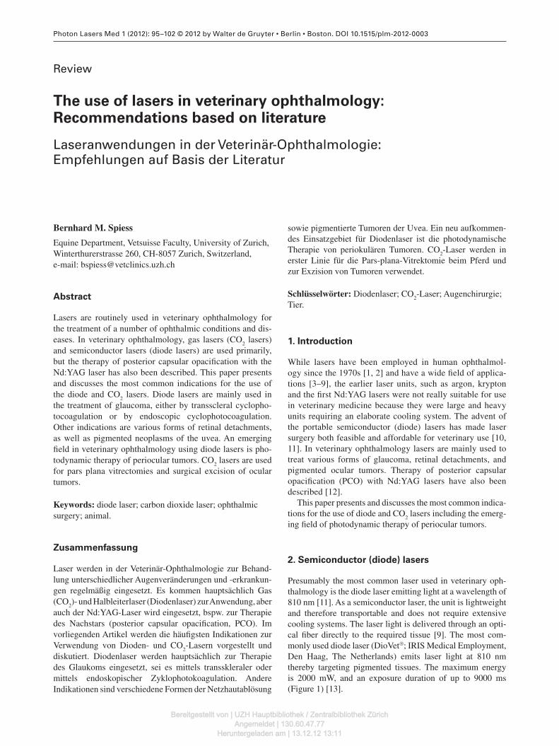
Presumably the most common laser used in veterinary oph-thalmology is the diode laser emitting light at a wavelength of 810 nm [11] . As a semiconductor laser, the unit is lightweight and therefore transportable and does not require extensive cooling systems. The laser light is delivered through an opti-cal fi ber directly to the required tissue [9] . The most com-monly used diode laser (DioVet ® ; IRIS Medical Employment, Den Haag, The Netherlands) emits laser light at 810 nm thereby targeting pigmented tissues. The maximum energy is 2000 mW, and an exposure duration of up to 9000 ms (Figure 1 ) [13] .
Bereitgestellt von | UZH Hauptbibliothek / Zentralbibliothek ZürichAngemeldet | 130.60.47.77
Heruntergeladen am | 13.12.12 13:11
96 B.M. Spiess: The use of lasers in veterinary ophthalmology
2.1. Transscleral laser cyclophotocoagulation
Diode lasers are routinely used to treat various forms of pri-mary glaucoma in dogs, cats and horses [11, 14 – 18] .
The goal is to partially destroy the non-pigmented cili-ary epithelium, to reduce aqueous humor secretion, and thus decrease intraocular pressure (IOP). The laser energy delivered is captured by the neighboring pigmented ciliary epithelium, and the resulting thermal effects also affect the non-pigmented epithelium.
In small animals, the laser probe is positioned perpendic-ular to the sclera and 3 mm posterior to the limbus (dorsal hemisphere), or 4 mm posterior to the limbus (ventral hemi-sphere). In horses the probe is positioned 4 – 6 mm posterior to the limbus (Figure 2 ) [16, 19, 20] .
The 3 o ’ clock and 9 o ’ clock positions and the posterior ciliary arteries are avoided [11] . The original settings were 1500 mW and 1500 ms for 36 spots. This energy level should cause a popping sound (photodisruption) in about 20 % of the spots, indicating an effective level of energy. However, photodisruption caused considerable postoperative uveitis, and consequently the settings were changed to 1000 mW and 3 – 5000 ms.
Photocoagulation, not photodisruption, is achieved with these settings [21] . Some surgeons treat only a small number of spots at a given time and repeat the treatment as needed to titrate IOP to the target pressure of < 20 mm Hg.
One of the pitfalls of transscleral laser photocoagulation is post-operative pressure spikes, which have to be carefully monitored and treated.
Other methods of cyclophotodestruction/-ablation include cyclocryotherapy. This is an established method in glauco-mas that are non-responsive to medical therapy. The expected destruction was rather unpredictable and sometimes short-lived. A serious complication of this treatment was the occur-rence of retinal detachments [22] .
2.2. Endocyclophotocoagulation
In recent years endoscopic laser cyclophotocoagulation [23] has been advocated but controlled studies of its long-term usefulness in animals are as yet lacking. This procedure involves the introduction of a diode laser probe and the trans-mission of laser light into the anterior chamber. The ciliary sulcus is extended with viscoelastic substances and the probe is advanced toward the ciliary processes. The ciliary epithe-lium is laser-treated in a controlled fashion over 270–360 degrees of the pars pilcata. Since cataract formation is a fre-quent complication of endolaser surgery, the procedure is best performed on aphakic or pseudophakic patients [24, 25] .
2.3. Transscleral laser retinopexy
Retinal detachments, so-called giant tears, are frequent com-plications of intracapsular extraction of luxated lenses in dogs and cats, but they can also occur spontaneously. The retinal disinsertions at the level of the ora ciliaris retinae can be avoided by prophylactic transscleral retinopexy [26] . In cer-tain breeds of dogs, rhegmatogenous retinal detachments are frequent complications after extracapsular cataract surgery and these can also be prevented by transscleral retinopexy. In general, 4–5 rows of spots are applied beginning 10 mm posterior to the limbus with a total of 65–75 spots. The rec-ommended settings are 800 mW and 1000 ms [26] .
Cryopexy of detached retinas has been used in canine vit-reoretinal surgery [27] , but has largely been replaced by laser retinopexy. The delivery of thermal energy to actively seal the retina to the underlying RPE appears to be more precise and effective with laser treatment.
2.4. Transpupillary laser retinopexy
Occasionally circumscribed bullous retinal detachments or localized retinal tears are encountered in animals. Usually these are incidental fi ndings since such localized lesions rarely lead to recognizable visual defi cits.
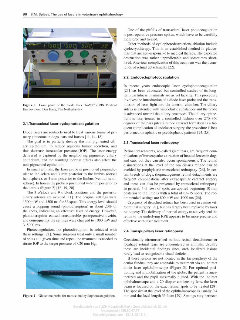
If these lesions are not located in the far periphery of the ocular fundus, they are amenable to treatment via an indirect diode laser ophthalmoscope (Figure 3 ). For optimal posi-tioning and immobilization of the globe, the patient is anes-thetized and the pupil maximally dilated. With the indirect ophthalmoscope and a 20 diopter condensing lens, the laser beam is focused on the exact retinal spots to be treated [28] . The spot size at the level of the ophthalmoscope is usually 0.4 mm and the focal length 35.6 cm [29] . Settings vary between
Figure 1 Front panel of the diode laser DioVet ® (IRIS Medical Employment, Den Haag, The Netherlands).
Figure 2 Glaucoma probe for transscleral cyclophotocoagulation.
Bereitgestellt von | UZH Hauptbibliothek / Zentralbibliothek ZürichAngemeldet | 130.60.47.77
Heruntergeladen am | 13.12.12 13:11
B.M. Spiess: The use of lasers in veterinary ophthalmology 97
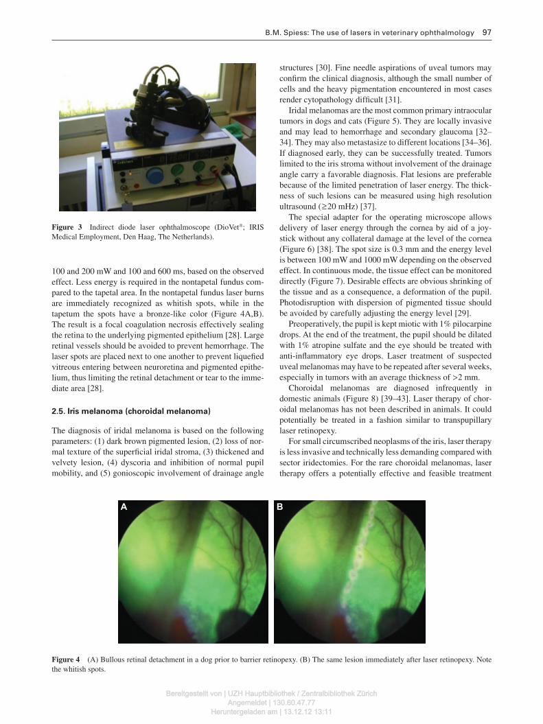
100 and 200 mW and 100 and 600 ms, based on the observed effect. Less energy is required in the nontapetal fundus com-pared to the tapetal area. In the nontapetal fundus laser burns are immediately recognized as whitish spots, while in the tapetum the spots have a bronze-like color (Figure 4 A,B). The result is a focal coagulation necrosis effectively sealing the retina to the underlying pigmented epithelium [28] . Large retinal vessels should be avoided to prevent hemorrhage. The laser spots are placed next to one another to prevent liquefi ed vitreous entering between neuroretina and pigmented epithe-lium, thus limiting the retinal detachment or tear to the imme-diate area [28] .
2.5. Iris melanoma (choroidal melanoma)
The diagnosis of iridal melanoma is based on the following parameters: (1) dark brown pigmented lesion, (2) loss of nor-mal texture of the superfi cial iridal stroma, (3) thickened and velvety lesion, (4) dyscoria and inhibition of normal pupil mobility, and (5) gonioscopic involvement of drainage angle
structures [30] . Fine needle aspirations of uveal tumors may confi rm the clinical diagnosis, although the small number of cells and the heavy pigmentation encountered in most cases render cytopathology diffi cult [31] .
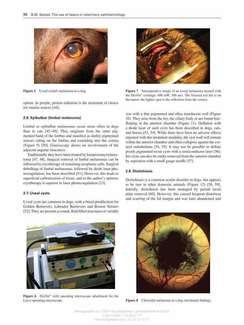
Iridal melanomas are the most common primary intraocular tumors in dogs and cats (Figure 5 ). They are locally invasive and may lead to hemorrhage and secondary glaucoma [32 – 34] . They may also metastasize to different locations [34 – 36] . If diagnosed early, they can be successfully treated. Tumors limited to the iris stroma without involvement of the drainage angle carry a favorable diagnosis. Flat lesions are preferable because of the limited penetration of laser energy. The thick-ness of such lesions can be measured using high resolution ultrasound ( ≥ 20 mHz) [37] .
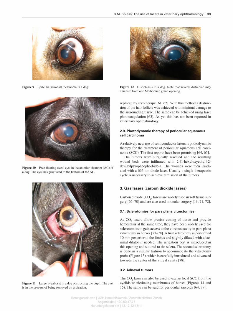
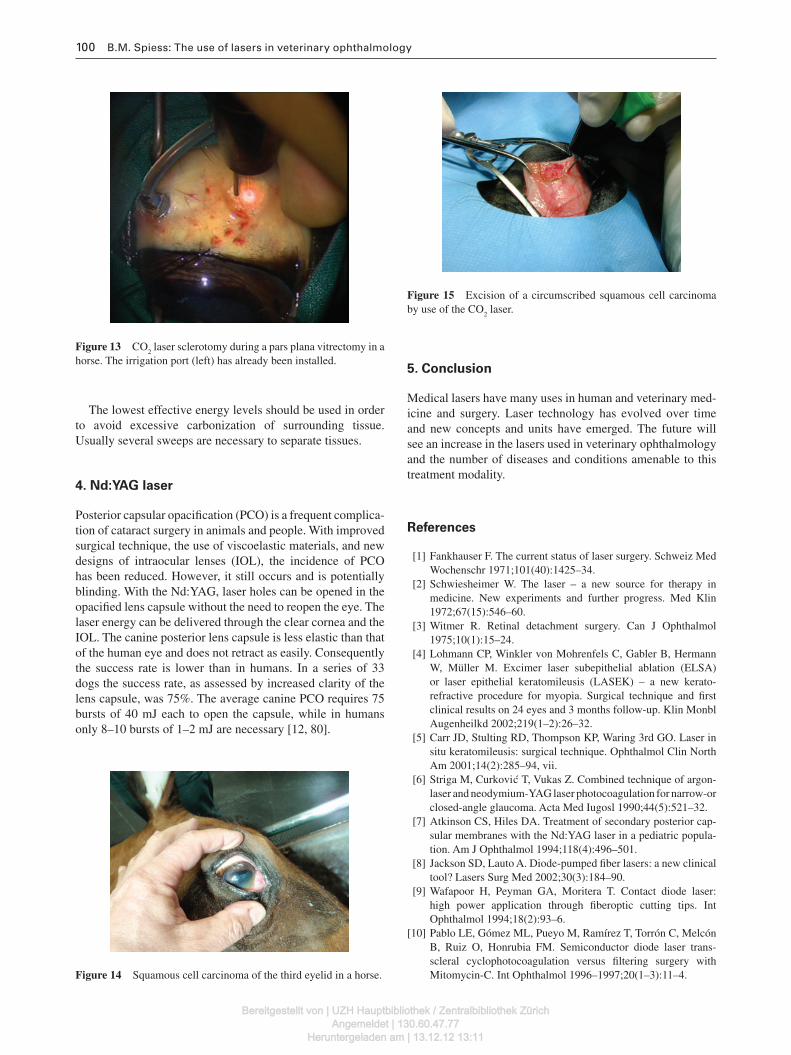
The special adapter for the operating microscope allows delivery of laser energy through the cornea by aid of a joy-stick without any collateral damage at the level of the cornea (Figure 6 ) [38] . The spot size is 0.3 mm and the energy level is between 100 mW and 1000 mW depending on the observed effect. In continuous mode, the tissue effect can be monitored directly (Figure 7 ). Desirable effects are obvious shrinking of the tissue and as a consequence, a deformation of the pupil. Photodisruption with dispersion of pigmented tissue should be avoided by carefully adjusting the energy level [29] .
Preoperatively, the pupil is kept miotic with 1 % pilocarpine drops. At the end of the treatment, the pupil should be dilated with 1 % atropine sulfate and the eye should be treated with anti-infl ammatory eye drops. Laser treatment of suspected uveal melanomas may have to be repeated after several weeks, especially in tumors with an average thickness of > 2 mm.
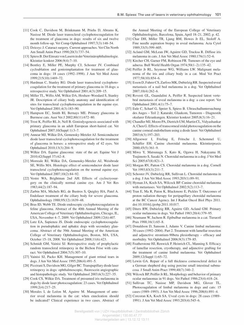
Choroidal melanomas are diagnosed infrequently in domestic animals (Figure 8 ) [39 – 43] . Laser therapy of chor-oidal melanomas has not been described in animals. It could potentially be treated in a fashion similar to transpupillary laser retinopexy.
For small circumscribed neoplasms of the iris, laser therapy is less invasive and technically less demanding compared with sector iridectomies. For the rare choroidal melanomas, laser therapy offers a potentially effective and feasible treatment
Figure 3 Indirect diode laser ophthalmoscope (DioVet ® ; IRIS Medical Employment, Den Haag, The Netherlands).
A B
Figure 4 (A) Bullous retinal detachment in a dog prior to barrier retinopexy. (B) The same lesion immediately after laser retinopexy. Note the whitish spots.
Bereitgestellt von | UZH Hauptbibliothek / Zentralbibliothek ZürichAngemeldet | 130.60.47.77
Heruntergeladen am | 13.12.12 13:11
98 B.M. Spiess: The use of lasers in veterinary ophthalmology
Figure 8 Choroidal melanoma in a dog (incidental fi nding). Figure 6 DioVet ® with operating microscope attachment for the Leica operating microscope.
Figure 7 Intraoperative image of an uveal melanoma treated with the DioVet ® (settings: 400 mW, 300 ms). The focused red dot is on the tumor, the lighter spot is the refl ection from the cornea.
Figure 5 Uveal (iridal) melanoma in a dog.
option. In people, proton radiation is the treatment of choice for similar tumors [44] .
2.6. Epibulbar (limbal melanoma)
Limbal or epibulbar melanomas occur more often in dogs than in cats [45 – 49] . They originate from the outer pig-mented band of the limbus and manifest as darkly pigmented masses riding on the limbus and extending into the cornea (Figure 9 ) [50] . Gonioscopy shows no involvement of the adjacent angular structures.
Traditionally they have been treated by keratectomy/sclerec-tomy [47, 48] . Surgical removal of limbal melanomas can be followed by cryotherapy of remaining neoplastic cells. Surgical debulking of limbal melanomas, followed by diode laser pho-tocoagulation, has been described [51] . However, this leads to superfi cial carbonization of tissue, and in the author ’ s opinion, cryotherapy is superior to laser photocoagulation [13] .
2.7. Uveal cysts
Uveal cysts are common in dogs, with a breed predilection for Golden Retrievers, Labrador Retrievers and Boston Terriers [52] . They are present as round, fl uid fi lled structures of variable
size with a thin pigmented and often translucent wall (Figure 10 ). They arise from the iris, the ciliary body or are found free-fl oating in the anterior chamber (Figure 11 ). Defl ation with a diode laser of such cysts has been described in dogs, cats, and horses [53, 54] . While there have been no adverse effects reported with this treatment modality, the cyst wall will remain within the anterior chamber and often collapses against the cor-neal endothelium [54, 55] . It may not be possible to defl ate poorly pigmented uveal cysts with a semiconductor laser [56] . Iris cysts can also be easily removed from the anterior chamber by aspiration with a small gauge needle [57] .
2.8. Distichiasis
Distichiasis is a common ocular disorder in dogs, but appears to be rare in other domestic animals (Figure 12 ) [58, 59] . Initially, distichiasis has been managed by partial tarsal plate removal [60] . However, this caused frequent distortion and scarring of the lid margin and was later abandoned and
Bereitgestellt von | UZH Hauptbibliothek / Zentralbibliothek ZürichAngemeldet | 130.60.47.77
Heruntergeladen am | 13.12.12 13:11
B.M. Spiess: The use of lasers in veterinary ophthalmology 99
Figure 9 Epibulbal (limbal) melanoma in a dog.
Figure 11 Large uveal cyst in a dog obstructing the pupil. The cyst is in the process of being removed by aspiration.
Figure 10 Free-fl oating uveal cyst in the anterior chamber (AC) of a dog. The cyst has gravitated to the bottom of the AC.
Figure 12 Distichiasis in a dog. Note that several distichiae may emanate from one Meibomian gland opening.
replaced by cryotherapy [61, 62] . With this method a destruc-tion of the hair follicle was achieved with minimal damage to the surrounding tissue. The same can be achieved using laser photocoagulation [63] . As yet this has not been reported in veterinary ophthalmology.
2.9. Photodynamic therapy of periocular squamous
cell carcinoma
A relatively new use of semiconductor lasers is photodynamic therapy for the treatment of periocular squamous cell carci-noma (SCC). The fi rst reports have been promising [64, 65] .
The tumors were surgically resected and the resulting wound beds were infi ltrated with 2-[1-hexyloxyethyl]-2-devinylpyropheophorbide-a. The wounds were then irradi-ated with a 665 nm diode laser. Usually a single therapeutic cycle is necessary to achieve remission of the tumors.
3. Gas lasers (carbon dioxide lasers)
Carbon dioxide (CO 2 ) lasers are widely used in soft tissue sur-gery [66 – 70] and are also used in ocular surgery [13, 71, 72] .
3.1. Sclerotomies for pars plana vitrectomies
As CO 2 lasers allow precise cutting of tissue and provide hemostasis at the same time, they have been widely used for sclerotomies to gain access to the vitreous cavity in pars plana vitrectomy in horses [73 – 78] . A fi rst sclerotomy is performed 10 mm posterior to the limbus and slightly dilated with a lac-rimal dilator if needed. The irrigation port is introduced in this opening and sutured to the sclera. The second sclerotomy is done in a similar fashion to accommodate the vitrectomy probe (Figure 13 ), which is carefully introduced and advanced towards the center of the vitreal cavity [78] .
3.2. Adnexal tumors
The CO 2 laser can also be used to excise focal SCC from the eyelids or nictitating membranes of horses (Figures 14 and 15 ). The same can be said for periocular sarcoids [64, 79] .
Bereitgestellt von | UZH Hauptbibliothek / Zentralbibliothek ZürichAngemeldet | 130.60.47.77
Heruntergeladen am | 13.12.12 13:11
100 B.M. Spiess: The use of lasers in veterinary ophthalmology
Figure 15 Excision of a circumscribed squamous cell carcinoma by use of the CO 2 laser.
Figure 13 CO 2 laser sclerotomy during a pars plana vitrectomy in a horse. The irrigation port (left) has already been installed.
Figure 14 Squamous cell carcinoma of the third eyelid in a horse.
The lowest effective energy levels should be used in order to avoid excessive carbonization of surrounding tissue. Usually several sweeps are necessary to separate tissues.
4. Nd:YAG laser
Posterior capsular opacifi cation (PCO) is a frequent complica-tion of cataract surgery in animals and people. With improved surgical technique, the use of viscoelastic materials, and new designs of intraocular lenses (IOL), the incidence of PCO has been reduced. However, it still occurs and is potentially blinding. With the Nd:YAG, laser holes can be opened in the opacifi ed lens capsule without the need to reopen the eye. The laser energy can be delivered through the clear cornea and the IOL. The canine posterior lens capsule is less elastic than that of the human eye and does n o t retract as easily. Consequently the success rate is lower than in humans. In a series of 33 dogs the success rate, as assessed by increased clarity of the lens capsule, was 75 % . The average canine PCO requires 75 bursts of 40 mJ each to open the capsule, while in humans only 8–10 bursts of 1–2 mJ are necessary [12, 80] .
5. Conclusion
Medical lasers have many uses in human and veterinary med-icine and surgery. Laser technology has evolved over time and new concepts and units have emerged. The future will see an increase in the lasers used in veterinary ophthalmology and the number of diseases and conditions amenable to this treatment modality.
References
[1] Fankhauser F. The current status of laser surgery. Schweiz Med Wochenschr 1971;101(40):1425 – 34.
[2] Schwiesheimer W. The laser – a new source for therapy in medicine. New experiments and further progress. Med Klin 1972;67(15):546 – 60.
[3] Witmer R. Retinal detachment surgery. Can J Ophthalmol 1975;10(1):15 – 24.
[4] Lohmann CP, Winkler von Mohrenfels C, Gabler B, Hermann W, M ü ller M. Excimer laser subepithelial ablation (ELSA) or laser epithelial keratomileusis (LASEK) – a new kerato-refractive procedure for myopia. Surgical technique and fi rst clinical results on 24 eyes and 3 months follow-up. Klin Monbl Augenheilkd 2002;219(1 – 2):26 – 32.
[5] Carr JD, Stulting RD, Thompson KP, Waring 3rd GO. Laser in situ keratomileusis: surgical technique. Ophthalmol Clin North Am 2001;14(2):285 – 94, vii.
[6] Striga M, Curkovi c T, Vukas Z. Combined technique of argon-laser and neodymium-YAG laser photocoagulation for narrow-or closed-angle glaucoma. Acta Med Iugosl 1990;44(5):521 – 32.
[7] Atkinson CS, Hiles DA. Treatment of secondary posterior cap-sular membranes with the Nd:YAG laser in a pediatric popula-tion. Am J Ophthalmol 1994;118(4):496 – 501.
[8] Jackson SD, Lauto A. Diode-pumped fi ber lasers: a new clinical tool ? Lasers Surg Med 2002;30(3):184 – 90.
[9] Wafapoor H, Peyman GA, Moritera T. Contact diode laser: high power application through fi beroptic cutting tips. Int Ophthalmol 1994;18(2):93 – 6.
[10] Pablo LE, G ó mez ML, Pueyo M, Ram í rez T, Torr ó n C, Melc ó n B, Ruiz O, Honrubia FM. Semiconductor diode laser trans-scleral cyclophotocoagulation versus fi ltering surgery with Mitomycin-C. Int Ophthalmol 1996 – 1997;20(1 – 3):11 – 4.
Bereitgestellt von | UZH Hauptbibliothek / Zentralbibliothek ZürichAngemeldet | 130.60.47.77
Heruntergeladen am | 13.12.12 13:11
B.M. Spiess: The use of lasers in veterinary ophthalmology 101
[11] Cook C, Davidson M, Brinkmann M, Priehs D, Abrams K, Nasisse M. Diode laser transscleral cyclophotocoagulation for the treatment of glaucoma in dogs: results of six and twelve month follow-up. Vet Comp Ophthalmol 1997;7(3):148 – 54.
[12] Dziezyc J. Cataract surgery. Current approaches. Vet Clin North Am Small Anim Pract 1990;20(3):737 – 54.
[13] Spiess B. Der Einsatz von Lasern in der Veterin ä rophthalmologie. Kleintier konkret 2006;9(4):7 – 10.
[14] Bentley E, Miller PE, Murphy CJ, Schoster JV. Combined cycloablation and gonioimplantation for treatment of glau-coma in dogs: 18 cases (1992 – 1998). J Am Vet Med Assoc 1999;215(10):1469 – 72.
[15] Hardman C, Stanley RG. Diode laser transscleral cyclophoto-coagulation for the treatment of primary glaucoma in 18 dogs: a retrospective study. Vet Ophthalmol 2001;4(3):209 – 15.
[16] Miller TL, Willis AM, Wilkie DA, Hoshaw-Woodard S, Stanley JR. Description of ciliary body anatomy and identifi cation of sites for transscleral cyclophotocoagulation in the equine eye. Vet Ophthalmol 2001;4(3):183 – 90.
[17] Hampson EC, Smith RI, Bernays ME. Primary glaucoma in Burmese cats. Aust Vet J 2002;80(11):672 – 80.
[18] Trost K, Peiffer RL Jr, Nell B. Goniodysgenesis associated with primary glaucoma in an adult European short-haired cat. Vet Ophthalmol 2007;10(Suppl 1):3 – 7.
[19] Annear MJ, Wilkie DA, Gemensky-Metzler AJ. Semiconductor diode laser transscleral cyclophotocoagulation for the treatment of glaucoma in horses: a retrospective study of 42 eyes. Vet Ophthalmol 2010;13(3):204 – 9.
[20] Wilkie DA. Equine glaucoma: state of the art. Equine Vet J 2010;42(Suppl 37):62 – 8.
[21] Morreale RJ, Wilkie DA, Gemensky-Metzler AJ, Weisbrode SE, Willis MA. Histologic effect of semiconductor diode laser transscleral cyclophotocoagulation on the normal equine eye. Vet Ophthalmol 2007;10(2):84 – 92.
[22] Vestre WA, Brightman 2nd AH. Effects of cyclocryosur-gery on the clinically normal canine eye. Am J Vet Res 1983;44(2):187 – 94.
[23] Zarbin MA, Michels RG, de Bustros S, Quigley HA, Patel A. Endolaser treatment of the ciliary body for severe glaucoma. Ophthalmology 1988;95(12):1639 – 48.
[24] Bras ID, Webb TE. Diode endoscopic cyclophotocoagulation in feline glaucoma. Abstract of the 40th Annual Meeting of the American College of Veterinary Ophthalmologists, Chicago, IL, USA, November 4 – 7, 2009. Vet Ophthalmol 2009;12(6):407.
[25] Lutz EA, Sapienza JS. Diode endoscopic cyclophotocoagula-tion in pseudophakic and aphakic dogs with secondary glau-coma. Abstract of the 39th Annual Meeting of the American College of Veterinary Ophthalmologists, Boston, MA, USA, October 15 – 18, 2008. Vet Ophthalmol 2008;11(6):423.
[26] Schmidt GM, Vainisi SJ. Retrospective study of prophylactic random transscleral retinopexy in the Bichon Frise with cata-ract. Vet Ophthalmol 2004;7(5):307 – 10.
[27] Vainisi SJ, Packo KH. Management of giant retinal tears in dogs. J Am Vet Med Assoc 1995;206(4):491 – 5.
[28] Pizzirani S, Davidson MG, Gilger BC. Transpupillary diode laser retinopexy in dogs: ophthalmoscopic, fl uorescein angiographic and histopathologic study. Vet Ophthalmol 2003;6(3):227 – 35.
[29] Cook CS, Wilkie DA. Treatment of presumed iris melanoma in dogs by diode laser photocoagulation: 23 cases. Vet Ophthalmol 1999;2(4):217 – 25.
[30] Morales I, de Le é on M, Aguirre M. Management of ante-rior uveal melanoma in the cat: when enucleation should be indicated ? Clinical experience in two cases. Abstract of
the Annual Meeting of the European College of Veterinary Ophthalmologists, Barcelona, Spain, April 18 – 21, 2002; p. 42.
[31] Char DH, Miller TR, Ljung BM, Howes Jr EL, Stoloff A. Fine needle aspiration biopsy in uveal melanoma. Acta Cytol 1989;33(5):599 – 605.
[32] Acland GM, McLean IW, Aguirre GD, Trucksa R. Diffuse iris melanoma in cats. J Am Vet Med Assoc 1980;176(1):52 – 6.
[33] Kircher CH, Garner FM, Robinson FR. Tumours of the eye and adnexa. Bull World Health Organ 1974;50(1 – 2):135 – 42.
[34] Peiffer Jr RL, Seymour WG, Williams LW. Malignant mela-noma of the iris and ciliary body in a cat. Mod Vet Pract 1977;58(10):854 – 6.
[35] Esson D, Fahrer CS, Zarfoss MK, Dubielzig RR. Suspected uveal metastasis of a nail bed melanoma in a dog. Vet Ophthalmol 2007;10(4):262 – 6.
[36] Rovesti GL, Guandalini A, Peiffer R. Suspected latent verte-bral metastasis of uveal melanoma in a dog: a case report. Vet Ophthalmol 2001;4(1):75 – 7.
[37] Eule C, Scharf G, Speier S, Spiess B. Ultraschalluntersuchung des Auges – Teil 2: Katarakt, Glaukom, Tumoren – Diagnostik okularer Erkrankungen. Kleintier konkret 2005;8(3):16 – 21.
[38] Chandler MJ, Moore PA, Dietrich UM, Martin CL, Vidyashankar A, Chen G. Effects of transcorneal iridal photocoagulation on the canine corneal endothelium using a diode laser. Vet Ophthalmol 2003;6(3):197 – 203.
[39] Allgoewer I, Frieling E, Fritsche J, Schemmel U, Sch ä ffer EH. Canine choroidal melanoma. Kleintierpraxis 2000;45(5):361 – 9.
[40] Miwa Y, Matsunaga S, Kato K, Ogawa H, Nakayama H, Tsujimoto S, Sasaki N. Choroidal melanoma in a dog. J Vet Med Sci 2005;67(8):821 – 3.
[41] Morgan RV, Patton CS. Choroidal melanoma in a dog. Cornell Vet 1993;83(3):211 – 7.
[42] Schoster JV, Dubielzig RR, Sullivan L. Choroidal melanoma in a dog. J Am Vet Med Assoc 1993;203(1):89 – 91.
[43] Hyman JA, Koch SA, Wilcock BP. Canine choroidal melanoma with metastases. Vet Ophthalmol 2002;5(2):113 – 7.
[44] Tran E, Ma R, Paton K, Blackmore E, Pickles T. Outcomes of proton radiation therapy for peripapillary choroidal melanoma at the BC Cancer Agency. Int J Radiat Oncol Biol Phys 2011. doi:10.1016/j.ijrobp.2011.10.017.
[45] Diters RW, Dubielzig RR, Aguirre GD, Acland GM. Primary ocular melanoma in dogs. Vet Pathol 1983;20(4):379 – 95.
[46] Neumann W, Juchem R. Epibulbar melanoma in a cat. Tierarztl Prax 1988;16(1):65 – 8.
[47] Donaldson D, Sansom J, Adams V. Canine limbal melanoma: 30 cases (1992 – 2004). Part 2. Treatment with lamellar resection and adjunctive strontium-90beta plesiotherapy – effi cacy and morbidity. Vet Ophthalmol 2006;9(3):179 – 85.
[48] Featherstone HJ, Renwick P, Heinrich CL, Manning S. Effi cacy of lamellar resection, cryotherapy, and adjunctive grafting for the treatment of canine limbal melanoma. Vet Ophthalmol 2009;12(Suppl 1):65 – 72.
[49] Lewin GA. Repair of a full thickness corneoscleral defect in a German shepherd dog using porcine small intestinal submu-cosa. J Small Anim Pract 1999;40(7):340 – 2.
[50] Wilcock BP, Peiffer Jr RL. Morphology and behavior of primary ocular melanomas in 91 dogs. Vet Pathol 1986;23(4):418 – 24.
[51] Sullivan TC, Nasisse MP, Davidson MG, Glover TL. Photocoagulation of limbal melanoma in dogs and cats: 15 cases (1989 – 1993). J Am Vet Med Assoc 1996;208(6):891 – 4.
[52] Corcoran KA, Koch SA. Uveal cysts in dogs: 28 cases (1989 – 1991). J Am Vet Med Assoc 1993;203(4):545 – 6.
Bereitgestellt von | UZH Hauptbibliothek / Zentralbibliothek ZürichAngemeldet | 130.60.47.77
Heruntergeladen am | 13.12.12 13:11
102 B.M. Spiess: The use of lasers in veterinary ophthalmology
[53] Gemensky-Metzler AJ, Wilkie DA, Cook CS. The use of semi-conductor diode laser for defl ation and coagulation of anterior uveal cysts in dogs, cats and horses: a report of 20 cases. Vet Ophthalmol 2004;7(5):360 – 8.
[54] Grahn BH, Wolfer J. Diagnostic ophthalmology. Distichiasis and uveal cysts. Can Vet J 1997;38(6):391 – 2.
[55] Bedford PG. The anterior uveal cyst as an unusual cause of corneal pigmentation in the dog. J Small Anim Pract 1980;21(2):97 – 101.
[56] Spiess BM, Bolliger JO, Guscetti F, Haessig M, Lackner PA, Ruehli MB. Multiple ciliary body cysts and secondary glau-coma in the Great Dane: a report of nine cases. Vet Ophthalmol 1998;1(1):41 – 5.
[57] Hendrix DVH. Diseases and surgery of the canine anterior uvea. In: Gelatt KN, editor. Essentials of veterinary ophthalmology. 2nd ed. Iowa: Blackwell Publishing; 2008, p. 189 – 216.
[58] Lawson DD. Canine distichiasis. J Small Anim Pract 1973;14(8):469 – 78.
[59] Bellhorn RW. Variation of canine distichiasis. J Am Vet Med Assoc 1970;157(3):342 – 3.
[60] Bedford PG. Distichiasis and its treatment by the method of partial tarsal plate excision. J Small Anim Pract 1973;14(1):1 – 5.
[61] Delaney MR, Rogers PA. A simplifi ed cryotherapy tech-nique for trichiasis and distichiasis. Aust J Ophthalmol 1984;12(2):163 – 6.
[62] Frueh BR. Treatment of distichiasis with cryotherapy. Ophthalmic Surg 1981;12(2):100 – 3.
[63] Pham RT, Biesman BS, Silkiss RZ. Treatment of trichiasis using an 810-nm diode laser: an effi cacy study. Ophthal Plast Reconstr Surg 2006;22(6):445 – 7.
[64] Giuliano EA. Equine periocular neoplasia: current concepts in aetiopathogenesis and emerging treatment modalities. Equine Vet J 2010;42(Suppl 37):9 – 18.
[65] Giuliano EA, MacDonald I, McCaw DL, Dougherty TJ, Klauss G, Ota J, Pearce JW, Johnson PJ. Photodynamic ther-apy for the treatment of periocular squamous cell carcinoma in horses: a pilot study. Vet Ophthalmol 2008;11(Suppl 1):27 – 34.
[66] Taney K, Smith MM. Resection of mast cell tumor of the lip in a dog. J Vet Dent 2009;26(1):28 – 34.
[67] Schick RO, Schick MP. CO 2 laser surgery in veterinary derma-tology. Clin Dermatol 1994;12(4):587 – 9.
[68] Clark GN, Sinibaldi KR. Use of a carbon dioxide laser for treat-ment of elongated soft palate in dogs. J Am Vet Med Assoc 1994;204(11):1779 – 81.
[69] Bergh A, Nyman G, Lundeberg T, Drevemo S. Effect of defo-cused CO 2 laser on equine tissue perfusion. Acta Vet Scand 2006;47:33 – 42.
[70] Bergh A, Ridderstr å le Y, Ekman S. Defocused CO 2 laser on equine skin: a histological examination. Equine Vet J 2007;39(2):114 – 9.
[71] Gilmour MA. Laser applications for corneal disease. Clin Tech Small Anim Pract 2003;18(3):199 – 202.
[72] Rayner SG, Van Zyl N. The use of mitomycin C as an adjunc-tive treatment for equine ocular squamous cell carcinoma. Aust Vet J 2006;84(1 – 2):43 – 6.
[73] Fr ü hauf B, Ohnesorge B, Deegen E, Boev é M. Surgical man-agement of equine recurrent uveitis with single port pars plana vitrectomy. Vet Ophthalmol 1998;1(2 – 3):137 – 51.
[74] Gilger BC, Michau TM. Equine recurrent uveitis: new methods of management. Vet Clin North Am Equine Pract 2004;20(2):417 – 27, vii.
[75] T ö m ö rdy E, H ä ssig M, Spiess BM. The outcome of parsplane vitrectomy in horses with equine recurrent uveitis with regard to the presence or absence of intravitreal antibodies against various serovars of Leptospira interrogans. Pferdeheilkunde 2010;26(2):251 – 4.
[76] Werry H, Gerhards H. The surgical therapy of equine recurrent uveitis. Tierarztl Prax 1992;20(2):178 – 86.
[77] Gelatt KN, Spiess BM, Gilger BC. Vitreoretinal surgery. In: Gelatt KN, Gelatt JP, editors. Veterinary ophthalmic surgery. St. Louis: Elsevier Saunders; 2011, p. 357 – 87.
[78] Spiess BM. Pars plana vitrectomy. In: Gilger BC, editor. Equine ophthalmology. 2nd ed. St. Louis: Elsevier Saunders; 2010, p. 344 – 6.
[79] English RV, Nasisse MP, Davidson MG. Carbon dioxide laser ablation for treatment of limbal squamous cell carcinoma in horses. J Am Vet Med Assoc 1990;196(3):439 – 42.
[80] Beale AB, Salmon J, Michau TM, Gilger BC. Effect of oph-thalmic Nd:YAG laser energy on intraocular lenses after posterior capsulotomy in normal dog eyes. Vet Ophthalmol 2006;9(5):335 – 40.
Received January 16, 2012; revised February 27, 2012; accepted March 1, 2012
Bereitgestellt von | UZH Hauptbibliothek / Zentralbibliothek ZürichAngemeldet | 130.60.47.77
Heruntergeladen am | 13.12.12 13:11