the structure of cyp101d2 unveils a potential path for ... · the structure of cyp101d2 unveils a...
TRANSCRIPT

Biochem. J. (2011) 433, 85–93 (Printed in Great Britain) doi:10.1042/BJ20101017 85
The structure of CYP101D2 unveils a potential path for substrate entry intothe active siteWen YANG*, Stephen G. BELL†1, Hui WANG‡, Weihong ZHOU*, Mark BARTLAM*1, Luet-Lok WONG† and Zihe RAO*‡*Tianjin Key Laboratory of Protein Science, College of Life Sciences, Nankai University, Tianjin 300071, China, †Inorganic Chemistry Laboratory, Department of Chemistry, University ofOxford, South Parks Road, Oxford OX1 3QR, U.K., and ‡Laboratory of Structural Biology, Tsinghua University, Beijing 100084, China
The cytochrome P450 CYP101D2 from Novosphingobiumaromaticivorans DSM12444 is closely related to CYP101D1from the same bacterium and to P450cam (CYP101A1) fromPseudomonas putida. All three are capable of oxidizing camphorstereoselectively to 5-exo-hydroxycamphor. The crystal structureof CYP101D2 revealed that the likely ferredoxin-binding site onthe proximal face is largely positively charged, similar to that ofCYP101D1. However, both the native and camphor-soaked formsof CYP101D2 had open conformations with an access channel.In the active site of the camphor-soaked form, the camphorcarbonyl interacted with the haem-iron-bound water. Two otherpotential camphor-binding sites were also identified from electrondensities in the camphor-soaked structure: one located in the
access channel, flanked by the B/C and F/G loops and the Ihelix, and the other in a cavity on the surface of the enzyme nearthe F helix side of the F/G loop. The observed open structuresmay be conformers of the CYP101D2 enzyme that enable thesubstrate to enter the buried active site via a conformationalselection mechanism. The second and third binding sites may beintermediate locations of substrate entry and translocation into theactive site, and provide insight into a multi-step substrate-bindingmechanism.
Key words: access channel, crystal structure, cytochromeP450, multi-step binding mechanism, Novosphingobiumaromaticivorans DSM12444, open conformation.
INTRODUCTION
The CYP (cytochrome P450) superfamily of haem-dependentmono-oxygenases is widely distributed and catalyses C−H bondoxidation in a broad range of organic molecules [1,2]. Throughthis chemically remarkable reaction and other activities, CYP en-zymes are involved in a variety of biochemical processes, includ-ing biosynthesis, biodegradation and xenobiotic metabolism [3,4].Despite low sequence identities, structurally characterized CYPenzymes have a similar overall fold, but considerable structuraldiversity within this fold leads to different substrate specificity [5].The structures of the substrate-free and substrate-bound forms ofCYP enzymes often have similar closed or open conformationswith little or no change upon substrate binding. CYP enzymeaccess channels have been classified via a systematic analysis ofstructurally characterized enzymes [6]. Substrate entry and bin-ding are mainly controlled by residues within the B, B′, F and Ghelices, and the B/C and F/G loops [6−8]. However, the detailsof how substrates are recognized and enter the active site areoften poorly defined. Molecular dynamic simulations of camphorbinding to CYP101A1 from Pseudomonas putida suggest thatprotein backbone conformational changes and aromatic side chainrotations occur to allow camphor entry into the active site, andexperiments have shown that salt-bridge interactions modulatesubstrate access [7,9,10]. On the basis of theoretical and NMRstudies, a second camphor-binding site, on the protein surface ofCYP101A1, and a two-step mechanism for camphor binding havebeen proposed [11]. Multi-step binding mechanisms and multiplesubstrate occupancy have been investigated in the mammalianenzymes CYP3A4 [12] and CYP1A2 [13].
Crystal structures of different substrate-bound forms ofCYP119 from Sulfolobus acidocalderius show significantconformational dissimilarities [14−16], and NMR studiesindicate that multiple conformations of the substrate-free enzymemay exist [17,18]. This may allow substrate binding to occurvia a process of conformation selection, as suggested fromkinetic analysis of substrate binding to P450 EryK (CYP113A1)from Saccharopolyspora erythraea [19]. Crystal structures ofmany forms of CYP101A1 have been determined in order tobetter understand P450 catalysis. These include the substrate andproduct complexes [20,21], the ferrous−carbon monoxide- andferrous−oxygen-bound forms [22−24] and mutant enzymestructures [25,26]. In the majority of these structures, a closedconformation is observed and structural information on substrateentry into the active site is not available. Open conformations ofCYP101A1 have been induced by binding adamantyl substrateslinked to dansyl fluorophores or ruthenium complexes that forceopen the enzyme structure [27−29]. Recently, substrate-freestructures of CYP101A1 have been reported in an open conform-ation similar to those observed with the linker substrates [30].
The CYP101A1 homologues CYP101D2 and CYP101D1from Novosphingobium aromaticivorans DSM12444 bothbind camphor and oxidize it stereoselectively to 5-exo-hydroxycamphor with high activity and coupling of productformation to NADH consumption. Both enzymes utilize a classI electron-transfer system, consisting of a NADH-dependentFAD-containing ferredoxin reductase ArR and a [2Fe−2S]ferredoxin Arx, which are analogous to PdR (putidaredoxinreductase) and Pdx (putidaredoxin) of the CYP101A1 system[31]. We have previously solved the crystal structures of ArR,
Abbreviations used: CYP, cytochrome P450; D-4-Ad, dansylbutyladamantane; D-8-Ad, dansyloctyladamantane; DEG, diethylene glycol; MR, molecularreplacement; PdR, putidaredoxin reductase; Pdx, putidaredoxin; PEG, poly(ethylene) glycol; RMSD, root mean square deviation; Ru-F8bp-Ad, rutheniumtris(bipyridine)-4,4′-octafluorobiphenyladamantane.
1 Correspondence may be addressed to either of these authors (email [email protected] or [email protected]).The co-ordinates for the crystal structures of native CYP101D2 and camphor-soaked CYP101D2 have been deposited in the PDB under the accession
codes 3NV5 and 3NV6 respectively.
c© The Authors Journal compilation c© 2011 Biochemical Society

86 W. Yang and others
Arx and CYP101D1, a complete physiological class I electron-transfer system [31]. The structures and the kinetic data indicatethat electrostatic interactions are important in protein−proteinrecognition. In the present paper we report the structure ofCYP101D2 in the native and camphor-soaked forms. Bothstructures have an open conformation with an access channel.The camphor-soaked structure contains three regions of electrondensity that could be modelled as camphor molecules. Oneputative camphor molecule was located in the access channelclose to the F/G and B/C loops, whereas the second was foundat the enzyme surface on the F helix side of the F/G loop. Thethird camphor molecule was bound within the active site where itscarbonyl oxygen interacted with the water ligand of the haem iron.
EXPERIMENTAL
Cloning, expression and purification
An N-terminal His6 tag was incorporated into CYP101D2by transferring the encoding gene into the expression vectorpET28a(+) (Novagen) using the NdeI and HindIII restrictionenzymes [32]. The recombinant plasmid was transformed intoEscherichia coli BL21(DE3). A single colony was cultured at37 ◦C in LB (Luria−Bertani) medium containing 50 μg · ml− 1
kanamycin, to an D600 of 0.6−0.8, and then induced with0.5 mM IPTG (isopropyl β-D-thiogalactopyranoside) for 18 h at20 ◦C. The cells were harvested by centrifugation (6000 g for15 min), resuspended in 1× PBS, pH 7.4, and then disrupted bysonication at 4 ◦C. The supernatant obtained by centrifugationat 27000 g for 30 min was loaded on to a Ni2+-chelating affinitycolumn pre-equilibrated with 1× PBS. After washing with 200 mlof 1× PBS, pH 7.4, containing 20 mM imidazole, the targetprotein was eluted from the column with 1× PBS, pH 7.4,containing 300 mM imidazole. The protein was concentratedand then applied to a Superdex-200 (GE Healthcare) columnequilibrated with 1× PBS. The red-coloured fractions werecollected and concentrated in buffer A [20 mM Tris/HCl,pH 8.0, and 1 mM DTT (dithiothreitol)]. The protein wasfurther purified using a Resource Q anion-exchange column(GE Healthcare), eluting with a linear gradient of 0−1 M NaClin buffer A. The purity of CYP101D2 was estimated to be>95% by SDS/PAGE analysis (Supplementary Figure S1 athttp://www.BiochemJ.org/bj/433/bj4330085add.htm).
Crystallization and substrate-soaking experiments
The purified CYP101D2 protein was concentrated to 50 mg · ml− 1
in crystallization buffer (20 mM Tris/HCl, pH 8.0, and 150 mMKCl). Crystallization was performed by the hanging-drop vapour-diffusion method at 18 ◦C. The protein solution (1 μl) was mixedwith an equal volume of reservoir solution and equilibratedwith 200 μl of reservoir solution. Crystal screening was carriedout with Hampton Research Crystal Screen kits. Initially, smallcrystals were obtained using 0.1 M Hepes, pH 7.5, 2% (v/v)PEG [poly(ethylene glycol)] 400 and 2.0 M ammonium sulfate(condition number 39 of Crystal Screen I) and further optimizationwas performed by adjusting the buffer, pH and concentration ofprecipitant. Good quality crystals were obtained after approx.1 week from 0.1 M Tris/HCl, pH 8.3, 2.1 M ammonium sulfateand 4% (v/v) PEG 400. The camphor substrate was soaked intocrystals of native CYP101D2 by adding solid camphor to the dropscontaining the crystals. Camphor saturated the crystal solutionafter several days and the soaking time was varied from 1 weekto 1 month.
Data collection and structure determination
Immediately prior to data collection all crystals werecryoprotected by the addition of 20% (v/v) glycerol. X-raydiffraction data were collected in-house at −173 ◦C on a mar345image plate using Cu Kα radiation (λ = 1.5418 Å; 1 Å = 0.1 nm)from a Rigaku MicroMax-007 rotating-anode X-ray generatoroperating at 40 kV and 20 mA. All diffraction data were indexed,integrated and scaled with the HKL2000 package [33].
The structure of native CYP101D2 was solved by the MR(molecular replacement) method using the program Phaser [34]in the CCP4 suite [35] with CYP101D1 as a search model (PDBcode 3LXH). The initial model was then rebuilt using Coot[36] and refined by Refmac5 [37]. The CYP101D2 camphor-soaked structure was solved by the MR method using the nativeCYP101D2 as a search model. The programs Coot and Refmac5were used for manual adjustment and refinement of the model.
The stereochemical quality of the structures was examinedwith the program PROCHECK [38]. The data collection andrefinement statistics are summarized in Table 1.
RESULTS
Structure of CYP101D2
The crystal structures of native and camphor-soaked CYP101D2were solved at 2.4 Å and 2.2 Å resolution respectively. The finalmodels of both structures contained a single CYP101D2 molecule.The native structure contained a single DEG (diethylene glycol;C4H10O3) molecule and was traced from residues 10−413 (out of416) with the exception of residues 92 and 93 in the B′ helix andresidues 189−191 in the F/G loop. The camphor-soaked structurewas also traced from residues 10−413, and the entire B′ helix andF/G loop could be modelled.
CYP101D2 had the characteristic trigonal prism-shaped, mixedα/β structure commonly found for CYP enzymes (Figure 1A).The organization of the secondary-structure elements of nativeand camphor-soaked CYP101D2 were virtually identical [RMSD(root mean square deviation) of 0.49 Å for 399 Cα atoms],and both were similar to CYP101D1 (PDB code 3LXH) andCYP101A1 (PDB code 2CPP) (Supplementary Figure S2 andSupplementary Table S1 at http://www.BiochemJ.org/bj/433/bj4330085add.htm). However, both structures had an openconformation resulting from shifts of the F helix (residues173−188), G helix (192−217), H helix (225−232) and theN-terminus of the I helix (241−261). These helices werepushed away from the haem, thus opening an access channelfrom the haem to the enzyme surface (Figures 1B–1D andSupplementary Figure S2C). These arrangements resemble theopen conformations found in the substrate-free (PDB code 3L61)[30] and linker-substrate-bound CYP101A1 structures {Ru-F8bp-Ad [ruthenium tris(bipyridine)-4,4′-octafluorobiphenyl-adamantane], PDB code 1K2O [28]; D-4-Ad (dansylbutyladam-antane), PDB code 1RF9; and D-8-Ad (dansyloctyladamantane),PDB code 1RE9 [29]} (Figure 1D and SupplementaryFigure S2).
The structure of native CYP101D2 contained electron densityin the active site that was modelled as DEG, which is present inthe PEG 400 from the crystallization buffer. The carbon atomsof the DEG molecule were in van der Waals contact with Leu250,Gly254, Thr258 and Val301 (Supplementary Figure S3 at http://www.BiochemJ.org/bj/433/bj4330085add.htm), whereas the O2 andO3 atoms were hydrogen-bonded to Wat-568 (3.1 Å), the sixthligand to the haem iron (Fe−O, 2.2 Å), and Glu303 (3.3 Å)respectively.
c© The Authors Journal compilation c© 2011 Biochemical Society

Crystal structure of CYP101D2 87
Table 1 X-ray data collection and structure refinement statistics for native (DEG-bound) and camphor-soaked CYP101D2
Values in parentheses are for the highest-resolution shell. Rmerge = � i|Ii-〈I〉|/�〈I〉, where Ii is an individual intensity measurement and 〈I〉 is the average intensity for all the reflections.Rwork/R free = �‖F o| − |F c‖/�|F o|, where F o and F c are the observed and calculated structure factors respectively.
Parameter CYP101D2 CYP101D2−camphor
Data collection statisticsWavelength 1.5418 1.5418Space group P 32 2 1 P 32 2 1Cell dimensions a/b/c (A), α/β/γ (◦) 86.6/86.6/124.7, 90.0/90.0/120.0 86.1/86.1/123.9, 90.0/90.0/120.0Resolution (A) 50−2.4 (2.49−2.40) 50−2.2 (2.28−2.20)Average I/σ (I) 25.7 (3.5) 33.2 (5.8)Completeness (%) 96.2 (98.4) 99.0 (99.5)Redundancy 5.4 (5.1) 4.7 (4.4)Rmerge (%) 6.0 (47.4) 4.3 (23.2)
Structure refinement statisticsResolution (A) 50−2.4 50−2.2Average B-factor (A2) 43.1 34.4Rwork/R free (%) 19.6/26.0 18.9/25.5RMSD bond lengths (A) 0.014 0.016RMSD bond angles (◦) 1.559 1.683
Ramachandran plotMost favoured (%) 88.8 90.5Allowed (%) 11.2 9.5Generously allowed (%) 0 0Disallowed (%) 0 0
Figure 1 Overall structure of CYP101D2
(A) The secondary structure of camphor-soaked CYP101D2. The B′ , F and G helices are coloured red, orange and salmon respectively. The haem moiety is shown in yellow. (B) An overlay of thecamphor-soaked form of CYP101D2 and CYP101D1 (grey) highlighting the movement of the F, G and H helices and the N terminus of the I helix. The B′ , F, G, H and I helices of CYP101D2 arecoloured red, orange, salmon, blue and magenta respectively. (C) The distal surface of camphor-soaked CYP101D2. The B′ , F and G helices and F/G loop (in green) open up to form an access channel(black circle). (D) Overlay of camphor-soaked CYP101D2 and an open CYP101A1 conformer with an adamantyl-linked ruthenium complex (Ru-F8bp-Ad) bound (PDB code 1K2O). The secondarystructure of CYP101A1 is shown in grey and the Ru-F8bp-Ad molecule is shown in green. The open form of CYP101A1 (PDB code 3L61) has a similar structure to that of Ru-F8bp-Ad-boundCYP101A1 and CYP101D2. The positions of the F, G, H and I helices of CYP101D2 closely resemble those in both these structures; the F/G loop and the residues at the N-terminus of the G helix inthe open forms of CYP101A1 are bent further back.
c© The Authors Journal compilation c© 2011 Biochemical Society

88 W. Yang and others
Figure 2 Active site, access channel and surface cavity of camphor-soaked CYP101D2
(A) The position of the three potential camphor electron densities relative to the active site and access channel. The electron density (F o − F c contoured at 3σ ) of the two potential camphor-bindingsites that are located in the access channel and on the enzyme surface are shown in orange. The electron density (2F o − F c contoured at 1σ ) of the camphor molecule bound in the active site isshown in blue. The camphor and haem molecules are shown in green and yellow respectively. (B) The structure around the camphor-recognition site. The electron density (Fo − F c contoured at 3σ )of the camphor is coloured orange. The helices are shown in grey and the neighbouring residues are shown in green. Asp257 forms a salt bridge with Arg186 in the camphor-soaked structure, whereasin the native structure this salt bridge is broken and the Arg186 side chain moves away from Asp257 towards the bulk solvent and forms a new salt bridge with Glu156 (residues Arg186 and Glu156 in thenative form are coloured in cyan). (C) The access channel of camphor-soaked CYP101D2. The electron density (F o − F c contoured at 3σ ) of the camphor molecule located in the access channel isshown in orange. The residues involved in camphor binding are shown in green. In the B′ helix the Tyr96 residue moves 5 A when compared with the native CYP101D2 structure to accommodate thecamphor in the access channel. The rotation of the side chain of Phe87 and the poor density map (2F o − F c contoured at 1σ ), coloured blue, in the camphor-soaked form are also shown (residuesTyr96 and Phe87 in the native form are coloured cyan). (D) The active site of camphor-soaked CYP101D2. The residues involved in substrate binding are shown in green and the water molecule(Wat-589) that occupies the sixth haem iron co-ordination site and which hydrogen-bonds to the camphor carbonyl is shown as a red sphere. The electron density (2Fo − F c contoured at 1σ ) ofthe camphor molecule is shown in blue.
Analysis of the crystal-packing interactions of CYP101D2revealed that the F and G helices and F/G loop had extensiveinteractions with neighbouring molecules (Supplementary FigureS4 at http://www.BiochemJ.org/bj/433/bj4330085add.htm). Thisis also the case for the open CYP101A1 structure (PDB code3L61) and these crystal-packing interactions may help to maintainthe open conformation observed in these crystal structures.
Structure of camphor-soaked CYP101D2
Co-crystallization experiments with CYP101D2 and differentcamphor concentrations were performed for >500 conditions.CYP101D2 always crystallized in an open conformation, similarto that described above for the native structure, rather than aclosed conformation as might be expected. The open camphor-soaked CYP101D2 structure showed three regions of differencein the electron-density map, compared with the native form,
consistent with the presence of bound camphor molecules. Onecamphor molecule was located in the active site, the second inthe access channel and the third in a groove on the enzymesurface (Figure 2A). The electron densities of the second andthird camphor molecules were weaker than that of the active-sitecamphor, presumably due to greater disorder and lower occupancyas the substrate became more exposed to the external solvent.
The camphor closest to the enzyme surface is located 15−16 Åfrom the haem iron, in a cavity on the F helix side of the F/G loopthat was defined by the residues Glu156, Pro159, Val160, Ser178, Ala181,Arg182, Thr185, Arg186, Leu256, Asp257 and Val260 (Figures 2A and2B, Supplementary Figures S5 and S6, and Supplementary TableS2 at http://www.BiochemJ.org/bj/433/bj4330085add.htm). Theelectron-density-difference map revealed a positive peak (5.7σ ),the size and shape of which was consistent with a camphormolecule; the contacts with surrounding residues were all within3.5−4 Å and only solid camphor was added to the soaking drop.
c© The Authors Journal compilation c© 2011 Biochemical Society
Crystal structure of CYP101D2 89
After refinement of the camphor-soaked CYP101D2 structure,the average B-factor of this camphor was ∼67.6 Å2. In thenative structure only a single water molecule could be identifiedin this cavity with a hydrogen bond with the carbonyl oxygenof Ser178 (3.2 Å). No camphor was detected in this cavity inthe crystal structures of CYP101A1 or CYP101D1, which wereinstead filled with several water molecules [20,31]. However, acamphor molecule was computationally docked in this cavity inCYP101A1, and a binding constant of 43 μM was calculated fora second camphor molecule using NMR techniques (∼16 Å fromthe haem iron, but of unknown location, although the distance isconsistent with the position of this surface cavity) [11].
The second camphor molecule in CYP101D2 was locatedabove the active site, in the access channel and approx. 12 Åfrom the haem iron (Figures 2A and 2C). The peak intensityin the difference map was similar to that located in thecavity on the enzyme surface (5.8σ ). Again, this electrondensity could be assigned to a camphor molecule despitebeing relatively poor, which may be the result of partialoccupancy and multiple binding conformations due to weakinteractions with the residues lining the access channel. Followingrefinement of the camphor-soaked CYP101D2 structure, theaverage B-factor of this second camphor was ∼67.6 Å2. ResiduesPhe87, Tyr96, Met98, Met184, Thr185, Leu199, Leu253 and Ile401
were found to be in contact with this camphor molecule(Figure 2C, Supplementary Figure S6 and Supplementary TableS3 at http://www.BiochemJ.org/bj/433/bj4330085add.htm), andthe camphor carbonyl oxygen atom could interact with watermolecules or the backbone carbonyls of Met184 and Thr185. Furtheranalysis on the possible orientations of these two camphormolecules is provided in Supplementary Figure S6.
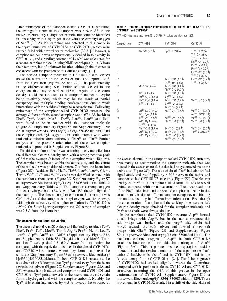
The third camphor molecule was unambiguously modelled intothe difference-electron-density map with a strong positive peakof 8.9σ (the average B-factor of this camphor was ∼40.4 Å2).The camphor was bound within the active site, and the centreof the molecule was positioned approx. 7 Å from the haem iron(Figure 2D). Residues Ile86, Met98, Thr101, Leu250, Leu253, Gly254,Thr258, Val301, Ile401 and Val402 were in van der Waals contact withthe camphor carbon atoms (Figure 2D, Supplementary Figure S7at http://www.BiochemJ.org/bj/433/bj4330085add.htm, Table 2and Supplementary Table S1). The camphor carbonyl oxygenformed a hydrogen bond (2.8 Å) with Wat-589, the sixth ligand ofthe haem iron. The closest camphor carbon to the iron atom wasC10 (4.9 Å) and the camphor carbonyl oxygen was 4.4 Å away.Although the selectivity of camphor oxidation by CYP101D2 is�99% for 5-exo-hydroxycamphor, the C5 carbon of camphorwas 7.5 Å from the haem iron.
The access channel and active site
The access channel was 20 Å deep and flanked by residues Tyr29,Phe87, Pro89, Tyr96, Met184, Thr185, Arg186, Pro187, Met195, Leu199,Asn203, Asp257, Val401 and Val402 (Supplementary Figure S3Aand Supplementary Table S3). The side chains of Met184, Thr185
and Leu199 were pushed 5.5−6.0 Å away from the active sitecompared with the equivalent residues in the closed CYP101D1and CYP101A1 structures, where they form a cap over thesubstrate (Supplementary Figure S8 at http://www.BiochemJ.org/bj/433/bj4330085add.htm). In both CYP101D2 structures, theside chain of the B′ loop residue, Tyr96 pointed away from the haemtowards the solvent (Figure 2C, Supplementary Figures S3A andS8), whereas in both native and camphor-bound CYP101D1 andCYP101A1 Tyr96 points inwards at the haem, and the side chainforms a hydrogen bond with the camphor carbonyl. Notably, theTyr96 side chain had moved by ∼5 Å towards the entrance of
Table 2 Protein−camphor interactions at the active site of CYP101D2,CYP101D1 and CYP101A1
CYP101D1 values are taken from [31], CYP101A1 values are taken from [20].
Camphor atom CYP101D2 CYP101D1 CYP101A1
O Wat-589 (2.8 A) Tyr97OH (2.6 A) Tyr96 OH (2.7 A)Tyr96 Cζ (3.5 A)Tyr96 Cε2 (3.4 A)Leu244 Cδ1(3.7 A)Phe87 Cζ (3.6 A)Phe87 Cε2 (3.7 A)
C1 Leu244 Cδ1 (3.8 A)Tyr96 OH (3.7 A)
C2 Leu251 Cδ1 (4.0 A) Leu244 Cδ1 (3.7 A)Trp88 CH2 (4.0 A) Tyr96 OH (3.4 A)
C3 Met98 Cε (4.4 A) Leu251 Cδ1 (4.1 A)Thr102 Cγ 2 (4.0 A)
C5 Ile86 Cδ1 (4.0 A) Leu251 Cδ1 (4.0 A)Thr101 Cγ 2 (4.2 A) Gly255 Cα (4.0 A)
C6 Thr101 Cγ 2 (4.3 A) Leu251 Cδ1 (4.2 A) Val247 Cγ 1 (3.9 A)Ile86 Cδ1 (4.5 A) Gly255 Cα (3.5 A)
Leu254 Cγ (4.2 A)C8 Val402 Cγ 2 (3.6 A) Val302 Cγ 1 (3.7 A) Val295 Cγ 1 (3.7 A)
Ile401 Cγ 2 (4.4 A) Asp297 Oδ2 (3.8 A)C9 Ile401 Cγ 2 (3.6 A) Val302 Cγ 2 (3.6 A) Val295 Cγ 1 (3.9 A)
Val301 Cγ 1 (4.4 A) Thr259 Cγ 2 (3.6 A) Val295 Cγ 2 (3.9 A)Glu303 Cβ (4.1 A)
C10 Thr258 Cγ 2 (4.6 A) Leu254 Cγ (3.9 A) Val247 Cγ 1 (3.6 A)Val301 Cγ 1 (4.2 A) Leu254 Cδ1 (3.8 A) Thr185 Cγ 2 (3.9 A)Val301 Cγ 2 (4.3 A) Thr186 Cγ 2 (3.7 A)
Val403 Cγ 2 (3.9 A)
the access channel in the camphor-soaked CYP101D2 structure,presumably to accommodate the camphor molecule that waslocated in the access channel, but that had not yet moved inside theactive site (Figure 2C). The side chain of Phe87 had also shiftedsignificantly and was flipped by ∼90 ◦ between the native andcamphor-soaked CYP101D2 structures (Figure 2C). The electrondensity of Phe87 in the camphor-soaked structure was less welldefined compared with the native structure. The lower resolutionof the Phe87 side chain and the second camphor molecule in thisstructure may be due to different camphor occupancy and bindingorientations resulting in different Phe87 orientations. Even thoughthe concentration of camphor and the soaking times were varied,electron-density maps obtained for the camphor molecule andPhe87 side chain were always similar.
In the camphor-soaked CYP101D2 structure, Asp257 formeda salt bridge with Arg186, but in the native structure thissalt bridge was broken and the Arg186 side chain hadmoved towards the bulk solvent and formed a new saltbridge with Glu156 (Figure 2B and Supplementary FigureS9 at http://www.BiochemJ.org/bj/433/bj4330085add.htm). Thebackbone carbonyl oxygen of Asp257 in both CYP101D2structures interacts with the side-chain nitrogen of Asn261
(Figure 3A). This aspartate residue−asparagine residueinteraction and the resultant rotation of the aspartate residue’scarbonyl backbone is also found in CYP101D1 and in theferrous deoxy form of CYP101A1 [24]. The I helix grooveof CYP101D2 had shifted slightly towards the N-terminuscompared with its position in closed CYP101A1 and CYP101D1structures, mirroring the shift of this groove in the openconformations of CYP101A1 (Supplementary Figure S10 athttp://www.BiochemJ.org/bj/433/bj4330085add.htm) [29]. Thesemovements in CYP101D2 resulted in a shift of the side chain of
c© The Authors Journal compilation c© 2011 Biochemical Society

90 W. Yang and others
Figure 3 Environment of the I helix, haem moiety and proximal face of CYP101D2
(A) Overlay of the residues in the I helix between CYP101D2 (green) and CYP101A1 (salmon). The conformation of the carbonyl oxygen of Asp257 is flipped by ∼90◦ towards Asn261 due to ahydrogen bond with the side-chain nitrogen of Asn261 (2.8 A). Two water molecules hydrogen bond with the Asp257 side chain and are shown as red spheres. (B) The haem environment in the nativeform of CYP101D2. The A-ring propionate group forms a salt bridge with Arg305. The O1 atom forms hydrogen bonds with two water molecules, Wat-565 and Wat-436, and Wat-565 forms anotherhydrogen bond with Wat-566. The O2 atom of the A-ring propionate group is hydrogen-bonded to the side-chains of Tyr75 and Glu303. The Oε1 atom of Glu303 is hydrogen-bonded to the DEGmolecule and the Oε2 atom is hydrogen-bonded to Wat-566 and the A-ring propionate group. The haem and the relevant residues are shown in yellow and green respectively. The water moleculesare shown as red spheres. The electron density (2F o − F c contoured at 1σ ) of the DEG molecule is shown in blue. (C) The haem environment in the camphor-soaked structure of CYP101D2.Compared with the native structure, the A-ring propionate group is rotated by approx. 90◦, and both the oxygen atoms of Glu303 are hydrogen-bonded to the propionate group. The propionate groupalso forms a salt bridge with Arg305 and hydrogen bonds with two water molecules, Wat-498 and Wat-464. Glu303 is hydrogen bonded to Tyr75. The haem and residues are shown in yellow and greenrespectively. The water molecules are shown as red spheres and the electron density (2F o − F c contoured at 1σ ) of the camphor molecule is shown in blue. (D) The proximal face of CYP101D2.Negatively and positively charged surface areas are coloured red and blue respectively. The positively charged proximal face is similar to that of CYP101D1 and more positively charged than that ofCYP101A1 (see Supplementary Figure S12 at http://www.BiochemJ.org/bj/433/bj4330085add.htm). The basic residues of CYP101D2 (Arg112, Arg125, Arg289, Arg349, Arg362 and Arg370) and Ser76
which aligns with Arg77 in CYP101D1 are highlighted.
Thr258 to a similar orientation to that observed for Thr252 in theferrous−oxy form of CYP101A1, but different from those seenin the closed structures of CYP101A1 and CYP101D1.
A hydrogen-bonded network of four water molecules waslocated in the vicinity of the haem propionate groups of thenative CYP101D2 structure (Figures 3B and 3C). In the camphor-soaked structure there are fewer water molecules in this regionand the Glu303 side chain and the A-ring propionate groupof the haem occupied different positions compared with thenative structure (Figure 3C). Several water molecules were alsolocated in the access channel of both structures of CYP101D2(Supplementary Figure S11 at http://www.BiochemJ.org/bj/433/bj4330085add.htm).
Proximal face
The haem-proximal surface of CYP101D2 had a preponderanceof positively charged residues, including Arg112, Arg125, Arg289,Arg349, Arg362 and Arg370, that are conserved spatially and insequence in CYP101D1 (Figure 3D and Supplementary FigureS12 at http://www.BiochemJ.org/bj/433/bj4330085add.htm). Bycomparison, the proximal face of CYP101A1 is less positivelycharged. The ferredoxin gene Arx is genomically associated withthe CYP101D2 gene of N. aromaticivorans, and together ArRand Arx support the mono-oxygenase activity of CYP101D2,CYP101D1 (a kcat of 39 and 41 s− 1 respectively) and three other
N. aromaticivorans CYP enzymes [31]. These basic residuesin CYP101D2 and CYP101D1 probably interact with acidicsurface residues on Arx in the competent electron-transfercomplex (Supplementary Figure S12). Electrostatic interactionsare important in Arx−CYP101D2 electron transfer, and the chargedistributions are consistent with the low cross-reactivity withthe CYP101A1 and CYP101D2 systems when the physiologicalferredoxins are exchanged [31,32,39].
DISCUSSION
CYP101D2 has a primary sequence identity of 62%with CYP101D1 and of 44% with CYP101A1 (SupplementaryFigure S13 at http://www.BiochemJ.org/bj/433/bj4330085add.htm and Supplementary Table S1). However, owing to crystalpacking interactions, the native and camphor-soaked structuresof CYP101D2 had an open conformation with a clearly definedaccess channel, in contrast with the closed structures observedwith CYP101D1 [31]. The helices of the native structure ofCYP101D2 overlaid closely with those of the open formsof CYP101A1. Unexpectedly, the carbonyl oxygen of the active-site camphor was hydrogen-bonded to the haem-iron-bound waterrather than the Tyr96 side chain, as observed in the closed structuresof camphor-bound CYP101A1 and CYP101D1 (Table 2). Soak-ing of crystals of the native open form of CYP101D2 in a camphorsolution or co-crystallization of camphor with CYP101D2 did
c© The Authors Journal compilation c© 2011 Biochemical Society

Crystal structure of CYP101D2 91
not result in a closed form in which the camphor in the activesite was completely shielded from the solvent. Camphor-bindingtitrations with CYP101D2 showed an approx. 40% shift to thehigh-spin form and a Kd of 3.1 μM (Supplementary Figure S14 athttp://www.BiochemJ.org/bj/433/bj4330085add.htm), similar tothat observed previously for CYP101D1 [31].
The binding orientation of the camphor molecule in the activesite is not consistent with the observed product profile of almostexclusive formation of 5-exo-hydroxycamphor. In addition, it isinhibitory towards the first electron-transfer step. The enzyme hasto undergo a change to a closed conformation with a concomitantshift in the camphor orientation, e.g. breaking the hydrogen bondwith the axial water and rotation to form a hydrogen bond withthe Tyr96 side chain to orient the camphor C5 over the haemiron. This suggests that crystal-packing interactions may haveeffectively locked CYP101D2 in the open conformation, resistingthe conversion into the closed structure expected upon substratebinding and resulting in the observed camphor-binding orienta-tion. In solution, conformational dynamics of the enzyme mayresult in rapid transition to the closed form, perhaps even duringcamphor entry into the active site, initially leading to differenttransient binding orientations of the camphor molecule before theenzyme−substrate complex reaches its equilibrium structure.
Multi-step binding mechanisms have been proposed forCYP3A4, CYP1A2, CYP101A1 and recently for CYP107L1(P450 PikC) [11−13,40,41]. In such mechanisms the substratebinds to a recognition site on the protein surface before enteringthe active site. The product could also leave via the same route,although alternative routes, such as via the F and G helices or theE/F loop, have been suggested [6]. Binding of multiple camphormolecules to CYP101A1 has been reported from NMR andUV studies [11,42−44]. The observation of a putative camphormolecule in a surface cavity, coupled to the presence of a secondcamphor in the access channel of camphor-soaked CYP101D2,may be further evidence of a multi-step binding mechanism withinitial substrate recognition occurring at the enzyme surface [11].Modelling and NMR relaxation studies led to the proposal that acamphor entry/recognition site (Kd of 43 μM) may be located inthis cavity in CYP101A1, and it is of note that these experimentsshowed that this surface-bound camphor molecule was in a highlyfluxional state [11]. The B-factors associated with the camphormolecules in the access channel and the surface cavity are similarto those observed for the solvent, whereas the camphor moleculein the active site has a B-factor similar to that of the protein.The less-specific interactions and mobile nature of the camphormolecules located in the CYP101D2 surface cavity and accesschannel compared with the camphor in the active site suggestthat these may be snapshots of intermediate binding positions inthe substrate entrance pathway into the active site (or of productrelease).
Many structurally characterized CYP enzymes have closedconformations with buried active sites [20,31]. However, clearlydefined access channels are not uncommon [45,46]. Other P450enzymes show open conformations in the absence of a boundsubstrate, but adopt a more closed conformation upon substratebinding, e.g. P450 EryK [19]. Double-jump substrate-bindingstudies of P450 EryK suggest a conformational selection modelfor substrate recognition and binding [19]. Crystal structures ofCYP119 reveal that the enzyme undergoes major rearrangementsat the active site to accommodate the binding of differentsubstrates, and NMR studies of this enzyme tagged with theunnatural amino acid 13C-p-methoxyphenylalanine showed that itcan exist in different conformations [15,17,18]. These conformersare thought to be important in substrate recognition and binding,and the equilibrium between them can be perturbed using
different substrates. The open structure of CYP101D2 may bea conformer that facilitates substrate entry and product exit fromthe active site. The movement of Tyr96 between the open andclosed conformers may assist in the recognition and binding ofcamphor. We note, however, that CYP101D2 is probably heldin this open conformation by crystal packing contacts and thecamphor concentration of ∼8 mM (1.2 g · l− 1) is higher thanthose in turnover reactions (1 mM). Therefore we cannot rule outadventitious binding at one or both of the locations outside theactive site. The substrate might enter (or the product exit)the active site that is opened up by normal thermal fluctuationsof the structure via the channel without forming an intermediatestate. On the other hand, no adventitious binding of camphor wasobserved to the surface cavity of the closed conformations ofCYP101D1 under a similar camphor-soaking regime [31].
The switch between the open and the closed forms ofCYP101A1 involves breaking and formation of inter-residuesalt bridges and hydrogen bonds, and the rearrangement ofhydrophobic interactions [29]. The presence or otherwise of theseinteractions, together with adventitious binding of molecules,such as DEG or deliberate introduction of longer substrates such asD-4-Ad, as well as crystal packing effects, may control whethercrystals with open or closed conformation are obtained. Salt-bridge interactions between the F helix or F/G loop and the I helixare less extensive in CYP101D2 (and CYP101D1) compared withCYP101A1 (Supplementary Table S2). A network of interactionsbetween Lys178, Asp182, Arg186 and Asp251 in CYP101A1 has beenshown to play a key role in the control of the diffusion step ofcamphor binding and provide a link from the I helix to the bulksolvent at the enzyme surface (Supplementary Table S2) [10,47].These interactions are broken in the open conformations ofCYP101A1 [29,30]. The Asp257−Arg186 salt bridge is conserved incamphor-soaked CYP101D2, but Ser178 and Arg182 replace Lys178
and Asp182. Ser178 is located in the access channel, whereas Arg182
points towards the enzyme surface (Figure 2B and SupplementaryFigure S5B). The Asp257−Arg186 salt bridge separates the camphormolecules in the surface recognition site and in the access channelsite. The breaking of this salt bridge and the movement of Arg186 tointeract with Glu156, which lines the surface recognition site, couldopen up a short route of substrate entry or product exit betweenthese two sites. This blocking of the proposed surface recognitionsite and the active-site camphor by the equivalent salt bridge inCYP101A1 has been noted previously [11], and in the substrate-free open CYP101A1 structure, Arg186 interacts with Glu156 [30].The structure and function of mutants with substitutions to breaksalt bridges will be of interest [10,47,48].
The proximal faces of CYP101D2 and CYP101D1 are verysimilar, and more positively charged than CYP101A1, whereasthe corresponding interaction region on the Arx ferredoxin ismore negatively charged compared with Pdx [31], consistent withelectrostatic interactions being important in Arx−CYP101D2 andArx−CYP101D1 binding and recognition. However, the preciserole of each of the acidic residues on Arx and the basic residues onthe CYP enzymes remains to be elucidated. The ArR/Arx systemalso supports the mono-oxygenase activity of at least three otherCYP enzymes from N. aromaticivorans (CYP101B1, CYP101C1and CYP111A2). The crystal structures of the other P450 enzymessupported by Arx will be required for a fuller analysis, and this isthe subject of ongoing investigations.
Conclusions
CYP101D2 and CYP101D1 are analogues of CYP101A1 thatcan catalyse the fast, efficient and stereoselective oxidationof camphor to 5-exo-hydroxycamphor. Although the structure
c© The Authors Journal compilation c© 2011 Biochemical Society

92 W. Yang and others
of CYP101D1 is similar to that of CYP101A1, the camphor-soaked structure of CYP101D2 has an open conformation andreveals potential camphor-binding sites at the active site, in theaccess channel and at the enzyme surface. This open structure maybe a conformer involved in substrate recognition and binding. Thecamphor-binding sites at the enzyme surface and in the accesschannel may be intermediate locations of the substrate in itsprogression into the active site of CYP101D2 via multi-step andconformational selection mechanisms.
AUTHOR CONTRIBUTION
Wen Yang, Stephen Bell and Hui Wang performed the protein production, purificationand biochemical assays. Wen Yang and Hui Wang performed the crystallization andsoaking experiments. Wen Yang, Weihong Zhou and Mark Bartlam conducted the structuredetermination and refinement. Stephen Bell, Wen Yang, Mark Bartlam, Luet Wong andZihe Rao designed the research, analysed the data and wrote the manuscript. All authorshelped with editing the manuscript before submission.
ACKNOWLEDGEMENT
We are grateful to Dr Zhiyong Lou (Tsinghua University, Beijing, China) for help in datacollection and processing.
FUNDING
This work was supported by the Ministry of Science and Technology of China Project 973[grant number 2007CB914301 (to M.B.)]; the Tianjin Municipal Science and TechnologyCommission [grant number 08SYSYTC00200]; and by the Higher Education FundingCouncil for England.
REFERENCES
1 Ortiz de Montellano, P. R. (2005) Cytochrome P450: Structure, Mechanism, andBiochemistry, Kluwer Academic/Plenum Press, New York
2 Sigel, A., Sigel, H. and Sigel, R. (2007) The Ubiquitous Roles of Cytochrome P450Proteins, John Wiley & Sons, Chichester
3 Cryle, M. J., Stok, J. E. and De Voss, J. J. (2003) Reactions catalyzed by bacterialcytochromes P450. Aust. J. Chem. 56, 749–762
4 Bernhardt, R. (2006) Cytochromes P450 as versatile biocatalysts. J. Biotechnol. 124,128–145
5 Denisov, I. G., Makris, T. M., Sligar, S. G. and Schlichting, I. (2005) Structure andchemistry of cytochrome P450. Chem. Rev. 105, 2253–2277
6 Cojocaru, V., Winn, P. J. and Wade, R. C. (2007) The ins and outs of cytochrome P450s.Biochim. Biophys. Acta 1770, 390–401
7 Wade, R. C., Winn, P. J., Schlichting, I. and Sudarko, I. (2004) A survey of active siteaccess channels in cytochromes P450. J. Inorg. Biochem. 98, 1175–1182
8 Li, H. and Poulos, T. L. (2004) Crystallization of cytochromes P450 andsubstrate−enzyme interactions. Curr. Top. Med. Chem. 4, 1789–1802
9 Winn, P. J., Ludemann, S. K., Gauges, R., Lounnas, V. and Wade, R. C. (2002)Comparison of the dynamics of substrate access channels in three cytochrome P450sreveals different opening mechanisms and a novel functional role for a buried arginine.Proc. Natl. Acad. Sci. U.S.A. 99, 5361–5366
10 Deprez, E., Gerber, N. C., Di Primo, C., Douzou, P., Sligar, S. G. and Hui Bon Hoa, G.(1994) Electrostatic control of the substrate access channel in cytochrome P-450cam.Biochemistry 33, 14464–14468
11 Yao, H., McCullough, C. R., Costache, A. D., Pullela, P. K. and Sem, D. S. (2007)Structural evidence for a functionally relevant second camphor binding site in P450cam:model for substrate entry into a P450 active site. Proteins 69, 125–138
12 Isin, E. M. and Guengerich, F. P. (2006) Kinetics and thermodynamics of ligand bindingby cytochrome P450 3A4. J. Biol. Chem. 281, 9127–9136
13 Sohl, C. D., Isin, E. M., Eoff, R. L., Marsch, G. A., Stec, D. F. and Guengerich, F. P. (2008)Cooperativity in oxidation reactions catalyzed by cytochrome P450 1A2: highlycooperative pyrene hydroxylation and multiphasic kinetics of ligand binding. J. Biol.Chem. 283, 7293–7308
14 Yano, J. K., Koo, L. S., Schuller, D. J., Li, H., Ortiz de Montellano, P. R. and Poulos, T. L.(2000) Crystal structure of a thermophilic cytochrome P450 from the archaeon Sulfolobussolfataricus. J. Biol. Chem. 275, 31086–31092
15 Nishida, C. R. and Ortiz de Montellano, P. R. (2005) Thermophilic cytochrome P450enzymes. Biochem. Biophys. Res. Commun. 338, 437–445
16 Park, S. Y., Yamane, K., Adachi, S., Shiro, Y., Weiss, K. E., Maves, S. A. and Sligar, S. G.(2002) Thermophilic cytochrome P450 (CYP119) from Sulfolobus solfataricus: highresolution structure and functional properties. J. Inorg. Biochem. 91, 491–501
17 Lampe, J. N., Brandman, R., Sivaramakrishnan, S. and Ortiz de Montellano, P. R. (2010)2D NMR and all-atom molecular dynamics of cytochrome P450 CYP119 reveal hiddenconformational substrates. J. Biol. Chem. 285, 9594–9603
18 Lampe, J. N., Floor, S. N., Gross, J. D., Nishida, C. R., Jiang, Y., Trnka, M. J. and Ortiz deMontellano, P. R. (2008) Ligand-induced conformational heterogeneity of cytochromeP450 CYP119 identified by 2D NMR spectroscopy with the unnatural amino acid13C-p-methoxyphenylalanine. J. Am. Chem. Soc. 130, 16168–16169
19 Savino, C., Montemiglio, L. C., Sciara, G., Miele, A. E., Kendrew, S. G., Jemth, P.,Gianni, S. and Vallone, B. (2009) Investigating the structural plasticity of a cytochromeP450: three-dimensional structures of P450 EryK and binding to its physiologicalsubstrate. J. Biol. Chem. 284, 29170–29179
20 Poulos, T. L., Finzel, B. C. and Howard, A. J. (1987) High-resolution crystal structure ofcytochrome P450cam. J. Mol. Biol. 195, 687–700
21 Li, H., Narasimhulu, S., Havran, L. M., Winkler, J. D. and Poulos, T. L. (1995) Crystalstructure of cytochrome P450cam complexed with its catalytic product,5-exo-hydroxycamphor. J. Am. Chem. Soc. 117, 6297–6299
22 Raag, R. and Poulos, T. L. (1989) Crystal structure of the carbon monoxide-substrate−cytochrome P-450cam ternary complex. Biochemistry 28, 7586–7592
23 Nagano, S. and Poulos, T. L. (2005) Crystallographic study on the dioxygen complex ofwild-type and mutant cytochrome P450cam. Implications for the dioxygen activationmechanism. J. Biol. Chem. 280, 31659–31663
24 Schlichting, I., Berendzen, J., Chu, K., Stock, A. M., Maves, S. A., Benson, D. E.,Sweet, R. M., Ringe, D., Petsko, G. A. and Sligar, S. G. (2000) The catalytic pathway ofcytochrome p450cam at atomic resolution. Science 287, 1615–1622
25 Chen, X., Christopher, A., Jones, J. P., Bell, S. G., Guo, Q., Xu, F., Rao, Z. and Wong, L. L.(2002) Crystal structure of the F87W/Y96F/V247L mutant of cytochrome P-450cam with1,3,5-trichlorobenzene bound and further protein engineering for the oxidation ofpentachlorobenzene and hexachlorobenzene. J. Biol. Chem. 277, 37519–37526
26 Vidakovic, M., Sligar, S. G., Li, H. and Poulos, T. L. (1998) Understanding the role of theessential Asp251 in cytochrome p450cam using site-directed mutagenesis,crystallography, and kinetic solvent isotope effect. Biochemistry 37, 9211–9219
27 Dmochowski, I. J., Crane, B. R., Wilker, J. J., Winkler, J. R. and Gray, H. B. (1999) Opticaldetection of cytochrome P450 by sensitizer-linked substrates. Proc. Natl. Acad. Sci.U.S.A. 96, 12987–12990
28 Dunn, A. R., Dmochowski, I. J., Bilwes, A. M., Gray, H. B. and Crane, B. R. (2001) Probingthe open state of cytochrome P450cam with ruthenium-linker substrates. Proc. Natl.Acad. Sci. U.S.A. 98, 12420–12425
29 Hays, A. M., Dunn, A. R., Chiu, R., Gray, H. B., Stout, C. D. and Goodin, D. B. (2004)Conformational states of cytochrome P450cam revealed by trapping of syntheticmolecular wires. J. Mol. Biol. 344, 455–469
30 Lee, Y. T., Wilson, R. F., Rupniewski, I. and Goodin, D. B. (2010) P450cam visits an openconformation in the absence of substrate. Biochemistry 49, 3412–3419
31 Yang, W., Bell, S. G., Wang, H., Zhou, W., Hoskins, N., Dale, A., Bartlam, M., Wong, L. L.and Rao, Z. (2010) Molecular characterization of a class I P450 electron transfer systemfrom Novosphingobium aromaticivorans DSM12444. J. Biol. Chem. 285, 27372–27384
32 Bell, S. G. and Wong, L. L. (2007) P450 enzymes from the bacterium Novosphingobiumaromaticivorans. Biochem. Biophys. Res. Commun. 360, 666–672
33 Otwinowski, Z. W. M. (1997) Processing of X-ray diffraction data collected in oscillationmode. Methods Enzymol. 276, 307–326
34 McCoy, A. J., Grosse-Kunstleve, R. W., Adams, P. D., Winn, M. D., Storoni, L. C. andRead, R. J. (2007) Phaser crystallographic software. J. Appl. Crystallogr. 40, 658–674
35 Collaborative Computational Project, Number 4 (1994) The CCP4 suite: programs forprotein crystallography. Acta Crystallogr. Sect. D Biol. Crystallogr. 50, 760–763
36 Emsley, P. and Cowtan, K. (2004) Coot: model-building tools for molecular graphics. ActaCrystallogr. Sect. D Biol. Crystallogr. 60, 2126–2132
37 Murshudov, G. N., Vagin, A. A. and Dodson, E. J. (1997) Refinement of macromolecularstructures by the maximum-likelihood method. Acta Crystallogr. Sect. D Biol. Crystallogr.53, 240–255
38 Laskowski, R. A., Macarthur, M. W., Moss, D. S. and Thornton, J. M. (1993) PROCHECK:a program to check the stereochemical quality of protein structures. J. Appl. Crystallogr.26, 283–291
39 Bell, S. G., Dale, A., Rees, N. H. and Wong, L. L. (2010) A cytochrome P450 class Ielectron transfer system from Novosphingobium aromaticivorans. Appl. Microbiol.Biotechnol. 86, 163–175
40 Isin, E. M. and Guengerich, F. P. (2008) Substrate binding to cytochromes P450. Anal.Bioanal. Chem. 392, 1019–1030
c© The Authors Journal compilation c© 2011 Biochemical Society

Crystal structure of CYP101D2 93
41 Li, S., Ouellet, H., Sherman, D. H. and Podust, L. M. (2009) Analysis of transient andcatalytic desosamine-binding pockets in cytochrome P-450 PikC from Streptomycesvenezuelae. J. Biol. Chem. 284, 5723–5730
42 Lee, H., Ortiz de Montellano, P. R. and McDermott, A. E. (1999) Deuterium magic anglespinning studies of substrates bound to cytochrome P450. Biochemistry 38,10808–10813
43 Marden, M. C. and Hoa, G. H. (1987) P-450 binding to substrates camphor and linaloolversus pressure. Arch. Biochem. Biophys. 253, 100–107
44 Narasimhulu, S. and Willcox, J. K. (2001) Temperature-jump relaxation kinetics ofsubstrate-induced spin-state transition in cytochrome P450 (comparison of the wild-typeand C334A mutant P450CAM and P4502B4). Arch. Biochem. Biophys. 388, 198–206
45 Ravichandran, K. G., Boddupalli, S. S., Hasermann, C. A., Peterson, J. A. andDeisenhofer, J. (1993) Crystal structure of hemoprotein domain of P450BM-3, aprototype for microsomal P450s. Science 261, 731–736
46 Bell, S. G., Xu, F., Forward, I., Bartlam, M., Rao, Z. and Wong, L. L. (2008) Crystalstructure of CYP199A2, a para-substituted benzoic acid oxidizing cytochrome P450 fromRhodopseudomonas palustris. J. Mol. Biol. 383, 561–574
47 Di Primo, C., Deprez, E., Sligar, S. G. and Hui Bon Hoa, G. (1997) Origin of thephotoacoustic signal in cytochrome P-450cam: role of the Arg186−Asp251−Lys178
bifurcated salt bridge. Biochemistry 36, 112–11848 Lounnas, V. and Wade, R. C. (1997) Exceptionally stable salt bridges in cytochrome
P450cam have functional roles. Biochemistry 36, 5402–5417
Received 9 July 2010/24 September 2010; accepted 15 October 2010Published as BJ Immediate Publication 15 October 2010, doi:10.1042/BJ20101017
c© The Authors Journal compilation c© 2011 Biochemical Society

Biochem. J. (2011) 433, 85–93 (Printed in Great Britain) doi:10.1042/BJ20101017
SUPPLEMENTARY ONLINE DATAThe structure of CYP101D2 unveils a potential path for substrate entry intothe active siteWen YANG*, Stephen G. BELL†1, Hui WANG‡, Weihong ZHOU*, Mark BARTLAM*1, Luet-Lok WONG† and Zihe RAO*‡*Tianjin Key Laboratory of Protein Science, College of Life Sciences, Nankai University, Tianjin 300071, China, †Inorganic Chemistry Laboratory, Department of Chemistry, University ofOxford, South Parks Road, Oxford OX1 3QR, U.K., and ‡Laboratory of Structural Biology, Tsinghua University, Beijing 100084, China
Figure S1 SDS/PAGE analysis of purified CYP101D2
Molecular masses are indicated in kDa.
1 Correspondence may be addressed to either of these authors (email [email protected] or [email protected]).The co-ordinates for the crystal structures of native CYP101D2 and camphor-soaked CYP101D2 have been deposited in the PDB under the accession
codes 3NV5 and 3NV6 respectively.
c© The Authors Journal compilation c© 2011 Biochemical Society

W. Yang and others
Figure S2 Structural comparison of the CYP101A1 family
Per residue RMSD difference diagrams. (A) Comparison between the camphor-boundCYP101D2 structure and other open conformation structures, including native CYP101D2(blue line), D-4-Ad-bound CYP101A1 (cyan line), D-8-Ad-bound CYP101A1 (red line) and thenative open form of CYP101A1 (green line). (B) Comparison between the camphor-bound CYP101D2 structure and two closed conformation structures, native CYP101D1 (blue)and camphor-bound CYP101A1 (red). The Cα RMSD of CYP101D1 and CYP101A1 comparedwith CYP101D2 are 1.36 A and 1.62 A respectively. (C) Overlay of camphor-soaked CYP101D2and an open CYP101A1 conformer (PDB code 3L61). The secondary structure of CYP101A1is shown in grey. The open form of CYP101A1 (PDB code 3L61) has a similar structure toRu-F8bp-Ad-bound CYP101A1 (see Figure 1D in the main paper). rms deviation = RMSD.
c© The Authors Journal compilation c© 2011 Biochemical Society

Crystal structure of CYP101D2
Figure S3 The access channel and the active site of native CYP101D2
(A) The access channel and the active site of native CYP101D2. The residues distributed along the hydrophobic channel, the active site and the DEG molecule are shown in green. The Tyr96 residueis found in the access channel and the side chain points away from the haem and the DEG molecule. The salt bridge between Arg186 and Asp257 has been broken, with Arg186 moving away from theaccess channel towards the surface recognition site. Co-crystallization experiments for CYP101D2 with camphor yielded crystals using the same crystallization conditions. The structure adoptedthe same open conformation, but no electron densities corresponding to camphor molecules could be identified and a DEG molecule was found in the active site as in the native structure. Differentcrystallization conditions were also attempted, but these resulted in no crystal formation. (B) The active site of native CYP101D2 with the DEG molecule bound. The active site of CYP101D2 isdefined by Ile86, Phe87, Met98, Thr101, Leu250, Leu253, Asp257, Thr258, Val301, Glu303, Ile401 and Val402. The DEG molecule and the residues distributed in this hydrophobic pocket are shown in greenand the five water molecules in the active site are shown as red spheres. The electron density (2F o − F c contoured at 1σ ) of the DEG molecule is shown in blue. Wat-568 is hydrogen-bonded to thecarbonyl oxygen of Gly254 (2.8 A, not shown), which is in turn hydrogen-bonded to the side chain oxygen of Thr258 (2.8 A).
c© The Authors Journal compilation c© 2011 Biochemical Society

W. Yang and others
Figure S4 Crystal packing comparison of CYP101 family members
(A) CYP101D2, (B) the CYP101A1 open structure (PDB code 3L61), (C) CYP101D1 and (D) the CYP101A1 closed structure (PDB code 2CPP). The secondary structures are shown as ribbonsand the symmetry molecules and the haem are coloured grey and yellow. The F, G and B′ helices are coloured orange, salmon and red respectively. (E) In CYP101D2, the F and G helices and theF/G loop interact with the J helix and the loop between K′ and L′ helices. In CYP101A1 these regions interact with the B helix and the loop between K′ and L′ helices. Some contact pairs thatmay contribute to the crystal packing in CYP101D2 include: Glu180–Trp343 (Lys344), Arg183–Glu341, Gln184–Glu341, Thr198–Glu280, Asp199–Lys344, Glu201–Pro275 and Gln205–Arg273. Only Glu280 isconserved in CYP101A1 and CYP101D1.
c© The Authors Journal compilation c© 2011 Biochemical Society

Crystal structure of CYP101D2
Figure S5 Topology of the camphor-recognition sites on the surface of CYP101 family members
(A) CYP101D2, (B) CYP101D1 and (C) CYP101A1. These cavities are located near the F helix side of the F/G loop. In CYP101D2 the Asp257–Arg186 salt bridge blocks substrate entry into the accesschannel. This salt bridge is conserved and formed by Arg186 and Asp251 in CYP101A1 and Arg187 and Asp258 in CYP101D1. Another salt bridge formed by Lys178 and Asp182 in CYP101A1 is notconserved in CYP101D1 and CYP101D2. The cavities in these structures are all of a similar size. In the open CYP101A1 structure, this cavity seems slightly flatter when compared with the others.
Figure S6 Modelling of the camphor molecules in camphor-soaked CYP101D2
(A) The modelling of a camphor molecule into the region of electron density of the access channel (upper panel) and the surface cavity (lower panel) of CYP101D2. The camphor molecule in theactive site is included. The camphor molecules are in yellow, the haem is in green, the electron density (F o − F c contoured at 3σ ) of the camphor molecules is shown in red and the interactingresidues are shown in grey. (B) The position of the three potential camphor molecules relative to the active site and access channel. The camphor molecules in the access channel and in the surfacecavity have been modelled using their electron densities. The camphor molecules and haem molecule are shown in yellow and green respectively.
c© The Authors Journal compilation c© 2011 Biochemical Society
W. Yang and others
Figure S7 Comparison of the active sites of CYP101 family members
(A) Camphor-soaked CYP101D2, (B) camphor-bound CYP101A1 (PDB code 2CPP), (C) camphor-bound CYP101D1 (PDB code 3LXI) and (D) the open form of CYP101A1 (PDB code 3L61).The residues involved in substrate binding are shown in green and the water molecules that occupy the sixth haem iron co-ordination site are shown as red spheres. The electron density(2F o − F c contoured at 1σ ) of the camphor molecule in camphor-soaked CYP101D2 is shown in blue.
c© The Authors Journal compilation c© 2011 Biochemical Society

Crystal structure of CYP101D2
Figure S8 Comparison of the CYP101D1 and CYP101D2 active site andaccess channel
A comparison between the open structure of CYP101D2 (green ribbon) and closed structure ofCYP101D1 (grey ribbon) to show the different positions of Tyr96, Met184, Thr185 and Leu199. Theactive-site residues in CYP101D2 and CYP101D1 are shown in green and wheat respectively.The movements of Met184, Thr185 and Leu199 are highlighted with pink arrows. Tyr96 also movesout of the active site. The haem moiety is shown in yellow.
Figure S9 Comparison of the active site, access channel and surface cavityof camphor-soaked and native CYP101D2
The position of the two potential camphor electron densities relative to the active site,substrate-access channel and active-site camphor molecule. The camphor and haem moleculeare shown in green and yellow respectively. The residues Phe87, Tyr96, Glu156 and Arg186 areshown in green in the camphor-soaked structure and cyan in the native structure.
c© The Authors Journal compilation c© 2011 Biochemical Society

W. Yang and others
Figure S10 Location of the I helix groove in different CYP101 family members
(A) The hydrogen-bonding network for the I helix groove is shown. The differences between camphor-bound CYP101A1 (closed conformation, PDB code 2CPP), camphor-bound CYP101D1(closed conformation, PDB code 3LXI), D-4-Ad-bound CYP101A1 (open conformation, PDB code 1RF9), native CYP101A1 (open conformation, PDB code 3L61) and camphor-soaked CYP101D2(open conformation, PDB code 3NV6) are highlighted in red. The I helix shift and the helical kink between the closed and open conformation are shown in pale green. (B) A comparison of the positionof the I helix groove of camphor-soaked CYP101D2 (yellow) with CYP101A1 (grey, PDB code 2CPP). The position of the groove is highlighted in each structure.
c© The Authors Journal compilation c© 2011 Biochemical Society
Crystal structure of CYP101D2
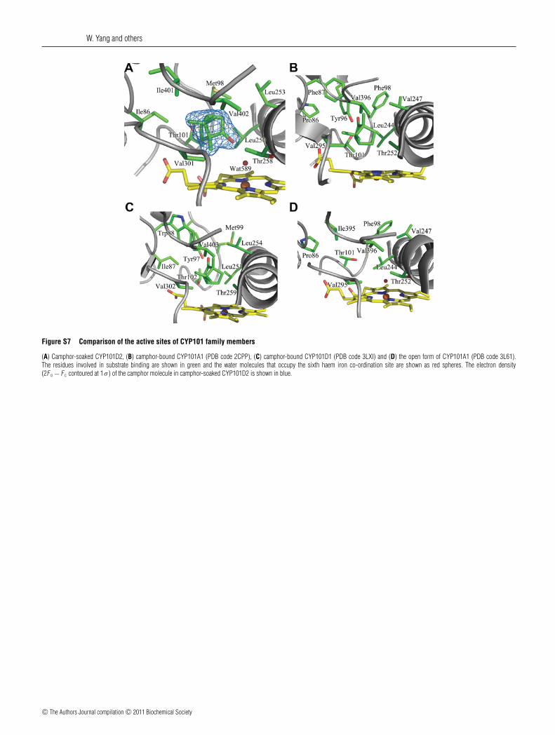
Figure S11 Substrate-access channel of (A) the native and (B) the camphor-soaked structures of CYP101D2
The chains of water molecules that lead from the protein surface to Asp257 in the I helix groove are shown as red spheres. The residues that interact with the water molecules are shown in green. Inthe native structure, the carboxy group of Asp257 forms a hydrogen bond with Wat-511, which is at the end of a chain of five hydrogen-bonded water molecules that lead to the surface of the enzymeand the bulk solvent. In the camphor-soaked structure, two water molecules are hydrogen-bonded to Asp257, and a further seven water molecules are located in the open channel, forming a chainleading to the external solvent. The final water of this chain Wat-513 is located on the enzyme surface and forms hydrogen bonds with the carbonyl oxygen of Tyr29 and the Nδ1 of His398. Tyr29 isconserved in CYP101D2, CYP101D1 and CYP101A1, whereas His398 is conserved in CYP101D1 and aligns with Lys392 in CYP101A1 (Supplementary Table S3). Networks of water molecules arealso found in the open conformations of CYP101A1 and the D-4-Ad- and D-8-Ad-bound structures of CYP101A1 [6,7].
Figure S12 A comparison of the proximal faces of (A) CYP101D1 and (B) CYP101A1
Negatively and positively charged surface areas are coloured red and blue respectively. The proximal faces of CYP101D2 (Figure 3D in the main paper) and CYP101D1 are more positively chargedthan that of CYP101A1. Highlighted are the positively charged residues of CYP101D1 (Arg77, Arg113, Arg126, Arg290, Arg350, Arg363 and Arg371). The corresponding residues of CYP101D2 (Ser76,Arg112, Arg125, Arg289, Arg349, Arg362 and Arg370) and of CYP101A1 (Glu76, Arg112, Asp125, Ala283, Gln343, Leu356 and Arg364) are also highlighted.
c© The Authors Journal compilation c© 2011 Biochemical Society
W. Yang and others
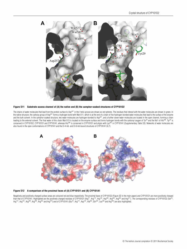
Figure S13 Sequence alignment of CYP101D2 and CYP101D1 from N. aromaticivorans and CYP101A1 from P. putida
The alignment was performed using ClustalW and ESPript [8,9]. Black arrows and cylinders indicate the β-sheets and α-helices respectively. Conserved residues are highlighted.
c© The Authors Journal compilation c© 2011 Biochemical Society

Crystal structure of CYP101D2
Figure S14 Analysis of camphor binding to CYP101D2
The enzyme concentration was 1.2 μM in 50 mM Tris/HCl, pH 7.4, at 30◦C. The results show agood fit to a hyperbolic function. Values for K d are 5.2 +− 0.7 and 3.1 +− 0.5 μM in the absenceand presence of 200 mM KCl respectively. E = ×10.
Table S1 Comparison of (a) primary sequence identities and (b) active siteresidues of CYP101D2, CYP101D1 and CYP101A1 (including the K+-bindingsite of CYP101A1)
Differences in CYP101D1 and CYP101D2 from CYP101A1 are underlined. K+ bindingstrengthens camphor binding by CYP101A1 (0.45 compared with 16 μM) [1,2], but theeffect is greatly reduced in CYP101D1 (K d of 5.9 compared with 9.1 μM) and CYP101D2(K d 3.1 compared with 5.2 μM) [3]. The K+-binding site in CYP101A1 is octahedral, withthe backbone carbonyl oxygens of Glu84, Gly93, Glu94 and Tyr96, and two water molecules, asligands [2,4]. These residues are conserved in CYP101D2, but the open conformation resultsin movement of Tyr96 and its carbonyl group points away from where the K+-binding site wouldbe expected to be. Glu84, which hydrogen bonds to one of the water ligands, is replaced withArg85 in CYP101D1, which results in an expanded and non-octahedral arrangement of ligands.Replacement of Glu84 with a lysine residue in the E84K mutant of CYP101A1 has been shownto abolish the effect of K+ ions [2].
(a)
N. aromaticivorans N. aromaticivoransProtein CYP101D1 CYP101D2
P. putida CYP101A1 (P450cam)Identities 187/418 (44 %) 184/398 (46 %)Positives 258/418 (61 %) 251/398 (63 %)
N. aromaticivorans CYP101D2Identities 254/404 (62 %) –Positives 310/404 (76 %) –
(b)
CYP101D2 CYP101D1 CYP101A1
Glu84 Arg85 Glu84
Phe87 Trp88 Phe87
Gly93 Gly94 Gly93
Glu94 Glu95 Glu94
Tyr96 Tyr97 Tyr96
Met98 Met99 Phe98
Thr101 Thr102 Thr101
Thr185 Thr186 Thr185
Leu250 Leu251 Leu244
Leu253 Leu254 Val247
Val301 Val302 Val295
Glu303 Asp304 Asp297
Ile401 Ile402 Ile395
Val402 Val403 Val396
c© The Authors Journal compilation c© 2011 Biochemical Society

W. Yang and others
Table S2 Comparison of the residues found in the substrate recognitionsite on the surfaces of CYP101D2, CYP101D1 and CYP101A1
Arg186 and Asp257 form a salt bridge in the camphor-soaked form of CYP101D2. This saltbridge is conserved in CYP101D1 (Arg187 and Asp258) and CYP101A1 (Arg186 and Asp251).In CYP101A1, Lys178 and Asp182 form part of salt bridge network with Arg186 and Asp251. InCYP101D2 and CYP101D1, Lys178 and Asp182 are replaced with Ser178 and Gly179, and Arg182
and Asn183 respectively. A water molecule hydrogen-bonds to Asp258 in CYP101D1 (2.63 A),replacing the salt bridge found between Asp251 and Lys178 in CYP101A1. Other salt bridges havebeen proposed to contribute to the stability of the CYP101A1 structure, with the Asp97–Lys197
salt bridge being particularly stable [5]. These salt bridge interactions are less well conservedin CYP101D1 and CYP101D2, with Gln97 and Asn203 replacing Asp97 and Lys197 in CYP101D2(Supplementary Table S3).
CYP101D2 CYP101D1 CYP101A1
Glu156 Thr157 Glu156
Pro159 Pro160 Pro159
Val160 Val161 Ile160
Ser178 Gly179 Lys178
Ala181 Ala182 Thr181
Arg182 Asn183 Asp182
Thr185 Thr186 Thr185
Arg186 Arg187 Arg186
Leu256 Leu257 Leu250
Asp257 Asp258 Asp251
Val260 Val261 Val254
Table S3 Comparison of the residues that comprise the access channeland binding site in CYP101D2 and their equivalent residues in CYP101D1and CYP101A1
CYP101D2 CYP101D1 CYP101A1
Tyr29 Tyr30 Tyr29
Phe87 Trp88 Phe87
Pro89 Pro90 Pro89
Tyr96 Tyr97 Tyr96
Gln97 Asp98 Asp97
Met98 Met99 Phe98
Met184 Met185 Met184
Thr185 Thr186 Thr185
Pro187 Pro188 Pro187
Met195 Gln196 –Leu199 Leu200 Phe193
Asn203 Asn204 Lys197
Leu253 Leu254 Val247
His398 His399 Lys392
Ile401 Ile402 Ile395
Val402 Val403 Val396
REFERENCES
1 Deprez, E., Gill, E., Helms, V., Wade, R. C. and Hui Bon Hoa, G. (2002) Specific andnon-specific effects of potassium cations on substrate–protein interactions in cytochromesP450cam and P450lin. J. Inorg. Biochem. 91, 597–606
2 Westlake, A. C., Harford-Cross, C. F., Donovan, J. and Wong, L. L. (1999) Mutations ofglutamate-84 at the putative potassium-binding site affect camphor binding and oxidationby cytochrome p450cam. Eur. J. Biochem. 265, 929–935
3 Yang, W., Bell, S. G., Wang, H., Zhou, W., Hoskins, N., Dale, A., Bartlam, M., Wong, L. L.and Rao, Z. (2010) Molecular characterization of a class I P450 electron transfer systemfrom Novosphingobium aromaticivorans DSM12444. J. Biol. Chem. 285, 27372–27384
4 Poulos, T. L., Finzel, B. C. and Howard, A. J. (1987) High-resolution crystal structure ofcytochrome P450cam. J. Mol. Biol. 195, 687–700
5 Lounnas, V. and Wade, R. C. (1997) Exceptionally stable salt bridges in cytochromeP450cam have functional roles. Biochemistry 36, 5402–5417
6 Dunn, A. R., Dmochowski, I. J., Bilwes, A. M., Gray, H. B. and Crane, B. R. (2001) Probingthe open state of cytochrome P450cam with ruthenium-linker substrates. Proc. Natl. Acad.Sci. U.S.A. 98, 12420–12425
7 Lee, Y. T., Wilson, R. F., Rupniewski, I. and Goodin, D. B. (2010) P450cam visits an openconformation in the absence of substrate. Biochemistry 49, 3412–3419
8 Chenna, R., Sugawara, H., Koike, T., Lopez, R., Gibson, T. J., Higgins, D. G. andThompson, J. D. (2003) Multiple sequence alignment with the Clustal series of programs.Nucleic Acids Res. 31, 3497–3500
9 Gouet, P., Courcelle, E., Stuart, D. I. and Metoz, F. (1999) ESPript: analysis of multiplesequence alignments in PostScript. Bioinformatics 15, 305–308
Received 9 July 2010/24 September 2010; accepted 15 October 2010Published as BJ Immediate Publication 15 October 2010, doi:10.1042/BJ20101017
c© The Authors Journal compilation c© 2011 Biochemical Society