the skeletal system: bones and...
TRANSCRIPT
1
6The Skeletal System: Bones
and JointsFOCUS: The extracellular matrix of bonecontains collagen, which lends flexible strength,and minerals, which give bone weight-bearingstrength. The two major types of bone arecompact bone and cancellous bone. Boneossification, growth, remodeling and repair aredynamic processes carried out by osteoblastsand osteoclasts. The skeletal system consists ofthe axial skeleton (skull, vertebral column, andrib cage) and the appendicular skeleton (limbs
and their girdles). The skull surrounds andprotects the brain; the vertebral columnsupports the head and trunk and protects thespinal cord; and the rib cage protects the heartand lungs. The pectoral girdle attaches theupper limbs to the trunk and allows a widerange of movement of the upper limbs. Thepelvic girdle attaches the lower limbs to thetrunk and is specialized to support the weight ofthe body.
Connective Tissue
❛❛Connective tissue consists of cells separated from each other by an extracellular matrix.❜❜
Match these terms with the Bone Ligamentscorrect statement or definition: Cartilage Tendons
1. Extracellular matrix is made up primarily of collagen.
2. Extracellular matrix is made up of collagen and proteoglycans.
3. Extracellular matrix is made up of collagen and minerals.
4. Attach muscles to bones.
CONTENT EARNINGL ACTIVITY

2
Most of the mineral in bone is in the form of calcium phosphate crystals calledhydroxyapatite.
General Features of Bone
❛❛There are four types of bone, described by their shape as long, short, flat, and irregular.❜❜
A. Match these terms with the Flat bones Long bonescorrect statement or definition: Irregular bones Short bones
1. Bones longer than they are wide, e.g., limb bones.
2. Bones as broad as they are long, e.g., ankle and wrist bones.
3. Bones such as the ribs, scapula, and sternum.
4. Bones such as the vertebrae and facial bones.
B. Using the terms provided, complete these statements:
Blood-forming FatDiaphysis MarrowEndosteum Medullary cavityEpiphysis OsteoblastsEpiphyseal line PeriosteumEpiphyseal plate
Each long bone consists of a shaft, called the (1) , anda(n) (2) at each end of the bone. A long bone that is stillgrowing has a(n) (3) , composed of cartilage, between eachepiphysis and the diaphysis. When bone growth stops, theepiphyseal plate is replaced by bone, and is called the (4) .The large cavity in the diaphysis is called the (5) . Thisspace, and other spaces are filled with soft tissue called (6) .Yellow marrow consists mostly of (7) , whereas red marrowconsists of (8) cells. Most of the outer surface of the bone iscovered by a connective tissue layer called the (9) , whichcontains blood vessels and nerves. The medullary cavity islined with a thinner connective tissue membrane, the (10) .The periosteum and endosteum contain (11) , which functionin the formation, repair, and remodeling of bone.
1.
2.
3.
4.
5.
6.
7.
8.
9.
10.
11.
C. Using the terms provided, complete these statements:
Canaliculi LamellaeLacunae Osteocytes
Bone is formed in thin sheets of extracellular matrix called (1) , with bone cells, called (2) between the lamellae. Theosteocytes are located within spaces called (3) . Cell processesextend from the osteocytes across the extracellular matrix of thelamellae within tiny canals called (4) .
1.
2.
3.
4.
�

3
Compact Bone and Cancellous bone
❛❛The two major types of bone are compact bone, which is mostly solid matrix and cells,❜❜ and cancellous bone, which consists of a lacy network of bone with many small, marrow-filled spaces.
A. Using the terms provided, complete these statements:
Blood vessels OsteonCanaliculi Periosteum andCentral canal endosteum
Most of the lamellae of compact bone are organized into setsof concentric rings with each set surrounding a (1) . Withinthe central canal are (2) that run parallel to the long axis ofthe bone. Each central canal with the lamellae andosteocytes surrounding it, is called a(n) (3) . The osteocytesare connected to each other by cell processes located in (4) .Blood vessels in the (5) supply blood to vessels in the centralcanal.
1.
2.
3.
4.
5.
B. Using the terms provided, complete these statements:
Blood vessels OsteocytesCanaliculi TrabeculaeMarrow
Cancellous (spongy) bone consists of delicate interconnectingrods or plates of bone called (1) . The spaces between thetrabeculae are filled with (2) . Each trabecula consists ofseveral lamellae, with (3) between the lamellae. Usually no(4) penetrate the trabeculae, and the trabeculae have nocentral canals. Nutrients pass by diffusion through the (5)to the osteocytes of the trabeculae.
1.
2.
3.
4.
5.
Bone Ossification
❛❛Ossification is the formation of bone by osteoblasts.❜❜
A. Match these terms with the Endochondral ossificationcorrect statement or definition: Intramembranous ossification
1. Bone formation that occurs within connective tissuemembranes.
2. Ossification process that occurs primarily in the flat bones ofthe skull.
3. Ossification process that produces most of the skeletal system.

4
B. Using the terms provided, complete these statements:
Calcified PrimaryCartilage model ossification centerConnective tissue SecondaryOsteoblasts ossification centersOsteoclasts
Intramembranous ossification occurs when osteoblasts beginto produce bone in ossification centers of (1) membranes.Endochondral ossification begins with a (2) , which has thegeneral shape of the mature bone. The chondrocytes of thecartilage model increase in number, hypertrophy, and die andthe cartilage matrix becomes (3) , forming an ossificationcenter. When chondrocytes die, (4) invade spaces in thecenter of the bone and produce bone matrix; (5) remove boneand calcified cartilage to form the medullary cavity. Thecenter part of the diaphysis where bone first begins to appearis called the (6) . Later, (7) form in the epiphyses.
1.
2.
3.
4.
5.
6.
7.
Bone Growth, Remodeling, and Repair
❛❛Bone growth, remodeling, and repair all involve deposition of new bone matrix by osteoblasts.❜❜
A. Using the terms provided, complete these statements:
Calcified OssificationChondrocytes OsteoblastsDiameter OsteoclastsEpiphyseal plate ProliferatingLength
As osteoblasts deposit new bone matrix on the surface ofbones between the periosteum and the existing bone, thebone increases in (1) . Growth in the (2) of a bone, which isthe major source of increased height in an individual, occursin the (3) . Just as in endochondral ossification, (4) increasein number. Within the (5) zone, the chondrocytes line up incolumns, hypertrophy, and die. The cartilage matrix is (6) ,and (7) start forming bone matrix on the surface of thecalcified cartilage. This process produces a zone of (8) onthe diaphyseal side of the epiphyseal plate.
1.
2.
3.
4.
5.
6.
7.
8.
5
B. Using the terms provided, complete these statements:
Calcium DecreaseCallus IncreaseCancellous OsteoblastsClot Osteoclasts
Bone remodeling involves the removal of old bone by (1) ,and the deposition of new bone by (2) . Bone is the majorstorage site for (3) in the body. Calcium is removed frombones when blood calcium levels (4) , and it is depositedwhen dietary calcium is adequate. When a bone is broken,the bone bleeds, and a (5) is formed in the damaged area.Cells from surrounding tissue invade and form a fibrousnetwork with islets of cartilage, which holds the bonefragments together. The zone of tissue repair between thetwo bone fragments is called a (6) . Osteoblasts enter thecallus and begin forming (7) bone, which is later remodeled.
1.
2.
3.
4.
5.
6.
7.
General Considerations of Bone Anatomy
❛❛Several common terms are used to describe features of bones.❜❜
Match these terms with the Canal or meatus Fossacorrect statement or definition: Condyle Process
Foramen Tubercle or tuberosity
1. Hole in a bone.
2. Tunnel-like passage through a bone.
3. Depression in a bone.
4. Lump on a bone.
5. Projection from a bone.
6. Smooth, rounded end of a bone, where it forms a joint withanother bone.
The axial skeleton is divided into the skull, vertebral column, and thoracic cage.�
6
Skull
❛❛The bones of the skull are divided into two portions, the cranial vault and the face.❜❜
A. Match these terms with the Cranial vault (braincase) Nasal septumcorrect statement or definition: Facial bones Orbit
Hyoid bone Paranasal sinusesNasal conchae Sella turcica
1. Subdivision of the skull that protects the brain.
2. Bones that form the structure of the face, but do not contributeto the cranial vault.
3. Bone that "floats" in the neck and is the attachment site forthroat and tongue muscles.
4. Structure in the skull that surrounds and protects the eye.
5. Three bony shelves of the nasal cavity that help to warm andmoisten the air.
6. Air-filled cavities that open into the nasal cavity.
7. Structure resembling a saddle that is occupied by the pituitarygland.
8. Perpendicular bone and cartilage that divide the nasal cavityinto right and left halves.
B. Match these openings or External auditory meatus Orbitdepressions with the correct Foramen magnum Orbital fissuresdescription: Mandibular fossa Optic foramen
Nasolacrimal canal
1. Large openings through which nerves or blood vesselscommunicate with the eye.
2. Opening through which the optic nerve passes into the skull.
3. Opening through which the spinal cord connects to the brain.
4. Depression where mandible articulates with temporal bone.
5. Cone-shaped fossa that surrounds the eye.
6. Opening that passes from the orbit into the nasal cavity.
7. Temporal bone canal; allows sound to reach the eardrum.

7
C. Match these terms with Coronal suture Occipital bonethe correct parts labeled External auditory meatus Parietal bonein figure 6.1: Frontal bone Sphenoid bone
Lambdoid suture Squamous sutureMandible Styloid processMastoid process Temporal boneMaxilla Zygomatic archNasal bone Zygomatic bone
1. 7. 12.
2. 8. 13.
3. 9. 14.
4. 10. 15.
5. 11. 16.
6.
Figure 6.1

8
D. Match these bone parts Horizontal plate of palatine Temporalwith structures to which Palatine process of maxilla Vomerthey contribute: Perpendicular plate Zygomatic
of ethmoid
1. Two parts that form the hard palate.
2. Two parts that form the nasal septum.
3. Two bones that form the zygomatic arch.
Vertebral Column
❛❛The vertebral column provides support, protection, a site for muscle attachment, and flexibility.❜❜
A. Match these terms with the Cervical Sacralcorrect statement or definition: Lumbar Thoracic
1. Two sections of the vertebral column that curve posteriorly.
2. There are seven of these vertebrae in the vertebral column.
3. There are twelve of these vertebrae in the vertebral column.
4. There are five of these vertebrae in the vertebral column.
B. Match the type of vertebra Atlas Lumbar vertebraewith the correct description: Axis Sacrum
Cervical vertebrae Thoracic vertebraeCoccyx
1. Have transverse foramina and partly split spinous processes.
2. First cervical vertebra; allows a "yes" motion of the head.
3. Articular facets for ribs present.
4. Superior articular facets of these vertebrae face medially and“lock” with laterally facing inferior articular facets of thevertebra above it.
5. Five fused vertebrae that have a median crest and a hiatus.
6. Tailbone, usually consisting of four fused vertebrae.

9
C. Match these terms with the Articular process Pediclecorrect statement or definition: Body Spinous process
Intervertebral disk Transverse processIntervertebral foramina Vertebral foramenLamina
1. Weight-bearing portion of the vertebra.
2. Two parts that form the vertebral arch.
3. Contains the spinal cord; all of them together form the vertebralcanal.
4. Where the spinal nerves exit the vertebral column.
5. Where vertebrae articulate with each other.
6. Lumps that can be seen and felt down the midline of the back.
7. Dense fibrous connective tissue that separates vertebrae.
D. Match these terms with Articular facet 1. the correct parts labeled Articular processin figure 6.2: Body 2.
LaminaPedicle 3. Spinous processTransverse process 4. Vertebral archVertebral foramen 5.
6.
7.
8.
9.
Spinous processes of the first four sacral vertebrae form the median sacral crest, but the fifthsacral vertebra does not form, leaving a sacral hiatus (opening) at the inferior end of thesacrum. The anterior edge of the first sacral vertebra forms the sacral promontory.
�
Figure 6.2

10
Thoracic Cage
❛❛The thoracic cage or rib cage protects the vital organs within the thorax and prevents the❜❜ collapse of the thorax during respiration.
A. Match these terms with the Body Manubriumcorrect statement or definition: False ribs Sternal angle
Floating ribs True ribsJugular notch Xiphoid process
1. First seven pairs of ribs that attach directly to the sternum.
2. Eleventh and twelfth ribs, which have no attachment to thesternum.
3. Middle part of the sternum.
4. Most superior part of the sternum.
5. Depression at the superior end of the sternum.
6. Slight elevation at the junction of the manubrium and body ofthe sternum; landmark for locating the second rib.
B. Match these terms with Body Manubriumthe correct parts labeled Costal cartilage Sternumin figure 6.3: False ribs True ribs
Floating ribs Xiphoid process
1.
2.
3.
4.
5
6.
7.
8.
Figure 6.3
11
Pectoral Girdle
❛❛The pectoral, or shoulder girdle consists of two pairs of bones that attach the upper limb to the❜❜ body: the scapulae, or shoulder blades, and the clavicles, or collar bones.
Match these terms with the Acromion process Glenoid fossacorrect statement or definition: Coracoid process Spine
1. Ridge that runs across the posterior surface of the scapula.
2. Projection from the scapular spine that forms the point of theshoulder; point of attachment of the clavicle.
3. Projection from the scapula that curves below the clavicle andprovides attachment for arm and chest muscles.
4. Depression where the head of the humerus articulates with thescapula.
Upper Limb
❛❛The upper limb consists of the bones of the arm, forearm, wrist, and hand.❜❜
A. Match the bone parts that Distal end of humerus Head of radiusarticulate with each other: Head of humerus Head of ulna
1. Glenoid fossa
2. Semilunar notch
3. Humerus and ulna
4. Carpals
The portion of the ulna proximal to the semilunar notch is the olecranon process, or pointof the elbow. Just distal to the semilunar notch is the coronoid process.
B. Match the bony part with Epicondyles Radial tuberosityits function: Greater and lesser tubercles Styloid processes
1. Location for shoulder muscles to attach to the humerus.
2. Location where forearm muscles attach to the humerus.
3. Location of attachment of the biceps brachii to the radius.
4. Attachments for the ligaments of the wrists.
The wrist is composed of eight carpal bones, the hand is composed of five metacarpals,and each finger, called a digit, is composed of three phalanges. The thumb has twophalanges.
�
�

12
Pelvic Girdle
❛❛The pelvic girdle is a ring of bones formed by the sacrum and two coxae.❜❜
Using the terms provided, complete these statements:
Acetabulum Iliac crestCoxa MaleFemale Obturator foramenAnterior superior iliac spine
Each (1) is formed by the fusion of the ilium, ischium, andpubis. The superior margin of the ilium is called the (2) ,and a(n) (3) is located at each ilium's anterior end. The (4)is the socket of the hip joint, and the (5) is the large hole inthe coxa. The pelvic inlet and outlet are larger and thepelvic inlet is more oval in the pelvis of the (6) .
1.
2.
3.
4.
5.
6.
Lower Limb
❛❛The lower limb consists of the bones of the thigh, leg, ankle, and foot.❜❜
A. Match the bone parts that Acetabulum Talusarticulate with each other: Condyles of femur Tibia
1. Head of the femur.
2. Proximal end of tibia.
3. Head of fibula.
4. Distal end of tibia and fibula.
B. Match the bony part with Calcaneus Patellaits function: Condyles Tibial tuberosity
Epicondyles TrochantersMalleolus
1. Projections lateral to the condyles on the distal end of femur.
2. Points of muscle attachment near the head of the femur.
3. Located within the major tendon of the thigh muscles; enablesthe tendon to turn the corner over the knee.
4. Location on the tibia where anterior thigh muscles attach.
5. Prominence on each side of the ankle that forms a partial socketfor the talus bone.
6. Inferior to the talus; protrudes posteriorly to form the heel.
13
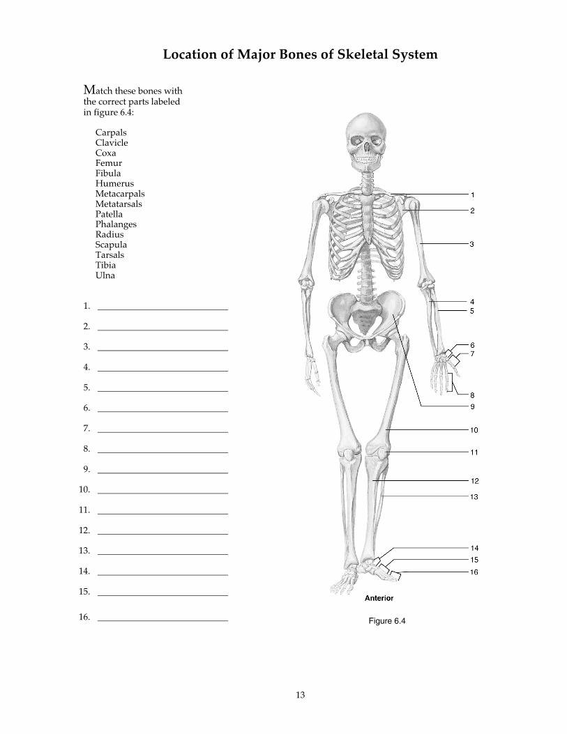
Location of Major Bones of Skeletal System
Match these bones withthe correct parts labeledin figure 6.4:
CarpalsClavicleCoxaFemurFibulaHumerusMetacarpalsMetatarsalsPatellaPhalangesRadiusScapulaTarsalsTibiaUlna
1.
2.
3.
4.
5.
6.
7.
8.
9.
10.
11.
12.
13.
14.
15.
16. Figure 6.4
14
Articulations
❛❛An articulation, or joint, is a place where two bones come together.❜❜
A. Match the class of joint with Cartilaginous Synovialthe correct definition: Fibrous
1. Two bones united by fibrous tissue; exhibit little or nomovement.
2. Two bones united by cartilage; only slight movement can occurat these joints.
3. Freely moving joints that contain fluid in a cavity surroundingthe ends of bones.
B. Match these terms with the Fontanels Suturescorrect statement or definition: Gomphoses Syndesmoses
1. Fibrous joints between the bones of the skull.
2. Wide sutures (soft spots) present in newborns.
3. Fibrous joints where bones are separated by some distance andare held together by ligaments.
4. Fibrous joints consisting of pegs fitted into sockets.
C. Match these terms with the Articular cartilage Joint cavitycorrect statement or definition: Bursa Synovial membrane
Joint capsule
1. Cartilage that provides a smooth surface where bones meet.
2. The space surrounding the ends of articulating bones.
3. Surrounds the joint cavity; portions may be thickened to formligaments.
4. Tissue that lines the joint capsule except over the articularcartilage; produces synovial fluid.
5. Extension of the synovial membrane that forms a pocket or sac;reduces friction where structures would rub together.
15
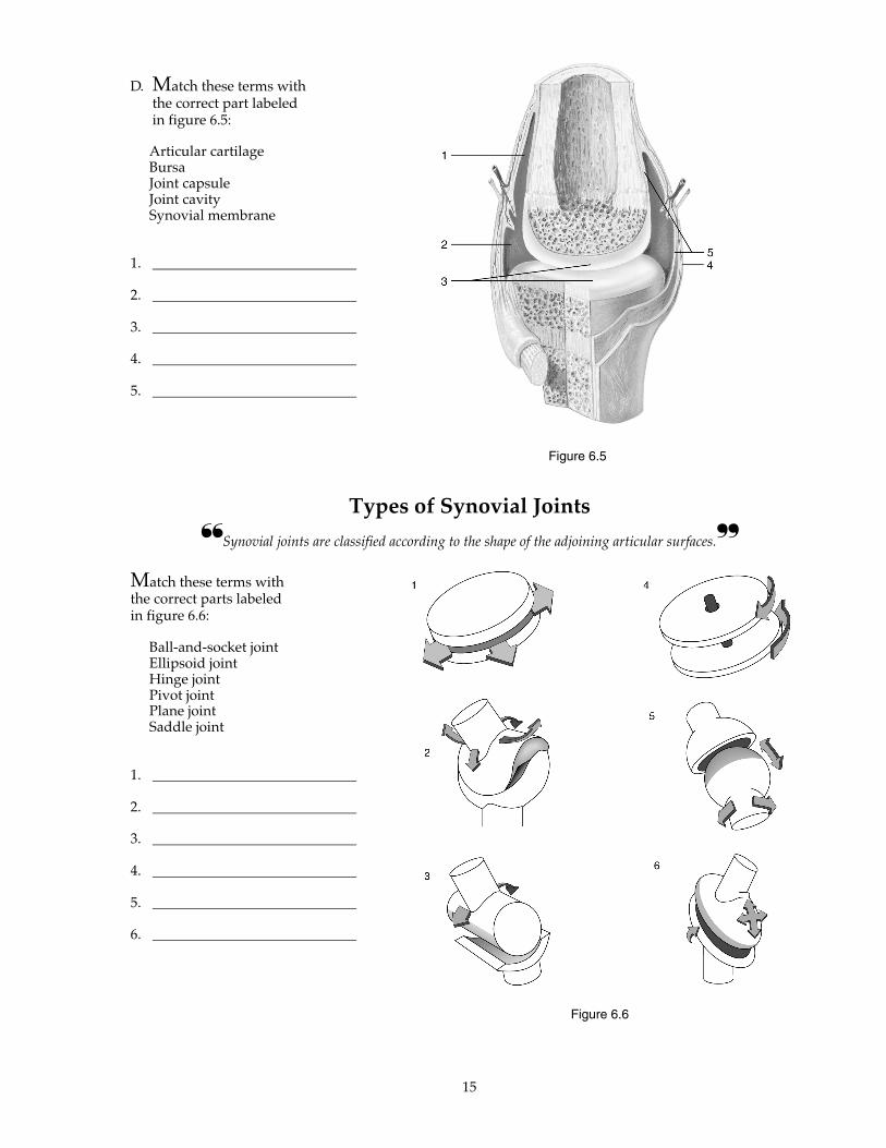
D. Match these terms withthe correct part labeledin figure 6.5:
Articular cartilageBursaJoint capsuleJoint cavitySynovial membrane
1.
2.
3.
4.
5.
Types of Synovial Joints
❛❛Synovial joints are classified according to the shape of the adjoining articular surfaces.❜❜
Match these terms withthe correct parts labeledin figure 6.6:
Ball-and-socket jointEllipsoid jointHinge jointPivot jointPlane jointSaddle joint
1.
2.
3.
4.
5.
6.
Figure 6.5
Figure 6.6
16
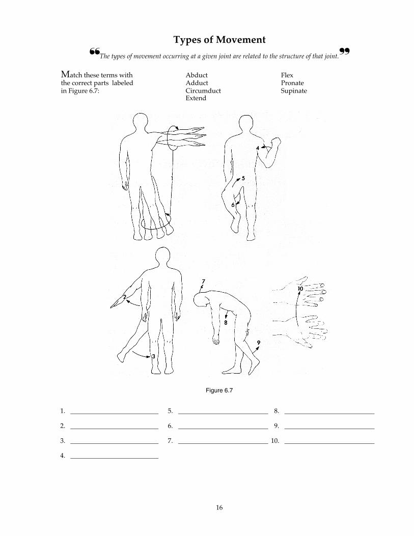
Types of Movement
❛❛The types of movement occurring at a given joint are related to the structure of that joint.❜❜
Match these terms with Abduct Flexthe correct parts labeled Adduct Pronatein Figure 6.7: Circumduct Supinate
Extend
1. 5. 8.
2. 6. 9.
3. 7. 10.
4.
Figure 6.7
17
1. List five functions of bone.
2. List three functions of cartilage.
3. List three types of bone cells, depending on their function.
4. List two types of bone, depending on their internal structure.
5. Name two types of bone ossification.
6. Explain how a bone grows in diameter and length.
7. Name the five types of vertebrae, and give the number of each found in the vertebralcolumn.
8. Name the three types of ribs according to their attachment, and give the number ofeach type.
QUICK RECALL
18
9. Name the bones of the pectoral and pelvic girdles.
10. Give the number of carpals, metacarpals, and phalanges in the upper limb, and give thenumber of tarsals, metatarsals, and phalanges in the lower limb.
11. List the three major classes of joints.
12. Name the six types of synovial joints and give an example of each.
Give an example of a new vocabulary word that contains each word part.
WORD PART MEANING EXAMPLE
chondr- cartilage 1.
oste- bone 2.
cancel- crossbar; lattice 3.
lacun- space; hollow 4.
styl- shaped like a pen 5.
artic- joint 6.
WORD PARTS
19
Place the letter corresponding to the correct answer in the space provided.
1. Which of these is a function of bone?a. internal support and protectionb. provide attachment for musclesc. mineral storaged. blood cell formatione. all of the above
2. Bone matrix containsa. collagen.b. calcium and phosphate.c. proteoglycan.d. chondrocytes.e. both a and b
3. A break in the shaft of a bone is abreak in thea. epiphysis.b. perichondrium.c. diaphysis.d. articular cartilage.
4. Which of these connective tissuestructures cover the surface ofmature bones?a. perichondriumb. periosteumc. hyaline cartilaged. b and c
5. In compact bone, the osteocytes areconnected to each other by tiny cellprocesses extending through tinycanals calleda. lamellae.b. lacunae.c. central canals.d. canaliculi.
6. Intramembranous ossificationa. occurs at the epiphyseal plate.b. gives rise to the flat bones of the
skull.c. is responsible for increased
diameter of bone.d. develops from a cartilage model.
7. Primary ossification centers of a longbone are found in thea. diaphysis.b. epiphysis.c. perichondrium.d. periosteum.e. articular cartilage.
8. As a long bone increases in diameter,the medullary cavitya. increases in size.b. decreases in size.c. does not change in size.
9. During bone growth at theepiphyseal plate, _____ increase innumber, hypertrophy, and die.a. osteocytes.b. osteoblasts.c. osteoclasts.d. chondrocytes.
10. The prime function of osteoclasts istoa. prevent osteoblasts from forming.b. break down bone.c. produce calcium salts and
collagen fibers.d. change spongy bone to cartilage.
11. In the healing of bone fracturesa. a blood clot forms around the
break.b. a callus is formed.c. cancellous bone is formed in the
callus.d. the callus may eventually
disappear.e. all of the above
12. Which of these is a tunnel-likepassage through a bone?a. canal or meatusb. condylec. foramend. fossae. process
MASTERY LEARNING ACTIVITY

20
13. Which of these is part of theappendicular skeleton?a. craniumb. ribsc. clavicled. sternume. vertebra
14. The perpendicular plate of theethmoid and the ______ form thenasal septum.a. zygomatic archb. nasal bonec. nasal conchaed. vomer
15. Which of these bones does NOTcontain a paranasal sinus?a. ethmoidb. sphenoidc. temporald. frontale. maxilla
16. The squamous suture joins thea. frontal and temporal bones.b. frontal and parietal bones.c. parietal and temporal bones.d. parietal and occipital bones.
17. The passageway that carries tearsfrom the eyes to the nasal cavity isa. the nasolacrimal canal.b. the optic foramen.c. the orbital fissure.d. the foramen magnum.
18. The weight-bearing portion of avertebra is thea. vertebral arch.b. articular process.c. body.d. transverse process.e. spinous process.
19. Transverse foramina are found onlyina. cervical vertebrae.b. thoracic vertebrae.c. lumbar vertebrae.d. the sacrum.e. the coccyx
20. Which of these parts of the upperlimb is NOT correctly matched withthe number of bones in that part?a. arm: 1b. forearm: 2c. wrist: 10d. palm of hand: 5e. fingers: 14
21. Process that forms the outer ankle?a. lateral condyleb. lateral epicondylec. lateral tuberosityd. lateral malleoluse. none of the above
22. Which of these pairs of bones orstructures do NOT articulate witheach other?a. mandible - temporal boneb. maxillary bone - palatine bonec. scapula - clavicled. head of the ulna - humeruse. acetabulum of coxa - femur
23. Which of these types of joints containfibrous connective tissue?a. syndesmosisb. suturec. gomphosisd. a and be. all of the above
24. Which of these is characteristic of asynovial joint?a. articular surfaces covered with
cartilageb. joint capsulec. synovial membraned. synovial fluide. all of the above
25. Once a doorknob is grasped with theright hand, what movement of theforearm is necessary to unlatch thedoor (turn in a clockwise direction)?a. pronationb. rotationc. flexiond. supinatione. extension
21
Use a separate sheet of paper to complete this section.
1. A doctor tells an elderly friend of yours thatthe cartilage in his joints is degenerating andhas lost its resiliency. Your friend asks youto explain what that means and what theconsequences might be. What do you say?
2. The length of the lower limbs of a 5-year-oldwere measured, and it was determined thatone limb was 2 cm shorter than the otherlimb. Would you expect the little girl toexhibit kyphosis, scoliosis, or lordosis?Explain.
3. Can you suggest a possible advantage inhaving the coccyx attached to the sacrum bya flexible cartilage joint?
4. What would be the likely consequences if afontanel in a baby's skull fused shortly afterbirth?
FINAL CHALLENGES✰ ✰