the science of transcranial magnetic stimulation · the science of transcranial magnetic...
TRANSCRIPT
279
CME
The Science of Transcranial Magnetic Stimulation
ABSTRACTTranscranial magnetic stimulation
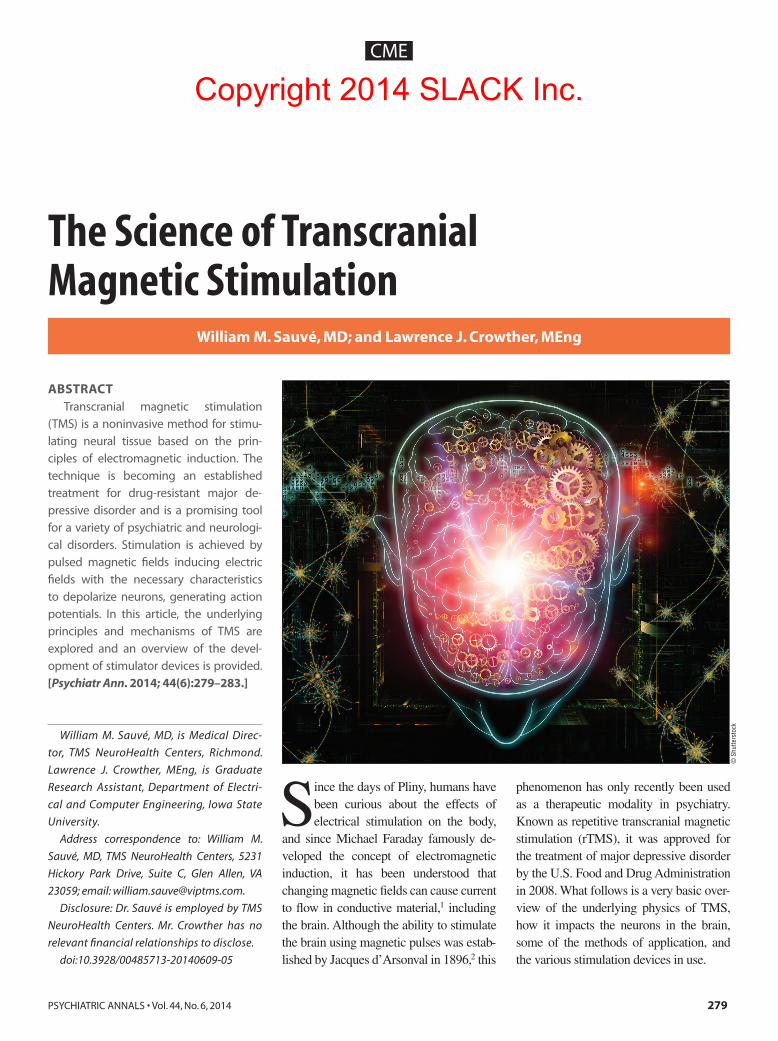
(TMS) is a noninvasive method for stimu-lating neural tissue based on the prin-ciples of electromagnetic induction. The technique is becoming an established treatment for drug-resistant major de-pressive disorder and is a promising tool for a variety of psychiatric and neurologi-cal disorders. Stimulation is achieved by pulsed magnetic !elds inducing electric !elds with the necessary characteristics to depolarize neurons, generating action potentials. In this article, the underlying principles and mechanisms of TMS are explored and an overview of the devel-opment of stimulator devices is provided. [Psychiatr Ann. 2014; 44(6):279–283.]
Since the days of Pliny, humans have been curious about the effects of electrical stimulation on the body,
and since Michael Faraday famously de-veloped the concept of electromagnetic induction, it has been understood that changing magnetic fields can cause current to flow in conductive material,1 including the brain. Although the ability to stimulate the brain using magnetic pulses was estab-lished by Jacques d’Arsonval in 1896,2 this
phenomenon has only recently been used as a therapeutic modality in psychiatry. Known as repetitive transcranial magnetic stimulation (rTMS), it was approved for the treatment of major depressive disorder by the U.S. Food and Drug Administration in 2008. What follows is a very basic over-view of the underlying physics of TMS, how it impacts the neurons in the brain, some of the methods of application, and the various stimulation devices in use.
William M. Sauvé, MD, is Medical Direc-
tor, TMS NeuroHealth Centers, Richmond.
Lawrence J. Crowther, MEng, is Graduate
Research Assistant, Department of Electri-
cal and Computer Engineering, Iowa State
University.
Address correspondence to: William M.
Sauvé, MD, TMS NeuroHealth Centers, 5231
Hickory Park Drive, Suite C, Glen Allen, VA
23059; email: [email protected].
Disclosure: Dr. Sauvé is employed by TMS
NeuroHealth Centers. Mr. Crowther has no
relevant financial relationships to disclose.
doi:10.3928/00485713-20140609-05
William M. Sauvé, MD; and Lawrence J. Crowther, MEng
© S
hutte
rsto
ck
Copyright 2014 SLACK Inc.
280
CME
FARADAY’S LAW OF ELECTROMAGNETIC INDUCTION
Both Michael Faraday and Joseph Henry independently discovered the con-cept of electromagnetic induction in 1831, but Faraday was the first to publish his findings. Simply put, a magnetic field that is in motion relative to a conductor brings about a current in said conductor. Hence, a changing magnetic field induces a flow of electric current in nearby conductors that, for the purposes of this article, in-clude human tissue.3 The most commonly used form of expression for this concept is the Maxwell-Faraday equation, also re-ferred to as Faraday’s Law.
Electromagnetic induction is the key principle in transcranial magnetic stimu-lation (TMS), taking advantage of the fact that every electric current has a magnetic field surrounding it, with alternating cur-rents bringing about fluctuating magnetic fields. Fluctuating magnetic fields in turn cause electric current to flow in conduc-tors placed within them; the conductors in the case of TMS being neurons in the brain, thus allowing for electrical stimula-tion of neurons within the brain in a non-invasive fashion.
DIRECT NEURONAL EFFECTS OF TMSIt has been demonstrated that a mag-
netic field pulsed adjacent to a volume conductor (such as the brain) induces an electrical field in that conductor. Although the brain is truly a heterogeneous con-ductor, with the white and gray matter as well as cerebrospinal fluid all having dif-ferent conductivities (0.48, 0.7 and 1.79 siemens/m, respectively),3 the resultant differences in the induced electric field are small enough that the brain can be thought of as a homogeneous volume conductor. Furthermore, the induced current is small enough so as not to have any effect on the magnetic field, thus eddy currents are not significant in this case, making the induc-tion of an electric field within the brain via TMS a one-way proposition.
When discussing the effect of TMS on
a neuron, two major factors include chro-naxie and rheobase. Chronaxie is defined as the minimum time for an electric cur-rent to double the strength of the rheobase of a neuron; rheobase is defined as the lowest intensity of current that can cause an action potential in said neuron.4
Thus, when a magnetic field pulses adjacent to the volume conductor, which in this case is the brain, an electrical field is generated at sufficient strength and du-ration to cause the neuron to depolarize, resulting in an action potential. When the motor cortex is stimulated in this way, the result is a motor evoked potential (MEP), leading to motor activity. Simi-lar single or paired pulse TMS delivered over the occipital cortex has also resulted in flashes of light being perceived by the subject. Pulses delivered over other parts of the brain may not be experienced on the conscious level but have resulted in measurable changes, such as on the sub-ject’s performance in a cognitive task. Practical application of TMS, however, is largely focused on rTMS, where such pulses are being delivered in trains at cer-tain frequencies that have been shown to generate more lasting effects. Generally, by mechanisms that are not well under-stood, low-frequency stimulation (!1 Hz) is thought to bring about reduced corti-cal excitability, whereas high-frequency stimulation (>5 Hz) increases cortical excitability and, in each case, the effect is maintained for some time after a num-ber of pulse trains have been completed. Again, the mechanism is not well un-derstood but may be partly explained by the phenomena of long-term potentiation (LTP) and long-term depression (LTD).5
LONG-TERM POTENTIATION/DEPRESSION
LTP refers to a process by which syn-aptic communication between neurons is made more efficient when said neurons fire in sequence. Often remembered in school by the saying “neurons that fire to-gether, wire together,” LTP is thought to
be an important part of learning.6 LTP was first described by Terje Lømo in 1966, showing that while a single electric stimu-lus delivered to presynaptic fibers resulted in excitatory postsynaptic potentials in the postsynaptic cells, high-frequency trains of stimuli delivered to the same resulted in an enhanced response over an extended period of time. He called this phenom-enon “long-lasting potentiation,”7 which was later changed to “long-term potentia-tion” by Douglas and Goddard in 1975.8
LTD is the opposing process to LTP, with the efficacy of neuronal synapses be-ing decreased after certain stimuli. LTD is thought to result mainly from a decrease in postsynaptic receptor density, with L-glutamate interacting with multiple re-ceptors to selectively weaken receptor strength. Some examples of the utility of LTD can be the possible clearing of old memory traces in the hippocampus9 and the concept of neuroplasticity in general, with LTP and LTD occurring in concert to selectively strengthen and weaken synap-tic connections in the brain. It is the pos-sible modulation of these phenomena by rTMS that may explain some of its lasting effects and clinical utility.
DEFINING PULSE SEQUENCESDetermining pulse sequences requires
that decisions be made about frequency, intensity, and duration of stimulation. Frequency of stimulation will be chosen based on the desired effect, either an in-crease or decrease in cortical excitability in the area being stimulated, with an increase typically brought about by high-frequency pulse trains, and a decrease brought about by low-frequency pulse trains. For exam-ple, the approved treatment for depression consists of 4-second pulse trains at 10 Hz delivered to the left-side dorsolateral pre-frontal cortex and is thought to generate an increase in cortical excitability in this area. Conversely, some small studies in the treatment of Tourette syndrome have used a frequency of 1 Hz stimulation over the supplementary motor area (SMA), with
Copyright 2014 SLACK Inc.
281
CME
the expectation that cortical excitability will be decreased as a result.10
Intensity of stimulation is affected by many variables but is largely dictated by the baseline excitability of the cortex, which can be measured by the minimum stimulation required to bring about an MEP. In clinical practice, this is often determined by the observation of muscle movement in the subject being stimulated and is called the resting motor threshold (RMT). Stimulus intensity in various pro-tocols will then be expressed as a percent-age of RMT (eg, the approved treatment for depression is typically performed at an intensity of 120% of RMT).
The duration of a pulse train may have an effect on the duration of the after ef-fects. In the motor cortex, a 15-minute train of rTMS at approximately 1 Hz re-duces cortical excitability for at least the subsequent 15 minutes, whereas single-pulse stimulations have been shown to only change cortical excitability for ap-proximately 200 ms.11
It is important to note, however, that many studies on cortical excitability follow-ing pulse sequences varying in frequency, intensity, and duration are either inconsis-tent or even contradictory. For practical pur-poses it is useful to work with the paradigm that high-frequency tends to increase corti-cal excitability and low frequency tends to decrease it, and longer durations of stimu-lus may increase the duration of after ef-fects; the exact mechanisms of all the above are not completely understood.5
THETA BURSTSTheta burst stimulation (TBS) proto-
cols consist of very high-frequency (ap-proximately 2500 Hz) pulses delivered in 100-Hz bursts at 5-Hz intervals, which is consistent with theta rhythm as measured on electroencephalography.12 TBS proto-cols can be divided into two main catego-ries, intermittent and continuous, with the effects being excitatory and inhibitory, re-spectively. Intermittent TBS is defined as 1840 ms of stimulation repeated every 10
seconds for a total of 191.84 seconds, or a total of 600 pulses, with continuous TBS being defined as three pulses at 50 Hz repeated every 200 ms for 20 or 40 sec-onds for a total of 300 or 600 pulses. TBS protocols remain in the investigational stage, with the main potential advantage being that similar effects to rTMS may be achieved with considerably shorter pro-tocols leading to similar or even greater duration of either excitatory or inhibitory after effects.
TMS STIMULATOR DESIGNTMS stimulators generate the pulsed
electrical current needed by TMS coils to produce the transient magnetic field necessary for stimulation of neural tissue. Energy is stored within a large capacitor that is discharged by a silicon-controlled rectifier switch designed to minimize losses and be capable of carrying currents of thousands of amps. The nature of the discharged current depends on the reso-nant frequency of the stimulator circuitry. In the case of TMS, the rate of change of current and subsequent magnetic field with respect to time is the primary con-sideration. Two main types of magnetic stimulators exist and are distinguished by the characteristics of the pulse they pro-duce: monophasic and biphasic. Mono-phasic stimulators are simpler in design and inadequate for generating the repeti-tive pulses required for therapeutic use. Biphasic stimulators enable shorter in-terpulse periods by using nonpolarized capacitors, allowing energy to be returned to the capacitor during each pulse. These stimulators have become more widely used, offering pulse repetition rates of up to 100 Hz13 as required for TBS.
EARLY TMS COIL DESIGNSFollowing the first demonstration of
noninvasive stimulation of the human mo-tor cortex by Barker et al.14 in 1985, TMS stimulator coils were predominantly of flat circular type. The greatest electric field is induced directly below the coil windings,
meaning circular coils do not produce a single area of maximum field. Circular coils do, however, offer the ability to stim-ulate both hemispheres at the same time to some degree by placing the coil at the cranial vertex, although the direction of induced current has an influence over the extent to which neuronal activation can be achieved in the motor cortex, with a pref-erence for currents flowing from posterior to anterior. Flat circular coils are still used and commercially produced15,16 but have been succeeded by more complex designs for therapeutic implementation. In 1998, Ueno et al.17 proposed the figure-8 coil, also known as a butterfly coil, as a method of achieving localized stimulation by plac-ing two coils side by side with currents flowing in the same direction where the two coils meet. The resulting induced elec-tric fields add together, allowing focused stimulation. Although the localization of stimulation can be greatly increased with a figure-8 coil, the decay of electric field within a homogeneous volume conduc-tor has been shown to occur more rapidly for a figure-8 coil compared to a circular coil,18,19 reducing its ability to stimulate deeper brain regions.
MODIFICATIONS TO TMS COILS AND USE OF IRON CORES
The double-cone coil is similar in ge-ometry to the figure-8 coil but rather than being flat, each side of the coil is rotated to form an angle. The coil is able to create higher intensities of electric field at depth than is possible with a standard figure-8 coil, with some studies showing it to be capable of stimulating the leg motor area, located 30 to 40 mm below the surface of the scalp.20,21 Roth et al.22 estimate the stimulation threshold of neurons to be 20 to 60 V/m, requiring 30% to 50% of the maximum output achievable with a com-mon commercial magnetic stimulator, when used with the double-cone coil. It is indicated that attempting stimulation of deeper-lying regions can be painful because of the high-intensity field being
Copyright 2014 SLACK Inc.
282
CME
induced in higher cortical areas and the possible stimulation of facial muscles. A drawback of the geometry of the double-cone coil is that it produces larger field in-tensities at the sides of the head. The field in these regions can approach 50% of the maximum field produced below the coil center when stimulator output is 150%.3 In this case, the field below the side loops is theoretically capable of stimulating brain tissue. Therefore, care must be taken when using the double-cone coil to ensure that only brain regions below the cen-ter of the coil are affected. Although the double-cone coil will improve the depth at which stimulation can be achieved, it will also increase the volume of tissue that is stimulated.
To reduce the field intensity away from the coil center in figure-8 coils, double-butterfly coils and later, eccentri-cally wound coils have been proposed.23,24 Other methods for manipulating the field produced by figure-8 coils have included the use of a conductive shielding plate25 and “active” shielding by magnetic fields produced by secondary coils.26 Layering multiple figure-8 coils has also been pro-posed.27 To achieve an effective “sham” coil for use in clinical studies, coils with the ability to engage a reverse-current mode have been developed,28 provid-ing the sensation of stimulation without producing a field of sufficient intensity for neuronal activation. Many TMS coils rely solely on the magnetic field produced by the current carrying conductor (typi-cally copper) in the coils to produce the stimulating field. However, coils making use of ferromagnetic iron cores have been proposed in coils of varying designs and sizes29-32 and used in widely used com-mercial systems.33
COILS FOR DEEP TMSThe ability to noninvasively stimulate
deep brain regions has proved challeng-ing as the intensity of electric field in the brain decays rapidly as a function of dis-tance from the stimulator coil.18,19,34-36 If
commonly used coil designs are used for stimulation of deep brain regions, the in-tensity of field that is required stimulates cortical regions and also facial nerves to an extent that can cause pain.22 However, the ability to stimulate deep brain regions noninvasively could lead to the develop-ment of various therapeutic applications for neurobehavioral disorders37 and non-invasive treatment of tremor arising from Parkinson’s disease and dystonia in place of deep brain stimulation where elec-trodes are inserted into the brain. When designing stimulator coils for this pur-pose, various factors must be considered. The stimulation threshold of neurons needs to be fully understood to ensure new coil designs are capable of achiev-ing stimulation where desired. Conflict-ing values of stimulation threshold can be found in the literature,22,38 with values of required intensity ranging from 20 to 100 V/m. Variations in this value are likely to occur due to the alignment of the neurons and the overlying gyral folding pattern. The limitations of the available magnetic stimulators must also be taken into ac-count, meaning new coils must conform to existing inductance values, typically in the range of 15 to 25 !H.
The Halo coil, a large circular coil capable of being placed over the head, was developed to increase the magnetic field at depth in the brain when used to-gether with an existing circular or figure-8 coil.39 The Halo coil has been shown to provide less decay of field as a function of distance than a figure-8 coil. Magnetic field measurements revealed that the Halo coil in combination with a circular coil increases the magnetic field strength by 10% at a depth of 20 mm and by 50% at a depth of 50 mm, when compared to the circular coil energized alone. Roth et al.22 have also proposed a coil design, termed the Hesed Coil (H-Coil), for the stimu-lation of deep brain regions, identifying that previously used coils mainly stimu-late the cortical brain regions only. The electric field induced by several of these
coil designs was calculated using the method proposed by Eaton,34 assuming a current discharge of 10 kA in 100 !sec, resulting in an optimized design for deep brain stimulation. Crucially, Roth et al.22 identified the effect of coil orientation on induced electric field, stating that coil ele-ments that are perpendicular to the brain tissue surface create an accumulation of surface charge, which adversely effects or cancels the perpendicular component of the induced electric field. For this rea-son, the H-Coil minimizes the presence of coil elements not tangential to the tissue surface. Zangen et al.40 report on use of a modified H-Coil for stimulation of the abductor pollicis brevis (APB) area of the motor cortex to test the efficacy of the coil. The motor threshold was measured in patients as the H-Coil was progres-sively moved away from the surface of the head. The intensity that was required for stimulation of the APB at various distanc-es from the scalp using the H-Coil and a figure-8 coil were compared. As distance from the scalp increases, the stimulator output that is required to achieve stimula-tion was shown to be reduced when us-ing the H-Coil. When using the maximum stimulator output available, the figure-8 coil was able to stimulate the APB at a dis-tance of 20 mm from the scalp, whereas the H-Coil was able to stimulate the APB at a distance of 55 mm. A comprehensive comparison of the H-Coil and a standard figure-8 coil is provided by Fadini et al., 41 indicating “no advantage of this coil with regard to depth of stimulation in compari-son to the figure-of-eight coil,” but also noting that more study is indicated.
CONCLUSIONAlthough the practice of using chang-
ing magnetic fields to stimulate the brain has been taking place for many years, using this phenomenon to modulate the brain and provide therapy is a practice still very much in infancy. The technology to do so is rapidly evolving and proliferat-ing, and it now will behoove psychiatrists
Copyright 2014 SLACK Inc.
283
CME
to become ever more facile with providing a procedure-based treatment and main-taining the technical know-how that goes with it. Although the entire field of brain stimulation is far too expansive to cover in just a few pages, we hope this overview has provided a good starting point.
REFERENCES 1. Fitzgerald PB, Daskalakis ZJ. Repetitive Tran-
scranial Magnetic Stimulation Treatment For Depressive Disorders: A Practical Guide. New York, NY: Springer; 2013.
2. Malmivuo J, Plonsey, R. Bioelectromagnetism: Principles and Applications of Bioelectric and Biomagnetic Fields. New York, NY: Oxford University Press; 1995.
3. Wasserman E, Epstein CM, Ziemann U. The Oxford Handbook of Transcranial Stimulation. New York, NY: Oxford University Press; 2008.
4. Irnich W. The chronaxie time and its practi-cal importance. Pacing and clinical elec-trophysiology. Pacing Clin Electrophysiol. 1980;3:292-301.
5. Fitzgerald PB, Fountain S, Daskalakis ZJ. A comprehensive review of the effects of rTMS on motor cortical excitability and inhibition. Clin Neurophysiol. 2006;117:2584-2596.
6. Stahl SM. Stahl’s Essential Psychopharmacol-ogy: Neuroscientific Basis and Practical Ap-plication. 4th ed. Cambridge, UK: Cambridge University Press; 2013.
7. Bliss TV, Lomo T. Long-lasting potentiation of synaptic transmission in the dentate area of the anaesthetized rabbit following stimulation of the perforant path. J Physiol. 1973;232:331-356.
8. Douglas RM, Goddard GV. Long-term po-tentiation of the perforant path-granule cell synapse in the rat hippocampus. Brain Res. 1975;86:205-215.
9. Nicholls RE, Alarcon JM, Malleret G, et al. Transgenic mice lacking NMDAR-dependent LTD exhibit deficits in behavioral flexibility. Neuron. 2008;58:104-117.
10. Le K, Liu L, Sun M, Hu L, Xiao N. Transcra-nial magnetic stimulation at 1 hertz improves clinical symptoms in children with Tourette syndrome for at least 6 months. J Clin Neuro-sci. 2013;20:257-262.
11. Robertson EM, Theoret H, Pascual-Leone A. Studies in cognition: the problems solved and created by transcranial magnetic stimulation. J Cogn Neurosci. 2003;15:948-960.
12. Cardenas-Morales L, Nowak DA, Kammer T, Wolf RC, Schonfeldt-Lecuona C. Mecha-nisms and applications of theta-burst rTMS on the human motor cortex. Brain Topogr. 2010;22:294-306.
13. The Magstim Company. Magstim Rapid2 (P/N MOP03-EN-01) Operating Manual. Whitland,
Wales, UK: The Magstim Company; 2011. 14. Barker AT, Jalinous R, Freeston IL. Non-
invasive magnetic stimulation of human motor cortex. Lancet. 1985;1:1106-1107.
15. Hovey C, Jalinous, R. The Guide to Magnetic Stimulation. Whitland, Wales, UK: The Mags-tim Company; 2006.
16. MagVenture A/S. MagPro by MagVenture [brochure]. Farum, Denmark: MagVenture A/S; 2014.
17. Ueno S, Tashiro T, Harada K. Localized stimu-lation of neural tissues in the brain by means of a paired configuration of time-varying mag-netic fields. J Appl Phys. 1988;64:5862-5864.
18. Cohen LG, Roth BJ, Nilsson J, et al. Effects of coil design on delivery of focal magnetic stimulation. Technical considerations. Electro-encephalogr Clin Neurophysiol. 1990;75:350-357.
19. Maccabee PJ, Eberle L, Amassian VE, Cracco RQ, Rudell A, Jayachandra M. Spatial dis-tribution of the electric field induced in vol-ume by round and figure ‘8’ magnetic coils: relevance to activation of sensory nerve fi-bers. Electroencephalogr Clin Neurophysiol. 1990;76:131-141.
20. Stokic DS, McKay WB, Scott L, Sherwood AM, Dimitrijevic MR. Intracortical inhibition of lower limb motor-evoked potentials after paired transcranial magnetic stimulation. Exp Brain Res. 1997;117:437-443.
21. Terao Y, Ugawa Y, Hanajima R, et al. Predomi-nant activation of I1-waves from the leg mo-tor area by transcranial magnetic stimulation. Brain Res. 2000;859:137-146.
22. Roth Y, Zangen A, Hallett M. A coil design for transcranial magnetic stimulation of deep brain regions. J Clin Neurophysiol. 2002;19:361-370.
23. Qingyao A, Jiangtao L, Meng L, Wei J, Bo W. A new transcranial magnetic stimulation coil design to improve the focality. 2010 3rd International Conference on Biomedical En-gineering and Informatics. 2010;4:1391-1395. doi:10.1109/BMEI.2010.5639400.
24. Kato T, Sekino M, Matsuzaki T, Nishikawa A, Saitoh Y, Ohsaki H. Fabrication of a pro-totype magnetic stimulator equipped with ec-centric spiral coils. Conf Proc IEEE Eng Med Biol Soc. 2011;2011:1985-1988. doi:10.1109/IEMBS.2011.6090559.
25. Dong-Hun K, Georghiou GE, Won C. Im-proved field localization in transcranial magnet-ic stimulation of the brain with the utilization of a conductive shield plate in the stimulator. IEEE Trans Biomed Eng. 2006;53:720-725.
26. Hernandez-Garcia L, Hall T, Gomez L, Mich-ielssen E. A numerically optimized active shield for improved transcranial magnetic stimulation targeting. Brain Stimul. 2010;3:218-225.
27. Talebinejad M, Musallam S. Effects of TMS coil geometry on stimulation specificity. Conf Proc IEEE Eng Med Biol Soc. 2010;2010:1507-1510. doi:10.1109/IEMBS.2010.5626840.
28. Ruohonen J, Ollikainen M, Nikouline V, Vir-tanen J, Ilmoniemi RJ. Coil design for real and sham transcranial magnetic stimulation. IEEE Trans Biomed Eng. 2000;47:145-148.
29. Kim DH, Sykulski JK, Loukaides N, Georghiou GE. Numerical investigation of the electric field distribution induced in the brain by transcranial magnetic stimulation. Science, Measurement and Technology, IEE Proceed-ings. 2004;151:479-483.
30. Davey KR, Riehl M. Suppressing the surface field during transcranial magnetic stimula-tion. IEEE Trans Biomed Eng. 2006;53:190-194.
31. Zhi-De D, Peterchev AV, Lisanby SH. Coil de-sign considerations for deep-brain transcranial magnetic stimulation (dTMS). Conf Proc IEEE Eng Med Biol Soc. 2008;2008:5675-5679. doi:10.1109/IEMBS.2008.4650502.
32. Salvador R, Miranda PC, Roth Y, Zangen A. High permeability cores to optimize the stimu-lation of deeply located brain regions using transcranial magnetic stimulation. Phys Med Biol. 2009;54:3113-3128.
33. Epstein CM, Davey KR. Iron-core coils for transcranial magnetic stimulation. J Clin Neu-rophysiol. 2002;19:376-381.
34. Eaton H. Electric field induced in a spherical volume conductor from arbitrary coils: appli-cation to magnetic stimulation and MEG. Med Biol Eng Comput. 1992;30:433-440.
35. Tofts PS. The distribution of induced currents in magnetic stimulation of the nervous system. Phys Med Biol. 1990;35:1119-1128.
36. Tofts PS, Branston NM. The measurement of electric field, and the influence of surface charge, in magnetic stimulation. Electroen-cephalogr Clin Neurophysiol. 1991;81:238-239.
37. Kirkcaldie MT, Pridmore SA, Pascual-Leone A. Transcranial magnetic stimulation as therapy for depression and other disorders. Aust N Z J Psychiatry. 1997;31:264-272.
38. Kammer T, Beck S, Thielscher A, Laubis-Herrmann U, Topka H. Motor thresholds in humans: a transcranial magnetic stimulation study comparing different pulse waveforms, current directions and stimulator types. Clin Neurophysiol. 2001;112:250-258.
39. Crowther LJ, Marketos P, Williams PI, Me-likhov Y, Jiles DC, Starzewski JH. Transcra-nial magnetic stimulation: improved coil de-sign for deep brain investigation. J Appl Phys. 2011;109:07B314-307B314-3.
40. Zangen A, Roth Y, Voller B, Hallett M. Tran-scranial magnetic stimulation of deep brain regions: evidence for efficacy of the H-coil. Clin Neurophysiol. 2005;116:775-779.
41. Fadini T, Matthaus L, Rothkegel H, et al. H-coil: Induced electric field properties and input/output curves on healthy volunteers, compari-son with a standard figure-of-eight coil. Clin Neurophysiol. 2009;120:1174-1182.
Copyright 2014 SLACK Inc.