the rstab system impacts virulence, motility, cell morphology,
TRANSCRIPT

fmicb-10-00897 April 23, 2019 Time: 16:5 # 1
ORIGINAL RESEARCHpublished: 24 April 2019
doi: 10.3389/fmicb.2019.00897
Edited by:Daniela De Biase,
Sapienza University of Rome, Italy
Reviewed by:Jose Ramos-Vivas,
Instituto de Investigación Marquesde Valdecilla (IDIVAL), Spain
Marino Prearo,Istituto Zooprofilattico Sperimentale
del Piemonte, Liguria e Valle d’Aosta,Italy
*Correspondence:Carlos R. [email protected]
Specialty section:This article was submitted to
Microbial Physiology and Metabolism,a section of the journal
Frontiers in Microbiology
Received: 14 January 2019Accepted: 08 April 2019Published: 24 April 2019
Citation:Terceti MS, Vences A,
Matanza XM, Barca AV, Noia M,Lisboa J, dos Santos NMS, do Vale A
and Osorio CR (2019) The RstABSystem Impacts Virulence, Motility,
Cell Morphology, Penicillin Toleranceand Production of Type II Secretion
System-Dependent Factorsin the Fish and Human Pathogen
Photobacterium damselae subsp.damselae. Front. Microbiol. 10:897.
doi: 10.3389/fmicb.2019.00897
The RstAB System ImpactsVirulence, Motility, Cell Morphology,Penicillin Tolerance and Productionof Type II SecretionSystem-Dependent Factors in theFish and Human PathogenPhotobacterium damselaesubsp. damselaeMateus S. Terceti1, Ana Vences1, Xosé M. Matanza1, Alba V. Barca1, Manuel Noia2,Johnny Lisboa3,4, Nuno M. S. dos Santos3,4, Ana do Vale3,4 and Carlos R. Osorio1*
1 Departamento de Microbioloxía e Parasitoloxía, Instituto de Acuicultura, Universidade de Santiago de Compostela – USC,Santiago de Compostela, Spain, 2 Departamento de Bioloxía Funcional, Facultade de Bioloxía-CIBUS, Universidadede Santiago de Compostela – USC, Santiago de Compostela, Spain, 3 Fish Immunology and Vaccinology Group,IBMC-Instituto de Biologia Molecular e Celular, Universidade do Porto, Porto, Portugal, 4 i3S – Instituto de Investigação eInovação em Saúde, Universidade do Porto, Porto, Portugal
The RstB histidine kinase of the two component system RstAB positively regulates theexpression of damselysin (Dly), phobalysin P (PhlyP) and phobalysin C (PhlyC) cytotoxinsin the fish and human pathogen Photobacterium damselae subsp. damselae, a marinebacterium of the family Vibrionaceae. However, the function of the predicted cognateresponse regulator RstA has not been studied so far, and the role of the RstAB systemin other cell functions and phenotypes remain uninvestigated. Here, we analyzed theeffect of rstA and rstB mutations in cell fitness and in diverse virulence-related features.Both rstA and rstB mutants were severely impaired in virulence for sea bream andsea bass fish. Mutants in rstA and rstB genes were impaired in hemolysis and inDly-dependent phospholipase activity but had intact PlpV-dependent phospholipaseand ColP-dependent gelatinase activities. rstA and rstB mutants grown at 0.5% NaClexhibited impaired swimming motility, enlarged cell size and impaired ability to separateafter cell division, whereas at 1% NaCl the mutants exhibited normal phenotypes.Mutation of any of the two genes also impacted tolerance to benzylpenicillin. Notably,rstA and rstB mutants showed impaired secretion of a number of type II secretionsystem (T2SS)-dependent proteins, which included the three major cytotoxins Dly, PhlyPand PhlyC, as well as a putative delta-endotoxin and three additional uncharacterizedproteins which might constitute novel virulence factors of this pathogenic bacterium.The analysis of the T2SS-dependent secretome of P. damselae subsp. damselae alsoled to the identification of RstAB-independent potential virulence factors as lipoproteins,
Frontiers in Microbiology | www.frontiersin.org 1 April 2019 | Volume 10 | Article 897

fmicb-10-00897 April 23, 2019 Time: 16:5 # 2
Terceti et al. RstAB Regulates Virulence and Physiology in P. damselae
sialidases and proteases. The RstAB regulon included plasmid, chromosome I andchromosome II-encoded genes that showed a differential distribution among isolatesof this subspecies. This study establishes RstAB as a major regulator of virulence anddiverse cellular functions in P. damselae subsp. damselae.
Keywords: RstAB, CarSR, Photobacterium damselae, damselysin, phobalysin, T2SS, TCS
INTRODUCTION
Two-component signal transduction systems enable bacteriato sense environmental stimuli and transfer this informationacross the cytoplasmic membrane to the cytoplasm (Stocket al., 2000). Such systems consist of a membrane-embeddedprotein kinase which acts as a sensory component, and itscognate response regulator, a cytoplasmic transcriptional factor.When the sensory component of the pair is stimulated by aspecific signal, it autophosphorylates a histidine residue and thentransfers the phosphate group to a conserved aspartate residue ofthe response regulator.
Photobacterium damselae subsp. damselae (hereafter Pdd)is a marine bacterium pathogenic for a variety of marineanimals as well as for humans, and represents an emergingthreat for fish species of financial importance in marineaquaculture (Rivas et al., 2013a; Terceti et al., 2016; Osorioet al., 2018). pPHDD1 plasmid encodes two major virulencefactors, the phospholipase-D damselysin (Dly) and the pore-forming toxin phobalysin P (PhlyP) (Rivas et al., 2011;Rivas et al., 2015a). Additional virulence factors are encodedwithin the chromosomes and include the pore-forming toxinphobalysin C (PhlyC), the phospholipase PlpV and thecollagenase ColP (Rivas et al., 2013b; Vences et al., 2017).While production of PhlyC and PlpV are almost ubiquitoustraits in this subspecies, only a fraction of the isolates producethe collagenase ColP (Vences et al., 2017). Mutants in thegene encoding EpsL protein, an inner membrane componentof the type II secretion system (T2SS) exhibit impairedhemolysis, phospholipase, and gelatinase activities, providingevidence that the T2SS secretes Dly, PhlyP, PhlyC, PlpV andColP enzymes (Rivas et al., 2015b; Vences et al., 2017).However, besides these cytotoxins, the secretome of Pdd remainslargerly uncharacterized.
Recently, a functional two-component regulatory system hasbeen reported in this pathogen which, based on its similarity tothe RstAB system originally described in Escherichia coli, wasdubbed RstAB (Terceti et al., 2017). The Pdd RstAB systemis thus predicted to consist of the histidine kinase RstB (locusVDA_000600) and its cognate cytoplasmic response regulatorRstA (locus VDA_000601). Single rstBmutants exhibited a strongimpairment in the expression of the three hemolysins Dly, PhlyPand PhlyC as well as in virulence in a sea bass fish model.However, the role of the putative cognate response regulator RstAhas not been studied to date, and nothing is known about the roleof RstAB system in the regulation of cell fitness and additionalvirulence traits.
In the present study, we have constructed single rstA mutantsin the pPHDD1-harboring Pdd strain RM-71, as well as rstA
and rstB mutants in the plasmidless strain LD-07. Notably, wefound that rstA mutation compromises virulence for fish andhemolytic activity at levels comparable to the rstB mutant. Inaddition, the RstAB system is essential for maintenance of cellshape and size and for full swimming motility under conditionsof low osmolarity, and tolerance to benzylpenicillin was impairedin rstA and rstB mutants. Mutation of either rstA or rstBstrongly compromised the secretion of Dly, PhyP and PhlyCas well as of a number of T2SS -dependent proteins, some ofwhich constitute potential novel virulence factors in P. damselae.The RstAB regulon comprised plasmid, chromosome I andchromosome II-encoded genes that showed a notable differentialdistribution among isolates of this subspecies. These resultsdemonstrate a major regulatory role of the RstAB system in thephysiology and in virulence of this important marine pathogen,and open new paradigms in the study of the RstAB regulon inmarine bacteria.
MATERIALS AND METHODS
Bacterial Plasmids, Strains, andCulture ConditionsThe bacterial strains and plasmids used in this study are listedin Table 1. In addition, 83 strains of Pdd from diverse isolationsources used in this study in the genetic screening of genesbelonging to the RstAB regulon are included in Figure 7.Pdd cells were routinely grown at 25◦C on tryptic soy agar(TSA) or broth (TSB) supplemented with 1% NaCl (TSA-1and TSB-1, respectively) unless otherwise stated. Escherichiacoli strains were grown at 37◦C in Luria-Bertani (LB) brothor LB agar. When necessary, antibiotics were used at thefollowing final concentrations: kanamycin (Km) at 50 µg mL−1,chloramphenicol (Cm) at 20 µg mL−1. For growth curve analysisat two NaCl concentrations (0.5 and 1%, respectively), threereplicates per strain were grown in 200 µl medium in a 96 wellplate inoculated 1:100 from exponentially growing precultures(OD600∼0.02) and analyzed using a Biotek plate reader bymeasuring OD600 at 2 h intervals.
Assays for Hemolysis, Phospholipaseand Gelatinase ActivitiesHemolysis assays on agar plates were conducted by picking acolony of each strain previously grown on TSA-1 and inoculatingit on sheep blood agar plates (Oxoid) followed by growth at25◦C. For the phospholipase/lecithinase activity assay, 3 µl ofovernight cultures in TSB-1 were spotted onto TSA-1 platessupplemented with 3% egg yolk extract (Oxoid), and results were
Frontiers in Microbiology | www.frontiersin.org 2 April 2019 | Volume 10 | Article 897

fmicb-10-00897 April 23, 2019 Time: 16:5 # 3
Terceti et al. RstAB Regulates Virulence and Physiology in P. damselae
TABLE 1 | Bacterial strains and plasmids used and constructed in this study.
Strain or plasmid Descriptiona References/Source
Strains
P. damselaesubsp. damselae
RM-71 Isolated from turbot; carriespPHDD1 plasmid
Fouz et al., 1992
MT151 RM-711rstB Terceti et al., 2017
MT319 RM-711rstA This study
MT157 MT151 with pRstAB(complemented mutant); CmR
Terceti et al., 2017
MT245 MT319 with pRstAB(complemented mutant); CmR
This study
LD-07 Isolated from giltheadseabream; does not carrypPHDD1plasmid
Vera et al., 1991
MT341 LD-071rstA This study
MT340 LD-071rstB This study
MT341C MT341 with pRstAB(complemented mutant); CmR
This study
MT340C MT340 with pRstAB(complemented mutant); CmR
This study
AR217 RM-711epsL Rivas et al., 2015b
AR129 RM-711hlyAch Rivas et al., 2013b
AR133 RM-711hlyApl Rivas et al., 2011
AR64 RM-711dly Rivas et al., 2011
AR158 RM-711hlyApl1hlyAch Rivas et al., 2013b
AR119 RM-711dly1hlyAch Rivas et al., 2013b
AR78 RM-711dly1hlyApl Rivas et al., 2011
AR89 (31) RM-711dly1hlyApl1hlyAch Rivas et al., 2013b
E. coli
DH5α Cloning strain Laboratory stock
S17-1-λpir RP4-2 (Km::Tn7,Tc::Mu-1)pro-82 λpir recA1 endA1 thiE1hsdR17 creC510
Herrero et al., 1990
β-3914 F− RP4-2-Tc::Mu1dapA::(erm-pir) gyrA462zei-298::Tn10 (KmR EmR TcR)
Le Roux et al., 2007
Plasmids
pMRB24 Cloning vector, mob, CmR Le Roux et al., 2011
pRstAB pMRB24 with rstAB genes;CmR
Terceti et al., 2017
pNidkan Suicide vector derived frompCVD442: KmR
Mouriño et al., 2004
aCmR, chloramphenicol resistance; KmR, kanamycin resistance: EmR,erythromycin resistance; TcR, tetracycline resistance; AmpR, ampicillin resistance;1, gene deletion.
evaluated after 24 h of culture at 25◦C. Hydrolysis of lecithin bythe phospholipase yields water-insoluble diglycerides that causethe appearance of an opaque precipitate. The gelatinase activityassay was carried out by spotting 3 µl of a TSB-1 overnightculture onto TSA-1 plates supplemented with 1% gelatin (Oxoid),and results were developed after 48 h of incubation at 25◦Cby covering the agar plate surface with a 12.5% (wt/vol) HgCl2solution. Hydrolysis of gelatin by the gelatinase enzyme causesthe appearance of a translucent halo around the bacterial colonyupon addition of HgCl2.
Motility AssaysMotility assays were carried out using motility agar, whichconsisted of TSB (0.5 or 1% NaCl) supplemented with 0.25%bacteriological agar. For this assay, 15 single colonies per assayedstrain from 18-h culture agar plates were picked with a sterileplastic tip, and stabbed into the motility agar. Plates wereincubated at 25◦C and diameter of motility haloes was measuredafter 24 h cultivation. Statistical significance of differencesbetween mean values was assessed with Student’s t-test; p ≤ 0.05was considered to indicate statistical significance. Mann-Whitneytest was used for non-parametric comparison of two mean values.
PCR AssaysRelevant PCR primers used in this study are listed inSupplementary Table S1. PCR reactions were routinelyperformed with Kapa Taq DNA polymerase (Kapa). Routinely,the following thermal cycling conditions were used: 95◦C for5 min, followed by 30 cycles of 95◦C for 30 s, 52.5◦C for 30 s andan elongation step of 1 min at 72◦C per kb.
Allelic-Exchange Deletion MutantConstruction andGene ComplementationNon-polar deletions of rstA and rstB genes were constructedusing PCR amplification of the 5′ and 3′ fragments of each gene,which, when fused together, would result in an in-frame deletionof more than 90% of the coding sequence. The primers usedare described in Supplementary Table S1. Amplification wascarried out using Hi-Fidelity Kapa Taq (Kapa). Allelic exchangewas performed using the KmR suicide vector pNidKan containingthe sacB gene, which confers sucrose sensitivity, and R6K ori,which requires the pir gene product for replication. The plasmidconstructs containing the deleted alleles were transferred fromE. coli S17-1-λpir into RM-71 strain. After conjugation for 48 hon TSA plates prepared with seawater, cells were scraped offthe plate and resuspended in TSB-1. Next, 100-µl aliquots ofserial decimal dilutions were spread on Thiosulfate citrate bilesalts sucrose (TCBS) agar supplemented with kanamycin to selectfor a first recombination event. Kanamycin resistant colonieswere subsequently selected on TSA plates supplemented withsucrose [15% (wt/vol)] for a second recombination event. Thisled to the Pdd mutant strains described in Table 1. Deletionswere confirmed by PCR and the genome region involvedwas sequenced to verify that the deletion was non-polar. Forcomplementation of the mutants, rstAB ORFs sequence togetherwith the respective promoter sequence was amplified by PCRusing Hi-Fidelity Kapa Taq, cloned into the CmR mobilizablevector pMRB24 and mobilized from E. coli S17-1-λpir intomutant strains MT319 (RM-71 1rstA), MT151 (RM-71 1rstB),MT341 (LD-07 1rstA) and MT340 (LD-07 1rstB).
Fish Virulence AssaysIn order to test the effect of rstA and rstB deletions in virulenceof Pdd for fish, we carried out virulence assays using Europeansea bass (Dicentrarchus labrax) and gilthead sea bream (Sparusaurata). Groups of 10 fish (6 ± 1.2 g for sea bass; 15 ± 3 g
Frontiers in Microbiology | www.frontiersin.org 3 April 2019 | Volume 10 | Article 897

fmicb-10-00897 April 23, 2019 Time: 16:5 # 4
Terceti et al. RstAB Regulates Virulence and Physiology in P. damselae
for sea bream) per strain tested and per dose were acclimated in100 l aquaria at 24◦C for 1 week before performing the assays.Fish were inoculated intraperitoneally with 0.1 ml of bacterialsuspensions of each strain in 0.85% NaCl solution at two differentdoses of 104 CFU/fish (for sea bass) and 107 CFU/fish (for seabream). Two control groups of 10 fish each were inoculated with0.1 ml of sterile 0.85% NaCl solution. Fish mortality was recordeddaily for 10 days after inoculation. Re-isolation and identificationof the bacteria from the kidney of dead fish were performed. Theprotocols of animal experimentation used in this study have beenreviewed and approved by the Animal Ethic Committee of theUniversidade de Santiago de Compostela.
E-Test AssayTo determine the susceptibility to benzylpenicillin, exponentiallygrown cultures of Pdd strains were adjusted to an optical densityat 600 nm (OD600) of 0.5 and seeded onto TSA-1 plates in thepresence of E-test gradient antibiotic strips (bioMérieux).
Scanning Electron MicroscopyExponentially growing cultures of Pdd strains in TSB with either0.5 or 1% NaCl were used for scanning electron microscopyobservation of cell shape and size. Bacteria were fixed for3 h at 4◦C in 4% paraformaldehyde and 2% glutaraldehyde in0.1 M phosphate buffer, pH 7.4, and postfixed for 1.5 h in 1%osmium tetroxide in the same buffer. Samples were then washedthree times in dH2O, dehydrated using a series of graded ethylalcohols, chemically dried using HMDS (hexamethyldisilazane)(Sigma), sputter-coated with iridium, before finally being viewedand photographed in an Ultra Plus ZEISS scanning electronmicroscope. Pictures were taken at different magnifications of5,000× and 15,000×. Cell width and length values were collectedfor 100 cells per strain tested. Box-plot diagrams were generatedusing the statistics program R.
Molecular Phylogenetic AnalysisPhylogenetic relationships of the sensor and regulator proteinsof the two component system RstAB of Pdd with homologousproteins of other bacteria were evaluated using MEGA6(Tamura et al., 2013). Phylogenetic trees were constructedusing the neighbor-joining method (Saitou and Nei, 1987),and evolutionary distances (number of residue substitutionsper site) were computed using the Maximum CompositeLikelihood method (Tamura et al., 2004). Numbers at the treenodes represent bootstrap values, expressed as a percentage of1,000 replications.
SDS-PAGETo identify the proteins secreted by the T2SS including thosewhich were under the control of the two component regulatorysystem RstAB, we collected extracellular products (ECPs) fromseveral replicates of liquid cultures of Pdd of the followingstrains: RM-71wt, 1rstA, 1rstB, a deletion mutant of epsL gene(1epsL) encoding a component of the T2SS, and differentmutant combinations for the genes encoding hemolysins Dly(dly gene), PhlyP (hlyApl gene) and PhlyC (hlyAch gene). The
ECPs were obtained from cultures grown in TSB-1 at 25◦C toan optical density at 600 nm (OD600) of 1.7, correspondingto the stationary phase of growth. Bacterial suspensions werecentrifuged (6000 × g, 5 min, 4◦C), cell pellets discarded andthe culture supernatants collected and filtered through 0.22 µm-pore size filters (Schleicher & Shuell, Dassel, Germany). Proteinsfrom 1.5 mL cell-free culture supernatants were precipitated with10% (wt/vol) trichloroacetic acid (TCA) for 30 min on ice andrecovered by centrifugation. Protein pellets were washed in 10%(wt/vol) TCA followed by a washing in acetone, and air-dried.Precipitated proteins were solubilized in SDS-sample buffer(50 mM Tris-HCl (pH 8.8), 2% SDS, 0.05% bromophenol blue,10% glycerol, 2 mM EDTA, and 100 mM DTT) and subjected toSDS-PAGE in 10 or 12% polyacrylamide gels using the Laemmlidiscontinuous buffer system (Laemmli, 1970). Proteins in the gelswere stained with Coomassie Brilliant Blue. For the identificationof T2SS-dependent proteins, protein bands were cut from severalindependent polyacrylamide gels. Thus, we also made sure thatthe secreted protein profiles generated were reproducible.
Proteomic AnalysisThe selected protein bands were excised from the gel, reducedwith DTT, and alkylated with IAA as previously described(Gomes et al., 2013), following digestion with trypsin. Theresulting peptides were desalted and concentrated using reversephase C18 tips (ZipTips, Millipore) following the manufacturer’sinstructions, eluted in 60% ACN/0.1% TFA, and allowed todry (SpeedVac, Thermo Scientific). The ressolubilized peptideswere analyzed by liquid chromatography (LC) coupled to anOrbitrap Q-Exactive mass spectrometer (Thermo Scientific)using a nano spray ionization source (Easy-Spray, ThermoScientific). Reverse phase peptide separation was performedwith an Ultimate 3000 system (Thermo Scientific) fitted with atrapping cartridge (Acclaim PepMap C18 100Å, 5 mm× 300 µmi.d., 160454, Thermo Scientific) in a mobile phase of 2% ACN,0.1% FA at 10 µL min−1. After 3 min of loading, the trapcolumn was switched in-line to a PepMap RSLC C18, 3 µm,75 µmi.d. × 15 cm EASY-Spray analytical column (ES800,Thermo Scientific). Separation was generated by mixing A: 0.1%FA, and B: 80% ACN, 0.1% FA at 300 nL min−1, with thefollowing gradient: 30 min (2.5% B to 35% B), 5 min (35% B to95% B), 5 min (hold 95% B). Data acquisition was controlledby Xcalibur 4.0 and Tune 2.8 software (Thermo Scientific). Themass spectrometer was operated in data-dependent (dd)positiveacquisition mode alternating between a full scan (m/z300-2000)and subsequent HCD MS/MS of the 10 most intense peaksfrom full scan (normalized collision energy of 27%). ESI sprayvoltage was 1.9 kV. Global settings: lock masses best (m/z445.12003), lock mass injection Full MS, chrom. peak width(FWHM) 15 s. Full scan settings: 70 k resolution (m/z200),AGC target 3e6, maximum injection time 100 ms.dd settings:minimum AGC target 1e3, intensity threshold 1e4, chargeexclusión (+) unassigned, 1, 5–8, >8, peptide match preferred,exclude isotopes on, dynamic exclusion 20 s. MS2 settings:microscans 1, resolution 17.5 k (m/z 200), AGC target 1e5,maximum injection time 100 ms, isolation window 2.0 m/z,isolation offset 0.0 m/z, spectrum data type profile. The raw
Frontiers in Microbiology | www.frontiersin.org 4 April 2019 | Volume 10 | Article 897

fmicb-10-00897 April 23, 2019 Time: 16:5 # 5
Terceti et al. RstAB Regulates Virulence and Physiology in P. damselae
data was processed using Proteome Discoverer 2.2.0.388 software(Thermo Scientific) and searched against the UniProt databasefor the taxonomic selection Photobacterium (September 2017release) and Rapid Annotations using Subsystems Technology(RAST) server (Aziz et al., 2008). The Sequest HT search enginewas used to identify tryptic peptides. The ion mass tolerancewas 10 ppm for precursor ions and 0.02 Da for fragment ions.Maximum allowed missing cleavage sites was set to 2. Cysteinecarbamidomethylation was set as a constant modification.Methionine oxidation and N-terminal protein acetylation weredefined as variable modifications. Gene nomenclature wasused following that of Pdd type strain CIP102761 (GenBankaccession number ADBS00000000.1). For those proteins absentin CIP102761, the gene nomenclature of strain RM-71 (GenBankaccession number NZ_LYBT00000000.1) was used instead.
RESULTS
Mutation of rstA or rstB ImpairsHemolytic Activity in PddIn a previous study, we generated a mini-Tn10 transposoninsertional library of Pdd RM-71, and identified a mutant thatexhibited impaired hemolytic activity. Such mutant contained adisrupted VDA_000600 locus, which encodes the histidine kinasepartner RstB of the two-component regulatory system RstAB(Terceti et al., 2017). The upstream locus VDA_000601 encodes aputative cognate response regulator RstA, but to date no mutantsof rstA gene have been constructed and assayed in Pdd.
Here, we constructed a rstA deletion mutant in RM-71 andfound that its hemolytic activity for sheep blood erythrocyteswas impaired at levels comparable to those of the 1rstB strain(Figure 1). Hemolysis of sheep erythrocytes by RM-71, and byall the Pdd strains that contain the virulence plasmid pPHDD1,is due to a synergistic effect of Dly phospholipase with the twopore-forming toxins PhlyP and PhlyC (Rivas et al., 2013b). Ofnote, the hemolytic activity of strains that do not harbor pPHDD1plasmid is exerted by the chromosome-encoded hemolysin PhlyC(encoded by hlyAch gene) (Vences et al., 2017). To demonstratethe role of RstAB system in the hemolytic activity of strainslacking pPHDD1 plasmid, we selected LD-07, a plasmidless strainof Pdd isolated from gilthead sea bream (Table 1) that doesnot produce Dly and PhlyP toxins. Deletion mutants of theRstAB system were made in strain LD-07, generating the singlemutants LD-071rstA and LD-071rstB that were seeded togetherwith the parental strain LD-07 on sheep blood agar plates.Mutation of either rstA or rstB abolished the hemolytic activityof LD-07 against sheep erythrocytes, demonstrating that RstABsystem is a positive regulator of hlyAch gene and, therefore, isessential for hemolytic activity in plasmidless isolates (Figure 1).Complementation of the LD-07 rstA and rstB mutants withplasmid pRstAB restored the hemolytic activity against sheeperythrocytes at levels of the parental strain (data not shown).
Of the three cytotoxins, only Dly has the ability to degradephospholipids on agar plates supplemented with lecithin, whichenables the study of impairment in Dly secretion. The effect ofrstA or rstB mutations in phospholipase activity in Pdd has not
FIGURE 1 | Hemolysis of sheep blood agar, lecithin degradation(phospholipase activity) and gelatin degradation (collagenase activity)phenotypes of P. damselae subsp. damselae parental strains RM-71 (toppanel) and LD07 (bottom panel), and their mutant derivatives 1rstA and1rstB. Scale bar, 1 cm.
been tested to date. Here, we show that single deletions of rstAand rstB in RM-71 caused a strong impairment in the ability todegrade phospholipids (Figure 1), which demonstrates that Dlyproduction is severely compromised in the absence of RstA orRstB. Introduction of the complementing plasmid pRstAB intothe rstA and the rstB mutants of strain RM-71 restored hemolyticand phospholipase activities at the levels of the parental strain(data not shown).
Mutations in rstA and rstB Do Not ImpairPlpV-Dependent Phospholipase andColP-Dependent Collagenase ActivitiesPdd strains encode an ubiquitous phospholipase dubbed PlpV,whose contribution to lecithin degradation and to virulence forfish is inferior to that of Dly toxin. Plasmidless strains do not
Frontiers in Microbiology | www.frontiersin.org 5 April 2019 | Volume 10 | Article 897

fmicb-10-00897 April 23, 2019 Time: 16:5 # 6
Terceti et al. RstAB Regulates Virulence and Physiology in P. damselae
produce Dly, and yield a thin phospholipase halo in lecithin-supplemented plates caused by the minor contribution of PlpV(Vences et al., 2017). Therefore, since RM-71 produces Dlyand PlpV (Vences et al., 2017), the residual precipitation haloobserved in the rstA and rstB mutants in the phospholipase assaymight be attributable to production of small amounts of Dly,to the contribution of PlpV, or to both. The role of the two-component system RstAB on regulation of PlpV phospholipasehas not been studied so far. In order to clarify this, we seededthe parental strain LD-07 and the respective single mutants LD-071rstA and LD-071rstB on agar plates supplemented withegg yolk emulsion. We observed that deletion of rstA and rstBdid not cause any impairment in the lecithin degradation halo,confirming that RstAB system is not a regulator of the plpVgene (Figure 1).
Recently, it was demonstrated that ColP collagenase has aminor contribution to virulence in LD-07 strain, and is theonly gene responsible for gelatin degradation on agar plate tests(Vences et al., 2017). Since RM-71 strain lacks colP gene, weassayed the collagenase activity of rstA and rstB deletion mutantsof strain LD-07 (colP-positive) on TSA-1 plates supplementedwith 1% gelatin. We observed that the RstAB system is notinvolved in the regulation of colP (Figure 1).
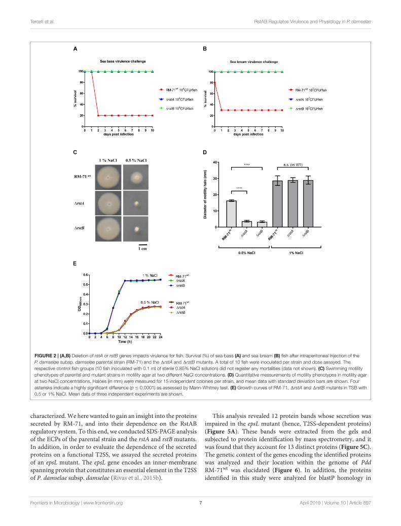
rstA and rstB Mutants Are Affected inVirulence for FishPreviously, it was shown that rstB mutation drasticallydiminished virulence of RM-71 in a European sea bass fishmodel (Terceti et al., 2017). Here we wanted to test whetherrstA mutation would compromise virulence of Pdd RM-71 forfish. In addition, we here extended the virulence tests to twodifferent fish host species of this pathogen, European sea bassand gilthead sea bream. Virulence tests clearly demonstrated thatthe single rstA and rstB mutants are strongly impaired in theirvirulence for European sea bass (Figure 2A) and for giltheadsea bream (Figure 2B). All these lines of evidence suggest thatRstA, the protein encoded by VDA_000601 in Pdd, is the cognateresponse regulator of the histidine kinase RstB and is necessaryfor hemolytic activity of Pdd and for virulence for fish.
rstAB Mutants Show Impaired Motilityand Aberrant Cell Shape and Size at0.5% NaCl, but Not at 1% NaClSwimming motility is believed to constitute an important factorin the pathogenicity of Pdd for fish and it has been demonstratedthat seawater transmits the disease (Fouz et al., 2000). Since theeffect of rstAB genes in motility has not been assayed so far, wehere investigated the behavior in motility agar of RM-71wt, RM-711rstA and RM-711rstB. We found that mutant strains werenot affected in swimming motility with respect to parental strainat 1% NaCl. However, when strains were assayed for motility at0.5% NaCl, both the rstA and the rstB mutants exhibited impairedswimming motility haloes (Figures 2C,D). Analysis of the growthcurves of RM-71wt, RM-711rstA and RM-711rstB did not revealdifferences in growth when the strains were exposed to the sameconditions of salinity (Figure 2E). This demonstrates that the
impaired motility haloes of the rstA and rstB mutants observed at0.5% NaCl are not due to differences in growth between parentalstrain and mutants.
It has been previously reported that single mutation of rstBdoes not affect cell morphology of RM-71 grown in TSB with1% NaCl (Terceti et al., 2017). The observation that motility isimpaired in rstAB mutants only under growth at low salinity(0.5% NaCl), prompted us to study the cell morphology ofparental strain and mutants by scanning electron microscopy.We observed that, at 1% NaCl, cells of the RM-711rstA andRM-711rstB mutants exhibited cell shapes and sizes similarto parental strain (data not shown). However, when cellswere cultured at 0.5% NaCl, differences in cell arrangement,morphology and size became evident (Figure 3). Both the rstAand rstB mutants exhibited longer and swollen cells, and mostoften formed chain-like structures, suggesting an impairmentin daughter cell separation upon cell division. Mobilization ofthe complementing plasmid pRstAB into the rstA and rstBmutants caused the reversion to normal cell shapes and size(Figure 3). These results suggest that the RstAB system isessential for optimal cell division and control of cell shape andsize, and for full swimming motility, under conditions of lowNaCl concentrations.
Mutation of rstA or rstB DecreasesTolerance to BenzylpenicillinPrevious studies reported that mutations in vprAB/carSR genes(homologs of Pdd rstAB) cause an increased sensitivity topolymyxin B in Vibrio cholerae (Herrera et al., 2014; Bilecenet al., 2015). However, rstB mutation in Pdd RM-71 was notfound to cause increased sensitivity to polymyxin B in a previousstudy (Terceti et al., 2017). As expected, we here found thatthe RM-711rstA mutant was as sensitive to polymyxin as RM-71wt and RM-711rstB (data not shown). Pdd, as other species ofthe Vibrionaceae family, exhibit high levels of intrinsic toleranceto typically bactericidal inhibitors of cell wall synthesis, as betalactams. A recent study reported that gene functions relatedto cell envelope synthesis and modification play a major rolein tolerance to beta lactams by V. cholerae (Weaver et al.,2018). Interestingly, we found that the RM-711rstA and RM-711rstB mutants showed a reduction in their tolerance tobenzylpenicillin in comparison to the parental strain whencultured with 1% NaCl, and this reduction was even higher whenthe test was conducted at 0.5% NaCl (Figure 4). These resultssuggest that the two-component RstAB system may be directlyor indirectly involved in the regulation of the Pdd toleranceto beta lactams.
rstA and rstB Mutants Are Impaired inProduction of Type II SecretionSystem-Dependent ProteinsThe four toxins Dly, PhlyP, PhlyC and PlpV are predicted tobe secreted via the T2SS since epsL mutants were shown to beimpaired in hemolytic and phospholipase activities in previousstudies (Rivas et al., 2015b; Vences et al., 2017). However,the T2SS-dependent secretome of RM-71 has not been further
Frontiers in Microbiology | www.frontiersin.org 6 April 2019 | Volume 10 | Article 897
fmicb-10-00897 April 23, 2019 Time: 16:5 # 7
Terceti et al. RstAB Regulates Virulence and Physiology in P. damselae
FIGURE 2 | (A,B) Deletion of rstA or rstB genes impacts virulence for fish. Survival (%) of sea bass (A) and sea bream (B) fish after intraperitoneal injection of theP. damselae subsp. damselae parental strain (RM-71) and the 1rstA and 1rstB mutants. A total of 10 fish were inoculated per strain and dose assayed. Therespective control fish groups (10 fish inoculated with 0.1 ml of sterile 0.85% NaCl solution) did not register any mortalities (data not shown). (C) Swimming motilityphenotypes of parental and mutant strains in motility agar at two different NaCl concentrations. (D) Quantitative measurements of motility phenotypes in motility agarat two NaCl concentrations. Haloes (in mm) were measured for 15 independent colonies per strain, and mean data with standard deviation bars are shown. Fourasterisks indicate a highly significant difference (p ≤ 0,0001) as assessed by Mann-Whitney test. (E) Growth curves of RM-71, 1rstA and 1rstB mutants in TSB with0.5 or 1% NaCl. Mean data of three independent experiments are shown.
characterized. We here wanted to gain an insight into the proteinssecreted by RM-71, and into their dependence on the RstABregulatory system. To this end, we conducted SDS-PAGE analysisof the ECPs of the parental strain and the rstA and rstB mutants.In addition, in order to evaluate the dependence of the secretedproteins on a functional T2SS, we assayed the secreted proteinsof an epsL mutant. The epsL gene encodes an inner-membranespanning protein that constitutes an essential element in the T2SSof P. damselae subsp. damselae (Rivas et al., 2015b).
This analysis revealed 12 protein bands whose secretion wasimpaired in the epsL mutant (hence, T2SS-dependent proteins)(Figure 5A). These bands were extracted from the gels andsubjected to protein identification by mass spectrometry, and itwas found that they account for 13 distinct proteins (Figure 5C).The genetic context of the genes encoding the identified proteinswas analyzed and their location within the genome of PddRM-71wt was elucidated (Figure 6). In addition, the proteinsidentified in this study were analyzed for blastP homology in
Frontiers in Microbiology | www.frontiersin.org 7 April 2019 | Volume 10 | Article 897

fmicb-10-00897 April 23, 2019 Time: 16:5 # 8
Terceti et al. RstAB Regulates Virulence and Physiology in P. damselae
FIGURE 3 | (A) Scanning electron microscopy of P. damselae subsp. damselae parental strain RM-71, 1rstA and 1rstB mutants, and complemented rstB mutant(MT157), grown at 25◦C in TSB with 0.5% NaCl. Note the enlarged cell size of the mutants, which also form chain-like structures likely due to an impairment indaughter cell separation upon cell division. Pictures from two magnifications, 5,000 × (5 K X) and 15,000 × (15 K X) are shown, and scale bars representing 2 µmare included in each picture. (B) Box plot graphs showing the comparison of cell width and cell length in exponential phase cultures. Whiskers indicate min andmax values.
other bacterial species (Supplementary Table S2) and for theirdomains by Pfam 31.0 (Supplementary Table S3).
The proteins related to bands 1, 2, 3, 8, and 11 are T2SSdependent but are not under control of the RstAB system,whereas the remaining bands are T2SS- and RstAB-dependent(Figure 5A). This observation indicates that mutation of theRstAB system does not prevent the correct function of the T2SSsince bands 1, 2, 3, 8, and 11 are equally present in the secretomeof the parental strain and in 1rstA and 1rstB mutants. In furthersupport of this idea, as described above (Figure 1), rstA and rstBmutants were not affected in the lecithinase activity attributableto PlpV (mutants in the plasmidless strain LD-07), an enzymeknown to be secreted via the T2SS (Vences et al., 2017).
Notably, secretion of seven T2SS-dependent proteins(included within bands 4, 5, 6, 7, 9, 10, and 12) was stronglyimpaired in rstA and rstB mutants, and for some of themsecretion was practically abolished (Figure 5A). One of the mostoutstanding features in the secretome pattern of RM-71 was thepresence of two major bands in the range of 60–80 kDa, thatwere absent in the rstA, rstB and epsL mutants. The molecularmasses of these bands suggest that they correspond to the
three hemolysins Dly, PhlyP and PhlyC, and we observedthat these two major bands were absent in the protein profileof a RM-71 triple mutant for genes dly, hlyApl and hlyAch(Figure 5A). To further determine which hemolysin(s) wascontained in each band, we carried out SDS-PAGE analysis ofall the combinations of mutants for these three hemolysins. As aresult, we corroborated that the upper intense band of ca. 75 kDacorresponded to Dly cytotoxin and the lower band of ca. 65 kDacorresponded to PhlyP plus PhlyC, with a major contributionof PhyP over PhlyC (Figure 5B). Thus, single mutation of eachof the RstAB system genes, practically abolishes Dly, PhlyP andPhlyC production. In addition, as previous studies had suggested(Rivas et al., 2015b), we demonstrate that mutation of epsL causesa strong impairment in Dly, PhlyP, and PhlyC secretion.
Among the additional secreted proteins whose productionwas impaired in the rstA and rstB mutants (Figure 5C) wefound potential novel virulence factors of Pdd. Of note, theparental strain produced high amounts of a small protein of11 kDa (identified as A0J47_07530) whose secretion was nearlyabolished in the rstAB and epsL mutants. Although the geneencoding A0J47_07530 protein is also present in the genome
Frontiers in Microbiology | www.frontiersin.org 8 April 2019 | Volume 10 | Article 897

fmicb-10-00897 April 23, 2019 Time: 16:5 # 9
Terceti et al. RstAB Regulates Virulence and Physiology in P. damselae
FIGURE 4 | E-tests for benzylpenicillin, showing a decreased tolerance inboth the rstA and the rstB mutants compared to parental strain RM-71.
of the type strain CIP102761, it was not initially annotated assuch in the GenBank database, although recently it has beengiven the locus tag VDA_RS10260. The analysis of the geneticcontext of A0J47_07530 unveiled that it is located upstream(and likely cotranscribed with) the gene encoding a putativedelta-endotoxin (VDA_002799) (Figure 6). Indeed, this delta-endotoxin was identified as the major component of bands 6and 7 (Figures 5A,C) and is also rstAB and epsL-dependent.MS analysis of band 6 identified delta-endotoxin peptidesranging from residues 197 to 529 whereas the delta-endotoxincorresponding to band 7 contained peptides ranging from residue197 to 474, suggesting that band 6 corresponds to non-truncateddelta-endotoxin, whereas band 7 corresponds to delta-endotoxintruncated at the C-terminus (Figure 5A).
Finally, two uncharacterized secreted proteins were also rstABdependent and corresponded to the pPHDD1 plasmid-encodedVDA_000112 and the chromosome II-encoded VDA_000358.Similarity searches failed to reveal well-characterized homologsfor these two proteins (Supplementary Table S2), and noconserved domains could be identified in a sequence analysis(Supplementary Table S3).
The P. damselae subsp. damselaeT2SS-Dependent Secretome IncludesAdditional Uncharacterized ProteinsSix additional proteins were identified as T2SS-dependentalthough their secretion was not affected in rstA and rstB mutants(Figures 5A,C). Since the secretome of Pdd has been poorlycharacterized so far, we judged of high interest to analyzethese six proteins. VDA002460 corresponds to a T2SS-dependentputative lipoprotein, although no Pfam-A matches to known
sequences were found (Supplementary Table S3). VDA_000694corresponds to a putative sialidase and contains two Sial-lect-insert domains and a BNR_2 domain (SupplementaryTables S2, S3). It is known that pathogenic bacteria can utilizehost sialic acid to form a protective coating that providesresistance to host immune response (Severi et al., 2007).
A0J47_15850 is a putative T2SS-dependent serine proteasewith peptidase S8 domain belonging to the family of subtilisin-like serine proteases (Bode et al., 1987). Subtilases are widespread,being found in eubacteria, archaebacteria, eukaryotes and viruses(Siezen and Leunissen, 1997). A0J47_09785 is also T2SS-dependent and corresponds to an uncharacterised Pdd proteinwith a trypsin-like domain (Supplementary Table S3) and withsimilarity (blastP) to proteases, metalloproteases and hemolysins(Supplementary Table S2). It also appears as Hit (19% identity)the VesB protease from Vibrio cholerae, which has been describedas a type II-secreted protease (Gadwal et al., 2014).
VDA_000966 is also T2SS dependent and is encoded withinchromosome II of Pdd. It has homology to hypotheticalproteins and porin family proteins (Supplementary Table S2)and analysis by Pfam31.0 revealed an OMP_b-brl domain(Supplementary Table S3). Another protein identified as T2SSdependent and encoded within chromosome II is VDA_000738which has homology with ComEA helix-hairpin-helix (HHH)repeat competence proteins of various species (SupplementaryTable S2). By analysis by Pfam31.0 we have found that thisprotein has a HHH domain which is a short DNA-bindingdomain belonging to CL0198 clan superfamily (SupplementaryTable S3) (Witte et al., 2008).
Genes of the RstAB Regulon ShowDifferential Presence in P. damselaesubsp. damselae IsolatesAs mentioned above, the RstAB regulon of RM-71 straincomprises at least 7 genes whose products are secreted by theT2SS. They include plasmid, chromosome I and chromosomeII-encoded genes (Figure 6). In order to assess the degree ofconservation of the RstAB regulon in the subspecies, a total of83 strains of Pdd isolated from a variety of geographical regionsand host species were tested for the presence of the followingtarget genes: hlyAch, hlyApl, dly, VDA_000112, VDA_002799,A0J47_07530 and VDA_000358 (Figure 7). Presence of rstABgenes was also tested in all the strains. Some of these markershad already been tested in a fraction of these 83 isolates inprevious studies (Rivas et al., 2011, 2014; Terceti et al., 2016;Terceti et al., 2018) and such previous information is alsoincluded in Figure 7.
All the genes of the RstAB regulon showed differentialdistribution among the isolates. The three genes dly, hlyApl andVDA_000112 were present in 34% (28/83) of the isolates, andalways co-occurred, an observation that is consistent with the factthat they are pPHDD1 plasmid-borne genes. The chromosomeI-borne hlyAch gene encoding PhlyC hemolysin is almostubiquitous in the subspecies (77/83), with the exception of threeTurkish and three Danish plasmidless isolates where this geneis truncated by an insertion sequence (Terceti et al., 2016, 2018).
Frontiers in Microbiology | www.frontiersin.org 9 April 2019 | Volume 10 | Article 897

fmicb-10-00897 April 23, 2019 Time: 16:5 # 10
Terceti et al. RstAB Regulates Virulence and Physiology in P. damselae
FIGURE 5 | (A) Representative SDS-PAGE of culture supernatants from RM-71WT, 1rstA, 1rstB, 31 (triple mutant for dly, hlyApl and hlyAch hemolysin genes) and1epsL (defective in T2SS) strains. The identified protein bands are denoted with numbers 1 to 12. (B) SDS-PAGE analysis of culture supernatants from RM-71WT,1hlyAch, 1hlyApl , 1dly, 1hlyAch1hlyApl , 1hlyAch1dly, 1hlyApl1dly and 31 (triple mutant). Comparative analysis of the protein profiles shows that band 4corresponds to Dly and band 5 to PhlyP+PhlyC. (C) Identification (gene barcodes) and characteristics of the T2SS-dependent proteins identified in this study,including those regulated by RstAB.
In addition, 63% (52/83) of the isolates were carriers of thegene encoding VDA_000358 (Figure 7). VDA_002799 (delta-endotoxin) and A0J47_07530 (the 11 kDa protein) always co-occurred and were present in 49% (41/83) of the assayed strains.
These two genes have been reported to be inserted into ahighly variable genome region that likely constitutes a hot spotfor the acquisition of horizontally transferred DNA in Pdd(Terceti et al., 2018) (Figure 6).
Frontiers in Microbiology | www.frontiersin.org 10 April 2019 | Volume 10 | Article 897

fmicb-10-00897 April 23, 2019 Time: 16:5 # 11
Terceti et al. RstAB Regulates Virulence and Physiology in P. damselae
FIGURE 6 | Genetic context and genomic location (chromosome I, II, or pPHDD1 plasmid) of the genes encoding the proteins identified in Figure 5.
Interestingly, rstAB genes were ubiquitous in the 83 isolatestested, demonstrating that this two-component regulatory systemis highly conserved in the subspecies. Thus, the RstAB systemmight be used as a weak point to control outbreaks caused by bothplasmid-containing and plasmidless strains. Of note, we foundthat homologs of the Pdd RstAB proteins are widely distributedin Vibrio and Photobacterium species, both pathogenic and non-pathogenic (Supplementary Figure S1).
DISCUSSION
Identification of the regulatory mechanisms in bacterialpathogens is of maximal interest in order to understand theenvironmental and host conditions that trigger the expression ofvirulence factors. Pdd is a generalist pathogen, capable of livingas a free-swimming bacterium that causes outbreaks in culturedfish species when the conditions become favorable (Osorio et al.,2018). This pathogen produces a variety of toxins which arethought to be secreted through the T2SS (Rivas et al., 2015b;Vences et al., 2017). So far, the only regulatory mechanism
characterized in this subspecies is the histidine kinase RstB,which is genetically linked to a putative response regulator RstA(Terceti et al., 2017).
Here we demonstrated that single mutation of rstA impairsvirulence in sea bass and sea bream, confirming the resultspreviously obtained with single rstB mutants. Studies of therole of RstAB system in fish pathogens are very scarce.Notably, mutation of rstB in Edwardsiella ictaluri causedimpaired colonization and virulence in a channel catfish model(Menanteau-Ledouble and Lawrence, 2013).
Pdd strains are able to grow through a range of temperaturesand of NaCl concentrations (Kreger, 1984; Fouz et al., 1992,1998). Mutation of rstA and rstB genes did not affect growthin comparison to the parental strain at 25◦C, either at 1%NaCl or at 0.5% NaCl. However, mutation of rstA or rstBimpaired swimming motility at 0.5% NaCl. It remains to beinvestigated whether this reduced motility phenotype is a directconsequence of impaired flagellar function, or whether it is aconsequence, at least in part, of the formation of the elongatedand multicellular structures in Pdd grown at 0.5% NaCl. Recentstudies demonstrated that salinity modulates Pdd swimming
Frontiers in Microbiology | www.frontiersin.org 11 April 2019 | Volume 10 | Article 897

fmicb-10-00897 April 23, 2019 Time: 16:5 # 12
Terceti et al. RstAB Regulates Virulence and Physiology in P. damselae
FIGURE 7 | Diagramm depicting the presence of rstAB genes and genes of the RstAB regulon, in 83 P. damselae subsp. damselae strains isolated from differentgeographical locations and from different hosts including marine animals and humans.
Frontiers in Microbiology | www.frontiersin.org 12 April 2019 | Volume 10 | Article 897

fmicb-10-00897 April 23, 2019 Time: 16:5 # 13
Terceti et al. RstAB Regulates Virulence and Physiology in P. damselae
motility and that mutation of the chemotaxis regulator cheAimpairs bacterial swimming (von Hoven et al., 2018). However,the connection between motility and the RstAB system in Pddis so far unknown. Recent studies have shown that silencingof the rstA or rstB genes result in impaired motility, hemolysisand virulence in Vibrio alginolyticus (Huang et al., 2018). InSalmonella typhimurium, FlhA and MglB proteins involved in cellmotility and chemotaxis are positively regulated by the RstABsystem and a rstB mutant in this species showed a significantreduction in motility (Tran et al., 2016).
rstA and rstB mutants cultured at 0.5% NaCl exhibitimpairment in cell separation upon cell division, as well asenlarged cell size. These mutants also exhibited decreasedtolerance to benzylpenicillin. In agreement with our results,overexpression of RstA in E. coli made this bacterium moreresistant to beta lactam antibiotics such as ampicillin (Hirakawaet al., 2003a,b). Similarly, E. coli rstAB mutants are hypersensitiveto ketoprofen, pridinol, and troleandomycin, although the basisfor these sensitivities has not been ascertained yet (Zhou et al.,2003). So far, the molecular mechanisms linking the RstABsystem with tolerance to beta lactam antibiotics and withmaintenance of cell size and shape in Pdd remain unknown.A recent study unveiled that tolerance to beta lactams byV. cholerae has a complex and pleiotropic nature, and that genefunctions related to cell envelope synthesis and modificationplay a major role in such tolerance (Weaver et al., 2018).Being Pdd a marine bacterium, it can be anticipated that thispathogen does not encounter conditions of low salinity (0.5%NaCl) in nature. However, the abnormal phenotypes observedin the present study in rstA and rstB mutants cultured underlow salinity conditions, will surely constitute the basis forfurther studies aimed at unveiling additional regulatory rolesof RstAB systems in bacterial pathogens. Among the T2SS-dependent proteins not regulated by the RstAB system we foundpredicted sialidases and proteases, among others. Identifyingthe function of these proteins constitutes a promising challengeand will require additional research. Fish cells secrete mucusglycoproteins containing sialic acid (Kimura et al., 1994), andpathogenic bacteria can use sialidases to remove sialic acidresidues from host cells and coat themselves, thus gainingresistance to components of the host’s innate immune system(Severi et al., 2007). In addition, pathogenic bacteria can bindto host sialic acid moiety to enhance adhesion and colonization.Thus, bacteria that produce sialidases might have a greater abilityto colonize and stabilize themselves in the skin, gills, scalesand intestines of animals that secrete mucosal glycoproteinscontaining sialic acids.
Mutation of rstA or rstB practically abolishes Dly, PhlyP andPhlyC production. These three hemolysins not only constitutemajor virulence factors for fish and mice (Rivas et al., 2011,2013b), but are also major components of the Pdd secretomeas revealed here in the proteome analysis. In support of thisobservation, a recent study on the Pdd transcriptome hasdemonstrated that the genes encoding Dly, PhlyP and PhlyCare among the most highly transcribed genes in this pathogen(Matanza and Osorio, 2018). That recent study also unveiledthat the PlpV phospholipase is expressed at very low levels in
comparison to the three aforementioned major hemolysins. Thisobservation can explain why the PlpV protein band was notidentified within the T2SS-dependent proteins in the presentstudy. In addition, this study has contributed to the identificationof 4 novel proteins belonging to the RstAB regulon, includingplasmid-, chromosome I- and chromosome II-encoded proteins.Therefore, the response regulator RstA is predicted to recognizetarget genes associated with the Pdd chromosomes as well asgenes that have been acquired by horizontal gene transfer viaconjugation. Notably, our study has brought to the forefronta number of hitherto uncharacterized, T2SS-dependent andRstAB-dependent proteins, and there are very few studies onthese except for the three hemolysins Dly, PhlyP and PhlyC(Kreger, 1984; Kothary and Kreger, 1985; Rivas et al., 2011,2013b, 2015a; Vences et al., 2017). Of special interest aretwo genes which are likely cotranscribed as an operon, andencode the delta-endotoxin VDA_002799 and an associated11 kDa small protein. These two genes are located within apotential hot spot for recombination within the genome ofPdd (Terceti et al., 2018). Delta-endotoxins, also called Cryand Cyt toxins, are pore-forming toxins predicted to cross thecytoplasmic membrane via the twin-arginine (Tat) translocationpathway (Li et al., 1991; Cygler et al., 1995; Galitsky et al.,2001). However, the role of this putative delta-endotoxin inPdd is still unknown. Last, VDA_000112 and VDA_000358 areT2SS- and RstAB-dependent uncharacterized proteins encodedwithin plasmid pPHDD1 and chromosome II, respectively.Surely all these proteins will deserve an in-depth characterizationin future studies.
rstAB genes are considered to be part of the PhoP/PhoQregulon (a Mg2+-sensing two-component system) (Oshima et al.,2002; Ogasawara et al., 2007). While some evidence suggeststhat the RstAB system might respond to acidic conditions(Ogasawara et al., 2007; Huang et al., 2018), the specific stimulithat trigger the activation of the sensor histidine kinase RstBremain unknown for all the RstAB-like systems studied to date(Ogasawara et al., 2007; Campbell et al., 2007; Flamez et al., 2008;Bilecen and Yildiz, 2009; Menanteau-Ledouble and Lawrence,2013; Tran et al., 2016; Huang et al., 2018). In contrast, anumber of genes and biological functions under control ofthe RstAB system have been identified, demonstrating that theRstAB regulon is not predictive since it shows a high diversityamong species. Functions regulated by homologous RstABsystems are as varied as pyrimidine metabolism, enterobactinbiosynthesis, ferrous iron uptake and motility in Salmonellaenterica (Tran et al., 2016), polysaccharide synthesis, biofilmformation and LPS modification in Vibrio cholerae (Bilecenand Yildiz, 2009; Herrera et al., 2014), or adhesion, biolfilmproduction, motility and hemolysis in Vibrio alginolyticus(Huang et al., 2018), among others.
This study has contributed to the knowledge of the bacterialRstAB regulon with a number of novel genes and functions.In addition, we here demonstrated that the RstAB regulon ofPdd RM-71 comprises genes of differential presence amongstrains, and no geographical or isolation source patternscould be identified. To summarize, the RstAB two-componentsystem plays a major role in regulation of virulence and of
Frontiers in Microbiology | www.frontiersin.org 13 April 2019 | Volume 10 | Article 897
fmicb-10-00897 April 23, 2019 Time: 16:5 # 14
Terceti et al. RstAB Regulates Virulence and Physiology in P. damselae
many aspects of cell physiology of Pdd. Ongoing studies areexpected to unveil the roles of the novel RstAB-regulated genesreported in this study.
ETHICS STATEMENT
The protocols of animal experimentation used in this study havebeen reviewed and approved by the Animal Ethic Committee ofthe Universidade de Santiago de Compostela.
AUTHOR CONTRIBUTIONS
MT conceived the study, designed and performed theexperiments, analyzed the data, and wrote the first draft of themanuscript. AV, XM, and AB performed analyses and interpretedthe results. MN performed the scanning electron microscopyanalyses. JL and NdS contributed to data analysis. AdV conceivedthe study, designed the protein analyses and data interpretation,and contributed to the writing of the manuscript. CO conceivedand supervised the study, designed the experiments, interpretedthe results, and wrote the manuscript. All the authors read andapproved the final manuscript.
FUNDING
This work has been supported by grant AGL2016-79738-R(AEI/FEDER, EU) from the State Agency for Research (AEI)
of Spain, and co-funded by the FEDER Programme fromthe European Union. The support of Xunta de Galicia(Spain) with grant ED431C 2018/18 is also acknowledged.MT thanks the Brazilian Ministry of Education and CAPES(Coordenaçao de Aperfeiçoamento de Pessoal de Nível Superior)for a predoctoral fellowship. XM thanks Xunta de Galiciafor a predoctoral fellowship. AdV was supported by theFCT fellowship SFRH/BPD/95777/2013. The mass spectrometrytechnique was performed at the Proteomics i3S ScientificPlatform with the assistance of Hugo Osório. This work hadsupport from the Portuguese Mass Spectrometry Network,integrated in the National Roadmap of Research Infrastructuresof Strategic Relevance (ROTEIRO/0028/2013; LISBOA-01-0145-FEDER-022125).
ACKNOWLEDGMENTS
We thank Bernardo Fernández Souto and the Instituto Galegode Formación en Acuicultura (IGaFA) (Illa de Arousa, Galicia,Spain) for their valuable support in providing sea bass for thevirulence experiments.
SUPPLEMENTARY MATERIAL
The Supplementary Material for this article can be foundonline at: https://www.frontiersin.org/articles/10.3389/fmicb.2019.00897/full#supplementary-material
REFERENCESAziz, R. K., Bartels, D., Best, A. A., DeJongh, M., Disz, T., Edwards, R. A., et al.
(2008). The RAST server: rapid annotations using subsystems technology. BMCGenomics 9:75. doi: 10.1186/1471-2164-9-75
Bilecen, K., Fong, J. C. N., Cheng, A., Jones, C. J., Zamorano-Sánchez, D., andYildiz, F. (2015). Polymyxin B resistance and biofilm formation in Vibriocholerae are controlled by the response regulator CarR. Infect. Immun. 83,1199–1209. doi: 10.1128/IAI.02700-14
Bilecen, K., and Yildiz, F. (2009). Identification of a calcium-controlled negativeregulatory system affecting Vibrio cholerae biofilm formation. Environ.Microbiol. 11, 2015–2029. doi: 10.1111/j.1462-2920.2009.01923.x
Bode, W., Papamokos, E., and Musil, D. (1987). The high-resolution X-ray crystalstructure of the complex formed between subtilisin Carlsberg and eglin c,an elastase inhibitor from the leech Hirudo medicinalis. Structural analysis,subtilisin structure and interface geometry. Eur. J. Biochem. 166, 673–692.doi: 10.1111/j.1432-1033.1987.tb13566.x
Campbell, T. L., Ederer, C. S., Allali-Hassani, A., and Brown, E. D. (2007). Isolationof the rstA gene as a multicopy suppressor of YjeE, an essential ATPase ofunknown function in Escherichia coli. J. Bacteriol. 189, 3318–3321. doi: 10.1128/jb.00131-06
Cygler, M., Borisova, S., Grochulski, P., Masson, L., Pusztai-Carey, M., Schwartz,J. L., et al. (1995). Bacillus thuringiensis CryIA(a) insecticidal toxin: crystalstructure and channel formation. J. Mol. Biol. 254, 447–464.
Flamez, C., Ricard, I., Arafah, S., Simonet, M., and Marceau, M. (2008). Phenotypicanalysis of Yersinia pseudotuberculosis 32777 response regulator mutants: newinsights into two-component system regulon plasticity in bacteria. Int. J. Med.Microbiol. 298, 193–207. doi: 10.1016/j.ijmm.2007.05.005
Fouz, B., Larsen, J. L., Nielsen, B., Barja, J. L., and Toranzo, A. E. (1992).Characterization of Vibrio damsela strains isolated from turbot Scophthalmusmaximus in Spain. Dis. Aquat. Organ. 12, 155–166. doi: 10.3354/dao012155
Fouz, B., Toranzo, A. E., Marco-Noales, E., and Amaro, C. (1998). Survival of fish-virulent strains of Photobacterium damselae subsp. damselae in seawater understarvation conditions. FEMS Microbiol. Lett. 168, 181–186.
Fouz, B., Toranzo, A. E., Milan, M., and Amaro, C. (2000). Evidence that watertransmits the disease caused by the fish pathogen Photobacterium damselaesubsp. damselae. J. Appl. Microbiol. 88, 531–535. doi: 10.1046/j.1365-2672.2000.00992.x
Gadwal, S., Korotkov, K. V., Delarosa, J. R., Hol, W. G., and Sandkvist, M. (2014).Functional and structural characterization of Vibrio cholerae extracellular serineprotease B, VesB. J. Biol. Chem. 289, 8288–8298. doi: 10.1074/jbc.M113.525261
Galitsky, N., Cody, V., Wojtczak, A., Ghosh, D., Luft, J. R., Pangborn, W.,et al. (2001). Structure of the insecticidal bacterial delta-endotoxin Cry3Bb1 ofBacillus thuringiensis. Acta Crystallogr. D 57, 1101–1109.
Gomes, C., Almeida, A., Ferreira, J. A., Silva, L., Santos-Sousa, H., Pinto-de-Sousa,J., et al. (2013). Glycoproteomic analysis of serum from patients with gastricprecancerous lesions. J. Proteome Res. 12, 1454–1466. doi: 10.1021/pr301112x
Herrera, C. M., Crofts, A. A., Henderson, J. C., Cassandra Pingali, S., Davies,B. W., and Trent, S. M. (2014). The Vibrio cholerae VprA-VprB two-componentsystem controls virulence through endotoxin modification. mBio 5, e2283-14.doi: 10.1128/mBio.02283-14
Herrero, M., de Lorenzo, V., and Timmis, K. N. (1990). Transposon vectorscontaining non-antibiotic resistance selection markers for cloning and stablechromosomal insertion of foreign genes in gram-negative bacteria. J. Bacteriol.172, 6557–6567. doi: 10.1128/jb.172.11.6557-6567.1990
Hirakawa, H., Nishino, K., Hirata, T., and Yamaguchi, A. (2003a). Comprehensivestudies of drug resistance mediated by overexpression of response regulators oftwo-component signal transduction systems in Escherichia coli. J. Bacteriol. 185,1851–1856. doi: 10.1128/jb.185.6.1851-1856.2003
Hirakawa, H., Nishino, K., Yamada, J., Hirata, T., and Yamaguchi, A. (2003b). Beta-lactam resistance modulated by the overexpression of response regulators oftwo-component signal transduction systems in Escherichia coli. J. Antimicrob.Chemother. 52, 576–582. doi: 10.1093/jac/dkg406
Frontiers in Microbiology | www.frontiersin.org 14 April 2019 | Volume 10 | Article 897

fmicb-10-00897 April 23, 2019 Time: 16:5 # 15
Terceti et al. RstAB Regulates Virulence and Physiology in P. damselae
Huang, L., Xu, W., Su, Y., Zhao, L., and Yan, Q. (2018). Regulatory role of theRstB-RstA system in adhesion, biofilm production, motility, and hemolysis.Microbiologyopen 23:e00599. doi: 10.1002/mbo3.599
Kimura, M., Hama, Y., Sumi, T., Asakawa, M., Rao, B. N. N., Horne, A. P.,et al. (1994). Characterization of a deaminated neuraminic acid-containingglycoprotein from the skin mucus of the loach, Misgurnus anguillicaudatus.J. Biol. Chem. 269, 32138–32143.
Kothary, M. H., and Kreger, A. S. (1985). Purification and characterization of anextracellular cytolysin produced by Vibrio damsela. Infect. Immun. 49, 25–31.
Kreger, A. S. (1984). Cytolytic activity and virulence of Vibrio damsela. Infect.Immun. 44, 326–331.
Laemmli, U. K. (1970). Cleavage of structural proteins during the assembly of thehead of bacteriophage T4. Nature 227, 680–685. doi: 10.1038/227680a0
Le Roux, F., Binesse, J., Saulnier, D., and Mazel, D. (2007). Construction of aVibrio splendidus mutant lacking the metalloprotease gene vsm by use of anovel counterselectable suicide vector. Appl. Environ. Microbiol. 73, 777–784.doi: 10.1128/aem.02147-06
Le Roux, F., Davis, B. M., and Waldor, M. K. (2011). Conserved small RNAs governreplication and incompatibility of a diverse new plasmid family from marinebacteria. Nucleic Acids Res. 39, 1004–1013. doi: 10.1093/nar/gkq852
Li, J. D., Carroll, J., and Ellar, D. J. (1991). Crystal structure of insecticidal delta-endotoxin from Bacillus thuringiensis at 2.5 A resolution. Nature 353, 815–821.doi: 10.1038/353815a0
Matanza, X. M., and Osorio, C. R. (2018). Transcriptome changes in response totemperature in the fish pathogen Photobacterium damselae subsp. damselae:clues to understand the emergence of disease outbreaks at increasedseawater temperatures. PLoS One 13:e0210118. doi: 10.1371/journal.pone.0210118
Menanteau-Ledouble, S., and Lawrence, M. L. (2013). Use of bioluminescencemutant screening for identification of Edwardsiella ictaluri genes involved inchannel catfish (Ictalurus punctatus) skin colonization. Vet. Microbiol. 162,724–730. doi: 10.1016/j.vetmic.2012.09.024
Mouriño, S., Osorio, C. R., and Lemos, M. L. (2004). Characterization of hemeuptake cluster genes in the fish pathogen Vibrio anguillarum. J. Bacteriol. 186,6159–6167. doi: 10.1128/jb.186.18.6159-6167.2004
Ogasawara, H., Hasegawa, A., Kanda, E., Miki, T., Yamamoto, K., and Ishihama, A.(2007). Genomic SELEX search for target promoters under the control of thePhoQP-RstBA signal relay cascade. J. Bacteriol. 189, 4791–4799. doi: 10.1128/jb.00319-07
Oshima, T., Aiba, H., Masuda, Y., Kanaya, S., Sugiura, M., Wanner, B. L.,et al. (2002). Transcriptome analysis of all two-component regulatory systemsmutants of Escherichia coli K-12. Mol. Microbiol. 46, 281–291. doi: 10.1046/j.1365-2958.2002.03170.x
Osorio, C. R., Vences, A., Matanza, X. M., and Terceti, M. S. (2018).Photobacterium damselae subsp. damselae, a generalist pathogen with uniquevirulence factors and high genetic diversity. J. Bacteriol. 200, e00002–e18.doi: 10.1128/JB.00002-18
Rivas, A. J., Balado, M., Lemos, M. L., and Osorio, C. R. (2011). The Photobacteriumdamselae subsp. damselae hemolysins damselysin and HlyA are encoded withina new virulence plasmid. Infect. Immun. 79, 4617–4627. doi: 10.1128/IAI.05436-11
Rivas, A. J., Labella, A., Borrego, J. J., Lemos, M. L., and Osorio, C. R. (2014).Evidences for horizontal gene transfer, gene duplication and genetic variationas driving forces of the diversity of haemolytic phenotypes in Photobacteriumdamselae subsp. damselae. FEMS Microbiol. Lett. 355, 152–162. doi: 10.1111/1574-6968.12464
Rivas, A. J., Balado, M., Lemos, M. L., and Osorio, C. R. (2013b). Synergistic andadditive effects of chromosomal and plasmid-encoded hemolysins contributeto hemolysis and virulence in Photobacterium damselae subsp. damselae. Infect.Immun. 81, 3287–3299. doi: 10.1128/IAI.00155-13
Rivas, A. J., Lemos, M. L., and Osorio, C. R. (2013a). Photobacterium damselaesubsp. damselae, a bacterium pathogenic for marine animals and humans.Front. Microbiol. 4:283. doi: 10.3389/fmicb.2013.00283
Rivas, A. J., Vences, A., Husmann, M., Lemos, M. L., and Osorio, C. R.(2015b). Photobacterium damselae subsp. damselae major virulence factorsDly, plasmid-encoded HlyA, and chromosome-encoded HlyA are secreted viathe type II secretion system. Infect. Immun. 83, 1246–1256. doi: 10.1128/IAI.02608-14
Rivas, A. J., von Hoven, G., Neukirch, C., Meyenburg, M., Qin, Q., Füser, S., et al.(2015a). Phobalysin, a small ß-pore-forming toxin of Photobacterium damselaesubsp. damselae. Infect. Immun. 83, 4335–4348. doi: 10.1128/IAI.00277-15
Saitou, N., and Nei, M. (1987). The neighbor-joining method: a new method forreconstructing phylogenetic trees. Mol. Biol. Evol. 4, 406–425.
Severi, E., Hood, D. W., and Thomas, G. H. (2007). Sialic acid utilization bybacterial pathogens. Microbiology 153, 2817–2822. doi: 10.1099/mic.0.2007/009480-0
Siezen, R. J., and Leunissen, J. A. (1997). Subtilases: the superfamily of subtilisin-like serine proteases. Protein Sci. 6, 501–523. doi: 10.1002/pro.5560060301
Stock, A. M., Robinson, V. L., and Goudreau, P. N. (2000). Two-component signaltransduction. Annu. Rev. Biochem. 69, 183–215.
Tamura, K., Nei, M., and Kumar, S. (2004). Prospects for inferring very largephylogenies by using the neighbor-joining method. Proc. Natl. Acad. Sci. U.S.A.101, 11030–11035. doi: 10.1073/pnas.0404206101
Tamura, K., Stecher, G., Peterson, D., Filipski, A., and Kumar, S. (2013). MEGA6:molecular evolutionary genetics analysis version 6.0. Mol. Biol. Evol. 30, 2725–2729. doi: 10.1093/molbev/mst197
Terceti, M. S., Ogut, H., and Osorio, C. R. (2016). Photobacterium damselae subsp.damselae, an emerging fish pathogen in the Black Sea: evidences of a multiclonalorigin. Appl. Environ. Microbiol. 82, 3736–3745. doi: 10.1128/AEM.00781-16
Terceti, M. S., Rivas, A. J., Alvarez, L., Noia, M., Cava, F., and Osorio, C. R. (2017).rstB regulates expression of the Photobacterium damselae subsp. damselaemajor virulence factors Damselysin, Phobalysin P and Phobalysin C. Front.Microbiol. 8:582. doi: 10.3389/fmicb.2017.00582
Terceti, M. S., Vences, A., Matanza, X. M., Dalsgaard, I., Pedersen, K., andOsorio, C. R. (2018). Molecular epidemiology of Photobacterium damselaesubsp. damselae outbreaks in marine rainbow trout farms reveals extensivehorizontal gene transfer and high genetic diversity. Front Microbiol. 9:2155.doi: 10.3389/fmicb.2018.02155
Tran, T. K., Han, Q. Q., Shi, Y., and Guo, L. (2016). A comparative proteomicanalysis of Salmonella typhimurium under the regulation of the RstA/RstB andPhoP/PhoQ systems. Biochim. Biophys. Acta 1864, 1686–1695. doi: 10.1016/j.bbapap.2016.09.003
Vences, A., Rivas, A. J., Lemos, M. L., Husmann, M., and Osorio, C. R.(2017). Chromosome-encoded hemolysin, phospholipase, and collagenase inplasmidless isolates of Photobacterium damselae subsp. damselae contribute tovirulence for fish. Appl. Environ. Microbiol. 83, e401-17.
Vera, P., Navas, J. I., and Fouz, B. (1991). First isolation of Vibrio damsela fromseabream (Sparus aurata). Bull. Eur. Ass. Fish Pathol. 11, 112–113. doi: 10.1111/jfd.12703
von Hoven, G., Neukirch, C., Meyenburg, M., Schmidt, S., Vences, A., Osorio,C. R., et al. (2018). Cytotoxin- and chemotaxis-genes cooperate to promoteadhesion of Photobacterium damselae subsp. damselae. Front. Microbiol. 9:2996.doi: 10.3389/fmicb.2018.02996
Weaver, A. I., Murphy, S. G., Umans, B. D., Tallavajhala, S., Onyekwere, I., Wittels,S., et al. (2018). Genetic determinants of penicillin tolerance in Vibrio cholerae.Antimicrob. Agents Chemother. 62, e1326-18. doi: 10.1128/AAC.01326-18
Witte, G., Hartung, S., Buttner, K., and Hopfner, K. P. (2008). Structuralbiochemistry of a bacterial checkpoint protein reveals diadenylate cyclaseactivity regulated by DNA recombination intermediates. Mol. Cell 30, 167–178.doi: 10.1016/j.molcel.2008.02.020
Zhou, L., Xiang-He, L., Bochner, B. R., and Wanner, B. L. (2003). Phenotypemicroarray analysis of Escherichia coli K-12 mutants with deletions of all two-component systems. J. Bacteriol. 185, 4956–4972. doi: 10.1128/jb.185.16.4956-4972.2003
Conflict of Interest Statement: The authors declare that the research wasconducted in the absence of any commercial or financial relationships that couldbe construed as a potential conflict of interest.
Copyright © 2019 Terceti, Vences, Matanza, Barca, Noia, Lisboa, dos Santos, do Valeand Osorio. This is an open-access article distributed under the terms of the CreativeCommons Attribution License (CC BY). The use, distribution or reproduction inother forums is permitted, provided the original author(s) and the copyright owner(s)are credited and that the original publication in this journal is cited, in accordancewith accepted academic practice. No use, distribution or reproduction is permittedwhich does not comply with these terms.
Frontiers in Microbiology | www.frontiersin.org 15 April 2019 | Volume 10 | Article 897