the role of albucasis in evolution of the history of ... · kitab al-tasrif li-man ajiza an...
TRANSCRIPT
Research ArticleVolume 2 Issue 4 - December 2016DOI: 10.19080/GJO.2016.02.555593
Glob J OtolaryngolCopyright © All rights are reserved by Faisal Dibsi
The Role of Albucasis in Evolution of the History of Otorhinolaryngology
Faisal Dibsi*Department of Otolaryngology-Head and Neck Surgery, Aleppo University, Iraq
Submission: September 29, 2016; Published: December 15, 2016
*Corresponding author: Faisal Dibsi, Department of Otolaryngology-Head and Neck Surgery, AL-KALIMAT HOSPITAL, Al-Sabil Area, Rezq-Allah Tahan Street, P.O.Box: 862, Aleppo city - Syria, Tel: 00963 21 2645909/00963944 488980; Email:
Abstract
“ALBUCASIS (936-1013 AD) author the first rational, complete, and illustrated treatises of surgery. The Surgery is the last of thirty treatises comprising his Kitab al-Tasrif li-man ajiza an al-Taʹalif, the excellent surgical textbook with illustration of surgical instruments in the Middle Ages. Most of the content was a repetition of the earlier contributions of Paul Aegina (7th Century) with modifications. This textbook was translated into Latin by Gerard of Cremona (12th Century) and greatly influenced Europe as Eastern Islamic countries. He was a working doctor and practical surgeon. This surgical textbook describes many operative procedures, manipulations and instruments in Otorhinolaryngology, explained the suture of new and old wounds in the Nose, Lip, and Ear. In the Ear Diseases include removing foreign bodies, performing operations for obstruction of the ear because of congenital aural atresia, scars and stenosis after injuries, polyps and granulations, extraction a creatures. Forward the Nose Diseases treatment fractures, nasal fistula, nasal polyps and tumors.
About mouth diseases explained extraction of fibroma on the lip, operation of the tongue-tie’s division, removal of tumors under the tongue, in the throat diseases, definition of Tonsils indication, contra indications, technique of tonsillectomy and its instruments. Excision of the throat’s tumors, removal of Uvula’s tumor that is called ‘UVA’, extraction of foreign bodies from the throat and esophagus, removal of leech sticking in the throat. In laryngeal diseases, he describes operation Tracheostomy which called it Laryngotomy, used cauterization to stop bleeding after wide excision of the cancer, and prophylactic a recurrence. Albucasis saved the surgery in the middle ages. Sought to revive the art of surgery and was the one of the founders of the renascence surgery can be ranked Albucasis with Hippocrates and Galen.
Keywords: History of Otolaryngology; History of Medicine; Albucasis
Glob J Otolaryngol 2(4): GJO.MS.ID.555593 (2016) 001
Introduction“ALBUCASIS” (936-1013 AD) is probably the commonest of
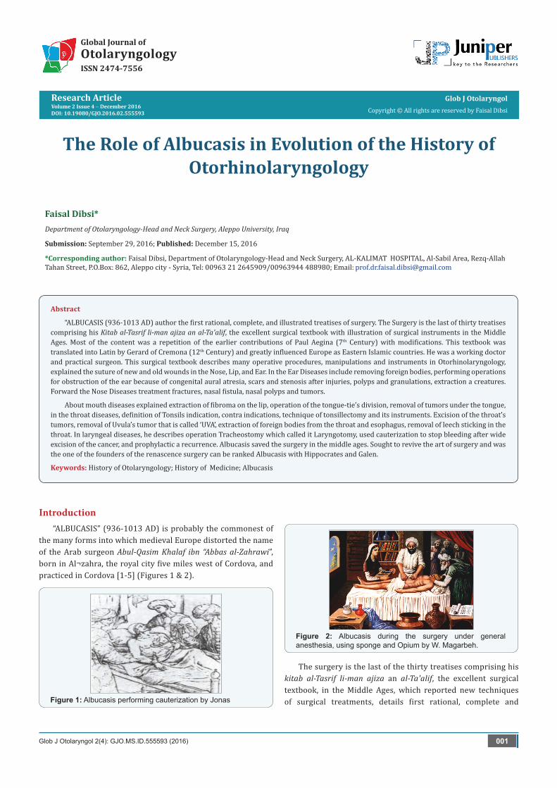
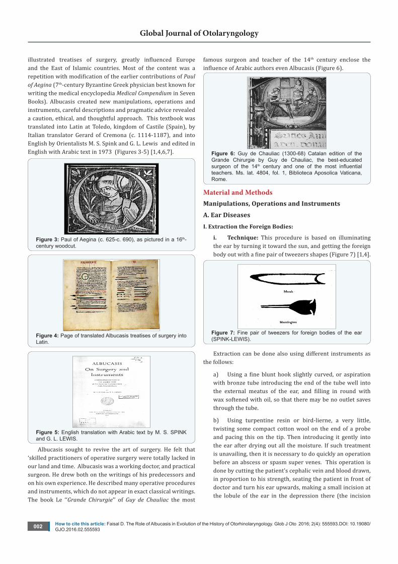
the many forms into which medieval Europe distorted the name of the Arab surgeon Abul-Qasim Khalaf ibn “Abbas al-Zahrawi”, born in Al¬zahra, the royal city five miles west of Cordova, and practiced in Cordova [1-5] (Figures 1 & 2).
Figure 1: Albucasis performing cauterization by Jonas
Figure 2: Albucasis during the surgery under general anesthesia, using sponge and Opium by W. Magarbeh.
The surgery is the last of the thirty treatises comprising his kitab al-Tasrif li-man ajiza an al-Taʹalif, the excellent surgical textbook, in the Middle Ages, which reported new techniques of surgical treatments, details first rational, complete and
Global Journal of OtolaryngologyISSN 2474-7556
How to cite this article: Faisal D. The Role of Albucasis in Evolution of the History of Otorhinolaryngology. Glob J Oto 2016; 2(4): 555593.DOI: 10.19080/GJO.2016.02.555593002
Global Journal of Otolaryngology
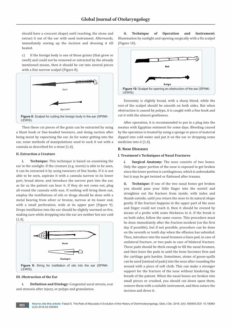
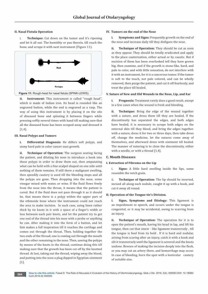
illustrated treatises of surgery, greatly influenced Europe and the East of Islamic countries. Most of the content was a repetition with modification of the earlier contributions of Paul of Aegina (7th-century Byzantine Greek physician best known for writing the medical encyclopedia Medical Compendium in Seven Books). Albucasis created new manipulations, operations and instruments, careful descriptions and pragmatic advice revealed a caution, ethical, and thoughtful approach. This textbook was translated into Latin at Toledo, kingdom of Castile (Spain), by Italian translator Gerard of Cremona (c. 1114-1187), and into English by Orientalists M. S. Spink and G. L. Lewis and edited in English with Arabic text in 1973 (Figures 3-5) [1,4,6,7].
Figure 3: Paul of Aegina (c. 625-c. 690), as pictured in a 16th-century woodcut.
Figure 4: Page of translated Albucasis treatises of surgery into Latin.
Figure 5: English translation with Arabic text by M. S. SPINK and G. L. LEWIS.
Albucasis sought to revive the art of surgery. He felt that ‘skilled practitioners of operative surgery were totally lacked in our land and time. Albucasis was a working doctor, and practical surgeon. He drew both on the writings of his predecessors and on his own experience. He described many operative procedures and instruments, which do not appear in exact classical writings. The book Le ‘‘Grande Chirurgie’’ of Guy de Chauliac the most
famous surgeon and teacher of the 14th century enclose the influence of Arabic authors even Albucasis (Figure 6).
Figure 6: Guy de Chauliac (1300-68) Catalan edition of the Grande Chirurgie by Guy de Chauliac, the best-educated surgeon of the 14th century and one of the most influential teachers. Ms. lat. 4804, fol. 1, Biblioteca Aposolica Vaticana, Rome.
Material and MethodsManipulations, Operations and Instruments
A. Ear Diseases
I. Extraction the Foreign Bodies:
i. Technique: This procedure is based on illuminating the ear by turning it toward the sun, and getting the foreign body out with a fine pair of tweezers shapes (Figure 7) [1,4].
Figure 7: Fine pair of tweezers for foreign bodies of the ear (SPINK-LEWIS).
Extraction can be done also using different instruments as the follows:
a) Using a fine blunt hook slightly curved, or aspiration with bronze tube introducing the end of the tube well into the external meatus of the ear, and filling in round with wax softened with oil, so that there may be no outlet saves through the tube.
b) Using turpentine resin or bird-lierne, a very little, twisting some compact cotton wool on the end of a probe and pacing this on the tip. Then introducing it gently into the ear after drying out all the moisture. If such treatment is unavailing, then it is necessary to do quickly an operation before an abscess or spasm super venes. This operation is done by cutting the patient’s cephalic vein and blood drawn, in proportion to his strength, seating the patient in front of doctor and turn his ear upwards, making a small incision at the lobule of the ear in the depression there (the incision
How to cite this article: Faisal D. The Role of Albucasis in Evolution of the History of Otorhinolaryngology. Glob J Oto 2016; 2(4): 555593.DOI: 10.19080/GJO.2016.02.555593003
Global Journal of Otolaryngology
should have a crescent shape) until reaching, the stone and extract it out of the ear with used instrument. Afterwards, immediately sewing up the incision and dressing it till healed.
c) If the foreign body is one of those grains (that grow or swell) and could not be removed or extracted by the already mentioned means, then it should be cut into several pieces with a fine narrow scalpel (Figure 8).
Figure 8: Scalpel for cutting the foreign body in the ear (SPINK-LEWIS).
Then these cut pieces of the grain can be extracted by using a blunt hook or fine-headed tweezers, and doing suction after being moist by vaporizing the ear. As for water getting into the ear, some methods of manipulations used to suck it out with a cannula as described for a stone [1,4].
II. Extraction a Creature
i. Technique: This technique is based on examining the ear in the sunlight. If the creature (e.g. worm) is able to be seen, it can be extracted it by using tweezers of fine hooks. If it is not able to be seen, aspirate it with a cannula narrow in its lower part, broad above, and introduce the narrow part into the ear, as far as the patient can bear it. If they do not come out, plug all-round the cannula with wax. If nothing will bring them out, employ the instillations of oils or drugs should be done with a metal bearing from silver or bronze, narrow at its lower end, with a small perforation, wide at its upper part (Figure 9). Drops instillation into the ear should be slightly warmed on fire, making sure while dropping into the ear are neither hot nor cold [1,4].
Figure 9: Siring for instillation of oils into the ear (SPINK-LEWIS).
III. Obstruction of the Ear
i. DefinitionandEtiology: Congenital aural atresia, scar and stenosis after injury, or polyps and granulation.
ii. Technique of Operation and Instrument: Illumination by sunlight and opening surgically with a fin scalpel (Figure 10).
Figure 10: Scalpel for opening an obstruction of the ear (SPINK-LEWIS).
Extremity is slightly broad, with a sharp blend, while the rest-of the scalpel should be smooth on both sides. But when obstruction is caused by polyps, it is caught with a fine hook and cut it with the utmost gentleness.
After operation, it is recommended to put in a plug into the meatus with Egyptian ointment for some days. Bleeding caused by the operation is treated by using a sponge or piece of material dipped into cold water and put it on the ear or dropping some medicine into it [1,4].
B. Nose DiseasesI. Treatment’s Techniques of Nasal Fractures
i. Surgical Anatomy: The nose consists of two bones. Only the upper portion of the nose is exposed to get broken since the lower portion is cartilaginous, which is unbreakable but it may be get twisted or flattened after trauma.
ii. Technique: If one of the two nasal bones get broken you should pass your little finger into the nostril and straighten out the fracture from inside, with index and thumb outside, until you return the nose to its natural shape gently. If the fracture happens in the upper part of the nose and finger could not reach it, then it should be evened by means of a probe with some thickness to it. If the break is on both sides, follow the same course. This procedure must be done immediately after the fraction incident (on the first day if possible), but if not possible, procedure can be done on the seventh or tenth day when the effusion has subsided. Then, introduce into the nasal foramen a linen pad, in case of unilateral fracture, or two pads in case of bilateral fracture. These pads should be thick enough to fill the nasal foramen, and then leave the pads in until the bone becomes firm and the cartilage gets harden. Sometimes, stems of goose-quills can be used (instead of pads) into the nose after rounding the wound with a piece of soft cloth. This can make a stronger support for the fracture of the nose without hindering the breath of the patient. When the nasal bones are broken into small pieces or crushed, you should cut down upon them, remove them with a suitable instrument, and then suture the incision and dress it.
How to cite this article: Faisal D. The Role of Albucasis in Evolution of the History of Otorhinolaryngology. Glob J Oto 2016; 2(4): 555593.DOI: 10.19080/GJO.2016.02.555593004
Global Journal of Otolaryngology
II. Nasal Fistula Operation
i. Technique: Cut down on the tumor and it’s ripening and let it all out. The humidity or pus therein, till reach the bone, and scrape it with next instrument (Figure 11).
Figure 11: Rough-head for nasal fistula (SPINK-LEWIS).
ii. Instrument: This instrument is called ‘‘rough head’’, which is made of Indian iron. Its head is rounded like an engraved button, while the end is engraved as a rasp. The way of using this instrument is by placing it on the site of diseased bone and spinning it between fingers while pressing softly several times with hand till making sure that all the diseased bone has been scraped away and dressed it [1,4].
III. Nasal Polyps and Tumors
i. Differential Diagnosis: He differs soft polyps, and stony hard pale in color cancer oust growth.
ii. Technique of Operation: The surgeon seating facing the patient, and dilating his nose to introduce a hook into these polyps in order to draw them out, then amputating what can be held with a fine scalpel shaped on one side until nothing of them remains. If still there a malignant swelling, then speedily cautery is used till the bleeding stops and all the polyps are gone. Then dropping into the nose, some vinegar mixed with water, or wine. If the fluid flows freely from the nose into the throat, it means that the patient is cured. But if the fluid does not pass through it as it should be, that means there is a polyp within the upper part of the ethmoide bone where the instrument could not reach the area to make incision. In such case, using linen rather thick by tie knots in it with a space of a finger’s width or less between each pair knots, and let the patient try to get one end of the thread into his nose with a probe or anything he can. After making it into the form of a button, and let him makes a full inspiration till it reaches the cartilage and comes out through the throat. Then, holding together the two ends of the thread; one is coming out through the mouth and the other remaining in the nose. Then, sawing the polyps by means of the knots in the thread, continue doing this till making sure that the growth has been cut off by the knotted thread. At last, taking out the thread, wiping away the blood, and putting into the nose a plug dipped in Egyptian ointment [1].
IV. Tumors on the end of the Nose
i. Symptoms and Signs: Frequently growth on the end of the nose and increase daily till they disfigure the nose.
ii. Technique of Operation: They should be cut as soon as they appear. They should be totally eradicated and apply to the place cauterization, either actual or by caustic. But if excision of them has been overlooked tell they have grown big, then examine, and if the growth is stone-like, hard, and pale in color, and with little sensation, do not interfere with it with an instrument, for it is a cancerous tumor. If the tumor is soft to the touch, not pale colored, and can be wholly removed, then purge the patient, and cut it off fearlessly, and treat the place till healed.
V. Suture of New and Old Wounds in the Nose, Lip, and Ear
i. Prognosis: Treatment rarely does a good result, except in a few cases when the wound is fresh and bleeding.
ii. Technique: Bring the edge of the wound together with a suture, and dress them till they are healed. If the discontinuity has separated the edges, and both edges have healed, it is necessary to scrape both edges on the external skin till they bleed, and bring the edges together. with a suture, dress it for two or three days, then take dress off, change the medicine, let the sutures come away of themselves, and afterward dress with ointment till healed. The manner of suturing is to close the discontinuity, either with a needle, or with a thread [1,4].
C. Mouth Diseases I. Extraction of Fibroma on the Lip
i. Signs: A little hard swelling inside the lips, some resemble the vetch grain.
ii. Technique of Operation: The lip should be inverted, incised all along each nodule, caught it up with a hook, and cut it away all round.
II. Operation of the Tongue-tie’s Division.
i. Signs, Symptoms and Etiology: This ligament is an impediment to speech, and occurs under the tongue is congenital, or it may be accidental, owing to scarring from an injury.
ii. Technique of Operation: The operation for it is to open the patient’s mouth, having his head in lap, and lift his tongue, then cut that sinew - like ligament transversely , till the tongue is feed from its hold . If it is hard and nodular, arising from scaring after an injury, catch it with a hook and slit it transversely until the ligament is severed and the knots undone. Beware of making the incision deeply into the flesh, or you may cut an artery there, and hemorrhage may occur. In case of bleeding, burn the spot with a lenticular cautery of suitable size.
How to cite this article: Faisal D. The Role of Albucasis in Evolution of the History of Otorhinolaryngology. Glob J Oto 2016; 2(4): 555593.DOI: 10.19080/GJO.2016.02.555593005
Global Journal of Otolaryngology
III.Removal of Tumors under the Tongue
i. Signs and Symptoms: Swellings occurs under the tongue resembling a small frog, which hinders the natural movements of the tongue. Sometimes grows so as even to fill the mouth.
ii. Technique of Operation: The technique of this operation is mainly based on opening the patient’s mouth in the full light of the sun and inspecting the tumor. If the diagnosis tells that the tumor has dark or black color, hard touch, and no sensation, that means cancer. When this tumor is pale and has fluid in it, a hook is used in order to separate the tumor completely [1,4].
D. Throat DiseasesI. Definition: The Tonsils is buboes in the throat
resemble the buboes occurring externally.
II.Operation of Tonsillectomy
i. Indications: Tonsils are pale colored, round, with a slender pedicle.
ii. Contra Indications: Acute inflammation of the tonsils, paratonsillar abscess that is a red color with a thick root, and cancer of the tonsils which is a hard tumor, dark and devoid of sensation, for off a hemorrhage.
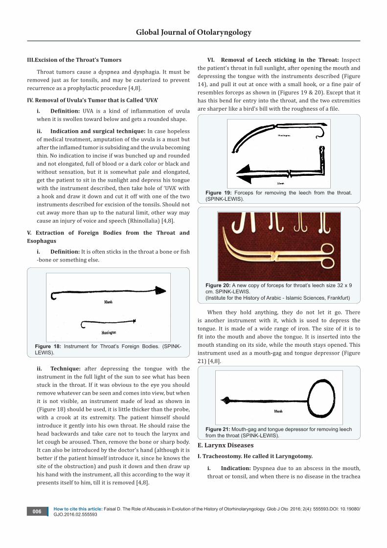
iii. Technique and instruments: Patient should be sitting down in the full light of the sun, his head in surgeon’s lap, open his mouth and assistant presses down the tongue with tongue depressor made from bronze or silver like a knife (Figures 12-14).
Figure 12: Page of Albucasisʹs treatises belongs to Bashir Aga in Istanbul No. 125 M about Tonsillectomy and its Instruments.
Figure 13: Tongue depressor made from bronze or silver like a knife (SPINK-LEWIS).
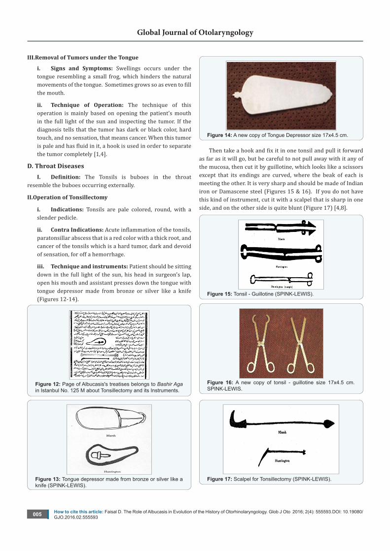
Figure 14: A new copy of Tongue Depressor size 17x4.5 cm.
Then take a hook and fix it in one tonsil and pull it forward as far as it will go, but be careful to not pull away with it any of the mucosa, then cut it by guillotine, which looks like a scissors except that its endings are curved, where the beak of each is meeting the other. It is very sharp and should be made of Indian iron or Damascene steel (Figures 15 & 16). If you do not have this kind of instrument, cut it with a scalpel that is sharp in one side, and on the other side is quite blunt (Figure 17) [4,8].
Figure 15: Tonsil - Guillotine (SPINK-LEWIS).
Figure 16: A new copy of tonsil - guillotine size 17x4.5 cm. SPINK-LEWIS.
Figure 17: Scalpel for Tonsillectomy (SPINK-LEWIS).
How to cite this article: Faisal D. The Role of Albucasis in Evolution of the History of Otorhinolaryngology. Glob J Oto 2016; 2(4): 555593.DOI: 10.19080/GJO.2016.02.555593006
Global Journal of Otolaryngology
III.Excision of the Throat’s Tumors
Throat tumors cause a dyspnea and dysphagia. It must be removed just as for tonsils, and may be cauterized to prevent recurrence as a prophylactic procedure [4,8].
IV. Removal of Uvula’s Tumor that is Called ‘UVA’
i. Definition: UVA is a kind of inflammation of uvula when it is swollen toward below and gets a rounded shape.
ii. Indication and surgical technique: In case hopeless of medical treatment, amputation of the uvula is a must but after the inflamed tumor is subsiding and the uvula becoming thin. No indication to incise if was bunched up and rounded and not elongated, full of blood or a dark color or black and without sensation, but it is somewhat pale and elongated, get the patient to sit in the sunlight and depress his tongue with the instrument described, then take hole of ‘UVA’ with a hook and draw it down and cut it off with one of the two instruments described for excision of the tonsils. Should not cut away more than up to the natural limit, other way may cause an injury of voice and speech (Rhinollalia) [4,8].
V. Extraction of Foreign Bodies from the Throat and Esophagus
i. Definition: It is often sticks in the throat a bone or fish -bone or something else.
Figure 18: Instrument for Throat’s Foreign Bodies. (SPINK-LEWIS).
ii. Technique: after depressing the tongue with the instrument in the full light of the sun to see what has been stuck in the throat. If it was obvious to the eye you should remove whatever can be seen and comes into view, but when it is not visible, an instrument made of lead as shown in (Figure 18) should be used, it is little thicker than the probe, with a crook at its extremity. The patient himself should introduce it gently into his own throat. He should raise the head backwards and take care not to touch the larynx and let cough be aroused. Then, remove the bone or sharp body. It can also be introduced by the doctor’s hand (although it is better if the patient himself introduce it, since he knows the site of the obstruction) and push it down and then draw up his hand with the instrument, all this according to the way it presents itself to him, till it is removed [4,8].
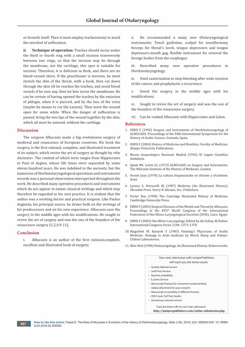
VI. Removal of Leech sticking in the Throat: Inspect the patient’s throat in full sunlight, after opening the mouth and depressing the tongue with the instruments described (Figure 14), and pull it out at once with a small hook, or a fine pair of resembles forceps as shown in (Figures 19 & 20). Except that it has this bend for entry into the throat, and the two extremities are sharper like a bird’s bill with the roughness of a file.
Figure 19: Forceps for removing the leech from the throat. (SPINK-LEWIS).
Figure 20: A new copy of forceps for throat’s leech size 32 x 9 cm. SPINK-LEWIS.(Institute for the History of Arabic - Islamic Sciences, Frankfurt)
When they hold anything, they do not let it go. There is another instrument with it, which is used to depress the tongue. It is made of a wide range of iron. The size of it is to fit into the mouth and above the tongue. It is inserted into the mouth standing on its side, while the mouth stays opened. This instrument used as a mouth-gag and tongue depressor (Figure 21) [4,8].
Figure 21: Mouth-gag and tongue depressor for removing leech from the throat (SPINK-LEWIS).
E. Larynx DiseasesI. Tracheostomy. He called it Laryngotomy.
i. Indication: Dyspnea due to an abscess in the mouth, throat or tonsil, and when there is no disease in the trachea
How to cite this article: Faisal D. The Role of Albucasis in Evolution of the History of Otorhinolaryngology. Glob J Oto 2016; 2(4): 555593.DOI: 10.19080/GJO.2016.02.555593007
Global Journal of Otolaryngology
or bronchi itself. Then it must employ tracheostomy to avoid the mischief of suffocation.
ii. Technique of operation: Trachea should incise under the third or fourth ring with a small incision transversely between two rings, so that the incision may be through the membrane, not the cartilage, this spot is suitable for incision. Therefore, it is deficient in flesh, and there are no blood-vessels there. If the practitioner is nervous, he must stretch the skin of the throat, with a hook, then cut down through the skin till he reaches the trachea, and avoid blood vessels if he sees any, then let him incise the membrane. He can be certain of having opened the trachea by the emission of phlegm, when it is pierced, and by the loss of the voice (maybe he means to cut the Larynx). Then leave the wound open for some while. When the danger of suffocation is passed, bring the two lips of the wound together by the skin, which all must be sutured, without the cartilage.
DiscussionThe surgeon Albucasis made a big evolutionon surgery of
medieval and renascence of European countries. His book the surgery, is the first rational, complete, and illustrated treatment of its subject, which revive the art of surgery as thought by «the Ancients». The content of which term ranges from Hippocrates to Paul of Aegina, whose life times were separated by some eleven hundred years. He was indebted to the ancients, but the numerous of Otorhinolaryngological operations and instruments records was a personal observation interspersed throughout the work. He described many operative procedures and instruments which do not appear in extant classical writings and which may therefore be regarded as his own practice. It is evident that the author was a working doctor and practical surgeon. Like Paulus Aegineta, his principal source, he draws both on the writings of his predecessors and on his own experience. Albucasis save the surgery in the middle ages with his modifications. He sought to revive the art of surgery and was the one of the founders of the renascence surgery [1,2,4,9-11].
Conclusioni. Albucasis is an author of the first rational,complete, excellent and illustrated book of surgery;
ii. He recommended a many new Otolaryngological instruments: Tonsil guillotine, scalpel for tonsillectomy, forceps for throat’s leech, tongue depressors and tongue depressors-mouth gag, flexible instrument for removal the foreign bodies from the esophagus.
iii. Described many new operative procedures in Otorhinolaryngology.
iv. Used cauterization to stop bleeding after wide excision of the cancer, and prophylactic a recurrence.
v. Saved the surgery in the middle ages with his modifications;
vi. Sought to revive the art of surgery and was the one of the founders of the renascence surgery.
vii. Can be ranked Albucasis with Hippocrates and Galen.
References1. DIBSI F (1992) Surgery and Instruments of Otorhinolaryngology of
ALBUCASIS. Proceedings of the Fifth International Symposium for the History of Arabic Science. Granada, Spain.
2. DIBSI F (2004) History of Medicine and Bioethics. Faculty of Medicine. Aleppo University Publications.
3. Museo Arqueologico Nacional, Madrid (1992) El Legato Cientifico Andalusia.
4. Spink MS, Lewis GL (1973) ALBUCASIS on Surgery and Instruments. The Welcome Institute of the History of Medicine, London.
5. Vernet Joan (1978) La cultura hispanoȧrabe en Oriente y Occidente. Ariel.
6. Lyonsa S, Petrucelli RJ (1987) Medicine (An Illustrated History). Abradale Press. Harry N Abrams, Inc., Publishers.
7. Porter Roy (1998) The Camridge Illustrated History of Medicine. Cambridge University Press.
8. DIBSI F (2002) Surgical Diseases of the Mouth and Throat by Albucasis. Proceedings of the XVIIth World Congress of the International Federation of Oto-Rhino-Laryngological Societies (IFOS), Cairo, Egypt.
9. DIBSI F (2003) Oto-Rhino-Laryngology. Edited by AG Zohny, RJ Ruben. International Congress Series 1240: 1375-1378.
10. Magarbeh W, Katayeh S (1983) Painting’s Physicians of Arabic Medicine. Homage to Arab medicine by Merck Sharp and Dohme-Chibret Laboratories.
11. Weir Neil (1990) Otolaryngology. An Illustrated History. Butterworths.
Your next submission with JuniperPublishers will reach you the below assets
• Quality Editorial service
• Swift Peer Review
• Reprints availability
• E-prints Service
• Manuscript Podcast for convenient understanding
• Global attainment for your research
• Manuscript accessibility in different formats
( Pdf, E-pub, Full Text, Audio)
• Unceasing customer service
Track the below URL for one-step submission
http://juniperpublishers.com/online-submission.php