the process of edc-nhs cross-linking of reconstituted collagen fibres increases collagen fibrillar...
TRANSCRIPT
The process of EDC-NHS cross-linking of reconstituted collagen fibres increasescollagen fibrillar order and alignmentD. V. Shepherd, J. H. Shepherd, S. Ghose, S. J. Kew, R. E. Cameron, and S. M. Best Citation: APL Materials 3, 014902 (2015); doi: 10.1063/1.4900887 View online: http://dx.doi.org/10.1063/1.4900887 View Table of Contents: http://scitation.aip.org/content/aip/journal/aplmater/3/1?ver=pdfcov Published by the AIP Publishing Articles you may be interested in Frictional properties of native and functionalized type I collagen thin films Appl. Phys. Lett. 103, 143703 (2013); 10.1063/1.4824685 Longitudinal variations in the Poisson’s ratio of collagen fibrils Appl. Phys. Lett. 98, 163707 (2011); 10.1063/1.3567784 Self-aligned patterns of multiple biomolecules printed in one step Appl. Phys. Lett. 93, 133901 (2008); 10.1063/1.2990045 Detailed evaluation of the performance of microfluidic T mixers using fluorescence and ultraviolet resonanceRaman spectroscopy Rev. Sci. Instrum. 77, 055105 (2006); 10.1063/1.2198800 A minimal model for stabilization of biomolecules by hydrocarbon cross-linking J. Chem. Phys. 124, 164907 (2006); 10.1063/1.2185645
This article is copyrighted as indicated in the article. Reuse of AIP content is subject to the terms at: http://aplmaterials.aip.org/about/rights_and_permissions
Downloaded to IP: 131.111.185.72 On: Sat, 22 Nov 2014 11:52:40

APL MATERIALS 3, 014902 (2015)
The process of EDC-NHS cross-linking of reconstitutedcollagen fibres increases collagen fibrillar orderand alignment
D. V. Shepherd,1,a J. H. Shepherd,1 S. Ghose,2 S. J. Kew,2 R. E. Cameron,1and S. M. Best11Department of Materials Science and Metallurgy, University of Cambridge,Cambridge, United Kingdom2Tigenix Ltd., Byron House, Cambridge Business Park,Cambridge, United Kingdom
(Received 4 September 2014; accepted 22 October 2014; published online 5 November 2014)
We describe the production of collagen fibre bundles through a multi-strand, semi-continuous extrusion process. Cross-linking using an EDC (1-ethyl-3-(3-dimethyl-aminopropyl)carbodiimide), NHS (N-hydroxysuccinimide) combination was consid-ered. Atomic Force Microscopy and Raman spectroscopy focused on how cross-linking affected the collagen fibrillar structure. In the cross-linked fibres, a clearfibrillar structure comparable to native collagen was observed which was notobserved in the non-cross-linked fibre. The amide III doublet in the Raman spectraprovided additional evidence of alignment in the cross-linked fibres. Raman spectros-copy also indicated no residual polyethylene glycol (from the fibre forming buffer)or water in any of the fibres. C 2014 Author(s). All article content, except whereotherwise noted, is licensed under a Creative Commons Attribution 3.0 UnportedLicense. [http://dx.doi.org/10.1063/1.4900887]
In 1989, Kato and Silver first described the extrusion of an acid swollen collagen gel underaqueous conditions on a laboratory scale.1 Since then there have been several studies of this processof self-assembly,2 with production parameters and cross-linking treatments3–6 considered in orderto optimise mechanical and biological characteristics. The acidic collagen suspension is typicallyextruded into a bath containing neutral fibre forming buffer, the collagen gels on contact with neutralpH as the fibrils, reconstitute and the macroscopic fibres form. Along the length of the bath, the threaddehydrates due to the osmotic gradient between the collagen and the fibre forming buffer.7
When collagen is generated in vivo, cross-linking occurs enzymatically and covalent inter- andintra-molecular bonds are formed that provide sufficient mechanical strength and proteolytic resis-tance.3 These same cross-links are not formed during the self-assembly of collagen in neutral pHand as such cross-linking routes are a particular area of consideration in the production of artificialcollagen fibre based structures. Cross-linking approaches include chemical (glutaraldehyde, isocy-anates, or carbodiimide based),6 physical (dehydrothermal),8,9 and enzymatic.10 The authors havepreviously settled upon the use of the zero length cross-linker carbodiimide 1-ethyl-3-(3-dimethyl-aminopropyl)carbodiimide (EDC) in combination with N-hydroxysuccinimide (NHS) as an effectiveroute for the cross-linking of extruded collagen fibres.11–13 This route results in a lower cross-linkingdensity than other chemical and physical cross-linking routes, but it has been shown to exhibit afavourable biological response14 unlike more aggressive techniques that have been associated withinflammatory response and reduced bioactivity.15
Whilst the mechanical properties,3,4,6,16 degradative characteristics,14,17,18 and surface micro-structure3,6 of extruded fibres have been considered quite extensively, any rigorous investigation
aAuthor to whom correspondence should be addressed. Electronic mail: [email protected]
2166-532X/2015/3(1)/014902/8 3, 014902-1 ©Author(s) 2014
This article is copyrighted as indicated in the article. Reuse of AIP content is subject to the terms at: http://aplmaterials.aip.org/about/rights_and_permissions
Downloaded to IP: 131.111.185.72 On: Sat, 22 Nov 2014 11:52:40

014902-2 Shepherd et al. APL Mater. 3, 014902 (2015)
into the fibrillar structure and the effect of cross-linking is more limited. Polarised light microscopyhas been carried out to provide a qualitative indication of alignment11,19 and Transmission Elect-ron Microscopy (TEM)6 and Small Angle X-Ray Scattering (SAXS)11,19 have also been applied toa limited extent, however, not with a view of investigating the effect of cross-linking on collagenstructure. Atomic force microscopy (AFM) has been considered quite extensively for the character-isation of native collagen20–24 but can also provide evidence as to the effect of cross-linking on thecollagen order and alignment in extruded collagen fibres. Raman spectroscopy may also provide auseful route for investigating orientation and alignment within the collagen structure.25–27 A recentstudy investigating bundles of collagen fibres from bovine Achilles tendon demonstrated that evennon-polarized Raman measurements (i.e., where a polarisation analyser is not present between thesample and detector) are sensitive to the laser polarisation direction.28 The amide III doublet at 1245and approximately 1270 cm−1 was shown to be highly sensitive to the orientation of the fibre.28
The aim of the work was to study the effects of cross-linking of collagen fibres extruded through asemi-continuous process13 using AFM and Raman spectroscopy. These techniques allowed the align-ment of the collagen fibres as well as the banding and chemistry to be analysed. This understandingof the basic structure is key to better understand fibre mechanics, degradation, and ultimately thebiological response to the material.
Collagen fibre “bundles” were extruded using a method applied previously by the authors13 andbased upon the route of Silver et al.29 The collagen source was an acid swollen gel type I collagenderived from bovine dermis (Devro Medical, Scotland). Briefly, the collagen was added to 2 mMHCl in a concentration of 6 mg/ml and left to swell overnight before blending and degassing. Sixchannels of the collagen slurry were then extruded into a long trough containing a fibre forming bufferof 20% by weight polyethylene glycol (PEG) (molecular weight 8000, Sigma Aldrich, UK) in 0.01Mphosphate buffered saline (PBS) solution. A peristaltic pump provided a constant flow rate of thefibre forming buffer, the 6 strands were brought together at the end of the trough and wound onto ahorizontally moving spool in the form of a 6 ply fibre. After winding onto the spools, the fibres wereair-dried overnight before washing or cross-linking.
Where bundles were cross-linked, fibres still on the spools were soaked in a cross-linkingsolution of 25 mM EDC (1-ethyl-3-(3-dimethylaminopropyl)carbodiimide) (Sigma-Aldrich, UK)and 12.5 mM NHS (N-hydroxysuccinimide) (Sigma-Aldrich, UK) in an 80/20 acetone/PBS solu-tion for 2 h. After cross-linking, fibres underwent a multi-stage washing process, with 2 half hourwashes in 0.01M PBS solution followed by 2 half hour washes in deionised water. Bundles wherecross-linking was not carried out underwent the multi-stage washing process in order to remove anyresidual PEG. All samples were then dried overnight before being removed from the spools andstored.
Scanning Electron Microscopy (SEM) was performed on sections of fibre secured onto a metalstub with carbon tape. Samples were sputter-coated with gold and viewed with a JEOL 820 SEM inthe secondary electron mode.
For AFM imaging, fibres were sectioned and secured onto microscope slides with silver-dag.Viewing was carried out in tapping mode on a Digital Instruments multimode AFM (VEECO) withNanoscope III control. ImageJ30 was used in order to measure the periodic D-spacing.
Raman spectroscopy was applied to understand the effect of the cross-linking process on thechemical and structural characteristics of the collagen fibre bundles and to give qualitative indicationsof any residual PEG or water within the fibre structure. Raman Spectroscopy was carried out usinga Renishaw Ramascope 1000 Raman spectrometer with a 633 nm He-Ne laser in combination witha Leica DM-LM microscope. An acquisition time of 10 s with 10 acquisitions was applied across awavenumber range of 800–3050 cm−1 on fibre samples with the fibre axis both parallel and perpen-dicular to the direction of polarization of the incident laser. Particular regions of interest were betweenwavenumbers of 1150 and 1350 cm−1 (for the amide III doublet28) and at the higher wavenumbers(2800–3050 cm−1), where the highest intensity PEG peaks are present.31 Additionally, literature sug-gests the δ and ν modes of the OH absorption within H2O to be present at 1640 and 3280 cm−1 sothese regions were considered to investigate any water presence.
The SEM images of Figure 1 suggest that cross-linking had no effect on the surface structureof the collagen fibres, with all images showing a comparatively smooth surface, with minimal and
This article is copyrighted as indicated in the article. Reuse of AIP content is subject to the terms at: http://aplmaterials.aip.org/about/rights_and_permissions
Downloaded to IP: 131.111.185.72 On: Sat, 22 Nov 2014 11:52:40

014902-3 Shepherd et al. APL Mater. 3, 014902 (2015)
FIG. 1. SEM imaging of extruded collagen fibres ((a) and (b)) with no cross-linking and ((c) and (d)) with EDC-NHScross-linking.
regular surface artifacts at the microstructural level. Despite the 6 ply nature of these fibres, a densehomogeneous fibre resulted, with the individual ply clearly adhering well during the winding pro-cess. Some evidence of the separately extruded collagen channels is evident across both cross-linkedand non-cross-linked fibres, in the contrast variations observed, probably a result of variations inthe tension applied to the collagen during the winding process. The horizontal motion of the spoolthroughout the winding process was designed to minimize overlapping of the collagen fibre, however,as clearly demonstrated in Figures 1(b) and 1(d), fibres, whilst separated over the spool rods couldbind together, away from them, resulting in the creation of a thicker fibre in places.
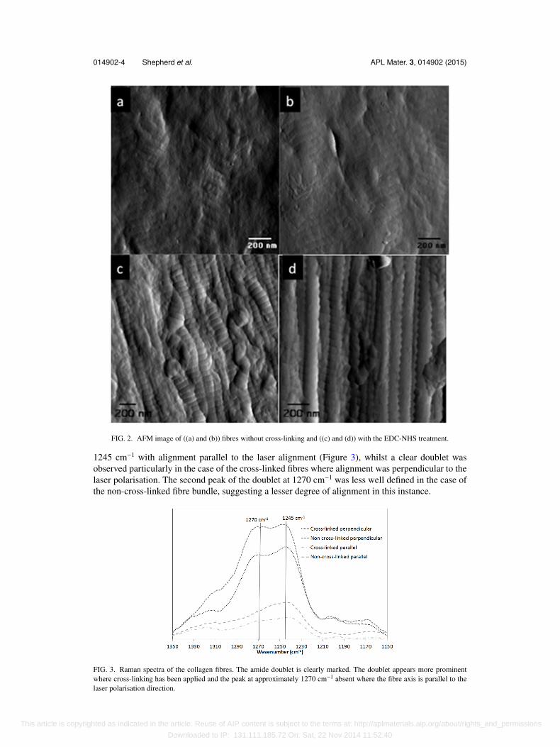
Figure 2 shows images of regions of collagen fibre taken in the tapping mode, without cross-linking ((a) and (b)) and with the EDC-NHS treatment ((c) and (d)). In the cross-linked material,the characteristic banding pattern of collagen fibrils, such as that found in native collagenous tissueis clearly observed. Measurement of a d-spacing of 66.1 ± 1.29 nm was also consistent with nativetissue. Individual collagen fibrils showed a relatively high degree of alignment within the structure.Slight tangling or clumping of collagen fibrils was evident and this is likely to explain surface de-fects observed during electron microscopy. Where fibres were not cross-linked, the degree of or-der was much less significant and the characteristic banding was evident only in very localized re-gions. It is possible that the non-cross-linked fibres are more swollen and hydrated (cross-linkingin acetone-based solution could further dehydrate the fibres). Water content was investigated usingRaman spectroscopy.
Figure 3 shows the Raman spectra for fibres oriented parallel and perpendicular to the direc-tion of polarization of the incident laser between wavenumbers of 1150 and 1350 cm−1 and Figure 4investigates the presence of PEG and water within the samples.
Anisotropic Raman scattering has previously been observed in collagen bundles from bovinetendon26,28 and human skin26 with differences particularly observed in the amide doublet at around1250 cm−1. Consistent with the work of Janko et al.,26 only a single peak was observed at around
This article is copyrighted as indicated in the article. Reuse of AIP content is subject to the terms at: http://aplmaterials.aip.org/about/rights_and_permissions
Downloaded to IP: 131.111.185.72 On: Sat, 22 Nov 2014 11:52:40
014902-4 Shepherd et al. APL Mater. 3, 014902 (2015)
FIG. 2. AFM image of ((a) and (b)) fibres without cross-linking and ((c) and (d)) with the EDC-NHS treatment.
1245 cm−1 with alignment parallel to the laser alignment (Figure 3), whilst a clear doublet wasobserved particularly in the case of the cross-linked fibres where alignment was perpendicular to thelaser polarisation. The second peak of the doublet at 1270 cm−1 was less well defined in the case ofthe non-cross-linked fibre bundle, suggesting a lesser degree of alignment in this instance.
FIG. 3. Raman spectra of the collagen fibres. The amide doublet is clearly marked. The doublet appears more prominentwhere cross-linking has been applied and the peak at approximately 1270 cm−1 absent where the fibre axis is parallel to thelaser polarisation direction.
This article is copyrighted as indicated in the article. Reuse of AIP content is subject to the terms at: http://aplmaterials.aip.org/about/rights_and_permissions
Downloaded to IP: 131.111.185.72 On: Sat, 22 Nov 2014 11:52:40
014902-5 Shepherd et al. APL Mater. 3, 014902 (2015)
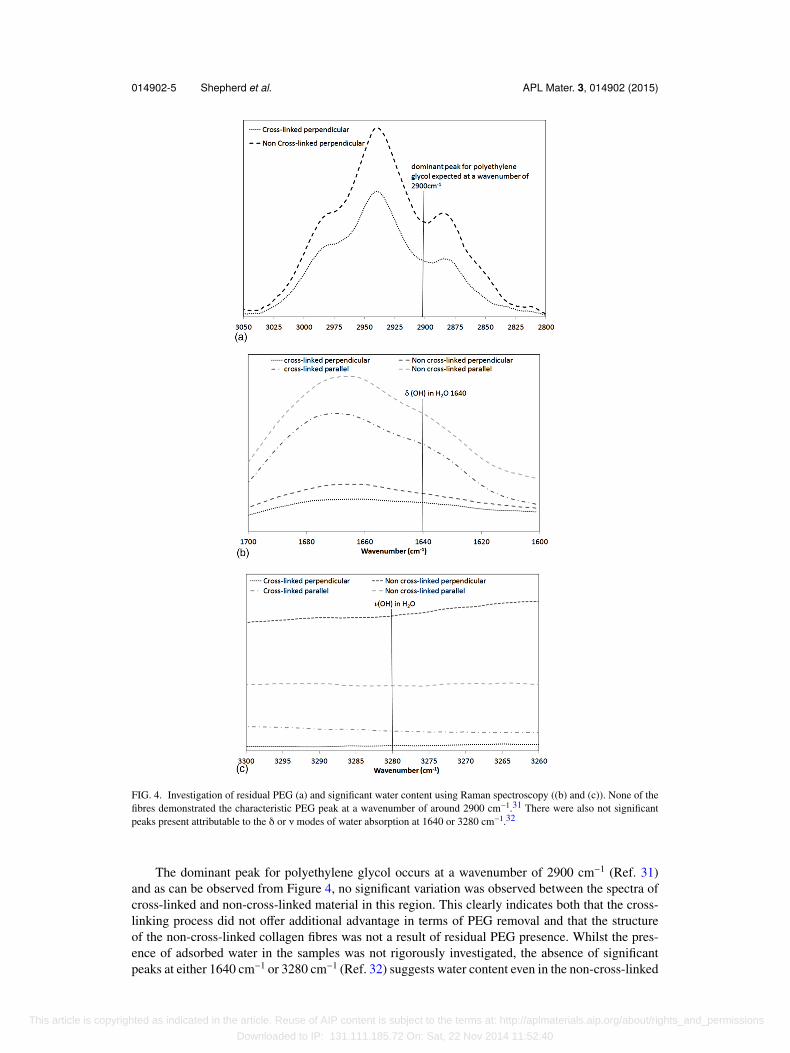
FIG. 4. Investigation of residual PEG (a) and significant water content using Raman spectroscopy ((b) and (c)). None of thefibres demonstrated the characteristic PEG peak at a wavenumber of around 2900 cm−1.31 There were also not significantpeaks present attributable to the δ or ν modes of water absorption at 1640 or 3280 cm−1.32
The dominant peak for polyethylene glycol occurs at a wavenumber of 2900 cm−1 (Ref. 31)and as can be observed from Figure 4, no significant variation was observed between the spectra ofcross-linked and non-cross-linked material in this region. This clearly indicates both that the cross-linking process did not offer additional advantage in terms of PEG removal and that the structureof the non-cross-linked collagen fibres was not a result of residual PEG presence. Whilst the pres-ence of adsorbed water in the samples was not rigorously investigated, the absence of significantpeaks at either 1640 cm−1 or 3280 cm−1 (Ref. 32) suggests water content even in the non-cross-linked
This article is copyrighted as indicated in the article. Reuse of AIP content is subject to the terms at: http://aplmaterials.aip.org/about/rights_and_permissions
Downloaded to IP: 131.111.185.72 On: Sat, 22 Nov 2014 11:52:40
014902-6 Shepherd et al. APL Mater. 3, 014902 (2015)
fibres was not significant and thus differences in structure as observed with AFM are unlikely to beattributable to water content.
This study builds on a body of literature that has focused on the extrusion of collagen fibres andthe effect of cross-linking, not least the previous work by Kew et al. which applied almost the sameapproach to fibre production.11 Whilst previous papers have considered the effect of cross-linkingagents on surface structure, mechanics, and biological response,2,3,6,11,16,33 this paper additionally con-siders an investigation of fibrillar arrangement using AFM and Raman spectroscopy and for the firsttime clearly shows that cross-linking brings about alignment of the collagen and shows characteristicbanding similar to native collagen with a d-spacing of 67 nm.
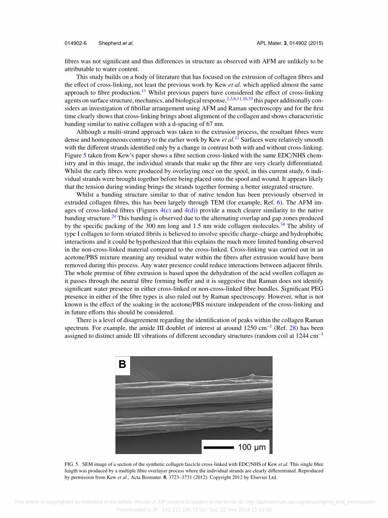
Although a multi-strand approach was taken to the extrusion process, the resultant fibres weredense and homogeneous contrary to the earlier work by Kew et al.11 Surfaces were relatively smoothwith the different strands identified only by a change in contrast both with and without cross-linking.Figure 5 taken from Kew’s paper shows a fibre section cross-linked with the same EDC/NHS chem-istry and in this image, the individual strands that make up the fibre are very clearly differentiated.Whilst the early fibres were produced by overlaying once on the spool, in this current study, 6 indi-vidual strands were brought together before being placed onto the spool and wound. It appears likelythat the tension during winding brings the strands together forming a better integrated structure.
Whilst a banding structure similar to that of native tendon has been previously observed inextruded collagen fibres, this has been largely through TEM (for example, Ref. 6). The AFM im-ages of cross-linked fibres (Figures 4(c) and 4(d)) provide a much clearer similarity to the nativebanding structure.24 This banding is observed due to the alternating overlap and gap zones producedby the specific packing of the 300 nm long and 1.5 nm wide collagen molecules.34 The ability oftype I collagen to form striated fibrils is believed to involve specific charge–charge and hydrophobicinteractions and it could be hypothesized that this explains the much more limited banding observedin the non-cross-linked material compared to the cross-linked. Cross-linking was carried out in anacetone/PBS mixture meaning any residual water within the fibres after extrusion would have beenremoved during this process. Any water presence could reduce interactions between adjacent fibrils.The whole premise of fibre extrusion is based upon the dehydration of the acid swollen collagen asit passes through the neutral fibre forming buffer and it is suggestive that Raman does not identifysignificant water presence in either cross-linked or non-cross-linked fibre bundles. Significant PEGpresence in either of the fibre types is also ruled out by Raman spectroscopy. However, what is notknown is the effect of the soaking in the acetone/PBS mixture independent of the cross-linking andin future efforts this should be considered.
There is a level of disagreement regarding the identification of peaks within the collagen Ramanspectrum. For example, the amide III doublet of interest at around 1250 cm−1 (Ref. 28) has beenassigned to distinct amide III vibrations of different secondary structures (random coil at 1244 cm−1
FIG. 5. SEM image of a section of the synthetic collagen fascicle cross-linked with EDC/NHS of Kew et al. This single fibrelength was produced by a multiple fibre overlayer process where the individual strands are clearly differentiated. Reproducedby permission from Kew et al., Acta Biomater. 8, 3723–3731 (2012). Copyright 2012 by Elsevier Ltd.
This article is copyrighted as indicated in the article. Reuse of AIP content is subject to the terms at: http://aplmaterials.aip.org/about/rights_and_permissions
Downloaded to IP: 131.111.185.72 On: Sat, 22 Nov 2014 11:52:40
014902-7 Shepherd et al. APL Mater. 3, 014902 (2015)
and triple helix at 1265 cm−1);35 to two regions of the polypeptide strands having different polarities;36
or to different positions of the proline residues in the structural motif typical of the collagen triplehelix.37 Bonifacio and Sergio suggest that because of the sensitivity to orientation, the 2 modes mustarise from vibrations taking place along different directions within the same molecular structure. Theyfound the intensity of the 1270 cm−1 band to be weaker when the fiber axis is oriented parallel tothe laser polarisation, suggesting that this mode involves vibrations along a direction perpendicularto the fibril axis.28 This finding is supported in the current work with the band at 1270 cm−1 beinglargely absent in the perpendicular direction. The intensity of this band also appears reduced in thecase of the non-cross-linked material supporting the less aligned structure observed for these fibreswith AFM.
While a significant body of literature has investigated the effect of EDC/NHS cross-linking onfibre mechanics, this is the first work to consider in any real detail the effect of this cross-linking onthe fibrillar alignment. The more aligned structure in the case of the cross-linked fibre, as demon-strated by both Raman spectroscopy and AFM is likely to support the superior mechanics,3,4,6 reducedswelling,3,4,14 and increased proteolytic resistance observed in previous studies of extruded collagenfibres.14,17,18
Homogeneous and dense collagen fibre bundles have been successfully produced through a multi-strand, broadly continuous extrusion process. Cross-linking with an EDC/NHS solution resulted ina highly aligned fibrillar structure with banding similar to that observed in native collagen. The non-cross-linked fibres on the other hand exhibited a more swollen structure with only isolated regions ofbanding evident. The difference in order and alignment between the two fibre types was also evidentin the amide III doublet in the Raman spectra. It is suggested that it is this difference in structure,in combination with the increased cross-linking concentration that explains frequently reported in-creases in mechanics and proteolytic resistance associated with cross-linking.
The authors would like to acknowledge the support of the Engineering and Physical SciencesResearch Council (EPSRC), UK through a Knowledge Transfer Secondment (KTS) (to J.H.S.), TheNational Institutes for Health Research (NIHR) through their i4i Grant to Tigenix Ltd., and the TSBGrant No. TP/8/BIO/6/I/Q0052.1 Y. P. Kato, D. L. Christiansen, R. A. Hahn, S.-J. Shieh, J. D. Goldstein, and F. H. Silver, “Mechanical properties of collagen
fibres: A comparison of reconstituted and rat tail tendon fibres,” Biomaterials 10, 38–42 (1989).2 G. D. Pins, D. L. Christiansen, R. Patel, and F. H. Silver, “Self-assembly of collagen fibers. Influence of fibrillar alignment
and decorin on mechanical properties,” Biophys. J. 73, 2164–2172 (1997).3 D. I. Zeugolis, G. R. Paul, and G. Attenburrow, “Cross-linking of extruded collagen fibers—A biomimetic three-dimensional
scaffold for tissue engineering applications,” J. Biomed. Mater. Res., Part A 89A, 895–908 (2009).4 G. D. Pins and F. H. Silver, “A self-assembled collagen scaffold suitable for use in soft and hard tissue replacement,” Mater.
Sci. Eng., C 3, 101–107 (1995).5 D. I. Zeugolis, R. G. Paul, and G. Attenburrow, “The influence of a natural cross-linking agent (Myrica rubra) on the
properties of extruded collagen fibres for tissue engineering applications,” Mater. Sci. Eng., C 30, 190–195 (2010).6 D. I. Zeugolis, R. G. Paul, and G. Attenburrow, “Post-self-assembly experimentation on extruded collagen fibres for tissue
engineering applications,” Acta Biomater. 4, 1646–1656 (2008).7 Y.-K. Seo, H.-H. Youn, C.-S. Park, K.-Y. Song, and J.-K. Park, “Reinforced bioartificial dermis constructed with collagen
threads,” Biotechnol. Bioprocess Eng. 13, 745–751 (2008).8 M. G. Haugh, M. J. Jaasma, and F. J. O’Brien, “The effect of dehydrothermal treatment on the mechanical and structural
properties of collagen-GAG scaffolds,” J. Biomed. Mater. Res., Part A 89A, 363–369 (2009).9 D. L. Ellis and I. V. Yannas, “Recent advances in tissue synthesis in vivo by use of collagen-glycosaminoglycan copolymers,”
Biomaterials 17, 291–299 (1996).10 D. Y. S. Chau, R. J. Collighan, E. A. M. Verderio, V. L. Addy, and M. Griffin, “The cellular response to transglutaminase-
cross-linked collagen,” Biomaterials 26, 6518–6529 (2005).11 S. J. Kew, J. H. Gwynne, D. Enea, R. Brookes, N. Rushton, S. M. Best et al., “Synthetic collagen fascicles for the regeneration
of tendon tissue,” Acta Biomater. 8, 3723–3731 (2012).12 D. Enea, J. Gwynne, S. Kew, M. Arumugam, J. Shepherd, R. Brooks et al., “Collagen fibre implant for tendon and ligament
biological augmentation. In vivo study in an ovine model,” Knee Surg., Sports Traumatol., Arthroscopy 21, 1783–1793(2013).
13 J. H. Shepherd, S. Ghose, S. J. Kew, A. Moavenian, S. M. Best, and R. E. Cameron, “Effect of fiber crosslinking oncollagen-fiber reinforced collagen–chondroitin-6-sulfate materials for regenerating load-bearing soft tissues,” J. Biomed.Mater. Res., Part A 101A, 176–184 (2013).
14 K. G. Cornwell, P. Lei, S. T. Andreadis, and G. D. Pins, “Crosslinking of discrete self-assembled collagen threads: Effectson mechanical strength and cell–matrix interactions,” J. Biomed. Mater. Res., Part A 80A, 362–371 (2007).
15 M. Chvapil, D. Speer, W. Mora, and E. Eskelson, “Effect of tanning agent on tissue reaction to tissue implanted collagensponge,” J. Surg. Res. 35, 402–409 (1983).
This article is copyrighted as indicated in the article. Reuse of AIP content is subject to the terms at: http://aplmaterials.aip.org/about/rights_and_permissions
Downloaded to IP: 131.111.185.72 On: Sat, 22 Nov 2014 11:52:40
014902-8 Shepherd et al. APL Mater. 3, 014902 (2015)
16 M.-C. Wang, G. D. Pins, and F. H. Silver, “Collagen fibres with improved strength for the repair of soft tissue injuries,”Biomaterials 15, 507–512 (1994).
17 K. S. Weadock, E. J. Miller, E. L. Keuffel, and M. G. Dunn, “Effect of physical crosslinking methods on collagen-fiberdurability in proteolytic solutions,” J. Biomed. Mater. Res. 32, 221–226 (1996).
18 A. B. Caruso and M. G. Dunn, “Functional evaluation of collagen fiber scaffolds for ACL reconstruction: Cyclic loading inproteolytic enzyme solutions,” J. Biomed. Mater. Res., Part A 69A, 164–171 (2004).
19 X. Cheng, U. A. Gurkan, C. J. Dehen, M. P. Tate, H. W. Hillhouse, G. J. Simpson et al., “An electrochemical fabricationprocess for the assembly of anisotropically oriented collagen bundles,” Biomaterials 29, 3278–3288 (2008).
20 S. Rele, Y. Song, R. P. Apkarian, Z. Qu, V. P. Conticello, and E. L. Chaikof, “D-periodic collagen-mimetic microfibers,” J.Am. Chem. Soc. 129, 14780–14787 (2007).
21 M. Jastrzebska, B. Barwinski, I. Mroz, A. Turek, J. Zalewska-Rejdak, and B. Cwalina, “Atomic force microscopy investi-gation of chemically stabilized pericardium tissue,” Eur. Phys. J. E: Soft Matter Biol. Phys. 16, 381–388 (2005).
22 I. Revenko, F. Sommer, D. T. Minh, R. Garrone, and J.-M. Franc, “Atomic force microscopy study of the collagen fibrestructure,” Biol. Cell 80, 67–69 (1994).
23 S. Rigozzi, A. Stemmer, R. Müller, and J. G. Snedeker, “Mechanical response of individual collagen fibrils in loaded tendonas measured by atomic force microscopy,” J. Struct. Biol. 176, 9–15 (2011).
24 D. R. Baselt, J. P. Revel, and J. D. Baldeschwieler, “Subfibrillar structure of type I collagen observed by atomic forcemicroscopy,” Biophys. J. 65, 2644–2655 (1993).
25 A. Masic, L. Bertinetti, R. Schuetz, L. Galvis, N. Timofeeva, J. W. C. Dunlop et al., “Observations of multiscale, stress-induced changes of collagen orientation in tendon by polarized Raman spectroscopy,” Biomacromolecules 12, 3989–3996(2011).
26 M. Janko, P. Davydovskaya, M. Bauer, A. Zink, and R. W. Stark, “Anisotropic Raman scattering in collagen bundles,” Opt.Lett. 35, 2765–2767 (2010).
27 Y. N. Wang, C. Galiotis, and D. L. Bader, “Determination of molecular changes in soft tissues under strain using laser Ramanmicroscopy,” J. Biomech. 33, 483–486 (2000).
28 A. Bonifacio and V. Sergo, “Effects of sample orientation in Raman microspectroscopy of collagen fibers and their impacton the interpretation of the amide III band,” Vib. Spectrosc. 53, 314–317 (2010).
29 F. H. Silver and Y. P. Kato, “Synthetic collagen orthopaedic structures such as grafts, tendons, and other structures,”U. S. patent US5171273 A (15 December 1992).
30 W. S. Rasband, ImageJ (U. S. National Institutes of Health, Bethesda, Maryland, USA, 1997–2011).31 A. Docoslis, K. Huszarik, G. Papageorgiou, D. Bikiaris, A. Stergiou, and E. Georgarakis, “Characterization of the distribu-
tion, polymorphism, and stability of nimodipine in its solid dispersions in polyethylene glycol by micro-Raman spectroscopyand powder x-ray diffraction,” AAPS J. 9, E361–E370 (2007).
32 L. Chrit, C. Hadjur, S. Morel, G. Sockalingum, G. Lebourdon, F. Leroy et al., “In vivo chemical investigation of humanskin using a confocal Raman fiber optic microprobe,” J. Biomed. Opt. 10, 044007 (2005).
33 V. Cheng and H. Screen, “The micro-structural strain response of tendon,” J. Mater. Sci. 42, 8957–8965 (2007).34 D. J. S. Hulmes, A. Miller, D. A. D. Parry, K. A. Piez, and J. Woodhead-Galloway, “Analysis of the primary structure of
collagen for the origins of molecular packing,” J. Mol. Biol. 79, 137–148 (1973).35 M. Wisniewski, A. Sionkowska, H. Kaczmarek, S. Lazare, V. Tokarev, and C. Belin, “Spectroscopic study of a KrF excimer
laser treated surface of the thin collagen films,” J. Photochem. Photobiol., A 188, 192–199 (2007).36 B. G. Frushour and J. L. Koenig, “Raman scattering of collagen, gelatin, and elastin,” Biopolymers 14, 379–391 (1975).37 A. Merlino, F. Sica, L. Mazzarella, A. Zagari, and A. Vergara, “Correlation between Raman and x-ray crystallography data
of (Pro-Pro-Gly)10,” Biophys. Chem. 137, 24–27 (2008).
This article is copyrighted as indicated in the article. Reuse of AIP content is subject to the terms at: http://aplmaterials.aip.org/about/rights_and_permissions
Downloaded to IP: 131.111.185.72 On: Sat, 22 Nov 2014 11:52:40